Abstract
Peptides, as potential therapeutics continue to gain importance in the search for active substances for the treatment of numerous human diseases, some of which are, to this day, incurable. As potential therapeutic drugs, peptides have many favorable chemical and pharmacological properties, starting with their great diversity, through their high affinity for binding to all sort of natural receptors, and ending with the various pathways of their breakdown, which produces nothing but amino acids that are nontoxic to the body. Despite these and other advantages, however, they also have their pitfalls. One of these disadvantages is the very low stability of natural peptides. They have a short half-life and tend to be cleared from the organism very quickly. Their instability in the gastrointestinal tract, makes it impossible to administer peptidic drugs orally. To achieve the best pharmacologic effect, it is desirable to look for ways of modifying peptides that enable the use of these substances as pharmaceuticals. There are many ways to modify peptides. Herein we summarize the approaches that are currently in use, including lipidization, PEGylation, glycosylation and others, focusing on lipidization. We describe how individual types of lipidization are achieved and describe their advantages and drawbacks. Peptide modifications are performed with the goal of reaching a longer half-life, reducing immunogenicity and improving bioavailability. In the case of neuropeptides, lipidization aids their activity in the central nervous system after the peripheral administration. At the end of our review, we summarize all lipidized peptide-based drugs that are currently on the market.
Keywords: Peptide therapeutics, structure modification, lipidization, therapeutic lipopeptides
Introduction
The interest of researchers and investors in peptides as potential therapeutics for various diseases has increased steadily in the recent years. Both natural or synthesized peptides and their analogues have shown promising pharmaceutical properties and other favorable features lacking in other substances such as small molecules. The main promise of peptides resides in their vast diversity, great biochemical selectivity and strong affinity to various cellular receptors. One significant benefit is that the degradation of peptides by hydrolysis leads to the formation of nothing more than amino acids which are nontoxic (Goodwin et al., 2012; Ahrens et al., 2012).
Peptides as therapeutics have a myriad of potential applications in the treatment of a broad range of diseases. These diseases include, for example, metabolic disorders such as obesity or type 2 diabetes mellitus (T2DM), various forms of cancer, inflammation-related and microbial diseases, immune dysfunction, hypertension, and others (Morozov & Khavinson, 1997; Goodwin et al., 2012; Mikulášková et al., 2016; LA Manna et al., 2018; Mahlapuu et al., 2020).
Despite their benefits, peptide and protein-based drugs face many challenges. The main one lies in their low stability in living organisms. Without special modification providing an increased half-life, most of them tend to be cleared from the organism in a matter of minutes. A short blood half-life results in a demand for increased and more frequent dosing, increasing the cost of the treatment and narrowing the therapeutic window. Moreover, such peptides and proteins in their natural form have a poor ability to permeate the cellular membrane (Menacho-Melgar et al., 2019).
It is evident that peptides need to be modified to be able to reach their target and fulfill their function, for instance, peptides involved in food intake regulation. The center controlling the food intake regulation is in the brain, specifically in the hypothalamus and brain stem (Mikulášková et al., 2016). Disturbances in food intake regulation lead to obesity or cachexia (Zhang et al., 2014). In our laboratory, we have demonstrated that anorexigenic (food intake lowering) peptides might be potential drugs for obesity (Kuneš et al., 2016; Pražienková et al., 2017). However, without modification, they cannot cross the blood brain barrier (hereafter BBB) and exert a central effect after peripheral administration (Maletínská et al., 2015; Kořínková et al., 2020). Lipidization, as one of the modification strategies, is a useful tool that results in increased stability and a longer half-life (Maletínská et al., 2015), slower biodegradation, and an ability to cross the BBB (Maletínská et al., 2015; Pražienková et al., 2017).
Considering the above, it is highly desirable to develop and test as many ways to modify these peptide or protein-based drugs as possible. This article provides a broad overview of possible peptide modifications that lead to the stabilization or general improvement of the peptide-based therapeutics. The approach to peptide stabilization which we focus on is lipidization. This review also presents biological aspects of lipidization such as prolonged half-life, bioavailability and improved drug delivery. Finally, we provide an overview summarizing the lipidized drugs that are currently on the market.
Methods of peptide stabilization
The above introduction highlights that there are many ways to improve and prolong the useful life of a peptide. Most of these modifications consist in adding a specific moiety to the original peptide chain.
PEGylation
The term PEGylation defines the modification of a non-peptide (Veronese & Pasut, 2005), peptide or protein molecule by linking one or more polyethylene glycol (PEG) chains to the original structure (Harris & Chess, 2003; Veronese & Pasut, 2005). PEG is a linear or branched polymer with nontoxic, non-immunogenic (Veronese & Mero, 2008; Roberts et al., 2002), non-antigenic properties that is highly soluble in aqueous solutions (Veronese & Pasut, 2005) and organic solvents (Roberts et al., 2002). PEGylation helps to increase drug stability and reduce proteolysis and renal excretion (Roberts et al., 2002; Veronese & Mero, 2008; Yadav & Dewangan, 2021). It can be used to modify drugs, food, or cosmetics as it has been approved by the Food and Drug Administration (FDA) (Harris & Chess, 2003; Veronese & Pasut, 2005). The FDA has approved PEG for all types of injectable, topical, rectal and nasal formulations. This modification shows little toxicity and PEGylated peptides or proteins are usually eliminated from the body by the kidneys (Harris & Chess, 2003).
To bind PEG to a molecule, it is first necessary to activate it. This can be easily done by preparing a molecule PEG that has a functional group at least at one terminus (Roberts et al., 2002). Originally, in the first-generation PEGylation processes, it was only possible to bind PEG to the desired amino acids. PEGylation was accomplished by alkylation or acylation of the target amino acid. In the case of proteins, PEG binds to reactive amino acids such as lysine, cysteine, histidine, arginine, aspartic acid, glutamic acid, serine, threonine, tyrosine, or N-terminal amino acid groups (Veronese, 2001; Roberts et al., 2002; Harris & Chess, 2003; Santos et al., 2018). In their first-generation, PEGylation processes consisted of the activation of PEG to make it suitable for its reaction with lysine and N-terminal amino acid groups and only linear PEGs were used. However, the bond between PEG and the peptide was unstable and led to the degradation and poor stability of the PEGylated peptide during manufacture and administration by injection (Harris & Chess, 2003). To increase the stability of the PEGylated peptide, methoxy-PEG (m-PEG) was used. However, m-PEG was contaminated with PEGdiol and resulted in protein crosslinking and formation of an inactive aggregate (Roberts et al., 2002; Harris & Chess, 2003). Second-generation PEGylation processes allow for the binding of PEG to the thiol, amide, or hydroxyl group (Veronese & Pasut, 2005). Researchers have come up with many changes to improve the PEGylated derivates and their bonds to drugs. The purpose of second-generation PEGylation processes is to make PEGylated drugs larger and more stable, thus improving their pharmacokinetic and pharmacodynamic properties (Roberts et al., 2002; Harris & Chess, 2003; Veronese & Mero, 2008; Santos et al., 2018).
PEGylation is without a doubt a significant modification in the scope of drug development. It changes both the physical and chemical properties of the molecule, such as conformation, hydrophobicity, size, or electrostatic binding (Veronese & Mero, 2008). These changes result in the improvement of pharmacokinetic and pharmacodynamic properties of the PEGylated peptide (Harris & Chess, 2003; Veronese & Mero, 2008). PEGylation reduces toxicity and renal clearance, simply by making the molecule bigger. The kidneys filter all molecules by their size, so increasing compound size by PEGylation leads to slower clearance (Harris et al., 2001). At the same time, the connection of PEG to a peptide or protein protects it against proteolytic enzymes (Harris et al., 2001; Harris & Chess, 2003).
In summary, PEGylation of a protein improves its pharmacokinetic and pharmacodynamic properties (Veronese, 2001; Harris et al., 2001; Roberts et al., 2002; Yadav & Dewangan, 2021), reduces its imunogenicity (Roberts et al., 2002; Veronese & Pasut, 2005), prevents degradation (Harris & Chess, 2003; Jevševar et al., 2010) and maintains the stability of the drug and increases the retention time of conjugates in the blood, possibly allowing reduced dosing frequency (Harris et al., 2001; Veronese & Pasut, 2005; Jevševar et al., 2010). Despite many advantages, PEGylation also has its inconveniences. Similarly to other substances, PEGylated proteins are polydisperse (i.e. composed of different numbers of monomers). This can possibly result in population of conjugates with potentially different biological behavior (Veronese & Pasut, 2005).
Today, despite their limitations, PEGylated drugs are being used and studied as treatments for many types of disease such as immunodeficiency or cancer (Harris & Chess, 2003), for example, the PEGylated drug peginesatide (Omontys). Peginesatide is a dimeric peptide composed of 21 amino acids, linked with a spacer linker to a PEGylation chain (methoxy polyethylene glycol-epoetin beta). Such modification significantly prolongs its half-life in vivo. This new analogue has no structural homology with the first and second generation of erythropoiesis-stimulating agents. This compound is approved by the FDA since 2012 and it is being used for the treatment of anemia (Bennett et al., 2012; Hermanson et al., 2016).
Glycosylation
Protein glycosylation entails the covalent attachment of a carbohydrate-based molecule (saccharide moiety) to the surface of a protein (Solá & Griebenow, 2010). It is one of the most frequent, post-translational modifications of a stereochemical complex (Solá & Griebenow, 2010; Jayaprakash & Surolia, 2017). Glycosylation improves the bioavailability of protein-based drugs and results in a better pharmacokinetic profile and improved pharmacodynamic properties. It has been shown to increase the ambient circulation level of a drugs and to prolong the duration of the effect. The improved pharmacokinetic properties are due to improved absorption and distribution, a longer half-time of circulation, and decreased hepatic clearance (Sinclair & Elliott, 2005; Solá & Griebenow, 2010).
The variety of glycans involved in protein glycosylation is greatly diverse. This is achieved by assembling a total of 41 bonds between up to thirteen monosaccharide units such as glucose, mannose, lactose, fucose, etc. and eight amino acids that participate in the glycosidic bond (Moharir et al., 2013; Jayaprakash & Surolia, 2017). There are therefore at least 31 glycan-amino acids combinations (Spiro, 2002). Glycosylation can be further divided into six types: N-linked glycosylation, O-linked glycosylation, C-linked mannosylation, S-linked glycosylation, phosphoglycation and glypiation (Spiro, 2002; Moharir et al., 2013; Jayaprakash & Surolia, 2017). However, the N-linked and O-linked glycosylation is the most relevant (Ohtsubo & Marth, 2006; Jayaprakash & Surolia, 2017).
O-linked glycosylation is usually mediated through the attachment of a single monosaccharide group (most likely N-acetylgalactosamine) to serine or threonine. By contrast, N-linked glycosylation is facilitated by the transfer of a 14-residue oligosaccharide to the nascent peptide, specifically to the amino acid Asn located in the Asn-X-Ser/Thr section. X can be any amino acid except Pro (Sinclair & Elliott, 2005; Reily et al., 2019).
Due to their positive effect on pharmacokinetic and pharmacodynamic properties, glycoproteins are increasingly being investigated and tested as potential drugs for various diseases (Reily et al., 2019).
Substitution of L-amino acid with D-amino acid
All amino acids forming natural peptides and proteins are L-amino acids (Mahalakshmi et al., 2006). D-amino acids (non-coded amino acids) are not usually found in natural proteins, and, maybe for this reason, they have certain noteworthy properties. For example, their different and unnatural stereochemistry gives them greater stability and resistance to proteolytic enzymes (Mitchell & Smith, 2003; Lu et al., 2020). Many articles have reported that modification consisting in replacing an L-amino acid with a D-amino acid increases the stability of the peptide, prolongs its half-life and reduces its cytotoxicity, without interfering with its biological activity (Molhoek et al., 2011; Carmona et al., 2013). Some peptides containing D-amino acids have even greater biological activity than their original L-residual analogue (Checco et al., 2018). Chen et al. have reported an improvement in the binding affinity and stability of peptide ligand, specifically, a bicyclic peptide inhibitor of the cancer-related protease plasminogen activator. Those parameters have been improved by exchanging the amino acid glycine for a D-amino acid, D-serine specifically (Chen et al., 2013). A great example of this type of modification is the synthetic analogue of vasopressin called desmopressin/DDAVP (deamino D-arginine vasopressin) (Vávra et al., 1968). The structure of desmopressin differs from that of vasopressin in two main modifications. The first one is deamination of the first amino acid and the second one a substitution of L-arginine with D-arginine at position 8. The original treatment with vasopressin was administered intramuscularly, the duration of action was up to 24 hours and the treatment was too painful. The amino acid substitution allows using subcutaneous or intravenous administration with the average duration of action of around eight to twelve hours (Drugs.com, 2022). Nowadays, a new and very attractive way to administer DDAVP is on the market, a nasal spray. DDAVP was originally approved for the treatment of diabetes insipidus. Since its first clinical use in 1977 DDAVP has also found application in the treatment of hemophilia A or Willebrand syndrome (Karanth et al., 2019). Since 2008 it has also been approved by the FDA in a tablet form. It is now being used for the treatment of mentioned diseases and many other health conditions such as heavy menstrual bleeding or kidney diseases (Lee et al., 1976; Kadir et al., 2002; Garrahy & Thompson, 2020).
Amino acid substitution
A useful way to stabilize or protect a peptide from fast degradation is to substitute amino acids and add different amino acids to the peptide backbone (Henikoff & Henikoff, 1992). Many peptides have been reported to have been modified with a different amino acid to protect the structure. One example in which this modification occurs is an anorexigenic prolactin-releasing peptide (PrRP). Maletínská et al. modified PrRP with norleucin at position 8. This modification prevents the fast oxidation of the original methionine. The same process has been applied to the cocaine- and amphetamine-regulated transcript (CART) peptide. In both these peptides, the change in amino acid was not found to affect biological activity (Maletínská et al., 2015; Pražienková et al., 2019). An example of this type of modification is an analogue of anorexigenic neuropeptide FF, called 1DMe (Mazarguil et al., 2001). The structure of this peptide contains several changes that lead to stabilization. The first one is a substitution of phenylalanine at position 1 with tyrosine. However, the newly added tyrosine at position 1 is not L-tyrosine, but D-tyrosine. The next change uses N-methylation of the third peptide bond. This modification is described in detail below (Xu et al., 1999).
N-methylation
Another way of stabilizing peptides is N-methylation. This type of modification consists in replacing the original natural amino acid with an N-methyl amino acid (NMAA). This, in effect, means that the amide bond in the original structure is methylated. N-methylation has been found to strongly enhance pharmaceutical properties such as oral availability, reduce enzymatic degradation, and improve the enzymatic stability of the modified structure (Biron et al., 2008; Sharma et al., 2018; Räder et al., 2018). Peptide modification by monomethylation has been used for years. However, because of the possible activity loss and difficulties during preparation multiply N-methylated peptides are now widely used instead of mono-N-methylated ones (Chatterjee et al., 2008). There are also dozens of naturally N-methylated peptides, for example, cyclosporine A. It is a hepta-N-methylated cyclic peptide with a strong immunosuppressive effect. Due to the seven methyl groups attached to nitrogen atoms, this compound is very stable and can be administered orally (Cohen et al., 1984; Tedesco & Haragsim, 2012; Chatterjee et al., 2013). Despite all the advantages, N-methylation has its own pitfalls. N-methylation has been reported to have a highly detrimental effect on the receptor binding affinity of some peptides (Gazdik et al., 2015).
Cyclization
A simple way to stabilize a peptide is its covalent peptide cyclizing. The essence of this process is the joining of the N and C termini, or any specific part of the peptide chain, to produce a cyclized main chain, or eventually the side chains can also be cyclized (Purkayastha & Kang, 2019). There are several approaches to generate a cyclic peptide, which include backbone cyclization, native chemical ligation and others. These structures can be formed using stable chemical bonds such as the amide, lactone, ether, thioether or disulfidic bond (Gilon et al., 1991; Qvit et al., 2017; Zhang et al., 2018). Such modification significantly improves resistance to thermal stress or proteolytic degradation. Moreover, these modifications provide stable analogues with improved pharmacodynamic properties (Moll et al., 2009). According to Zhang et al. (2018), cyclic peptides could be used as therapeutics with oral administration. There are also many naturally occurring cyclic peptides that can be associated with diverse biological activities such as hemolytic, anti-HIV, cytotoxic activity, and many others (Zhang et al., 2018).
Encapsulation in nanoparticles or microparticles
A relatively novel way of stabilizing peptides utilize colloidal systems consisting of nanoparticles or microparticles. These particles are designed to encapsulate (protect) the bioactive peptide and help to deliver it to the desired location. Nanoparticles or microparticles are usually obtained from food grade ingredients such as polysaccharides, mineral oils, or lipids. These colloidal particles are designed to allow the drug to be administered orally (Mcclements, 2015). However, polymeric nanoparticles also play an important role in the delivery of peptide drugs. Polymeric nanoparticles are capable of gradual releasing peptides over long periods of time, leading to a significant decrease in the frequency of administration. Encapsulated peptides have the potential to provide enhanced therapeutic activity, prolonged stability, and better bioavailability (Yadav et al., 2011).
PASylation
One promising way of peptide stabilization appears to be PASylation. PASylation is based on conjugation, achieved by genetic fusion or chemical coupling, of certain pharmaceutically active compounds, such as peptides, proteins or small molecules, with a hydrophilic amino acid polymer comprising proline, alanine and/or serine (hence PAS). These PAS sequences are uncharged with a random coil and possess with hydrophilic properties. Such a structure has a large hydrodynamic volume, which leads to the retardation of kidney filtration, thus slowing down the excretion of the compound from the organism, similarly to PEGylation (Binder & Skerra, 2017; Zvonova et al., 2017; Ahmadpour & Hosseinimehr, 2018).
PASylation dramatically prolongs pharmacokinetics in vivo. PASylated peptides show high stability in the plasma and no immunogenicity. This kind of modification has already been utilized to alter several peptides, such as leptin, interferon β-1, interferon α and erythropoietin, in all cases leading to the stabilization of the original molecule and increased therapeutic efficiency in vivo (Bolze et al., 2016; Zhang et al., 2023).
Lipidization
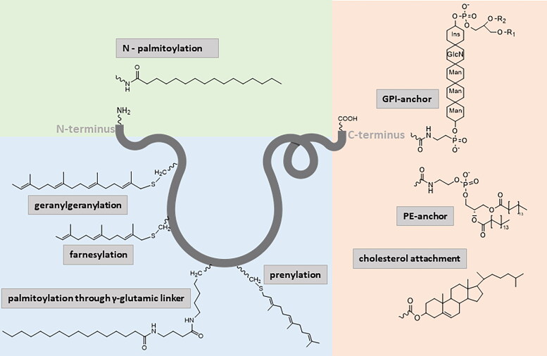
About thirty years ago, researchers came up with a new way, to stabilize a peptide/protein-based drug called lipidization (Pardridge, 1992). Lipidization entails of the attachment of a lipid group to a protein. There are various types of lipid modification that can be performed on a protein/peptide chain. These modifications include: prenylation on cysteine (e.g. S-geranylgeranyl lipids), fatty acylation at either the N-terminus or at the lateral amino acid group (e.g. N-palmitoyl, O-palmitoyl or N-myristoyl lipids); cholesterol, glycosylphosphatidylinositol (GPI), or phosphatidylethanolamine (PE) can also be used for the attachment of an additional moiety to the C-terminus of a peptide. In general, lipidization is a post-translational modification that yields a lipophilic prodrug derived from the original hydrophilic peptide. The process of lipidization falls in the groups of post-translational modifications called acylation. Lipidization is an approach that may allow modified peptides to cross the blood-brain barrier (BBB) (Jain et al., 2013; Erak et al., 2018; Menacho-Melgar et al., 2019; Botti et al., 2021; Hanna et al., 2022). This modification is also FDA-approved (Menacho-Melgar et al., 2019), so it is highly desirable to invent peptide drugs using this modification to achieve the best pharmacokinetic and pharmacodynamic properties. A good example of a naturally lipidized peptide is ghrelin. It is the only known peptide synthesized in the stomach that acts centrally and has a strong orexigenic effect (Kojima et al., 1999). The structure of ghrelin contains the amino acid serine acylated with n-octanoic acid, which is essential for its biological activity (Bednarek et al., 2000; Maletínská et al., 2012; Müller et al., 2015; Zemenova et al., 2017).
One type of lipidization is prenylation. Prenylation is a post-translational protein/peptide modification characterized by the irreversible covalent attachment of one or several isoprenoid units to conserved Cys residues at or close to the protein C-terminus. The binding of these units is mediated through a thioether bond. There are several types of prenylation, for example, geranyl (10-carbon, containing two isoprenoid units) farnesylation (15-carbon, containing three isoprenoid units) and geranylgeranylation (20-carbon, containing four isoprenoid units) (Zhang & Casey, 1996; Hannoush & Sun, 2010; Wang & Casey, 2016; Hanna et al., 2022). Although prenyl groups are most commonly attached through sulfur via a thioether bond, this attachment can also be mediated by oxygen or nitrogen. Such modification yield peptides with a lipophilic C-terminus, thanks to which they acquire an increased capacity to interact with cell membranes (Alhassan et al., 2014; Wang & Casey, 2016; Hanna et al., 2022). The addition of a prenyl chain to the peptide significantly alters the pharmacological properties of the original peptide. Prenylated compounds usually possess improved biological properties, so they are being studied as potential compounds useful in the treatment of varying diseases. Despite these benefits, there are still no FDA-approved prenylated peptides on the market. One of the investigated substances is Salirasib, which contains a farnesyl group modifying salicylic acid. However, this substance is still being investigated in preclinical studies (Alhassan et al., 2014).
Another type of protein lipidization is the attachment of a GPI anchor (glypiation). It is a widely used and well-known post-translational modification that usually occurs also at the C-terminus of a protein. This whole process stems from the fact that GPI is a signal peptide with the C-terminus cleaved from the original protein. Subsequently, the new C-terminus is linked to the amino group of the ethanolamine residue in the GPI precursors (Chatterjee & Mayor, 2001; Ikezawa, 2002; Mayor & Riezman, 2004; Hanna et al., 2022). This type of lipidization usually takes place naturally in cells and occurs in the endoplasmic reticulum (Ikezawa, 2002). GPI-anchored proteins play important roles in various biological conditions, although GPI-anchored proteins have diverse applications, their synthesis remains complicated, which slows their research greatly (Zhu & Guo, 2017).
Lipidization can be also mediated by a phosphatidylethanolamine (PE) anchor. This is a post-translational modification that is made through a covalently attached C-terminal Gly residue of a protein and an amino group of the PE. This attachment is done through an amide bond. This type of lipidization is quite rare and is not very well studied (Ichimura et al., 2000; Mejuch & Waldmann, 2016; Hanna et al., 2022).
Another lipophilic molecule that can be attached to a peptide for its stabilization is cholesterol. It can be coupled to the peptide through an ester or thioester linkage, and this bond can be achieved on the N-terminus of the peptide, the C-terminus and also in the middle of a peptide chain (Creanga et al., 2012; Erak et al., 2018; Hanna et al., 2022). Although there are still no FDA approved peptide drugs with the cholesterol attachment, some of them are in the preclinical trials. For instance, Ingallinella et al. synthetized a peptide, HIV inhibitor, derivatized with cholesterol. This compound possesses higher stability in blood plasma in vitro and prolonged half-life in vivo (tested in mice). The attachment of cholesterol also causes higher affinity to its receptors (Ingallinella et al., 2009).
The most widespread, the most advantageous and the easiest type of lipidization is fatty acid acylation. This adjustment is based on the covalent attachment of diverse fatty acids to the target peptide or protein. There are long, medium and short fatty acids that can be attached to peptides and each of them provides a different biochemical property. Usually, fatty acids such as caprylic (C8), myristic (C14), palmitic (C16), or stearic acid (C18) are used. However, myristic acid and palmitic acid are the most common modifying groups (Hannoush & Sun, 2010; Peng et al., 2016; Resh, 2016; Garst et al., 2021). The choice of fatty acid, the type of bond that attaches the fatty acid to the peptide, or the use of a linker that binds the peptide and the fatty acid are important factors that influence the physicochemical properties, binding affinity to different receptors, and bioactivity of the final lipidized peptide. Lipidization can be classified into three groups based on the type of lipid bond formation with the peptide chain and the fatty acid: amidation, esterification (S- or O-) and S bond (ether or disulfide) (Zhang & Bulaj, 2012). Amidation and a disulfide bond provide a strong covalent bond unlike S- and O-esterification, which are not so stable. The O-ester bond is formed through the hydroxyl group of the carboxylic acid present in the amino acid. (Hannoush & Sun, 2010; Zhang & Bulaj, 2012). The two most commonly occurring types of lipidization are, N-acylation (i.e. N-myristoylation, N-palmitoylation) and S-palmitoylation (Walsh et al., 2005).
All the aforementioned types of lipidization and their structural design are summarized in (Figure 1).
Figure 1.
Examples of peptide lipidization; GlcN: glukoseamine; GPI: glypiation, ins: inositol; man: mannose; PE: phosphatidylethanolamine. Note: the figure was drawn by the authors.
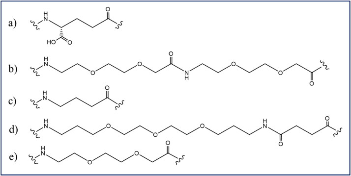
There are many ways to connect a chosen fatty acid to the peptide backbone. As mentioned above, the common bonds used for this are amide, ester, or disulfide bonds. However, there are also other types of bonds that can be used. Another way to attach a fatty acid to a peptide is via a linker/spacer. One widely used linker is a γ-glutamyl linker, namely gamma glutamic acid (Zhang & Bulaj, 2012; Pražienková et al., 2019). It has been shown (by a dose-response study of interactions with specific receptors) to enhance the potency of a lipidized peptide in comparison to other linkers or bonds. The γ-glutamic acid linker can be used to attach a fatty acid to the termini or in the middle of a peptide (Hutchinson et al., 2018). Most commonly the γ-glutamic acid linker binds to the amino acid lysine, to which it is attached on the secondary amino group (Ward et al., 2013; Hutchinson et al., 2018).
The next possibility of attaching a fatty acid to a peptide is through a short chain of polyethylene glycol (1,13-diamino-4,7,10-trioxadecan-succinamic acid) called the TTDS linker (Pražienková et al., 2019; DE Prins et al., 2020). This type of linker is usually attached to lysine at the peptide backbone. Furthermore, a series of linkers have been tested in search of potential new types of peptide lipidization. Some examples of them are: gamma-aminobutyric acid (GABA), 8-amino-3,6-dioxactanoic acid and OEG-OEG. Figure 2 presents the individual types of linkers that can be used for lipidization.
Figure 2.
Structure of linkers used for peptide lipidization; (a) γ-glutamyl linker, (b) OEG-OEG, (c) gamma-aminobutyric linker, (d) TTDS linker, (e) 8-amino-3,6-dioxactanoic acid. Note: the figure was drawn by the authors.
Improved pharmacological properties of lipidized drugs
Lipidization significantly improves the stability and many biological properties of peptides, such as bioavailability, enables new methods of administration, improves drug delivery, prolongs the half-life. A list of biological properties affected by the lipidization process is detailed below.
Prolonged half-life
One of the properties that needs to be improved in peptide-based drugs is their half-life in the blood plasma. Because of their small size and hydrophilic properties, unmodified peptides usually have a short plasma half-life. It is caused by fast renal clearance and fast enzymatic degradation that occurs in the systemic blood circulation (Werle & Bernkop-Schnürch, 2006). Such processes require more frequent administration of these nonmodified peptides and thus make the overall treatment more expensive (Morrow & Felcone, 2004). Lipidization gives inherently hydrophilic peptides an amphiphilic nature. Lipidized peptides than bind more strongly to serum albumin, which helps the modified peptide reach the target tissue and prolongs its half-life in the blood plasma (Wang et al., 2002). The albumin structure has seven binding sites, but under usual physiological circumstances, only two of them are occupied. This fact eliminates the threat of the lipidized drug competing for the binding site of albumin (Markussen et al., 1996). In general, the longer the fatty acid attached to the peptide, the stronger the binding to albumin, and the longer the half-life of a peptide. As mentioned above, lipidization can be performed at both ends of the peptide or anywhere on the peptide chain. It has been proven that the location of the fatty acid has no effect on binding to the albumin. At the same time peptides derivatized with two fatty acids were synthetized and tested, specifically di-palmitoylated peptides. The results clearly showed decreased affinity to particular receptors (Knudsen et al., 2000; Pražienková et al., 2017; Holubová et al., 2018; Menacho-Melgar et al., 2019).
One example of lipidized compound with a prolonged half-life (from minutes to hours) is liraglutide (Victoza – T2DM treatment/Saxenda – obesity treatment, Novo Nordisk) (Drucker et al., 2010). This lipidized drug is described in more detail below.
Decreased immunogenicity
Peptide-based drugs are likely to start a defensive immune reaction. Unmodified peptide drugs, including those with 100% structural homology to natural human peptides, are known to be capable of initiating a host immune response, also known as immunogenicity. Because of this, it is necessary to modify the respective peptide to make it unrecognizable by the immune system (Tornesello et al., 2020). Several studies show that lipidization is a powerful tool that can give rise to peptides with lower immunogenicity. The length of the attached fatty acid plays an important role in the process. The longer the fatty acid, the more significant a reduction in immunogenicity occurs (Kowalczyk et al., 2017).
Improved drug delivery
Peptide-based drugs are hydrophilic in unmodified form. This fact significantly influences their ability to cross the cell membrane or any membrane in general
(Pavan & Dalpiaz, 2011). In recent years, neuropeptides, such as PrRP, the CART peptide and, very importantly GLP-1 agonists, have been persistently studied due to their potential for use in the treatment of obesity, T2DM or neurodegeneration (Maletínská et al., 2008; Popelová et al., 2018; Holubová et al., 2019). Worth mentioning are dual GLP-1/glucose-dependent insulinotropic polypeptide (GIP) agonists and triple GLP-1/GIP/glucagon. GLP-1 agonists are being investigated for the potential treatment of overweight or obese T1DM patients. Also tested as possible anti-obesity therapeutics are GLP-1/GIP coagonist peptides, also called dual agonists, and s triple GLP-1/GIP/glucagon agonists (Janzen et al., 2016; Tan, 2023; Tran et al., 2023). However, in their natural form, they cannot cross the BBB and induce the desired effect on the respective receptors in the brain. The BBB, together with the blood-cerebrospinal fluid barrier (CSF, BCSFB) and the arachnoid barrier, forms the main barrier between the CNS and blood (Abbott et al., 2010). Lipidization helps overcome this challenge and induces a central effect after peripheral administration (Zemenová et al., 2017). In the case of PrRP, novel lipidized analogues, palmitoylated PrRP, were synthesized, palmitoylated PrRP in position 11 using a γ-glutamic linker (palm11-PrRP). Testing this analogue in mouse and rat models found it to attenuate body weight. In conclusion, lipidization aids drug delivery, specifically by helping peptides pass through physiological membranes.
New potential routes of administration
The change in peptide character from hydrophilic to hydrophobic allows lipidized peptides to penetrate through the cell membrane. Because of that, the use of new routes of administration is allowed, for example oral, topical or pulmonary (Menacho-Melgar et al., 2019). Although most lipidized therapeutics focus on subcutaneous administration (s.c.), acylation specifically offers several potential benefits for oral administration for peptide-based therapeutics (Trier et al., 2015). Such a drug administration option would naturally be much more convenient for patients than s.c. administration. It has also been demonstrated that acylation has the ability of peptide drugs to permeate intestinal tissue. Earlier in this review, we mention liraglutide, a lipidized GLP-1 receptor agonist. Another example of an acylated GLP-1 receptor agonist is semaglutide. Semaglutide was designed as a long-acting GLP-1 agonist with potential administration once a week s.c. It has 94% structural homology with natural GLP-1 with three main improvements: substitution of alanine at position 8 with 2-aminoisobutyric acid, substitution of lysine with arginine at position 34 and attachment of a palmitic acid using a γ-glutamic linker at position 26. Semaglutide has an even longer half-life compared to liraglutide (183 vs 11 – 15 h) after s.c. injection (Christou et al., 2019; Kalra & Sahay, 2020). Initially, semaglutide was approved for the treatment of T2DM with s.c. administration. Subsequently, a new formulation allowing oral administration was developed, followed by a novel tablet formulation with an absorption enhancer invented to cross the gastric epithelium. Semaglutide is now available as an oral formulation and has been approved since 2019 by the FDA to treat T2DM (Eliaschewitz & Canani, 2021).
Improved bioavailability
When using peptide-based drugs, it is common to encounter another pitfall, which is low bioavailability. Bioavailability is one of the main pharmacological properties. It is closely related to the method of administration. When a drug is administered intravenously (i.v.), its bioavailability is by definition 100%, whereas via another route, its bioavailability decreases (Toutain & Bousquet, 2004). Lipidization has been proven to help achieve improved bioavailability and even increased CNS bioavailability after intraperitoneal (i.p.) administration (Zhang et al., 2009; Green et al., 2010). Wang et al. (2003) have mentioned another example of improved bioavailability where a lipidized peptide achieves four times greater bioavailability after subcutaneous administration than the unmodified peptide (Wang et al., 2003).
Lipidized drugs on the market
As already stated above, the number of lipidized drugs has been steadily growing over the last few years. We present an overview of the lipidized peptide-based therapeutics currently available on the market. The following table summarizes these compounds, their indication, the routes of their administration and the parental molecules of which they are analogues (Table 1).
Table 1.
Currently approved lipidized peptide-based therapeutics.
| Drug | Type of modification | Parental molecule | Indication | Routes of administration | Reference |
|---|---|---|---|---|---|
| Liraglutide | Acylation | GLP-1 | T2DM | s.c. injection | Iepsen et al. (2015) |
| Obesity | s.c. injection | NovoNordisk (2020) | |||
| Semaglutide | Acylation | GLP-1 | T2DM | s.c. injection Oral | Scheen (2020) |
| Obesity | s.c. injection | FDA (2021) | |||
| Detemir | Acylation | Insulin | Diabetes | s.c. injection | Home and Kurtzhals (2006) |
| Degludec | Acylation | Insulin | Diabetes | s.c. injection | Jarosinski et al. (2021) |
| Xultophy | Acylation | Insulin + GLP-1 | T2DM | s.c. injection | Steyn (2022) |
| Somapacitan | Acylation | Human growth hormone | Growth hormone deficiencies | s.c. injection | FDA (2020) |
| Tesamorelin | Acylation | Human growth hormone releasing hormone | HIV-associated lipodystrophy | s.c. injection | Stanley et al. (2019) |
| Tirzepatide | Acylation | GLP-1/GIP | T2DM | s.c. injection | Syed (2022) |
| Mifamurtide | PE-anchor attachment + acylation | Muramyl dipeptide | Osteosarcoma | i.v. injection | Frampton (2010) |
| Polymyxin B | Acylation | Bacterial infection | i.v. injection/inhalation/topical | Moffatt et al. (2019) | |
| Daptomycin | Acylation | Bacterial infection | i.v. injection | Heidary et al. (2017) |
Liraglutide
Liraglutide (Saxenda, Novo Nordisk) is a glucagon like peptide-1 (GLP-1) receptor agonist. It was discovered during studies of GLP-1 derivatives with the intention to create an analogue with an increased plasma half-life compared to human GLP-1, which is approximately two minutes (Drucker et al., 2010; Mehta et al., 2017). This GLP-1 analogue has a 97% structural homology with the natural human GLP-1 hormone. The structure of liraglutide differs in amino acid position 34 where liraglutide has an arginine instead of a lysine. Unlike natural GLP-1, liraglutide is lipidized with palmitic acid attached through a glutamic spacer at position 26. Such modifications prolong the plasma half-life of liraglutide from two minutes to thirteen hours (assuming subcutaneous administration). Liraglutide was originally developed for the treatment of T2DM, but it has also exhibited great dose dependent anti-obesity properties. Liraglutide was approved by the FDA in 2010 as an injectable GLP-1 receptor agonist for the treatment of T2DM. Later in 2013 it was approved by the FDA as an anti-obesity treatment (Astrup et al., 2009; Drucker et al., 2010; Knudsen, 2010; Ng & Wilding, 2014).
Semaglutide
Semaglutide (Ozempic/Rybelsus or Wegovy, Novo Nordisk), is similarly to liraglutide, a GLP-1 receptor agonist with a prolonged plasma half-life (Kalra & Sahay, 2020). A big advantage of semaglutide is its once weekly dosing; liraglutide, by contrast, is administered once a day (Linderoth et al., 2021). Semaglutide is being used for the treatment of both T2DM (Ozempic or Rybelsus) and obesity (Wegovy). It is available in tablets and injections for the treatment of T2TDM. For the treatment of obesity, only s.c. administration has been approved by the FDA (Bergmann et al., 2023). The structure of semaglutide is described in detail above.
Insulin detemir
Another example of a commercially available acylated drug is insulin detemir (Levemir, Novo Nordisk). This drug is an analogue of natural insulin that has undergone modifications such as: the removal of the amino acid threonine in chain B at position 30 and acylation with the myristoyl fatty acid at position 29 on lysine (Havelund et al., 2004). Detemir has a prolonged half-life in comparison with neutral Protamine Hagedorn (NPH) insulin (Novolin N/Humulin N, Novo Nordisk) also known as isophane insulin. Acylated insulin shows a stable pharmacokinetic and pharmacodynamic profile, meaning that it causes lesser and less frequent fluctuations of the mean plasma glucose concentration and fewer overall hypoglycemic events than NPH insulin (Athanasiadou et al., 2022). Insulin detemir is prescribed to treat T1DM and T2DM, and it has been approved by the FDA since June 2005. The only route of administration is subcutaneous (Home & Kurtzhals, 2006; Hermansen & Davies, 2007).
Insulin degludec
Commercially available under the name of Tresiba (Novo Nordisk) is another insulin analogue called insulin degludec. Degludec is an ultra-long-acting basal insulin which is, produced by modifying the base structure of insulin. In this case the modification consists in adding the hexadecanedioic fatty acid to lysine at position B29 and the removal of threonine at position 30 (Steensgaard et al., 2013; Keating, 2013). Degludec was approved by the FDA in September 2005 and is being used for the treatment of both T1DM and T2DM in adults, adolescents and children. The administration of this substance is subcutaneous and thanks to its long half-life (24.5 hours) it can be administered once daily at any time of day (Traynor, 2015; Marso et al., 2017). It has been proven the effect of insulin degludec is equally distributed over the course of the day, with approximately 25% every six hours (Kalra, 2013).
Xultophy
In November 2016 the FDA approved the first fixed-ratio combination of basal insulin, insulin degludec and liraglutide, so called IDegLira (Xultophy, Novo Nordisk). The usual recommended starting dosage is 16 units/mL of IDeg and 0.6 mg of liraglutide once daily using the subcutaneous administration (Rodbard et al., 2016). It can be taken regardless of meal times and the time of day, alone or as an add-on therapy together with an oral antidiabetic drug. Some studies also report the beneficial influence of IDegLira on cardiovascular and renal function. Simultaneously, IDegLira seems to be suitable for diabetic patients with a chronic kidney disease (Bando et al., 2022). This drug is more effective than other basal insulin regimens in reaching individualized glycated hemoglobin target, with a lower dose of insulin per day. It also has a favorable effect on body weight and reduces overnight hypoglycemia compared to basal insulin. IDegLira has less gastrointestinal adverse effects with respect to liraglutide itself (Scheen & Mathieu, 2018).
Somapacitan
The active compound somapacitan (Sogroya, Novo Nordisk) is the first growth hormone deficiency (GHD) therapeutic. GHD in children is defined primarily by diminished height gain velocity or height below the normal range or the range expected considering the parent’s height. Somapacitan is a once-weekly subcutaneously administered treatment for both, children and adults. Unlike the natural growth hormone, somapacitan is modified with a single amino acid substitution and the addition of a short fatty acid. These changes enable somapacitan to reversibility and non-covalently bind to the albumin, which significantly prolongs its half-life and reduces clearance (Battelino et al., 2017; Sävendahl et al., 2020; Johannsson et al., 2020). The first growth hormone analogue somapacitan was approved by the FDA in August 2020 (FDA, 2020).
Tesamorelin
Another analogue of the growth hormone-releasing hormone is tesamorelin (Egrifta, Theratechnologies/EMD Serono). However, this compound is being used for the treatment of HIV-infected patients also undergoing antiretroviral therapy. These patients often tend to develop changes in body composition. Such changes mainly include an excess of abdominal visceral fat and a reduction of abdominal subcutaneous fat, which is then followed by reduced quality of life, possibly contributes to body dysmorphia anxiety. Tesamorelin induces increased growth hormone secretion, reduces excessive abdominal visceral fat and improves metabolic abnormalities (Falutz et al., 2010; Grunfeld et al., 2011). The structure of tesamorelin is made up of 44 amino acids and contains a trans-3-hexenoyl fatty acid group, which is responsible for the compound’s prolonged half-life caused by strong binding to serum albumin. This fatty acid is anchored on tyrosine at position 1 at the N-terminus of the compound. Tesamorelin was approved by the FDA in November 2010 (Mateo et al., 2011).
Tirzepatide
Tirzepatide (Mounjaro, Eli Lilly) is a new compound on the market, approved by the FDA May 2022 for the treatment of T2DM. It is also in phase III development for heart failure, obesity and cardiovascular disorders in T2DM and in phase II of development for nonalcoholic steatohepatitis. Tirzepatide is a dual GLP-1/GIP agonist acting on both the respective endogenous receptors (Syed, 2022; Karagiannis et al., 2022). This lipopeptide was tested in several studies, and at a dose 5 mg it proved superior to semaglutide at a dose of 1 mg with respect to how it reduced the level of glycated hemoglobin in patients with T2DM, receiving metformin. Tirzepatide was also superior to semaglutide at reducing of body weight (Frías et al., 2021).
Mifamurtide
Mifamurtide (Mepact, Takeda) is also known as a liposomal muramyl tripeptide phosphatidylethanolamine (L-MPT-PE). It is being used for the treatment of osteosarcoma. Osteosarcoma is an ultra-orphan disease that affects about 1,000 new people worldwide each year. The arrival of mifamurtide resulted in a decreased number of deaths by one-third when used in combination with chemotherapy. Since March 2009, mifamurtide has been approved by the FDA for the treatment of osteosarcoma in combination with chemotherapy (Meyers, 2009; Anderson et al., 2010). The structure of mifamuretide has two palmitoyl phosphatidylethanolamine groups, which cause increased lipophilicity of the compound and prolongation of its half-life in the organism (Meyers, 2009).
Polymyxin B
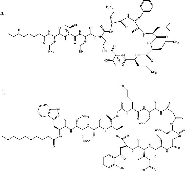
Aerosporin (GlaxoSmithKline), Metamyxin (Pola Pharma), Polomyxin B (Fuji Yakuhin), Poly-RX (X-Gen), Polyfax (Intra) or Polyxx (Celon) are different commercial names for polymyxin B. The polymyxin B lipopeptide is an antibiotic isolated from Bacillus polymyxa. Its structure contains various fatty acids: Polymyxin B1 contains 6-methyloctanoic acid, polymyxin B2 6-methylheptanoic acid, polymyxin B3 octanoic acid, and polymyxin B4 heptanoic acid. All types of polymyxins act as bactericidal agents with a detergent-like mechanism of action. They interact with lipopolysaccharides of the outer membrane of Gram-negative bacteria causing this membrane to be penetrated and their subsequent death. Polymyxins are used for the treatment of infections of the urinary tract, the meninges or the blood stream. Polymyxin B was officially approved for the medical use in the United States in 1964; however, it was approved by the FDA in 2015 for topical administration only (Zavascki et al., 2007; DrugBank, 2019).
Daptomycin
Daptomycin (Cubicin, Pfizer) is a lipopeptide antibiotic produced by the bacterium Streptomyces roseosporus. It is clinically used for the treatment of severe infections by Gram-positive bacteria. The mechanism of its function resides in the permeabilization and depolarization of the bacterial cell membrane, which leads to cell death. The structure of daptomycin contains one N-terminally attached caprylic fatty acid. Daptomycin was approved for use in September 2003 (FDA, 2003; Taylor & Palmer, 2016).
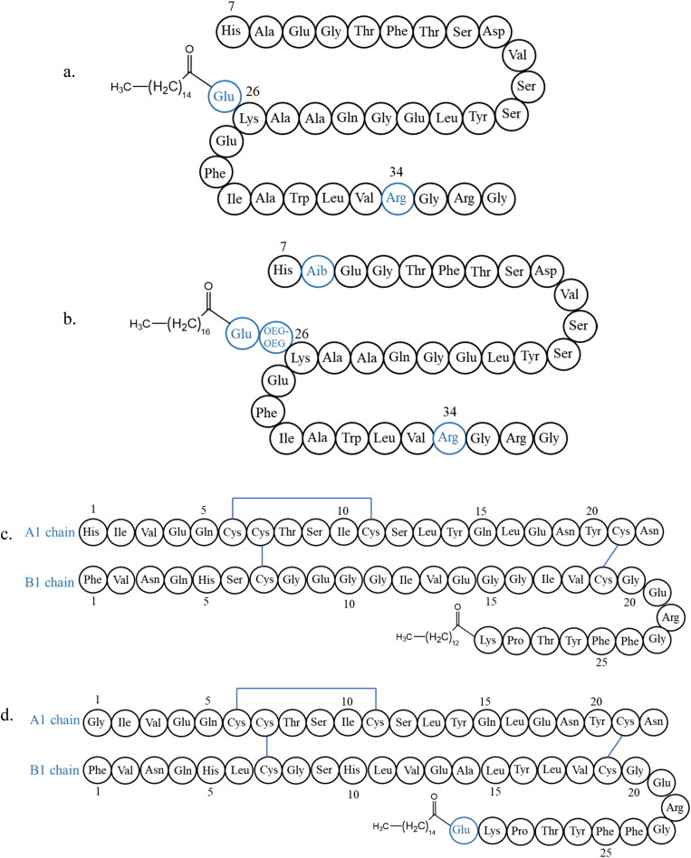
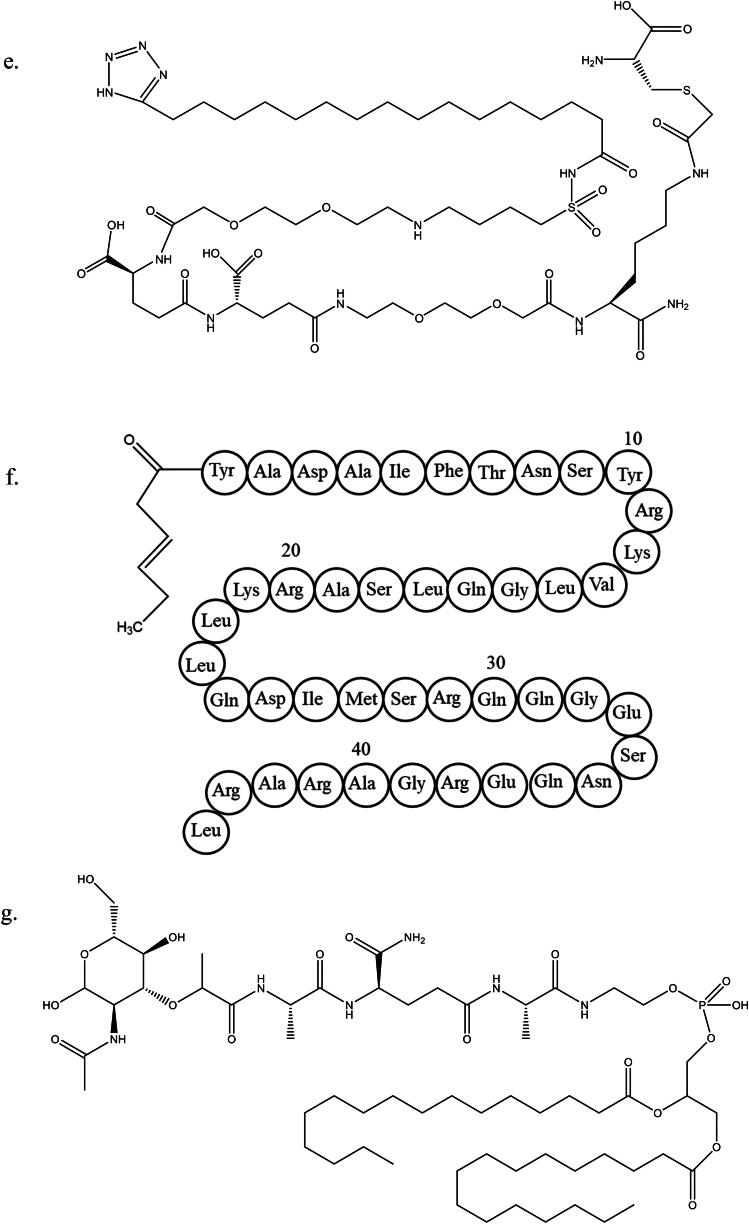
Chemical structures of all the lipidized peptides mentioned above are presented in Figure 3.
Figure 3.
Structures of lipidized peptides on the market; (a) liraglutide, (b) semaglutide, (c) insulin detemir, (d) insulin degludeg, (e) somapacitan, (f) tesamorelin, (g) mifamurtide, (h) polymyxin B1, (i) daptomycin. Note: the figure was drawn by the authors.
Conclusion
Peptide therapeutics have become a unique category of compounds with great potential for the treatment of various diseases. They include a low immune reaction, bind to natural receptors and, last but not least, are relatively easy to synthesize. All in all, peptides are in many ways more promising than small molecules. Despite all the benefits, peptides in their natural form generally suffer from poor stability in vivo and impermeability through cell membranes. Ongoing studies have explored various ways to overcome this pitfall, including peptide modifications such as PEGylation, glycosylation, aminoacidic substitution or insertion, encapsulation by nanoparticles, cyclization, N-methylation and lipidization. Lipidization appears to be of special significance, and several lipidized drugs are currently available on the pharmaceutical market. Lipidization changes the properties of peptides from hydrophilic to lipophilic, helping them better elicit pharmacological effects. So far, fatty acid acylation seems to be the most frequently taken approach to lipidization.
The development of peptide therapeutics has seen a great progress in recent years. The pathways that lead to peptide stabilization are increasingly diverse and due to their significant benefits, peptide drugs serve as potential therapeutics for the treatment of many diseases. Numerous commercially available drugs are made by the process of peptide lipidization and lipopeptides not only have a salient place on the pharmaceutical market, but have a great potential, for instance, in the treatment of hitherto incurable diseases.
Funding Statement
This work was supported by the Czech Science Foundation, projects 21-03691S, 22-11155S, and by the Czech Academy of Sciences, projects RVO: 61388963 and RVO: 67985823.
Authors’ contributions
A.M. was involved in the conception and drafting of the manuscript including figures, D.S. participated in drafting the manuscript, he made final revisions and approval of the manuscript version submitted, J.K. revised the manuscript, and L.M. revised the manuscript. All authors agree to be accountable for all aspects of the work.
Disclosure statement
The authors report there are no competing interests to declare.
References
- Abbott NJ, Patabendige AAK, Dolman DEM, et al. (2010). Structure and function of the blood–brain barrier. Neurobiol Dis 37:1–16. doi: 10.1016/j.nbd.2009.07.030. [DOI] [PubMed] [Google Scholar]
- Ahmadpour S, Hosseinimehr SJ. (2018). PASylation as a powerful technology for improving the pharmacokinetic properties of biopharmaceuticals. Curr Drug Deliv 15:331–41. doi: 10.2174/1567201814666171120122352. [DOI] [PubMed] [Google Scholar]
- Ahrens VM, Bellmann-Sickert K, Beck-Sickinger AG. (2012). Peptides and peptide conjugates: therapeutics on the upward path. Future Med Chem 4:1567–86. doi: 10.4155/fmc.12.76. [DOI] [PubMed] [Google Scholar]
- Alhassan AM, Abdullahi MI, Uba A, Umar A. (2014). Prenylation of aromatic secondary metabolites: a new frontier for development of novel drugs. Trop J Pharm Res 13:307–14. doi: 10.4314/tjpr.v13i2.22. [DOI] [Google Scholar]
- Anderson PM, Tomaras M, Mcconnell K. (2010). Mifamurtide in osteosarcoma–a practical review. Drugs Today (Barc) 46:327–37. doi: 10.1358/dot.2010.46.5.1500076. [DOI] [PubMed] [Google Scholar]
- Astrup A, Rössner S, VAN Gaal L, et al. (2009). Effects of liraglutide in the treatment of obesity: a randomised, double-blind, placebo-controlled study. Lancet 374:1606–16. doi: 10.1016/S0140-6736(09)61375-1. [DOI] [PubMed] [Google Scholar]
- Athanasiadou KI, Paschou SA, Stamatopoulos T, et al. (2022). Safety and efficacy of insulin detemir versus NPH in the treatment of diabetes during pregnancy: systematic review and meta-analysis of randomized controlled trials. Diabetes Res Clin Pract 190:110020. doi: 10.1016/j.diabres.2022.110020. [DOI] [PubMed] [Google Scholar]
- Bando H, Iwatsuki N, Ogawa T, Sakamoto K. (2022). Investigation for daily profile of blood glucose by the administration of canagliflozin and xultophy (Ideglira). Int J Endocrinol Diabetes 5:129. [Google Scholar]
- Battelino T, Rasmussen MH, DE Schepper J, GROUP, T. N.-S, et al. (2017). Somapacitan, a once-weekly reversible albumin-binding GH derivative, in children with GH deficiency: a randomized dose-escalation trial. Clin Endocrinol (Oxf) 87:350–8. & doi: 10.1111/cen.13409. [DOI] [PubMed] [Google Scholar]
- Bednarek MA, Feighner SD, Pong SS, et al. (2000). Structure-function studies on the new growth hormone-releasing peptide, ghrelin: minimal sequence of ghrelin necessary for activation of growth hormone secretagogue receptor 1a. J Med Chem 43:4370–6. doi: 10.1021/jm0001727. [DOI] [PubMed] [Google Scholar]
- Bennett CL, Spiegel DM, Macdougall IC, et al. (2012). A review of safety, efficacy, and utilization of erythropoietin, darbepoetin, and peginesatide for patients with cancer or chronic kidney disease: a report from the Southern Network on Adverse Reactions (SONAR). Seminars in thrombosis and hemostasis, 2012. Semin Thromb Hemost 38:783–96. doi: 10.1055/s-0032-1328884. [DOI] [PMC free article] [PubMed] [Google Scholar]
- Bergmann NC, Davies MJ, Lingvay I, Knop FK. (2023). Semaglutide for the treatment of overweight and obesity: a review. Diabetes Obes Metab 25:18–35. doi: 10.1111/dom.14863. [DOI] [PMC free article] [PubMed] [Google Scholar]
- Binder U, Skerra A. (2017). PASylation®: a versatile technology to extend drug delivery. Curr Opin Colloid Interf Sci 31:10–7. doi: 10.1016/j.cocis.2017.06.004. [DOI] [Google Scholar]
- Biron E, Chatterjee J, Ovadia O, et al. (2008). Improving oral bioavailability of peptides by multiple N-methylation: somatostatin analogues. Angew Chem Int Ed Engl 47:2595–9. doi: 10.1002/anie.200705797. [DOI] [PubMed] [Google Scholar]
- Bolze F, Morath V, Bast A, et al. (2016). Long-acting PASylated leptin ameliorates obesity by promoting satiety and preventing hypometabolism in leptin-deficient Lepob/ob mice. Endocrinology 157:233–44. doi: 10.1210/en.2015-1519. [DOI] [PubMed] [Google Scholar]
- Botti G, Dalpiaz A, Pavan B. (2021). Targeting systems to the brain obtained by merging prodrugs, nanoparticles, and nasal administration. Pharmaceutics 13:1144. doi: 10.3390/pharmaceutics13081144. [DOI] [PMC free article] [PubMed] [Google Scholar]
- Carmona G, Rodriguez A, Juarez D, et al. (2013). Improved protease stability of the antimicrobial peptide Pin2 substituted with D-amino acids. Protein J 32:456–66. doi: 10.1007/s10930-013-9505-2. [DOI] [PubMed] [Google Scholar]
- Cohen DJ, Loertscher R, Rubin MF, et al. (1984). Cyclosporine: a new immunosuppressive agent for organ transplantation. Ann Intern Med 101:667–82. doi: 10.7326/0003-4819-101-5-667. [DOI] [PubMed] [Google Scholar]
- Creanga A, Glenn TD, Mann RK, et al. (2012). Scube/You activity mediates release of dually lipid-modified Hedgehog signal in soluble form. Genes Dev 26:1312–25. doi: 10.1101/gad.191866.112. [DOI] [PMC free article] [PubMed] [Google Scholar]
- DE Prins A, VAN Eeckhaut A, Smolders I, et al. (2020). Neuromedin U: an overview on peptidomimetic ligands and structural analogs. Curr Med Chem 27:6744–68. doi: 10.2174/0929867326666190916143028. [DOI] [PubMed] [Google Scholar]
- Drucker DJ, Dritselis A, Kirkpatrick P. (2010). Liraglutide. Nat Rev Drug Discov 9:267–8. doi: 10.1038/nrd3148. [DOI] [PubMed] [Google Scholar]
- DRUGBANK. (2019). Polymyxin B [Online]. DrugBank. Available at: https://go.drugbank.com/drugs/DB00781 [accessed].
- DRUGS.COM. (2022). DDAVP Prescribing Information [Online]. Drugs.com. Available at: https://www.drugs.com/pro/ddavp.html#s-34090-1 [accessed 2008].
- Eliaschewitz FG, Canani LH. (2021). Advances in GLP-1 treatment: focus on oral semaglutide. Diabetol Metab Syndr 13:99. doi: 10.1186/s13098-021-00713-9. [DOI] [PMC free article] [PubMed] [Google Scholar]
- Erak M, Bellmann-Sickert K, Els-Heindl S, Beck-Sickinger AG. (2018). Peptide chemistry toolbox–transforming natural peptides into peptide therapeutics. Bioorg Med Chem 26:2759–65. doi: 10.1016/j.bmc.2018.01.012. [DOI] [PubMed] [Google Scholar]
- Falutz J, Mamputu J-C, Potvin D, et al. (2010). Effects of tesamorelin (TH9507), a growth hormone-releasing factor analog, in human immunodeficiency virus-infected patients with excess abdominal fat: a pooled analysis of two multicenter, double-blind placebo-controlled phase 3 trials with safety extension data. J Clin Endocrinol Metab 95:4291–304. doi: 10.1210/jc.2010-0490. [DOI] [PubMed] [Google Scholar]
- FDA. (2003). Cubicin (Daptomycin) Injection [Online]. FDA. Available at: https://www.accessdata.fda.gov/drugsatfda_docs/nda/2003/21-572_Cubicin.cfm [accessed Sept 2003].
- FDA. (2020). FDA approves weekly therapy for adults growth hormone deficiency [Online]. FDA. Available at: https://www.fda.gov/drugs/news-events-human-drugs/fda-approves-weekly-therapy-adult-growth-hormone-deficiency [accessed 09/01 2020].
- FDA. (2021). FDA Approves New Drug Treatment for Chronic Weight Management, First SInce 2014 [Online]. FDA. Available at: https://www.fda.gov/news-events/press-announcements/fda-approves-new-drug-treatment-chronic-weight-management-first-2014 [Accessed 06/04/2021 2021].
- Frampton JE. (2010). Mifamurtide: a review of its use in the treatment of osteosarcoma. Paediatr Drugs 12:141–53. doi: 10.2165/11204910-000000000-00000. [DOI] [PubMed] [Google Scholar]
- Frías JP, Davies MJ, Rosenstock J, et al. (2021). Tirzepatide versus semaglutide once weekly in patients with type 2 diabetes. N Engl J Med 385:503–15. doi: 10.1056/NEJMoa2107519. [DOI] [PubMed] [Google Scholar]
- Garrahy A, Thompson CJ. (2020). Management of central diabetes insipidus. Best Pract Res Clin Endocrinol Metab 34:101385. doi: 10.1016/j.beem.2020.101385. [DOI] [PubMed] [Google Scholar]
- Garst EH, DAS T, Hang HC. (2021). Chemical approaches for investigating site-specific protein S-fatty acylation. Curr Opin Chem Biol 65:109–17. doi: 10.1016/j.cbpa.2021.06.004. [DOI] [PMC free article] [PubMed] [Google Scholar]
- Gazdik M, O’Neill MT, Lopaticki S, et al. (2015). The effect of N-methylation on transition state mimetic inhibitors of the Plasmodium protease, plasmepsin V. Med Chem Commun 6:437–43. doi: 10.1039/C4MD00409D. [DOI] [Google Scholar]
- Gilon C, Halle D, Chorev M, et al. (1991). Backbone cyclization: a new method for conferring conformational constraint on peptides. Biopolymers: Original Res Biomol 31:745–50. doi: 10.1002/bip.360310619. [DOI] [PubMed] [Google Scholar]
- Goodwin D, Simerska P, Toth I. (2012). Peptides as therapeutics with enhanced bioactivity. Curr Med Chem 19:4451–61. doi: 10.2174/092986712803251548. [DOI] [PubMed] [Google Scholar]
- Green BR, White KL, Mcdougle DR, et al. (2010). Introduction of lipidization–cationization motifs affords systemically bioavailable neuropeptide Y and neurotensin analogs with anticonvulsant activities. J Pept Sci 16:486–95. doi: 10.1002/psc.1266. [DOI] [PubMed] [Google Scholar]
- Grunfeld C, Dritselis A, Kirkpatrick P. (2011). Tesamorelin. Nat Rev Drug Discov 10:95–6. doi: 10.1038/nrd3362. [DOI] [PubMed] [Google Scholar]
- Hanna CC, Kriegesmann J, Dowman LJ, et al. (2022). Chemical synthesis and semisynthesis of lipidated proteins. Angew Chem Int Ed Engl 61:e202111266. doi: 10.1002/anie.202111266. [DOI] [PMC free article] [PubMed] [Google Scholar]
- Hannoush RN, Sun J. (2010). The chemical toolbox for monitoring protein fatty acylation and prenylation. Nat Chem Biol 6:498–506. doi: 10.1038/nchembio.388. [DOI] [PubMed] [Google Scholar]
- Harris JM, Chess RB. (2003). Effect of pegylation on pharmaceuticals. Nat Rev Drug Discov 2:214–21. doi: 10.1038/nrd1033. [DOI] [PubMed] [Google Scholar]
- Harris JM, Martin NE, Modi M. (2001). Pegylation. Clin Pharmacokinet 40:539–51. doi: 10.2165/00003088-200140070-00005. [DOI] [PubMed] [Google Scholar]
- Havelund S, Plum A, Ribel U, et al. (2004). The mechanism of protraction of insulin detemir, a long-acting, acylated analog of human insulin. Pharm Res 21:1498–504. doi: 10.1023/b:pham.0000036926.54824.37. [DOI] [PubMed] [Google Scholar]
- Heidary M, Khosravi AD, Khoshnood S, et al. (2017). Daptomycin. J Antimicrob Chemother 73:1–11. doi: 10.1093/jac/dkx349. [DOI] [PubMed] [Google Scholar]
- Henikoff S, Henikoff JG. (1992). Amino acid substitution matrices from protein blocks. Proc Natl Acad Sci USA 89:10915–9. doi: 10.1073/pnas.89.22.10915. [DOI] [PMC free article] [PubMed] [Google Scholar]
- Hermansen K, Davies M. (2007). Does insulin detemir have a role in reducing risk of insulin-associated weight gain? Diabetes Obes Metab 9:209–17. doi: 10.1111/j.1463-1326.2006.00665.x. [DOI] [PubMed] [Google Scholar]
- Hermanson T, Bennett CL, Macdougall IC. (2016). Peginesatide for the treatment of anemia due to chronic kidney disease–an unfulfilled promise. Expert Opin Drug Saf 15:1421–6. doi: 10.1080/14740338.2016.1218467. [DOI] [PMC free article] [PubMed] [Google Scholar]
- Holubová M, Blechová M, Kákonová A, et al. (2018). In vitro and in vivo characterization of novel stable peptidic ghrelin analogs: beneficial effects in the settings of lipopolysaccharide-induced anorexia in mice. J Pharmacol Exp Ther 366:422–32. doi: 10.1124/jpet.118.249086. [DOI] [PubMed] [Google Scholar]
- Holubová M, Hrubá L, Popelová A, et al. (2019). Liraglutide and a lipidized analog of prolactin-releasing peptide show neuroprotective effects in a mouse model of β-amyloid pathology. Neuropharmacology 144:377–87. doi: 10.1016/j.neuropharm.2018.11.002. [DOI] [PubMed] [Google Scholar]
- Home P, Kurtzhals P. (2006). Insulin detemir: from concept to clinical experience. Expert Opin Pharmacother 7:325–43. doi: 10.1517/14656566.7.3.325. [DOI] [PubMed] [Google Scholar]
- Hutchinson JA, Burholt S, Hamley IW, et al. (2018). The effect of lipidation on the self-assembly of the gut-derived peptide hormone PYY3–36. Bioconjug Chem 29:2296–308. doi: 10.1021/acs.bioconjchem.8b00286. [DOI] [PubMed] [Google Scholar]
- Chatterjee J, Gilon C, Hoffman A, Kessler H. (2008). N-methylation of peptides: a new perspective in medicinal chemistry. Acc Chem Res 41:1331–42. doi: 10.1021/ar8000603. [DOI] [PubMed] [Google Scholar]
- Chatterjee J, Rechenmacher F, Kessler H. (2013). N-methylation of peptides and proteins: an important element for modulating biological functions. Angew Chem Int Ed Engl 52:254–69. doi: 10.1002/anie.201205674. [DOI] [PubMed] [Google Scholar]
- Chatterjee S, Mayor S. (2001). The GPI-anchor and protein sorting. Cell Mol Life Sci 58:1969–87. doi: 10.1007/PL00000831. [DOI] [PMC free article] [PubMed] [Google Scholar]
- Checco JW, Zhang G, Yuan W-D, et al. (2018). Molecular and physiological characterization of a receptor for D-amino acid-containing neuropeptides. ACS Chem Biol 13:1343–52. doi: 10.1021/acschembio.8b00167. [DOI] [PMC free article] [PubMed] [Google Scholar]
- Chen S, Gfeller D, Buth SA, et al. (2013). Improving binding affinity and stability of peptide ligands by substituting glycines with d-amino acids. Chembiochem 14:1316–22. doi: 10.1002/cbic.201300228. [DOI] [PubMed] [Google Scholar]
- Christou GA, Katsiki N, Blundell J, et al. (2019). Semaglutide as a promising antiobesity drug. Obes Rev 20:805–15. doi: 10.1111/obr.12839. [DOI] [PubMed] [Google Scholar]
- Iepsen EW, Torekov SS, Holst JJ. (2015). Liraglutide for type 2 diabetes and obesity: a 2015 update. Expert Rev Cardiovasc Ther 13:753–67. doi: 10.1586/14779072.2015.1054810. [DOI] [PubMed] [Google Scholar]
- Ichimura Y, Kirisako T, Takao T, et al. (2000). A ubiquitin-like system mediates protein lipidation. Nature 408:488–92. doi: 10.1038/35044114. [DOI] [PubMed] [Google Scholar]
- Ikezawa H. (2002). Glycosylphosphatidylinositol (GPI)-anchored proteins. Biol Pharm Bull 25:409–17. doi: 10.1248/bpb.25.409. [DOI] [PubMed] [Google Scholar]
- Ingallinella P, Bianchi E, Ladwa NA, et al. (2009). Addition of a cholesterol group to an HIV-1 peptide fusion inhibitor dramatically increases its antiviral potency. Proc Natl Acad Sci USA 106:5801–6. doi: 10.1073/pnas.0901007106. [DOI] [PMC free article] [PubMed] [Google Scholar]
- Jain A, Jain A, Gulbake A, et al. (2013). Peptide and protein delivery using new drug delivery systems. Critical Rev™ Therapeutic Drug Carrier Sys 30:293–329. [DOI] [PubMed] [Google Scholar]
- Janzen KM, Steuber TD, Nisly SA. (2016). GLP-1 agonists in type 1 diabetes mellitus. Ann Pharmacother 50:656–65. doi: 10.1177/1060028016651279. [DOI] [PubMed] [Google Scholar]
- Jarosinski MA, Dhayalan B, Chen YS, et al. (2021). Structural principles of insulin formulation and analog design: a century of innovation. Mol Metab 52:101325. doi: 10.1016/j.molmet.2021.101325. [DOI] [PMC free article] [PubMed] [Google Scholar]
- Jayaprakash NG, Surolia A. (2017). Role of glycosylation in nucleating protein folding and stability. Biochem J 474:2333–47. doi: 10.1042/BCJ20170111. [DOI] [PubMed] [Google Scholar]
- Jevševar S, Kunstelj M, Porekar VG. (2010). PEGylation of therapeutic proteins. Biotechnol J 5:113–28. doi: 10.1002/biot.200900218. [DOI] [PubMed] [Google Scholar]
- Johannsson G, Gordon MB, Højby Rasmussen M, et al. (2020). Once-weekly somapacitan is effective and well tolerated in adults with GH deficiency: a randomized phase 3 trial. J Clin Endocrinol Metab 105:e1358-76–e1376. doi: 10.1210/clinem/dgaa049. [DOI] [PMC free article] [PubMed] [Google Scholar]
- Kadir RA, Lee CA, Sabin CA, et al. (2002). DDAVP nasal spray for treatment of menorrhagia in women with inherited bleeding disorders: a randomized placebo-controlled crossover study. Haemophilia 8:787–93. doi: 10.1046/j.1365-2516.2002.00678.x. [DOI] [PubMed] [Google Scholar]
- Kalra S. (2013). Insulin degludec: a significant advancement in ultralong-acting basal insulin. Diabetes Ther 4:167–73. doi: 10.1007/s13300-013-0047-6. [DOI] [PMC free article] [PubMed] [Google Scholar]
- Kalra S, Sahay R. (2020). A review on semaglutide: an oral glucagon-like peptide 1 receptor agonist in management of type 2 diabetes mellitus. Diabetes Ther 11:1965–82. doi: 10.1007/s13300-020-00894-y. [DOI] [PMC free article] [PubMed] [Google Scholar]
- Karagiannis T, Avgerinos I, Liakos A, et al. (2022). Management of type 2 diabetes with the dual GIP/GLP-1 receptor agonist tirzepatide: a systematic review and meta-analysis. Diabetologia 65:1251–61. doi: 10.1007/s00125-022-05715-4. [DOI] [PMC free article] [PubMed] [Google Scholar]
- Karanth L, Barua A, Kanagasabai S, Nair NS. (2019). Desmopressin acetate (DDAVP) for preventing and treating acute bleeds during pregnancy in women with congenital bleeding disorders. Cochrane Database Syst Rev 2:Cd009824. doi: 10.1002/14651858.CD009824.pub4. [DOI] [PMC free article] [PubMed] [Google Scholar]
- Keating GM. (2013). Insulin degludec and insulin degludec/insulin aspart: a review of their use in the management of diabetes mellitus. Drugs 73:575–93. doi: 10.1007/s40265-013-0051-1. [DOI] [PubMed] [Google Scholar]
- Knudsen L. (2010). Liraglutide: the therapeutic promise from animal models. Int J Clin Pract Suppl 64:4–11. doi: 10.1111/j.1742-1241.2010.02499.x. [DOI] [PubMed] [Google Scholar]
- Knudsen LB, Nielsen PF, Huusfeldt PO, et al. (2000). Potent derivatives of glucagon-like peptide-1 with pharmacokinetic properties suitable for once daily administration. J Med Chem 43:1664–9. doi: 10.1021/jm9909645. [DOI] [PubMed] [Google Scholar]
- Kojima M, Hosoda H, Date Y, et al. (1999). Ghrelin is a growth-hormone-releasing acylated peptide from stomach. Nature 402:656–60. doi: 10.1038/45230. [DOI] [PubMed] [Google Scholar]
- Kořínková L, Holubová M, Neprašová B, et al. (2020). Synergistic effect of leptin and lipidized PrRP on metabolic pathways in ob/ob mice. J Mol Endocrinol 64:77–90. doi: 10.1530/JME-19-0188. [DOI] [PubMed] [Google Scholar]
- Kowalczyk R, Harris PW, Williams GM, et al. (2017). Peptide lipidation–a synthetic strategy to afford peptide based therapeutics. Peptides and peptide-based biomaterials and their biomedical applications, 185–227. [DOI] [PMC free article] [PubMed]
- Kuneš J, Pražienková V, Popelová A, et al. (2016). Prolactin-releasing peptide: a new tool for obesity treatment. J Endocrinol 230:R51–8. doi: 10.1530/JOE-16-0046. [DOI] [PubMed] [Google Scholar]
- LA Manna S, DI Natale C, Florio D, Marasco D. (2018). Peptides as therapeutic agents for inflammatory-related diseases. Int J Mol Sci 19:2714. doi: 10.3390/ijms19092714. [DOI] [PMC free article] [PubMed] [Google Scholar]
- Lee WP, Lippe BM, LA Franchi SH, Kaplan SA. (1976). Vasopressin analog DDAVP in the treatment of diabetes insipidus. Am J Dis Child 130:166–9. doi: 10.1001/archpedi.1976.02120030056010. [DOI] [PubMed] [Google Scholar]
- Linderoth L, Kofoed J, Kodra JT, et al. (2021). GLP-1 receptor agonists for the treatment of type 2 diabetes and obesity. Successful Drug Discovery, 87–110.
- Lu J, Xu H, Xia J, et al. (2020). D-and unnatural amino acid substituted antimicrobial peptides with improved proteolytic resistance and their proteolytic degradation characteristics. Front Microbiol 11:563030. doi: 10.3389/fmicb.2020.563030. [DOI] [PMC free article] [PubMed] [Google Scholar]
- Mahalakshmi R, Balaram P, et al. (2006). The use of D-amino acids in peptide design. In: Konno R., eds. D-amino acids: a new frontier in amino acid and protein research (1st edn). Hauppauge: Nova Science Publisher, 415–30. [Google Scholar]
- Mahlapuu M, Björn C, Ekblom J. (2020). Antimicrobial peptides as therapeutic agents: opportunities and challenges. Crit Rev Biotechnol 40:978–92. doi: 10.1080/07388551.2020.1796576. [DOI] [PubMed] [Google Scholar]
- Maletínská L, Maixnerová J, Matyšková R, et al. (2008). Synergistic effect of CART (cocaine- and amphetamine-regulated transcript) peptide and cholecystokinin on food intake regulation in lean mice. BMC Neurosci 9:101. doi: 10.1186/1471-2202-9-101. [DOI] [PMC free article] [PubMed] [Google Scholar]
- Maletínská L, Nagelová V, Tichá A, et al. (2015). Novel lipidized analogs of prolactin-releasing peptide have prolonged half-lives and exert anti-obesity effects after peripheral administration. Int J Obes (Lond) 39:986–93. doi: 10.1038/ijo.2015.28. [DOI] [PubMed] [Google Scholar]
- Maletínská L, Pýchová M, Holubová M, et al. (2012). Characterization of new stable ghrelin analogs with prolonged orexigenic potency. J Pharmacol Exp Ther 340:781–6. doi: 10.1124/jpet.111.185371. [DOI] [PubMed] [Google Scholar]
- Markussen J, Havelund S, Kurtzhals P, et al. (1996). Soluble, fatty acid acylated insulins bind to albumin and show protracted action in pigs. Diabetologia 39:281–8. doi: 10.1007/BF00418343. [DOI] [PubMed] [Google Scholar]
- Marso SP, Mcguire DK, Zinman B, et al. (2017). Efficacy and safety of degludec versus glargine in type 2 diabetes. N Engl J Med 377:723–32. doi: 10.1056/NEJMoa1615692. [DOI] [PMC free article] [PubMed] [Google Scholar]
- Mateo MG, Gutiérrez MDM, Domingo P. (2011). Tesamorelin for the treatment of excess abdominal fat in HIV-1-infected patients with lipodystrophy. Expert Rev Endocrinol Metab 6:21–30. doi: 10.1586/eem.10.83. [DOI] [PubMed] [Google Scholar]
- Mayor S, Riezman H. (2004). Sorting GPI-anchored proteins. Nat Rev Mol Cell Biol 5:110–20. doi: 10.1038/nrm1309. [DOI] [PubMed] [Google Scholar]
- Mazarguil H, Gouardères C, Tafani J-AM, et al. (2001). Structure-activity relationships of neuropeptide FF: role of C-terminal regions. Peptides 22:1471–8. doi: 10.1016/s0196-9781(01)00468-5. [DOI] [PubMed] [Google Scholar]
- Mcclements DJ. (2015). Encapsulation, protection, and release of hydrophilic active components: Potential and limitations of colloidal delivery systems. Adv Colloid Interface Sci 219:27–53. doi: 10.1016/j.cis.2015.02.002. [DOI] [PubMed] [Google Scholar]
- Mehta A, Marso SP, Neeland I. (2017). Liraglutide for weight management: a critical review of the evidence. Obes Sci Pract 3:3–14. doi: 10.1002/osp4.84. [DOI] [PMC free article] [PubMed] [Google Scholar]
- Mejuch T, Waldmann H. (2016). Synthesis of lipidated proteins. Bioconjug Chem 27:1771–83. doi: 10.1021/acs.bioconjchem.6b00261. [DOI] [PubMed] [Google Scholar]
- Menacho-Melgar R, Decker JS, Hennigan JN, Lynch MD. (2019). A review of lipidation in the development of advanced protein and peptide therapeutics. J Control Release 295:1–12. doi: 10.1016/j.jconrel.2018.12.032. [DOI] [PMC free article] [PubMed] [Google Scholar]
- Meyers PA. (2009). Muramyl tripeptide (mifamurtide) for the treatment of osteosarcoma. Expert Rev Anticancer Ther 9:1035–49. doi: 10.1586/era.09.69. [DOI] [PubMed] [Google Scholar]
- Mikulášková B, Maletínská L, Zicha J, Kuneš J. (2016). The role of food intake regulating peptides in cardiovascular regulation. Mol Cell Endocrinol 436:78–92. doi: 10.1016/j.mce.2016.07.021. [DOI] [PubMed] [Google Scholar]
- Mitchell JB, Smith J. (2003). D-amino acid residues in peptides and proteins. Proteins Struct Funct Bioinf 50:563–71. doi: 10.1002/prot.10320. [DOI] [PubMed] [Google Scholar]
- Moffatt JH, Harper M, Boyce JD. (2019). Mechanisms of polymyxin resistance. Polymyxin antibiotics: From laboratory bench to bedside, 55–71. [DOI] [PubMed]
- Moharir A, Peck SH, Budden T, Lee SY. (2013). The role of N-glycosylation in folding, trafficking, and functionality of lysosomal protein CLN5. PLoS One 8:e74299. doi: 10.1371/journal.pone.0074299. [DOI] [PMC free article] [PubMed] [Google Scholar]
- Molhoek EM, VAN Dijk A, Veldhuizen EJ, et al. (2011). Improved proteolytic stability of chicken cathelicidin-2 derived peptides by D-amino acid substitutions and cyclization. Peptides 32:875–80. doi: 10.1016/j.peptides.2011.02.017. [DOI] [PubMed] [Google Scholar]
- Moll G, Kuipers A, DE Vries L, et al. (2009). A biological stabilization technology for peptide drugs: enzymatic introduction of thioether-bridges. Drug Discovery Today: Technol 6:e13–e18. doi: 10.1016/j.ddtec.2009.03.001. [DOI] [PubMed] [Google Scholar]
- Morozov VG, Khavinson VK. (1997). Natural and synthetic thymic peptides as therapeutics for immune dysfunction. Int J Immunopharmacol 19:501–5. doi: 10.1016/s0192-0561(97)00058-1. [DOI] [PubMed] [Google Scholar]
- Morrow T, Felcone LH. (2004). Defining the difference: what makes biologics unique. Biotechnol Healthc 1:24–9. [PMC free article] [PubMed] [Google Scholar]
- Müller TD, Nogueiras R, Andermann ML, et al. (2015). Ghrelin. Mol Metab 4:437–60. doi: 10.1016/j.molmet.2015.03.005. [DOI] [PMC free article] [PubMed] [Google Scholar]
- Ng SYA, Wilding JPH. (2014). Liraglutide in the treatment of obesity. Expert Opin Biol Ther 14:1215–24. doi: 10.1517/14712598.2014.925870. [DOI] [PubMed] [Google Scholar]
- NOVONORDISK. (2020). FDA approves Saxenda® for the treatment of obesity in adolescents aged. 12–7. [Online]. Novo Nordisk. Available at: https://www.novonordisk-us.com/media/news-archive/news-details.html?id=39225
- Ohtsubo K, Marth JD. (2006). Glycosylation in cellular mechanisms of health and disease. Cell 126:855–67. doi: 10.1016/j.cell.2006.08.019. [DOI] [PubMed] [Google Scholar]
- Pardridge WM. (1992). Recent developments in peptide drug delivery to the brain. Pharmacol Toxicol 71:3–10. doi: 10.1111/j.1600-0773.1992.tb00512.x. [DOI] [PubMed] [Google Scholar]
- Pavan B, Dalpiaz A. (2011). Prodrugs and endogenous transporters: are they suitable tools for drug targeting into the central nervous system? Curr Pharm Des 17:3560–76. doi: 10.2174/138161211798194486. [DOI] [PubMed] [Google Scholar]
- Peng T, Thinon E, Hang HC. (2016). Proteomic analysis of fatty-acylated proteins. Curr Opin Chem Biol 30:77–86. doi: 10.1016/j.cbpa.2015.11.008. [DOI] [PMC free article] [PubMed] [Google Scholar]
- Popelová A, Kákonová A, Hrubá L, et al. (2018). Potential neuroprotective and anti-apoptotic properties of a long-lasting stable analog of ghrelin: an in vitro study using SH-SY5Y cells. Physiol Res 67:339–46. doi: 10.33549/physiolres.933761. [DOI] [PubMed] [Google Scholar]
- Pražienková V, Holubová M, Pelantová H, et al. (2017). Impact of novel palmitoylated prolactin-releasing peptide analogs on metabolic changes in mice with diet-induced obesity. PLoS One 12:e0183449. doi: 10.1371/journal.pone.0183449. [DOI] [PMC free article] [PubMed] [Google Scholar]
- Pražienková V, Popelová A, Kuneš J, Maletínská L. (2019). Prolactin-releasing peptide: physiological and pharmacological properties. Int J Mol Sci 20:5297. doi: 10.3390/ijms20215297. [DOI] [PMC free article] [PubMed] [Google Scholar]
- Purkayastha A, Kang TJ. (2019). Stabilization of proteins by covalent cyclization. Biotechnol Bioproc E 24:702–12. doi: 10.1007/s12257-019-0363-4. [DOI] [Google Scholar]
- Qvit N, Rubin SJ, Urban TJ, et al. (2017). Peptidomimetic therapeutics: scientific approaches and opportunities. Drug Discov Today 22:454–62. doi: 10.1016/j.drudis.2016.11.003. [DOI] [PMC free article] [PubMed] [Google Scholar]
- Räder AF, Reichart F, Weinmüller M, Kessler H. (2018). Improving oral bioavailability of cyclic peptides by N-methylation. Bioorg Med Chem 26:2766–73. doi: 10.1016/j.bmc.2017.08.031. [DOI] [PubMed] [Google Scholar]
- Reily C, Stewart TJ, Renfrow MB, Novak J. (2019). Glycosylation in health and disease. Nat Rev Nephrol 15:346–66. doi: 10.1038/s41581-019-0129-4. [DOI] [PMC free article] [PubMed] [Google Scholar]
- Resh MD. (2016). Fatty acylation of proteins: the long and the short of it. Prog Lipid Res 63:120–31. doi: 10.1016/j.plipres.2016.05.002. [DOI] [PMC free article] [PubMed] [Google Scholar]
- Roberts MJ, Bentley MD, Harris JM. (2002). Chemistry for peptide and protein PEGylation. Adv Drug Deliv Rev 54:459–76. doi: 10.1016/s0169-409x(02)00022-4. [DOI] [PubMed] [Google Scholar]
- Rodbard HW, Buse JB, Woo V, et al. (2016). Benefits of combination of insulin degludec and liraglutide are independent of baseline glycated haemoglobin level and duration of type 2 diabetes. Diabetes Obes Metab 18:40–8. doi: 10.1111/dom.12574. [DOI] [PMC free article] [PubMed] [Google Scholar]
- Santos JHPM, Torres-Obreque KM, Meneguetti GP, et al. (2018). Protein PEGylation for the design of biobetters: from reaction to purification processes. Braz J Pharm Sci 54:e01009. doi: 10.1590/s2175-97902018000001009. [DOI] [Google Scholar]
- Sävendahl L, Battelino T, Brod M, et al. (2020). Once-weekly somapacitan vs daily GH in children with GH deficiency: results from a randomized phase 2 trial. J Clin Endocrinol Metab 105:e1847–e1861. doi: 10.1210/clinem/dgz310. [DOI] [PMC free article] [PubMed] [Google Scholar]
- Sharma A, Kumar A, Abdel Monaim SA, et al. (2018). N-methylation in amino acids and peptides: scope and limitations. Biopolymers 109:e23110. doi: 10.1002/bip.23110. [DOI] [PubMed] [Google Scholar]
- Scheen A, Mathieu C. (2018). Basal insulin degludec-liraglutide fixed ratio combination (Xultophy®). Rev Med Liege 73:526–32. [PubMed] [Google Scholar]
- Scheen AJ. (2020). Oral semaglutide in Japanese versus non-Japanese patients with type 2 diabetes. Lancet Diabetes Endocrinol 8:350–2. doi: 10.1016/S2213-8587(20)30079-6. [DOI] [PubMed] [Google Scholar]
- Sinclair AM, Elliott S. (2005). Glycoengineering: the effect of glycosylation on the properties of therapeutic proteins. J Pharm Sci 94:1626–35. doi: 10.1002/jps.20319. [DOI] [PubMed] [Google Scholar]
- Solá RJ, Griebenow K. (2010). Glycosylation of therapeutic proteins. BioDrugs 24:9–21. doi: 10.2165/11530550-000000000-00000. [DOI] [PMC free article] [PubMed] [Google Scholar]
- Spiro RG. (2002). Protein glycosylation: nature, distribution, enzymatic formation, and disease implications of glycopeptide bonds. Glycobiology 12:43R–56R. doi: 10.1093/glycob/12.4.43r. [DOI] [PubMed] [Google Scholar]
- Stanley TL, Fourman LT, Feldpausch MN, et al. (2019). Effects of tesamorelin on non-alcoholic fatty liver disease in HIV: a randomised, double-blind, multicentre trial. Lancet HIV 6:e821–e830. doi: 10.1016/S2352-3018(19)30338-8. [DOI] [PMC free article] [PubMed] [Google Scholar]
- Steensgaard DB, Schluckebier G, Strauss HM, et al. (2013). Ligand-controlled assembly of hexamers, dihexamers, and linear multihexamer structures by the engineered acylated insulin degludec. Biochemistry 52:295–309. doi: 10.1021/bi3008609. [DOI] [PubMed] [Google Scholar]
- Steyn L. (2022). Xultophy. SA Pharmaceutical J 89:36–9. [Google Scholar]
- Syed YY. (2022). Tirzepatide: first approval. Drugs 82:1213–20. doi: 10.1007/s40265-022-01746-8. [DOI] [PubMed] [Google Scholar]
- Tan TM-M. (2023). Co-agonist therapeutics come of age for obesity. Nat Rev Endocrinol 19:66–7. doi: 10.1038/s41574-022-00788-y. [DOI] [PubMed] [Google Scholar]
- Taylor SD, Palmer M. (2016). The action mechanism of daptomycin. Bioorg Med Chem 24:6253–68. doi: 10.1016/j.bmc.2016.05.052. [DOI] [PubMed] [Google Scholar]
- Tedesco D, Haragsim L. (2012). Cyclosporine: a review. J Transplant 2012:230386–7. doi: 10.1155/2012/230386. [DOI] [PMC free article] [PubMed] [Google Scholar]
- Tornesello AL, Tagliamonte M, Tornesello ML, et al. (2020). Nanoparticles to improve the efficacy of peptide-based cancer vaccines. Cancers (Basel) 12:1049. doi: 10.3390/cancers12041049. [DOI] [PMC free article] [PubMed] [Google Scholar]
- Toutain P-L, Bousquet MA. (2004). Bioavailability and its assessment. J Vet Pharmacol Ther 27:455–66. doi: 10.1111/j.1365-2885.2004.00604.x. [DOI] [PubMed] [Google Scholar]
- Tran H, Aihara E, Mohammed FA, et al. (2023). In vivo mechanism of action of sodium caprate for improving the intestinal absorption of a GLP1/GIP coagonist peptide. Mol Pharm 20:929–41. doi: 10.1021/acs.molpharmaceut.2c00443. [DOI] [PubMed] [Google Scholar]
- Traynor K. (2015). Insulin degludec products approved for diabetes. Am J Health Syst Pharm 72:1834. doi: 10.2146/news150070. [DOI] [PubMed] [Google Scholar]
- Trier S, Linderoth L, Bjerregaard S, et al. (2015). Acylation of salmon calcitonin modulates in vitro intestinal peptide flux through membrane permeability enhancement. Eur J Pharm Biopharm 96:329–337. doi: 10.1016/j.ejpb.2015.09.001. [DOI] [PubMed] [Google Scholar]
- Vávra I, Machová A, Holecek V, et al. (1968). Effect of a synthetic analogue of vasopressin in animals and in patients with diabetes insipidus. Lancet 1:948–952. doi: 10.1016/s0140-6736(68)90904-5. [DOI] [PubMed] [Google Scholar]
- Veronese FM. (2001). Peptide and protein PEGylation: a review of problems and solutions. Biomaterials 22:405–417. doi: 10.1016/s0142-9612(00)00193-9. [DOI] [PubMed] [Google Scholar]
- Veronese FM, Mero A. (2008). The impact of PEGylation on biological therapies. BioDrugs 22:315–329. doi: 10.2165/00063030-200822050-00004. [DOI] [PubMed] [Google Scholar]
- Veronese FM, Pasut G. (2005). PEGylation, successful approach to drug delivery. Drug Discov Today 10:1451–1458. doi: 10.1016/S1359-6446(05)03575-0. [DOI] [PubMed] [Google Scholar]
- Walsh CT, Garneau-Tsodikova S, Gatto GJ. Jr (2005). Protein posttranslational modifications: the chemistry of proteome diversifications. Angew Chem Int Ed Engl 44:7342–7372. doi: 10.1002/anie.200501023. [DOI] [PubMed] [Google Scholar]
- Wang J, Chow D, Heiati H, Shen W-C. (2003). Reversible lipidization for the oral delivery of salmon calcitonin. J Control Release 88:369–380. doi: 10.1016/s0168-3659(03)00008-7. [DOI] [PubMed] [Google Scholar]
- Wang J, Wu D, Shen W-C. (2002). Structure-activity relationship of reversibly lipidized peptides: studies of fatty acid-desmopressin conjugates. Pharm Res 19:609–614. doi: 10.1023/a:1015397811161. [DOI] [PubMed] [Google Scholar]
- Wang M, Casey PJ. (2016). Protein prenylation: unique fats make their mark on biology. Nat Rev Mol Cell Biol 17:110–122. doi: 10.1038/nrm.2015.11. [DOI] [PubMed] [Google Scholar]
- Ward BP, Ottaway NL, Perez-Tilve D, et al. (2013). Peptide lipidation stabilizes structure to enhance biological function. Mol Metab 2:468–479. doi: 10.1016/j.molmet.2013.08.008. [DOI] [PMC free article] [PubMed] [Google Scholar]
- Werle M, Bernkop-Schnürch A. (2006). Strategies to improve plasma half life time of peptide and protein drugs. Amino Acids 30:351–67. doi: 10.1007/s00726-005-0289-3. [DOI] [PubMed] [Google Scholar]
- Xu M, Kontinen VK, Panula P, Kalso E. (1999). Effects of (1DMe)NPYF, a synthetic neuropeptide FF analogue, in different pain models. Peptides 20:1071–1077. doi: 10.1016/s0196-9781(99)00100-x. [DOI] [PubMed] [Google Scholar]
- Yadav D, Dewangan HK. (2021). PEGYLATION: an important approach for novel drug delivery system. J Biomater Sci Polym Ed 32:266–280. doi: 10.1080/09205063.2020.1825304. [DOI] [PubMed] [Google Scholar]
- Yadav SC, Kumari A, Yadav R. (2011). Development of peptide and protein nanotherapeutics by nanoencapsulation and nanobioconjugation. Peptides 32:173–187. doi: 10.1016/j.peptides.2010.10.003. [DOI] [PubMed] [Google Scholar]
- Zavascki AP, Goldani LZ, Li J, Nation RL. (2007). Polymyxin B for the treatment of multidrug-resistant pathogens: a critical review. J Antimicrob Chemother 60:1206–1215. doi: 10.1093/jac/dkm357. [DOI] [PubMed] [Google Scholar]
- Zemenova J, Sykora D, Adamkova H, et al. (2017). Novel approach to determine ghrelin analogs by liquid chromatography with mass spectrometry using a monolithic column. J Sep Sci 40:1032–1039. doi: 10.1002/jssc.201601141. [DOI] [PubMed] [Google Scholar]
- Zemenová J, Sýkora D, Freislebenová A, Maletínská L. (2017). LC–MS/MS analysis of lipidized analogs of prolactin-releasing peptide utilizing a monolithic column and simple sample preparation. Bioanalysis 9:1319–1328. doi: 10.4155/bio-2017-0125. [DOI] [PubMed] [Google Scholar]
- Zhang FL, Casey PJ. (1996). Protein prenylation: molecular mechanisms and functional consequences. Annu Rev Biochem 65:241–269. doi: 10.1146/annurev.bi.65.070196.001325. [DOI] [PubMed] [Google Scholar]
- Zhang L, Bulaj G. (2012). Converting peptides into drug leads by lipidation. Curr Med Chem 19:1602–1618. doi: 10.2174/092986712799945003. [DOI] [PubMed] [Google Scholar]
- Zhang L, Lee HK, Pruess TH, et al. (2009). Synthesis and applications of polyamine amino acid residues: improving the bioactivity of an analgesic neuropeptide, neurotensin. J Med Chem 52:1514–7. doi: 10.1021/jm801481y. [DOI] [PMC free article] [PubMed] [Google Scholar]
- Zhang Q, Li S, Wu W, et al. (2023). PASylation improves pharmacokinetic of liposomes and attenuates anti-PEG IgM production: an alternative to PEGylation. Nanomedicine 47:102622. doi: 10.1016/j.nano.2022.102622. [DOI] [PubMed] [Google Scholar]
- Zhang R-Y, Thapa P, Espiritu MJ, et al. (2018). From nature to creation: going around in circles, the art of peptide cyclization. Bioorg Med Chem 26:1135–1150. doi: 10.1016/j.bmc.2017.11.017. [DOI] [PubMed] [Google Scholar]
- Zhang Y, Liu, J, Yao J, Ji, et al. (2014). Obesity: pathophysiology and intervention. Nutrients 6:5153–5183. doi: 10.3390/nu6115153. [DOI] [PMC free article] [PubMed] [Google Scholar]
- Zhu S, Guo Z. (2017). Chemical synthesis of GPI glycan–peptide conjugates by traceless Staudinger ligation. Org Lett 19:3063–3066. doi: 10.1021/acs.orglett.7b01132. [DOI] [PMC free article] [PubMed] [Google Scholar]
- Zvonova EA, Ershov AV, Ershova OA, et al. (2017). PASylation technology improves recombinant interferon-β1b solubility, stability, and biological activity. Appl Microbiol Biotechnol 101:1975–1987. doi: 10.1007/s00253-016-7944-3. [DOI] [PubMed] [Google Scholar]


