Abstract
Introduction:
The current study aimed to compare the quality of root canal obturation performed with cold lateral condensation with other obturation techniques.
Materials and Methods:
Diverse Search was conducted using six electronic/academic databases following PICOS (i.e. population, intervention, control, outcomes, and study design) strategy: (P) Extracted mature permanent teeth; (I) Obturation techniques except for cold lateral condensation; (C) Cold lateral condensation tyechnique; (O) Quality of root canal obturation; and (S) In vitro studies assessing parameters using micro-computed tomography. The statistical method used for the meta-analyses was the “inverse variance DerSimonian-Laird test”. The heterogeneity data was calculated using the T2, Cochran Q test, and I2 statistics.
Results:
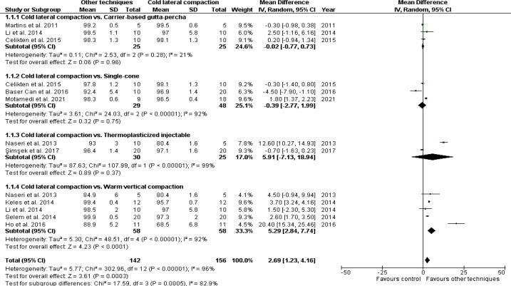
Fifteen studies were included for the final analysis; one had a low risk of bias, eight a moderate risk, and six a high risk of bias. Ten studies were selected for meta-analyses; three studies comparing cold lateral condensation with carrier-based gutta-percha techniques [P=0.96; mean difference (MD)=-0.02; confidence interval (CI): (-0.77, 0.73); I2=21%]; three comparing cold lateral condensation with single-cone techniques [P=0.75; MD=-0.39; CI: (-2.77, 1.99); I2=92%]; two comparing cold lateral condensation and thermo-plasticized injectable techniques [P=0.37; MD=5.91; CI: (-7.13,18.94); I2=99%]; and five comparing cold lateral condensation with warm vertical condensation techniques [P<0.0001; MD=5.29; CI=(2.84, 7.74); I2=92%]. The overall effect reported significant results [P=0.0003; MD=2.69; CI=(1.23, 4.16); I2=96%]; favoring fewer voids and gaps for the other used obturation techniques.
Conclusions:
Cold lateral condensation and single-cone techniques presented no statistical differences. Nonetheless, Warm vertical condensation technique had better results compared to cold lateral condensation.
Key Words: Cold Lateral Condensation, Endodontics, Quality of Obturation, Root Canal Obturation, Systematic Review, Warm Vertical Condensation
Introduction
Root canal treatment eliminates necrotic tissues, bacteria, and endotoxins and three-dimensionally fills the root canal system [1]. Materials used during obturation must fill the root canal complexities, such as ramifications and isthmi [2], to prevent bacterial proliferation and migration into the canals and toward the periodontium [3].
Studies have shown that several aspects can improve endodontic treatment outcomes [4, 5]. The presence of filling material up to the last 1-2 mm of the radiographic apex, homogeneous obturation, without empty spaces visible on periapical radiographs or cone-beam computed tomography (CBCT), is linked to better outcomes [6].
The presence of gaps (i.e., spaces between the filling material and the canal walls) and voids (i.e., spaces within the obturation material) can jeopardize the success of the endodontic treatment by permitting bacterial movement toward the major apical foramen or other foramina, potentially leading to the development or persistence of apical periodontitis [7]. Thus, the obturation technique should promote the adaptation of filling materials to the canal walls and fill the entire length of the canal [8].
Cold lateral condensation (CLC) is the most frequently used technique for root canal obturation. However, it has been reported that this technique is related to poor homogeneous fillings [9, 10]. In an attempt to optimize the filling of the root canal, other techniques have been proposed, such as the warm vertical condensation (WVC) [11, 12], Tagger's hybrid [13, 14], single-cone (SC) [15, 16], carrier-based gutta-percha [13, 17] and thermo-plasticized injectable (TI) techniques [12].
The quality of root canal filling has been clinically, radiographically, or topographically evaluated. More recently, micro-computed tomography (micro-CT) has been used in Endodontics to assess the quality of root canal filling in in vitro studies [18]. Micro-CT allows volumetric quantification of empty spaces without specimen destruction [19]. Currently, micro-CT is the most accurate imaging method for evaluating the quality of obturations in laboratory conditions. [20]
Mainly due to the importance of the quality of root canal filling for the success of the endodontic treatment and the availability of several techniques for the same purpose, this systematic review aimed to address the following question: Is the CLC as effective as other obturation techniques on the quality of root canal obturation?
Material and Methods
This systematic review was conducted following Preferred Reporting Items for Systematic Review and Meta-Analysis (PRISMA) recommendations (http://www.prisma-statement.org). A PROSPERO registration was not achievable since this is a systematic review of in vitro studies.
Search methodology
The literature search was conducted in six electronic databases and source (MEDLINE/PubMed, Cochrane, Scopus, Web of Science, EMBASE, and Grey Literature). The search included articles published until February 2022 without language or year restriction. The search strategy was developed using Medical Subject Heading terms and text words, combining the following terms: "Cold Lateral Condensation"; "Cold Lateral Condensation"; "Lateral Condensation"; "Lateral Condensation"; "Warm Vertical Condensation"; "Vertical Condensation"; "Warm Vertical Condensation"; "Vertical Condensation"; "Single-Cone"; "Carrier-Based"; "Thermoplasticized"; "Continuous Wave"; "Hybrid Tagger"; "Tagger"; "Root Canal Filling"; "Gaps"; "Voids"; "Empty Spaces"; "Quality"; "Microcomputed Tomography"; "Micro-CT"; "MicroCT". The Boolean operators "AND" and "OR" were applied to combine the terms and create the search results.
The search strategies for each database and findings are presented in Table 1. All articles selected were imported into the Zotero reference manager to catalog references and exclude duplicates.
Table 1.
Search strategy in each database and source
| Database | Search strategy | Findings |
|---|---|---|
| MEDLINE/PubMed | #1: (((Cold Lateral Compaction) OR (Cold Lateral Condensation)) OR (Lateral Compaction)) OR (Lateral Condensation) | 3.877 |
| #2: (((((((((Warm Vertical Compaction) OR (Vertical Compaction)) OR (Warm Vertical Condensation)) OR (Vertical Condensation)) OR (Single Cone)) OR (Carrier-Based)) OR (Thermoplasticized)) OR (Continuous Wave)) OR (hybrid Tagger)) OR (Tagger) | 30.703 | |
| #3: ((((Root Canal Filling) OR (Gaps)) OR (Voids)) OR (Empty Spaces)) OR (Quality) | 1.539.030 | |
| #4: ((Microcomputed tomography) OR (Micro-CT)) OR (MicroCT) | 24.624 | |
| #1 AND #2 AND #3 AND #4 | 32 | |
| Cochrane Library | #1: Cold Lateral Compaction OR Cold Lateral Condensation OR Lateral Compaction OR Lateral Condensation | 225 |
| #2: Warm Vertical Compaction OR Vertical Compaction OR Warm Vertical Condensation OR Vertical Condensation OR Single Cone OR Carrier-Based OR Thermoplasticized OR Continuous Wave OR Hybrid Tagger OR Tagger | 2.169 | |
| #3: Root Canal Filling OR Gaps OR Voids OR Empty Spaces OR Quality | 204.130 | |
| #4: Microcomputed Tomography OR Micro-CT OR MicroCT | 345 | |
| #1 AND #2 AND #3 AND #4 | 4 | |
| Scopus | #1: (TITLE-ABS-KEY (cold AND lateral AND compaction) OR TITLE-ABS-KEY (cold AND lateral AND condensation) OR TITLE-ABS-KEY (lateral AND compaction) OR TITLE-ABS-KEY (lateral AND condensation)) | 4.321 |
| #2: (TITLE-ABS-KEY (warm AND vertical AND compaction) OR TITLE-ABS-KEY (vertical AND compaction) OR TITLE-ABS-KEY (warm AND vertical AND condensation) OR TITLE-ABS-KEY (vertical AND condensation) OR TITLE-ABS-KEY (single AND cone) OR TITLE-ABS-KEY (carrier-based) OR TITLE-ABS-KEY (thermoplasticized) OR TITLE-ABS-KEY (continuous AND wave) OR TITLE-ABS-KEY (hybrid AND tagger) OR TITLE-ABS-KEY (tagger)) | 133.550 | |
| #3: (TITLE-ABS-KEY (root AND canal AND filling) OR TITLE-ABS-KEY (gaps) OR TITLE-ABS-KEY (voids) OR TITLE-ABS-KEY (empty AND spaces) OR TITLE-ABS-KEY (quality)) | 5.367.277 | |
| #4: (TITLE-ABS-KEY (microcomputed AND tomography) OR TITLE-ABS-KEY (micro-ct) OR TITLE-ABS-KEY (microct)) | 21.575 | |
| #1 AND #2 AND #3 AND #4: | 26 | |
| Web of Sciences | #1: TS=(Cold Lateral Compaction OR Cold Lateral Condensation OR Lateral Compaction OR Lateral Condensation) | 2.930 |
| #2: TS=(Warm Vertical Compaction OR Vertical Compaction OR Warm Vertical Condensation OR Vertical Condensation OR Single Cone OR Carrier-Based OR Thermoplasticized OR Continuous Wave OR Hybrd Tagger OR Tagger) | 95.660 | |
| #3: TS=(Root Canal Filling OR Gaps OR Voids OR Empty Spaces OR Quality) | 3.788.079 | |
| #4: TS=(Microcomputed Tomography OR Micro-CT OR MicroCT) | 18.703 | |
| #1 AND #2 AND #3 AND #4: | 28 | |
| EMBASE | #1: cold AND lateral AND compaction OR (cold AND lateral AND condensation) OR (lateral AND compaction) OR (lateral AND condensation) | 1.779 |
| #2: warm AND vertical AND compaction OR (vertical AND compaction) OR (warm AND vertical AND condensation) OR (vertical AND condensation) OR (single AND cone) OR 'carrier based' OR thermoplasticized OR (continuous AND wave) OR (hybrid AND tagger) OR tagger | 29.528 | |
| #3: root AND canal AND filling OR gaps OR voids OR (empty AND spaces) OR quality | 2.352.569 | |
| #4: microcomputed AND tomography OR 'micro ct' OR microct | 31.663 | |
| #1 AND #2 AND #3 AND #4: | 26 | |
| Grey Literature | #1: Cold Lateral Compaction OR Cold Lateral Condensation OR Lateral Compaction OR Lateral Condensation | 0 |
| #2: Warm Vertical Compaction OR Vertical Compaction OR Warm Vertical Condensation OR Vertical Condensation OR Single Cone OR Carrier-Based OR Thermoplasticized OR Continuous Wave OR Hybrid Tagger OR Tagger | 0 | |
| #3: Root Canal Filling OR Gaps OR Voids OR Empty Spaces OR Quality | 0 | |
| #4: Microcomputed Tomography OR Micro-CT OR MicroCT | 0 | |
| #1 AND #2 AND #3 AND #4: | 0 |
Inclusion criteria
The eligibility criteria were performed using the PICOS strategy (PRISMA-P 2015) [21, 22] following this scheme:
-P: Extracted mature permanent teeth;
-I: Other obturation techniques rather than CLC (i.e., SC, carrier-based gutta-percha, TI, continuous wave of condensation (CWC), hybrid Tagger, and WVC);
-C: CLC technique;
-O: Quality of root canal obturation (volume of filling material, presence of voids and/or gaps);
-S: In vitro studies assessing the investigated parameters using micro-CT.
Exclusion criteria
In vitro studies that evaluated the same parameters on immature and/or primary teeth, performed root canal preparation with hand files, and performed root canal filling using reparative materials (i.e. MTA, Biodentine) were excluded. Studies that did not use Micro-CT to assess the quality of root canal filling were also excluded.
Selection of studies
Two authors (N.B.A. and G.B.S.) selected studies and examined the retrieved titles and abstracts. Duplicates were identified and excluded. The full text was assessed when judging the studies by title and abstract was impossible. The next stage consisted of reading the full texts based on the eligibility criteria through the PICOS strategy. A third experienced author (T.W.) assessed the study in case of disagreement on study inclusion.
Synthesizing data
Two authors (N.B.A. and G.B.S.) independently collected the data from the included studies. Disagreements were solved by a third author (T.W.). The following data were extracted: author name(s), year of publication, sample size, group of teeth, root canal preparation technique, obturation technique, micro-CT scan parameters, evaluated parameters, outcomes, and main findings. In cases of missing data, the authors were contacted by e-mail at least three times.
Quality assessment
Due to the absence of a specific tool to evaluate the risk of bias of in vitro studies, the risk of bias of the included studies was evaluated using an adapted methodology based on previous systematic reviews [7, 23]. The following parameters were assessed:
1. Description of sample size calculation;
2. Selection and pairing of samples by micro-CT;
3. Description of micro-CT parameters:
-The following information were considered: Description of the device used, kilovoltage (kV), mA/micro-ampere (µA), voxel/pixel size, rotation angle, rotation step, exposure time, filters, and/or corrections;
4. Description of obturation technique:
-The following information were considered: Description of the endodontic sealer, description of the core material, description of the obturation technique;
5. Blinding of evaluators;
6. Description of statistical analysis.
Each included study was judged with "yes" when parameters were found and "no" in case of absence. In studies presenting partial data, authors were contacted at least three times by e-mail. If data could not be achieved, parameters presenting partial data were judged as "no". Studies with only one or two parameters were classified as a high risk of bias, three or four parameters as a moderate risk of bias, and five or more parameters as a low risk of bias. Two authors (N.B.A. and G.B.S) independently evaluated each study's methodological quality, and a third author (T.W.) validated the analysis.
Meta-analysis
The Review Manager Software (RevMan, Version 5.3, The Nordic Cochrane Centre, The Cochrane Collaboration, Copenhagen, Denmark, 2014) was used for the meta-analysis, considering a random-effect model. Meta-analyses were performed for studies that presented data (mean and standard deviation) on the filling material volume regarding the root canal's overall volume. Since this is a continuous variable, the effect measure was the mean difference, and the statistical method was the inverse variance DerSimonian-Laird test.
Heterogeneity was calculated using the T2, Cochran Q test, and I2 statistics. An I2 statistic below 30% was considered irrelevant, between 30% and 60% was regarded as moderate heterogeneity, between 50% and 75% as substantial heterogeneity, and over 90% was regarded as considerable heterogeneity [24, 25].
A P-value of less than 5% was considered significant. As for publication bias, it can be assessed visually by generating funnel plots when ten or more studies are included in a meta-analysis [26].
Results
Study Selection
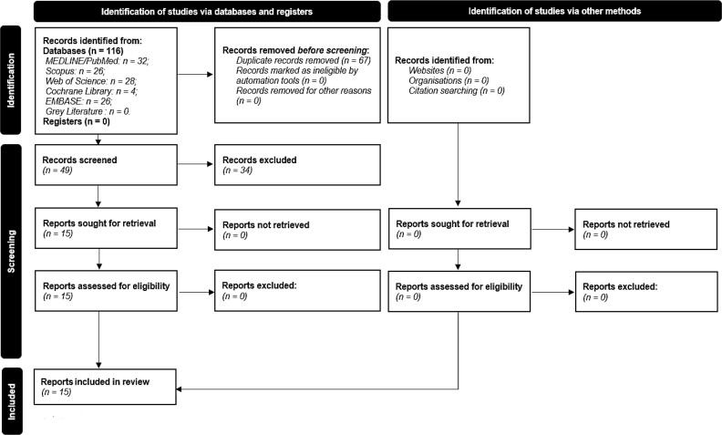
Figure 1 presents the flow diagram of the search strategy. Initial identification through database searching resulted in 116 studies, of which 67 were excluded as they were duplicates. From 49 studies, 34 were excluded after the title and abstract reading. Fifteen records met the inclusion criteria and were selected for full-text reading [3, 4, 12-16, 18, 27-33]. Of these studies, all were included in the present systematic review.
Figure 1.
Flow diagram of the systematic literature search according to PRISMA 2020 guidelines
Data extraction
Table 2 presents the characteristics and main findings of the included studies.
Table 2.
Characteristics extracted from the included studies
| Author(s) | Sample Size (Per Group) | Group of teeth | Root canal preparation technique | Obturation Technique | Micro-CT Scan Parameters | Evaluated Parameters | Outcomes | Main Findings |
|---|---|---|---|---|---|---|---|---|
| Alim & Berker [15] | N=60 (n=15) | Mandibular first molars | ProTaper Next (25.06) | CLC: Cold Lateral Compaction (Gutta-Percha+AH Plus) SC: Single-Cone (Gutta-percha+AH-Plus) CWC: Continuous Wave of Condensation (Gutta-percha+AH-Plus) CB: Carrier-Based Gutta-percha (GuttaCore+AH-Plus) |
Voxel Size: 20 μm kV: 70 µA: 114 Exposure time: 600 msec |
Void area | Percentage of filled area at 2mm was the lowest for SC; At 5mm, CLC and CB presented lower number of voids compared to SC and CWC; At 8 mm, filled area was similar for all techniques |
At 2 and 5mm, a smaller number of voids were verified for CLC and CB, respectively |
| Ba ş er Can et al. [28] | N=30 (n=10) | Single-rooted premolar | Endosequence (40.06) | CLC1: Cold Lateral Compaction (Gutta-percha+EndoREZ) CLC2: Cold Lateral Compaction (Gutta-percha+AH-Plus) SC: Single-Cone (ActiV Gutta-percha Root Canal Obturation System) |
Voxel size: 13.68μm kV: 100 µA: 100 Exposure time: 2000 msec Rotational angle: 180o Rotation step: 0.4° |
Percentage volume of canal filling and voids | Percentage volume of filling material in SC was lower than in the CLC groups, without differences between them; Percentage volume of voids in the SC was higher than in the CLC groups, without differences between them; Analysis of middle thirds showed no significant difference among groups |
None of the systems achieved completely void-free root fillings; SC was associated with a higher percentage volume of voids than the other groups |
| Celikten et al . [29] | N=30 (n=10) | Mandibular first premolars | ProTaper F3 (30.09) | SC: Single-Cone (Gutta-percha+EndoSequence BC Sealer) CLC: Cold Lateral Compaction (Gutta-percha+EndoSequence BC Sealer) CB: Carrier-Based Gutta-percha (Thermafil+EndoSequence BC Sealer) |
Voxel size: 13.47 μm kV: 100 µA: 100 Rotational angle: 360o |
Presence and volume of filling material and voids | There were no differences in relation to filling material volume or presence of voids; SC technique had the largest void volumes, and CB the smallest void volumes, at all levels |
Voids were present for all obturation techniques; however, SC had the largest void volume |
| Ho et al. [18] | N=33 (n=11) | Mandibular first molars | ProFile (30.06) |
CLC: Cold Lateral (Gutta-percha without sealer) WLC-U: Warm Lateral with Ultrasonic Spreader (Gutta-percha without sealer) TI: Thermo-plasticized Injectable Technique (Gutta-percha+Obtura II) |
Voxel size: 7.9 μm kV: 100 µA: 100 Rotational angle: 360o Rotation step: 1.5° |
Percentage volume of filling material | Volume of gutta-percha was lower in CLC than in WLC-U and TT, without differences between them; Density of gutta-percha increased towards the coronal third, in WLC-U and TT |
WLC-U and TI produced a greater root canal filling volume compared to CLC |
| Kele ş et al. [30] | N=24 (n=12) | Single-rooted maxillary premolars | Revo-S (25.06) |
CLC: Cold Lateral Compaction (Gutta-percha+AH Plus) WVC: Warm Vertical Compaction (Gutta-percha Dia-Gun Obturation System+AH-Plus) |
Voxel size: 12.5 μm kV: 90 µA: 112 Exposure time: 2600 msec Rotational angle: 180o Rotation step: 0.6° |
Percentage of canal filling and voids | WVC had a smaller volume of voids than CLC in the coronal and middle thirds; No differences between groups were observed in the volume of filling material in the middle third; In the apical third, there was no differences in the percentage volume of filling material and voids between groups |
No obturation technique produced void-free root canal fillings; WVC was associated with a lower percentage volume of voids than CLC |
| Li et al . [31] | N=30 (n=10) | Single-rooted premolars | Vortex Blue (40.04) | CB: Carrier-Based Gutta-percha (GuttaCore Obturator+ThermaSeal Plus) WVC: Warm Vertical Compaction (Gutta-percha+ThermaSeal Plus) CLC: Cold Lateral Compaction (Gutta-Percha+ThermaSeal Plus) |
Voxel size: 14.52 μm kV: 50 µA: 800 Exposure time: 4000 msec Rotational angle: 360o Rotation step: 0.9° |
Gaps and voids | There were no differences in the volumetric distribution of gaps and voids between CLC and WVC techniques, and between WVC and CB; Higher percentages of gaps, interfacial gaps and voids were found in CLC, when compared with CB |
None of the obturation techniques produced completely gap- and void-free root fillings; Both CB and WVC had lower incidences of gaps and voids than CLC |
| Martins et al . [13] | N=15 (n=5) | Mandibular molars | ProTaper F3 (30.09) | CLC: Cold Lateral Compaction (Gutta-Percha+AH-26) HT: Hybrid Tagger (Gutta-percha+AH-26) CB: Carrier-Based Gutta-percha (Thermafil+ AH26) |
Voxel size: 5 μm kV: 100 µA: 100 |
Volume of voids | There were no differences among techniques | None of the tested techniques allowed a void-free root filling |
| Moeller et al . [32] | N=67 (n=34; n=33) |
Mandibular molars, premolars and canines | Profile (35.04) |
CLC: Cold Lateral Compaction (Gutta-percha+AH-Plus) HT: Hybrid technique (Gutta-Percha master cone with AH-Plus+Thermafil) |
Voxel size: 10 μm kV: 70 µA: 85 |
Proportion and distribution of voids | CLC resulted in fewer voids in the apical than in the cervical third; HT resulted in fewer voids in the cervical than in the apical third; In relation to the proportion of voids, the obturation techniques did not differ |
No difference was observed in percentage of voids between techniques |
| Moinzadeh et al . [33] | N=20 (n=10) | Maxillary and mandibular canines | MTwo (40.06) |
SC: Single-Cone (Gutta-percha+Smartpaste Bio) CLC: Cold Lateral Compaction (Gutta-percha+Smartpaste Bio) |
Voxel size: 10 μm kV: 70 µA: 114 Exposure time: 300 msec |
Percentage volume of voids and filling material | SC exhibited less percentage of voids in the coronal and middle thirds, and lower median percentage of voids than CLC | Percentage of voids with CLC was higher than SC |
| Motamedi et al. [35] | N=36 (n=9) |
Single-rooted mandibular premolars | ProTaper F3 (30.09) |
CLC1: Cold Lateral Compaction (Gutta-percha 0.02+AdSeal) CLC2: Cold Lateral Compaction (Gutta Percha 0.04+AdSeal) SC: Single Cone (Gutta-Percha+Adseal) SC-U: Single Cone with Ultrasonic Activation (Gutta-percha+Adseal) |
Voxel size: 19 μm kV: 80 µA: 100 |
Percentage volume of voids | Total percentage volume of voids was lower in the SC-U group compared to all other groups; There were no differences between the CLC1 and CLC2 |
SC-U showed the least number of voids amongst all the obturation techniques |
| Naseri et al . [12] | N=20 (n=5) | Maxillary first molars | ProTaper F3 (30.09) |
CLC: Cold Lateral Compaction (Gutta-percha+AH-26) WVC: Warm Vertical Compaction (Gutta-percha+AH-26) TI1: Thermo-plasticized Injectable Technique (Obtura II+Gutta-percha+AH-26) TI2: Thermo-plasticized Injectable Technique (Gutta Flow+AH-26) |
Voxel size: 19.5 μm Exposure time: 3000 msec Rotational angle: 180o Rotation step: 0.9o |
Volume percentage of voids and obturation materials | Highest percentage of filling material was observed in TI2 followed by TI1, without differences; Voids were detected in all samples; CLC and TI2 had the highest and the lowest percentage of voids, respectively; In the apical third, CLC and TI1 had the highest and the lowest percentage of voids, and the lowest and highest percentage of gutta-percha, respectively |
None of the techniques were void-free; TI was associated to a lowest percentage of voids than CLC |
| Nhata et al . [14] | N=30 (n=10) | Mandibular incisors | Hero Files (45.02) | CLC: Cold Lateral Compaction (Gutta-percha+AH-Plus) CWC: Continuous Wave of Condensation (Gutta-Percha+AH Plus) HT: Hybrid Tagger (Gutta-percha+AH-Plus) |
Voxel size: 22.6 μm kV: 70 µA: 140 |
Presence of voids | CWC and HT showed less presence of voids compared to CLC | CWC and HT had less voids compared to CLC |
| Selem et al. [34] | N=40 (n=10) | Single-rooted premolars | K3XF (40.04–40.06) |
WVC-NGP: Warm Vertical compaction (Non-Gutta-percha Material+Sealer) WVC-GP: Warm Vertical Compaction (Gutta-percha+AH-Plus) CLC-NGP: Cold Lateral Compaction (Non-Gutta-Percha Material+Sealer) CLC-GP: Cold Lateral Compaction (Gutta-percha+AH-Plus) |
Voxel size: 14.52 μm kV: 50 µA: 800 Exposure time: 4000 msec Rotational angle: 360o Rotation step: 0.9° |
Volumertic percentage of gaps and voids | Differences in the presence of voids among all groups; There was no difference in the void area percentage distribution between the WVC or CLC groups; More canal areas were occupied by voids when CLC was used compared with WVC groups; CLC-NGP exhibited more gaps than the other groups |
None of the groups produced completely gap and void-free fillings; More canal areas were occupied by voids when CLC was used |
| Şimşek et al . [36] | N=40 (n=10) | Mandibular first molars | SAF (Self-adjusting file) Revo-S (25.06) |
TI: Thermo-plasticized Injectable Technique (Gutta-percha+AH-Plus) CLC: Cold Lateral Compaction (Gutta-percha+AH-Plus) |
Voxel size: 13.7 μm kV: 100 µA: 100 Exposure time: 2800 msec Rotational angle: 180° Rotation step: 0.6° |
Volume of filling material, voids and gaps | All techniques produced voids and gaps; There were no differences in voids and gaps regarding preparation technique |
None of the techniques produced void- or gap-free fillings |
| Suassuna et al . [37] | N=45 (n=15) | Mandibular premolars | Reciproc Files (40.06-50.05) | CLC: Cold Lateral Compaction (Gutta-percha+AH-Plus) SC: Single-Cone (Gutta-percha+AH-Plus) HT: Hybrid Tagger Technique (Gutta-perch+AH-Plus) |
Voxel size: 11 μm kV: 80 µA: 222 |
Presence of voids | Higher number of voids were detected for CLC group | CLC produced more voids |
The corresponding authors of the studies presenting insufficient data were contacted by e-mail, but no additional information was obtained.
Regarding teeth evaluated, four studies performed their evaluations on mandibular molars [14, 15, 18, 33]; one on maxillary molars [12]; three studies on mandibular premolars [16, 28, 32]; one study on maxillary premolars [29]; and three studies on premolars without specifying [3, 4, 27]; one study evaluated mandibular and maxillary canines [31]; one study used mandibular incisors [14]; and one study performed their evaluations on a group of teeth-mandibular molars, premolars and canines [30].
Regarding the instruments used for root canal preparation, three studies used instruments with a #25 tip diameter ProTaper Next (Dentsply Maillefer, Ballaigues, Switzerland) #25.06 [15], Revo-S rotary files (MicroMega,
Cedex, Besancon, France) #25.06 [29, 33]; four studies used instruments up to a #30 tip diameter ProTaper #30.09 [12, 13, 28, 32], one study up to a #35 tip diameter ProFile (Dentsply Tulsa Dental, Tulsa, OK, USA) #35.04 [30]; five studies prepared up to a #40 tip diameter Endosequence (Brasseler, Savannah, GA, USA) [16] #40.06 [27], Vortex Blue (Dentsply Sirona, Ballaigues, Switzerland) #40.04 [4], MTwo (VDW, Munich, Germany) #40.06 [31], K3XF (SybronEndo, Glendora, CA, USA) #40.04-#40.06 [3], Reciproc (VDW, Munich, Germany) #40.06 [16]; one study up to a #45 tip diameter Hero Shaper Files (Micro-Mega, Besançon, France) #45.02 [14]; one study also prepared up to a #50 tip diameter-Reciproc #50.05; and one study used the Self Adjusting File [33].
Besides the CLC technique, six studies evaluated the SC technique [15, 16, 27, 28, 31, 32]; two studies evaluated the CWC technique [14, 15]; four studies the carrier-based gutta-percha technique [4, 13, 15, 28], four studies the WVC technique [12, 30, 31, 34]; four studies performed their evaluation using the Tagger's hybrid technique [13, 14, 16, 30]; three studies used thermo-plasticized techniques [12, 18, 33]; and two studies performed ultrasonic activations associated to an obturation technique=warm lateral condensation [18] and SC technique [32].
Most studies used gutta-percha (GP) and AH-Plus to perform root canal obturation [3, 14-16, 27, 29, 30, 33]. GP and Endosequence BC Sealer (Brasseler USA, Savannah, GA, USA), Thermafil (Dentsply Maillefer, Ballaigues, Switzerland) and Endosequence BC Sealer [28], GP only, GP and Obtura II (Obtura Spartan, Fenton, MO, USA) [18], Other materials involved GP and EndoREZ, ActiV GP (Bras, seler USA, Savannah, GA, USA) system GuttaCore (Dentsply Tulsa Dental Specialties, Tulsa, OK, USA) and Thermaseal Plus (Dentsply Tulsa Dental Specialties), GP and Thermaseal Plus [4], GP and AH-26 [12, 13], Thermafil (Dentsply Maillefer, Ballaigues, Switzerland) and AH-Plus (Dentsply Maillefer, Ballaigues, Switzerland) [13], Obtura II (Obtura Spartan, Fenton, MO, USA), GP and AH-26 (Dentsply Maillefer, Ballaigues, Switzerland), GuttaFlow (Coltène/Whaledent, Altstätten/Switzerland) and AH-26 [12], GP, AH-Plus (DentsplyMaillefer, Ballaigues, Switzerland) and Thermafil [30], GP and SmartPaste Bio (Smartpaste Bio®, CRD Ltd, Stamford, UK) [31], GP and AdSeal (Metabiomed, Cheongju, Korea) [32] and Non-GP material [3].
As for the micro-CT parameters, the majority of studies presented information on voxel size [12-16, 18, 27-36]; kV and µA [13-16, 18, 27-36]. Other information included exposure time [12, 15, 28, 30, 31, 33, 34, 36]; rotational angle [12, 18, 27-29, 33]; and rotation step [12, 18, 28, 30, 31, 34, 36]. None of the included studies presented information on using filters or corrections.
Regarding the evaluated outcomes, most studies evaluated aspects related to voids [3, 4, 12-16, 27-33]; seven studies investigated aspects related to the volume of filling material [12, 18, 28-30, 33, 36]; and three studies evaluated aspects related to gaps [31, 34, 36].
As for the main findings presented by the included studies, three studies presented results favoring the CLC technique when compared to the SC technique [15, 28, 29] and the CWC technique [15]; two studies showed results favoring the carrier-based (CB) technique when compared to the SC technique [15, 29], the CWC [15], and the CLC techniques [31]. Three studies showed results favoring the WVC technique when compared to the CLC technique [30, 31, 34]; two studies had results favoring the TI technique over the CLC technique [12, 18]. Two studies presented better results for the SC technique than the CLC technique [16, 33]; two studies presented results favoring the hybrid Tagger (HT) technique when compared to the CLC technique [14, 16]. One study showed better results for the CWC technique when compared to the CLC technique [14]; two studies presented results favoring obturation techniques-warm lateral condensation and SC-associated with ultrasonic activation when compared to the CLC technique [18] and CLC and SC without activation [35], respectively. Finally, three studies did not present differences among the tested techniques [13, 32, 36].
Quality assessment
Table 3 summarizes the risk of bias in the included studies. Of the fifteen included studies, six were classified as having a high risk of bias [12-15, 18, 32], with four domains (sample size calculation, selection and pairing of samples, micro-CT scan parameters and blinding of evaluators) presenting some concerns; eight studies were classified as having a moderate risk of bias [16, 28-34, 36], with several domains presenting concerns. Only one study was classified as having a low risk of bias [29], with only one domain (blinding of evaluators) presenting some concerns.
Table 3.
Risk of bias assessment of the included studies
| Author(s) | Sample Size Calculation | Selection and Pairing of Samples | Micro-CT Scan Parameters | Obturation Technique | Blinding of Evaluators | Statistical Analysis | Risk of Bias |
|---|---|---|---|---|---|---|---|
| Alim & Berker [ 15 ] | No | No | No | Yes | No | Yes | HIGH |
| Başer Can et al. [ 28 ] | Yes | No | Yes | Yes | No | Yes | MODERATE |
| Celikten et al . [29] | No | No | No | Yes | Yes | Yes | MODERATE |
| Ho et al. [ 18 ] | No | No | No | Yes | No | Yes | HIGH |
| Keleş et al . [30] | Yes | Yes | Yes | Yes | No | Yes | LOW |
| Li et al . [ 31 ] | No | No | Yes | Yes | No | Yes | MODERATE |
| Martins et al . [13] | No | No | No | Yes | No | Yes | HIGH |
| Moeller et al . [32] | No | No | No | Yes | Yes | Yes | MODERATE |
| Moinzadeh et al . [ 33 ] | Yes | No | No | Yes | No | Yes | MODERATE |
| Motamedi et al. [35] | No | No | No | Yes | No | Yes | HIGH |
| Naseri et al . [12] | No | No | No | Yes | No | Yes | HIGH |
| Nhata et al . [ 14 ] | No | No | No | Yes | No | Yes | HIGH |
| Selem et al . [ 34 ] | No | No | Yes | Yes | No | Yes | MODERATE |
| Şimşek et al . [ 36 ] | No | No | Yes | Yes | Yes | Yes | MODERATE |
| Suassuna et al . [ 16 ] | No | No | No | Yes | Yes | Yes | MODERATE |
Meta-analysis
Figure 2 presents the results of all meta-analyses performed. Not all studies were included in the meta-analyses because of the high heterogeneity of the reported data.
Figure 2.
Forest plot depicting the comparisons between the cold lateral condensation and other obturation techniques
For the comparison between CLC and carrier-based gutta-percha techniques, three studies [13, 29, 31] were included for further analysis. No statistical differences were observed between these techniques [P=0.96; mean difference (MD)=-0.02; confidence interval (CI): (-0.77, 0.73); I2=21%].
Three studies [28, 29, 35] were included for the comparison between CLC and SC techniques. Again, no statistical differences were observed between the two techniques [P=0.75; MD=-0.39; CI: (-2.77, 1.99); I2=92%].
As for the comparison between CLC and TI techniques, two studies [12, 36] were included. Once again, no statistical differences were observed [P=0.37; MD=5.91; CI: (-7.13, 18. 94); I2=99%].
Five studies compared the CLC and WVC techniques [12, 18, 30, 31, 34]. In this comparison, a statistical difference was observed, favoring better results for the WVC technique [P<0.0001; MD=5.29; CI=(2.84, 7.74); I2=92%]. Mainly due to this result, the overall effect presented significant results [P=0.0003; MD=2.69; CI=(1.23, 4.16); I2=96%]. However, it is important to emphasize that this does not reflect the results for each comparison.
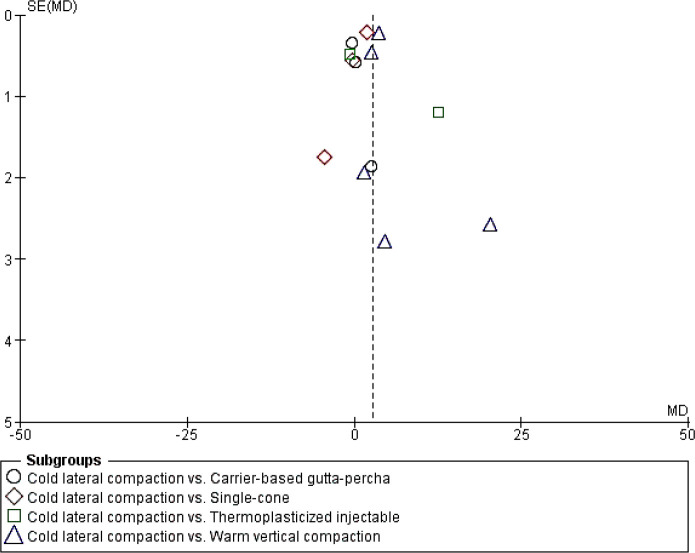
Ten studies were included in the meta-analyses. Therefore, it was possible to verify for publication bias. As presented in Figure 3 the funnel plot is approximately symmetrical, implying that publication bias does not seem significant to the research's validity.
Figure 3.
Funnel plot of the included studies showing homogeneity amongst studies
Discussion
Empty spaces (i.e., voids and gaps) in root canal obturation can cause bacterial leakage and possibly an endodontic failure [6, 7]. For this reason, evaluating the impact of the several obturation techniques available on the incidence of empty spaces is necessary. Because CLC is the most frequently used obturation technique, in the present systematic review, we aimed to evaluate studies that compared the quality of obturations performed using the CLC technique to other obturation techniques, assessed by using micro-CT.
This selection criteria were based on studies indicating that micro-CT assessment presents more reliable and accurate data on the quality of the root canal filling [38-40], enabling the tridimensional and volumetric evaluation of the root canal filling and, consequently, determining the presence of voids and gaps [41, 42].
The meta-analysis provides the interrelation of results from two or more independent studies on the same research question. Outcomes from a meta-analysis may include a more precise estimate of the effect of treatment than any individual study contributing to the pooled analysis [43]. In the present study, only ten studies were included in the meta-analysis because the data heterogeneity and the dificulty in control the multiple variables. The difficulty in standardizing the study variables such as the group of teeth, preparation, and obturation technique could compromise data analysis. We were able to extract essential data when analyzing a study with a limited pool; however, the results must be evaluated with caution due to the potential for misleading.
When assessing the main findings, two studies reported more voids when using the SC technique rather than CLC [15, 27], and two studies reported that the CLC technique had more voids than the SC technique [16, 31]. Yet, the meta-analysis presented no statistical differences in studies [28, 29, 35]. Several factors can explain these controversial results. First, a non-uniform filling is produced due to the difficulty of controlling the amount of sealer around the GP cone, causing poor sealer adaptation for the SC technique [44]. Another hypothesis is the difficult fitting of a single round cone in an irregularly-shaped canal, without any gaps [45]. However, the SC technique is more effective when using a paired GP cone [46]. Furthermore, the filling achieved by the SC technique can be improved by using endodontic sealers with excellent dimensional stability (i.e., epoxy resin-based sealer) or hygroscopic expansion (i.e., calcium silicate-based sealers) instead of zinc oxide and eugenol-based sealers that contract after setting [36, 47]. Additionally, ultrasonic activation of the sealer, prior to the placement of the single GP cone, can promote a better flow and filling of the canal irregularities, therefore, decreasing the presence of gaps and voids [48-50].
Only one study reported a lower percentage of voids using carrier-based gutta-percha [31], and the meta-analysis did not show differences among the evaluated studies [13, 29, 31]. Carrier-based gutta-percha systems comprise a two-phase GP (i.e., alpha- and beta-phase). In the alpha phase, the GP that covers the core is more adhesive and, when heated, becomes highly flowable to promote an improved adaptation to the canal walls and filling of irregularities [22], which can explain the present findings.Warm vertical condensation promoted gaps and voids in three studies [30, 31, 35]. However, the meta-analyses showed a statistical difference favoring the WVC technique, suggesting that the CLC technique promotes a lower volume of filling material and, therefore, a more significant number of gaps and voids [12, 18, 30, 31, 35]. The possible explanation is that using a heater during condensation enables the filling material to flow toward the canal irregularities and improves the filling material's adaptation with the principal and lateral canals during warm vertical condensation [51, 52].
In addition, two studies reported a lower percentage of empty spaces associated with the TI techniques [12, 18]; no differences were found in the meta-analysis [12, 36]. This can be explained by the better flow of the GP when used in injectable systems. When plasticized, GP produces a homogeneous mass, promoting fewer voids and better adaptation to the root canal walls [53]. During CLC, GP cones are generally tightly pressed together but are still separate [5], and a poorer adaptation of the GP cone to the canal walls can be the result of an inadequate pressure during condensation with finger spreaders [10]. Also, cold GP does not adhere to the canal walls and can be dislodged during condensation. Finally, due to its poorer flow ability than the sealer, GP would not fill the canal irregularities, leaving voids and gaps in an irregularly-shaped canal [44].
Based on previous systematic reviews [23, 24], a specific tool had to be created to assess the risk of bias in the in vitro studies. The authors selected the evaluated parameters, considering important methodological aspects for evaluating the quality of root canal obturations [38].
Of the fifteen studies included in this systematic review, only one was considered to present a low risk of bias [30]. Eight studies were considered as having a moderate risk of bias [16, 28, 29, 31-34, 36] , with significant concerns regarding the sample size calculation, selection and pairing of the samples, and blinding of evaluators. Six studies were considered as having a high risk of bias [12-15, 18, 33], with significant concerns regarding the sample size calculation, selection and pairing of the samples, description of the micro-CT scan parameters, and blinding of evaluators.
Sample size calculation is a crucial factor because it can prevent the occurrence of type II statistical error (i.e., when statistical differences are not observed because the sample size was so small that the test could not detect them) [54]. As for the pairing of samples, there needs to be an anatomical matching among samples to avoid misinterpretation of the outcomes and, consequently, poor internal validity [38]. The description of micro-CT parameters allows reproducibility among studies and the comparison of different results [40], and blinding evaluators is essential for avoiding bias in data assessment. Only the description of statistical analysis was informed by all studies. All of these parameters must be considered when evaluating the main findings from studies about the quality of obturation. Also, these must be considered for future research on the same topic.
Considering the methodological variability among the included studies (i.e., group of teeth, canal preparation technique, endodontic sealers), a high heterogeneity is unavoidable and, therefore, is a limitation of the present systematic review. Additionally, the success of the endodontic treatment depends on several aspects, such as the patient's systemic health, the pulp and periapical pathological condition, the presence of anatomical complexities, apical limit of instrumentation, the quality of the root canal obturation, and coronal sealing. Therefore, we must emphasize that the quality of the root canal obturation cannot be considered as the only determinant factor for the treatment's success.
So far, based on the results of the present systematic review, it is possible to affirm that none of the obturation techniques were gap-free; this fact is in accordance with another study [47, 53]. However, WVC was the only obturation technique with fewer voids and gaps than CLC. This result should be cautiously interpreted since most studies had a moderate or high risk of bias, and only a few studies compared other obturation techniques to CLC.
Conclusion
Due to the limitation of this study, it is possible to conclude that the WVC technique presented a lower incidence of voids and gaps when compared to the CLC technique. Furthermore, no differences were observed among the other investigated techniques compared to the CLC. However, the results should be interpreted with caution because of the small number of studies included in the meta-analysis and the risk of misleading. The presented evidence is based on studies with a moderate or high risk of bias. Further research is needed to fill the gap of well-designed and standardized studies to increase the evidence base in this domain.
Acknowledgments
None.
Conflict of interest
None.
Funding support
No funding was received for this study.
Authors' contributions
Conceptualization: Só GB, Abrahão NB, Weissheimer T. Data Curation: Só GB, da Rosa RA, Só MVR. Formal Analysis: Weissheimer T, Só MVR, da Rosa RA, Lenzi TL. Investigation: Abrahão NB, Weissheimer T, da Rosa RA. Methodology: Só GB, Abrahão NA. Project administration: Só GB, Abrahão NB, Weissheimer T. Supervision: Só MVR, da Rosa RA, Lenzi TL. Writing-original draft: Abrahão NB, Só GB. Writing-review & editing: da Rosa RA, Só MVR, Weissheimer T, Lenzi TL.
References
- 1.Azim AA, Griggs JA, Huang GT. The Tennessee study: factors affecting treatment outcome and healing time following nonsurgical root canal treatment. Int Endod J. 2016;49(1):6–16. doi: 10.1111/iej.12429. [DOI] [PubMed] [Google Scholar]
- 2.Cruse WP, Bellizzi R. A historic review of endodontics, 1689-1963, part 2. J Endod. 1980;6(4):532–5. doi: 10.1016/S0099-2399(80)80201-9. [DOI] [PubMed] [Google Scholar]
- 3.Kalender A, Orhan K, Aksoy U, Basmaci F, Er F, Alankus A. Influence of the quality of endodontic treatment and coronal restorations on the prevalence of apical periodontitis in a Turkish Cypriot population. Med Princ Pract. 2013;22(2):173–7. doi: 10.1159/000341753. [DOI] [PMC free article] [PubMed] [Google Scholar]
- 4.Imura N, Pinheiro ET, Gomes BP, Zaia AA, Ferraz CC, Souza-Filho FJ. The outcome of endodontic treatment: a retrospective study of 2000 cases performed by a specialist. J Endod. 2007;33(11):1278–82. doi: 10.1016/j.joen.2007.07.018. [DOI] [PubMed] [Google Scholar]
- 5.Ng YL, Mann V, Rahbaran S, Lewsey J, Gulabivala K. Outcome of primary root canal treatment: systematic review of the literature -- Part 2 Influence of clinical factors. Int Endod J. 2008;41(1):6–31. doi: 10.1111/j.1365-2591.2007.01323.x. [DOI] [PubMed] [Google Scholar]
- 6.Wu MK, van der Sluis LW, Ardila CN, Wesselink PR. Fluid movement along the coronal two-thirds of root fillings placed by three different gutta-percha techniques. Int Endod J. 2003;36(8):533–40. doi: 10.1046/j.1365-2591.2003.00685.x. [DOI] [PubMed] [Google Scholar]
- 7.Gillen BM, Looney SW, Gu LS, Loushine BA, Weller RN, Loushine RJ, Pashley DH, Tay FR. Impact of the quality of coronal restoration versus the quality of root canal fillings on success of root canal treatment: a systematic review and meta-analysis. J Endod. 2011;37(7):895–902. doi: 10.1016/j.joen.2011.04.002. [DOI] [PMC free article] [PubMed] [Google Scholar]
- 8.Schilder H. Filling root canals in three dimensions. Dent Clin North Am. . 1967:723–44. [PubMed] [Google Scholar]
- 9.De-Deus G, Murad C, Paciornik S, Reis CM, Coutinho-Filho T. The effect of the canal-filled area on the bacterial leakage of oval-shaped canals. Int Endod J. 2008;41(3):183–90. doi: 10.1111/j.1365-2591.2007.01320.x. [DOI] [PubMed] [Google Scholar]
- 10.Peng L, Ye L, Tan H, Zhou X. Outcome of root canal obturation by warm gutta-percha versus cold lateral condensation: a meta-analysis. J Endod. 2007;33(2):106–9. doi: 10.1016/j.joen.2006.09.010. [DOI] [PubMed] [Google Scholar]
- 11.Alshehri M, Alamri HM, Alshwaimi E, Kujan O. Micro-computed tomographic assessment of quality of obturation in the apical third with continuous wave vertical compaction and single match taper sized cone obturation techniques. Scanning. 2016;38(4):352–6. doi: 10.1002/sca.21277. [DOI] [PubMed] [Google Scholar]
- 12.Naseri M, Kangarlou A, Khavid A, Goodini M. Evaluation of the quality of four root canal obturation techniques using micro-computed tomography. Iran Endod J. 2013;8(3):89–93. [PMC free article] [PubMed] [Google Scholar]
- 13.Martins SC, Mello J, Martins CC, Maurício A, Ginjeira AJRPdE. Medicina Dentária e Cirugia Maxilofacial. Comparação da obturação endodôntica pelas técnicas de condensação lateral, híbrida de Tagger e Thermafil: estudo piloto com Micro-tomografia computorizada. 2011;52(2):59–69. [Google Scholar]
- 14.Nhata J, Machado R, Vansan LP, Batista A, Sidney G, Rosa TP, Leal Silva EJ. Micro-computed tomography and bond strength analysis of different root canal filling techniques. Indian J Dent Res. 2014;25(6):698–701. doi: 10.4103/0970-9290.152164. [DOI] [PubMed] [Google Scholar]
- 15.Alim BA, Garip Berker Y. Evaluation of different root canal filling techniques in severely curved canals by micro-computed tomography. Saudi Dent J. 2020;32(4):200–5. doi: 10.1016/j.sdentj.2019.08.009. [DOI] [PMC free article] [PubMed] [Google Scholar]
- 16.Suassuna FCM, Maia AMA, Melo DP, Antonino ACD, Gomes ASL, Bento PM. Comparison of microtomography and optical coherence tomography on apical endodontic filling analysis. Dentomaxillofac Radiol. 2018;47(2):20170174. doi: 10.1259/dmfr.20170174. [DOI] [PMC free article] [PubMed] [Google Scholar]
- 17.Zogheib C, Naaman A, Sigurdsson A, Medioni E, Bourbouze G, Arbab-Chirani R. Comparative micro-computed tomographic evaluation of two carrier-based obturation systems. Clin Oral Investig. 2013;17(8):1879–83. doi: 10.1007/s00784-012-0875-1. [DOI] [PubMed] [Google Scholar]
- 18.Ho ES, Chang JW, Cheung GS. Quality of root canal fillings using three gutta-percha obturation techniques. Restor Dent Endod. 2016;41(1):22–8. doi: 10.5395/rde.2016.41.1.22. [DOI] [PMC free article] [PubMed] [Google Scholar]
- 19.Iglecias EF, Freire LG, de Miranda Candeiro GT, Dos Santos M, Antoniazzi JH, Gavini G. Presence of Voids after Continuous Wave of Condensation and Single-cone Obturation in Mandibular Molars: A Micro-computed Tomography Analysis. J Endod. 2017;43(4):638–42. doi: 10.1016/j.joen.2016.11.027. [DOI] [PubMed] [Google Scholar]
- 20.Page MJ, McKenzie JE, Bossuyt PM, Boutron I, Hoffmann T, Mulrow CD, Shamseer L, Moher D. Mapping of reporting guidance for systematic reviews and meta-analyses generated a comprehensive item bank for future reporting guidelines. J Clin Epidemiol. 2020;118:60–8. doi: 10.1016/j.jclinepi.2019.11.010. [DOI] [PubMed] [Google Scholar]
- 21.Maia LC, Antonio AG. Systematic reviews in dental research A guideline. J Clin Pediatr Dent. 2012;37(2):117–24. doi: 10.17796/jcpd.37.2.h606137vj3826v61. [DOI] [PubMed] [Google Scholar]
- 22.Moher D, Shamseer L, Clarke M, Ghersi D, Liberati A, Petticrew M, Shekelle P, Stewart LA. Preferred reporting items for systematic review and meta-analysis protocols (PRISMA-P) 2015 statement. Syst Rev. 2015;4(1) doi: 10.1186/2046-4053-4-1. [DOI] [PMC free article] [PubMed] [Google Scholar]
- 23.Gorman CM, Ray NJ, Burke FM. The effect of endodontic access on all-ceramic crowns: A systematic review of in vitro studies. J Dent. 2016;53:22–9. doi: 10.1016/j.jdent.2016.08.005. [DOI] [PubMed] [Google Scholar]
- 24.Silva E, Rover G, Belladonna FG, De-Deus G, da Silveira Teixeira C, da Silva Fidalgo TK. Impact of contracted endodontic cavities on fracture resistance of endodontically treated teeth: a systematic review of in vitro studies. Clin Oral Investig. 2018;22(1):109–18. doi: 10.1007/s00784-017-2268-y. [DOI] [PubMed] [Google Scholar]
- 25.Higgins JP, Thompson SG, Deeks JJ, Altman DG. Measuring inconsistency in meta-analyses. BMJ. 2003;327(7414):557–60. doi: 10.1136/bmj.327.7414.557. [DOI] [PMC free article] [PubMed] [Google Scholar]
- 26.Deeks JJ, Higgins JP, Altman DG. nterventions CSMGJChfsro. Analysing data and undertaking meta‐analyses. 2019. pp. 241–84. [Google Scholar]
- 27.Page MJ, Higgins JP, Sterne JAJChfsroi. Assessing risk of bias due to missing results in a synthesis. 2019:349–74. [Google Scholar]
- 28.Başer Can ED, Keleş A, Aslan B. Micro-CT evaluation of the quality of root fillings when using three root filling systems. Int Endod J. 2017;50(5):499–505. doi: 10.1111/iej.12644. [DOI] [PubMed] [Google Scholar]
- 29.Celikten B, C FU, A IO, Tufenkci P, Misirli M, K OD, Orhan K. Micro-CT assessment of the sealing ability of three root canal filling techniques. J Oral Sci. 2015;57(4):361–6. doi: 10.2334/josnusd.57.361. [DOI] [PubMed] [Google Scholar]
- 30.Keleş A, Alcin H, Kamalak A, Versiani MA. Micro-CT evaluation of root filling quality in oval-shaped canals. Int Endod J. 2014;47(12):1177–84. doi: 10.1111/iej.12269. [DOI] [PubMed] [Google Scholar]
- 31.Li GH, Niu LN, Selem LC, Eid AA, Bergeron BE, Chen JH, Pashley DH, Tay FR. Quality of obturation achieved by an endodontic core-carrier system with crosslinked gutta-percha carrier in single-rooted canals. J Dent. 2014;42(9):1124–34. doi: 10.1016/j.jdent.2014.04.008. [DOI] [PMC free article] [PubMed] [Google Scholar]
- 32.Moeller L, Wenzel A, Wegge-Larsen AM, Ding M, Kirkevang LL. Quality of root fillings performed with two root filling techniques An in vitro study using micro-CT. Acta Odontol Scand. 2013;71(3-4):689–96. doi: 10.3109/00016357.2012.715192. [DOI] [PMC free article] [PubMed] [Google Scholar]
- 33.Moinzadeh AT, Zerbst W, Boutsioukis C, Shemesh H, Zaslansky P. Porosity distribution in root canals filled with gutta percha and calcium silicate cement. Dent Mater. 2015;31(9):1100–8. doi: 10.1016/j.dental.2015.06.009. [DOI] [PubMed] [Google Scholar]
- 34.Selem LC, Li GH, Niu LN, Bergeron BE, Bortoluzzi EA, Chen JH, Pashley DH, Tay FR. Quality of obturation achieved by a non-gutta-percha-based root filling system in single-rooted canals. J Endod. 2014;40(12):2003–8. doi: 10.1016/j.joen.2014.07.032. [DOI] [PubMed] [Google Scholar]
- 35.Kalantar Motamedi MR, Mortaheb A, Zare Jahromi M, Gilbert BE. Micro-CT Evaluation of Four Root Canal Obturation Techniques. Scanning. 2021;2021:6632822. doi: 10.1155/2021/6632822. [DOI] [PMC free article] [PubMed] [Google Scholar]
- 36.Şımşek N, Keleş A, Ahmetoğlu F, Akinci L, Er K. 3D Micro-CT Analysis of Void and Gap Formation in Curved Root Canals. Eur Endod J. 2017;2(1):1–5. doi: 10.14744/eej.2017.17004. [DOI] [PMC free article] [PubMed] [Google Scholar]
- 37.Alcalde MP, Bramante CM, Vivan RR, Amorso-Silva PA, Andrade FB, Duarte MAH. Intradentinal antimicrobial action and filling quality promoted by ultrasonic agitation of epoxy resin-based sealer in endodontic obturation. J Appl Oral Sci. 2017;25(6):641–9. doi: 10.1590/1678-7757-2017-0090. [DOI] [PMC free article] [PubMed] [Google Scholar]
- 38.Guimarães BM, Amoroso-Silva PA, Alcalde MP, Marciano MA, de Andrade FB, Duarte MA. Influence of ultrasonic activation of 4 root canal sealers on the filling quality. J Endod. 2014;40(7):964–8. doi: 10.1016/j.joen.2013.11.016. [DOI] [PubMed] [Google Scholar]
- 39.Combe EC, Cohen BD, Cummings K. Alpha- and beta-forms of gutta-percha in products for root canal filling. Int Endod J. 2001;34(6):447–51. doi: 10.1046/j.1365-2591.2001.00415.x. [DOI] [PubMed] [Google Scholar]
- 40.Tanomaru-Filho M, Silveira GF, Reis JM, Bonetti-Filho I, Guerreiro-Tanomaru JM. Effect of compression load and temperature on thermomechanical tests for gutta-percha and Resilon®. Int Endod J. 2011;44(11):1019–23. doi: 10.1111/j.1365-2591.2011.01910.x. [DOI] [PubMed] [Google Scholar]
- 41.Venturi M, Di Lenarda R, Breschi L. An ex vivo comparison of three different gutta-percha cones when compacted at different temperatures: rheological considerations in relation to the filling of lateral canals. Int Endod J. 2006;39(8):648–56. doi: 10.1111/j.1365-2591.2006.01133.x. [DOI] [PubMed] [Google Scholar]
- 42.De-Deus G, Gurgel-Filho ED, Magalhães KM, Coutinho-Filho T. A laboratory analysis of gutta-percha-filled area obtained using Thermafil, System B and lateral condensation. Int Endod J. 2006;39(5):378–83. doi: 10.1111/j.1365-2591.2006.01082.x. [DOI] [PubMed] [Google Scholar]
- 43.Marciano MA, Ordinola-Zapata R, Cunha TV, Duarte MA, Cavenago BC, Garcia RB, Bramante CM, Bernardineli N, Moraes IG. Analysis of four gutta-percha techniques used to fill mesial root canals of mandibular molars. Int Endod J. 2011;44(4):321–9. doi: 10.1111/j.1365-2591.2010.01832.x. [DOI] [PubMed] [Google Scholar]
- 44.Akobeng AK. Understanding type I and type II errors, statistical power and sample size. Acta Paediatr. 2016;105(6):605–9. doi: 10.1111/apa.13384. [DOI] [PubMed] [Google Scholar]
- 45.Babb BR, Loushine RJ, Bryan TE, Ames JM, Causey MS, Kim J, Kim YK, Weller RN, Pashley DH, Tay FR. Bonding of self-adhesive (self-etching) root canal sealers to radicular dentin. J Endod. 2009;35(4):578–82. doi: 10.1016/j.joen.2009.01.005. [DOI] [PubMed] [Google Scholar]
- 46.Sadr S, Golmoradizadeh A, Raoof M, Tabanfar MJ. Microleakage of Single-Cone Gutta-Percha Obturation Technique in Combination with Different Types of Sealers. Iran Endod J. 2015;10(3):199–203. doi: 10.7508/iej.2015.03.011. [DOI] [PMC free article] [PubMed] [Google Scholar]
- 47.Yang G, Yuan G, Yun X, Zhou X, Liu B, Wu H. Effects of two nickel-titanium instrument systems, Mtwo versus ProTaper universal, on root canal geometry assessed by micro-computed tomography. J Endod. 2011;37(10):1412–6. doi: 10.1016/j.joen.2011.06.024. [DOI] [PubMed] [Google Scholar]
- 48.De-Deus G, Souza EM, Silva E, Belladonna FG, Simões-Carvalho M, Cavalcante DM, Versiani MA. A critical analysis of research methods and experimental models to study root canal fillings. Int Endod J. 2022;55 Suppl 2:384–445. doi: 10.1111/iej.13713. [DOI] [PubMed] [Google Scholar]
- 49.Moazami F, Naseri M, Malekzadeh P. Different Application Methods for Endoseal MTA Sealer: A Comparative Study. Iran Endod J. 2020;15(1):44–9. doi: 10.22037/iej.v15i1.27568. [DOI] [PMC free article] [PubMed] [Google Scholar]
- 50.Sousa-Neto MD, Silva-Sousa YC, Mazzi-Chaves JF, Carvalho KKT, Barbosa AFS, Versiani MA, Jacobs R, Leoni GB. Root canal preparation using micro-computed tomography analysis: a literature review. Braz Oral Res. 2018;32(suppl 1) doi: 10.1590/1807-3107bor-2018.vol32.0066. [DOI] [PubMed] [Google Scholar]
- 51.Pereira TM, Piva E, de Oliveira da Rosa WL, da Silva Nobreza AM, Pivatto K, Aranha AMF, Pécora JD, Borges Á H. Physicomechanical Properties of Tertiary Monoblock in Endodontics: A Systematic Review and Meta-analysis. Iran Endod J. 2021;16(3):139–49. doi: 10.22037/iej.v16i3.26787. [DOI] [PMC free article] [PubMed] [Google Scholar]
- 52.Rodrigues FP, Li J, Silikas N, Ballester RY, Watts DC. Sequential software processing of micro-XCT dental-images for 3D-FE analysis. Dent Mater. 2009;25(6):e47–55. doi: 10.1016/j.dental.2009.02.007. [DOI] [PubMed] [Google Scholar]
- 53.Kablnl S, Moodley D, Parker M, Patel NJSADJ. An in-vitro comparative micro-computed tomographic evaluation of three obturation systems. South African dental journal. 2018;73(4):216–20. [Google Scholar]


