

Abstract
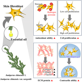
Essential oils derived from plants are major ingredients in the medical and cosmetic industry. Here, we evaluated nine types of plant essential oils to identify potential candidates with antioxidant and elasticity‐enhancing properties. Seven essential oils showed at least 10% radical scavenging activity at the highest concentration. Essential oils extracted from Aster glehnii, Cinnamomum cassia, Citrus unshiu, Juniperus chinensis L., and Juniperus chinensis var. sargentii significantly enhanced fibroblast viability, and oils from Cit. unshiu, J. chinensis L., and J. chinensis var. sargentii significantly increased cell proliferation and migration. Expression of extracellular matrix proteins, including collagen 1, collagen 3, and elastin, were upregulated by J. chinensis L. and J. chinensis var. sargentii oil, which also significantly enhanced the contractile activity of skin cells in a three‐dimensional gel contraction assay. The results suggest that J. chinensis L. and J. chinensis var. sargentii essential oils may be potential anti‐wrinkling and anti‐oxidative agents for future consideration of use in the medical and cosmetic industry.
Keywords: antioxidant, essential oils, Juniperus chinensis L., Juniperus chinensis var. sargentii , medicinal plant, skin elasticity
This study evaluated nine types of plant essential oils to select ingredients with antioxidant and elasticity‐enhancing properties. Juniperus chinensis L. and J. chinensis var. sargentii essential oils increased the antioxidant ability of fibroblasts and promoted their proliferation, thereby increasing production of proteins related to skin elasticity and contractile ability. These essential oils are potential anti‐oxidative agents.
Abbreviations
- 3D
three‐dimensional
- ABTS
2,2′‐azino‐bis‐(3‐ethylbenzothiazoline‐6‐sulfonate)
- BrdU
5‐bromo‐2‐deoxyuridine
- DW
distilled water
- ECM
extracellular matrix
- FBS
fetal bovine serum
- H2O2
hydrogen peroxide
- IC50
half maximal inhibitory concentrations
- MEM
modified Eagles' medium
- MTT
3‐(4,5‐dimethylthiazol‐2‐yl)‐2,5‐diphenyl tetrazolium bromide
- NHDFs
normal human dermal fibroblasts
- OD
optical densities
- ROS
reactive oxygen species
- SD
standard deviation
The medical and cosmetics market for skin health products has expanded in parallel with improved living standards and increased average life expectancy [1]. Antioxidant ability is the most important function required for functional cosmetic products that improve skin health [2]. When the skin is exposed to internal factors such as aging or external factors such as ultraviolet rays, reactive oxygen species (ROS) are generated in skin, and this ROS inhibits fibroblast proliferation in the dermal layer and the ability of fibroblasts to produce extracellular matrix (ECM) [3, 4]. Fibroblasts maintain skin elasticity, an indicator of skin health, by producing ECM proteins, such as collagen and elastin. Therefore, a decline in fibroblast activity results in a failure to synthesize ECM proteins and a consequent decrease in dermal ECM protein levels [5]. Of the various types of ECM proteins, collagen 1, collagen 3, and elastin account for the largest amount of protein in the dermal layer and are the primary skin components responsible for maintaining skin elasticity [6, 7]. Therefore, it is important for skin care products and cosmetics to contain ample collagen to eliminate ROS and enhance skin elasticity.
Currently, the active anti‐oxidative and anti‐wrinkle ingredients of functional cosmetics include tocopheryl acetate, tocopherol, ascorbic acid, niacinamide, and retinyl palmitate [8]. However, these are mostly organic synthetic materials that can cause skin irritation or have stability problems, and consequently, their use is being increasingly prohibited or restricted [9, 10]. Additionally, as the demand for mild and nature‐friendly raw materials has increased, interest in natural products is rising, and cosmetic companies are actively researching to replace problematic organic synthetics with natural alternatives [11].
Natural products are mostly composed of plant, animal, and microorganism secondary metabolites and some primary metabolites [12] and are biodegradable and usually have mild continuous actions, which results in few side effects and environmental friendliness [13, 14]. Plant natural products include many substances with antioxidant activities, such as essential oils [15]. These oils have strong aromatic properties and widely varied compositions and contents, which are contingent upon the unique characteristics of the plants [16]. For example, essential oils extracted from Pistacia atlantica have antioxidant effects on skin cells, while those extracted from lemons, juniper, and grapefruit have anti‐wrinkle effects [16, 17]. Also, essential oils extracted from the Manuka tree and Zingiber officinale Roscoe have anti‐wrinkle effects in mice [18].
As mentioned above, many essential plant oils have beneficial effects on skin. In this study, we screened Korean traditional native plants that showed potential for antioxidant efficacy from literature references [19, 20, 21, 22, 23, 24, 25]. Afterwards, plants for which the optimal concentration that could exhibit functionality in skin cells without causing cytotoxicity had not been identified were selected and used in this experiment. Thus, we analyzed and compared the antioxidant and physiological effects of nine plant essential oils on skin.
Materials and methods
Preparation of essential oils from nine types of plant
As shown in Table 1, the essential oils of nine medicinal plants were prepared by hydrodistilling different plant parts (National Institute of Forest Science, Republic of Korea). Each sample was mixed with distilled water (DW) in a ratio of 1 : 10 (kg : L). Mixtures were then heated at 102 °C using a heating mantle (cat. no. MS‐DM608; Misung Scientific, Gyeonggi‐do, Korea), and volatiles were condensed using a Dean‐Stark trap (National Institute of Forest Science, Republic of Korea). Finally, the essential oils were dehydrated using anhydrous sodium sulfite and stored at 4 °C until required.
Table 1.
Scientific names, abbreviations, common names, and plant parts used.
| No. | Scientific name | Abbreviation | Common name | Parts used |
|---|---|---|---|---|
| 1 | Aster glehnii F. Schmidt | A. glehnii | Ezo‐goma‐na | Grass clumps |
| 2 | Artemisia capillaris | Ar. capillaris | Yin Chen Hao | Grass clumps |
| 3 | Cinnamomum cassia | C. cassia | Chinese cinnamon | Leaves |
| 4 | Citrus natsudaidai Hayata | Cit. natsudaidai | Natsumikan | Peels |
| 5 | Citrus pseudo gulgul | Cit. pseudo gulgul | Hill lemon | Peels |
| 6 | Citrus unshiu | Cit. unshiu | Satsuma orange | Peels |
| 7 | Juniperus chinensis L. | J. chinensis L. | Chinese juniper | Leaves |
| 8 | Juniperus chinensis var. sargentii | J. chinensis var. sargentii | Sargent juniper | Leaves |
| 9 | Zanthoxylum piperitum | Z. piperitum | Japanese pepper | Fruits |
Cells and cell culture
Normal human dermal fibroblasts (NHDFs) were purchased from American Type Culture Collection (Manassas, VA, USA) and cultured in modified Eagles' medium (MEM; Welgene, Daegu, Republic of Korea) containing 10% fetal bovine serum (FBS; Sigma‐Aldrich, St. Louis, MO, USA) and 1% streptomycin/penicillin in a 5% CO2 incubator at 37 °C until the cell confluence reaches 100%. Cells were counted, and 5 × 10⁴ cells/well were cultured in 24‐well plates for MTT and BrdU assays for 24 h, and 2 × 10⁵ cells/well were cultured in 6‐well plates for western blot for 24 h. The cells were then treated with the essential oils for 24 h.
Determination of half maximal inhibitory concentrations (IC50) and highest non‐toxic concentration
An 3‐(4,5‐dimethylthiazol‐2‐yl)‐2,5‐diphenyl tetrazolium bromide (MTT) assay was performed to determine cell viabilities and IC50 values of the nine essential oils, as we previously described [26]. NHDFs were treated with the nine essential oils at various concentrations (Table 2) in culture medium in a 5% CO2 incubator for 24 h at 37 °C. After removing culture media, cells were treated with 500 μL of 0.5 mg·mL−1 MTT (Sigma‐Aldrich) and incubated in a 5% CO2 incubator for 4 h at 37 °C. MTT solution was then removed, 300 μL of DMSO was added to dissolve formazan crystals, and optical densities (OD) were measured at 570 nm using a microplate reader (Agilent Technologies, Waldbronn, Germany). Cell viability ratios were calculated using: OD sample/OD control × 100, and IC50 values were calculated using sigmaplot v.10.0 (Systat Software, San Jose, CA, USA).
Table 2.
Concentrations of plant essential oils used for each experiments.
| No. | Essential oil | Tested concentrations (p.p.m.) | ||||||
|---|---|---|---|---|---|---|---|---|
| MTT assay (IC50) | ABTS assay | MTT assay (H2O2) | BrdU assay | Scratch migration assay | Western blot analysis | Collagen gel contraction assay | ||
| 1 | Aster glehnii | 6.25, 12.5, 25, 50, 100 | 25 | 25 | 25 | 25 | 25 | 25 |
| 2 | Artemisia capillaris | 1.5625, 3.125, 6.25, 12.5, 25 | 3.125 | 3.125 | – | – | – | – |
| 3 | Cinnamomum cassia | 12.5, 25, 50, 100, 200 | 50 | 50 | 50 | 50 | 50 | 50 |
| 4 | Citrus natsudaidai | 6.25, 12.5, 25, 50, 100 | 12.5 | 12.5 | – | – | – | – |
| 5 | Citrus pseudo gulgul | 6.25, 12.5, 25, 50, 100 | 25 | 25 | – | – | – | – |
| 6 | Citrus unshiu | 6.25, 12.5, 25, 50, 100 | 25 | 25 | 25 | 25 | 25 | 25 |
| 7 | Juniperus chinensis L. | 12.5, 25, 50, 100, 200 | 25 | 25 | – | – | – | – |
| 8 | Juniperus chinensis var. sargentii | 12.5, 25, 50, 100, 200 | 25 | 25 | 25 | 25 | 25 | 25 |
| 9 | Zanthoxylum piperitum | 25, 50, 100, 200, 400 | 25 | 25 | 25 | 25 | 25 | 25 |
| Fig. 1 | Fig. 2 | Fig. 3 | Fig. 4 | Fig. 5 | Fig. 6 | Fig. 7 | ||
Measurement of the antioxidant effect of essential oils in the absence of cells
An 2,2′‐azino‐bis‐(3‐ethylbenzothiazoline‐6‐sulfonate) (ABTS) assay was used to assess the free radical scavenging activities of the essential oils. To determine oil concentrations, working ABTS solution (Sigma‐Aldrich) was diluted to different extents with distilled water to an OD of 0.7. Working ABTS solution and essential oils were mixed in the ratio 99 : 1 and incubated for 10 min in the dark at 37 °C, and then, 200 μL of each mixture was transferred to a 96‐well plate. OD values were measured at 734 nm.
Antioxidant effects of the essential oils against H2O2‐induced cell oxidative stress
Normal human dermal fibroblasts were seeded in 24‐well plates and cultured for 24 h in MEM containing 10% FBS and 1% streptomycin/penicillin at 37 °C. Cells treated with the essential oils at highest non‐toxic concentrations (refer to Table 2). To determine the antioxidant effects of the essential oils, 150 μm of hydrogen peroxide (H2O2; Sigma‐Aldrich), a negative control, was co‐treated with essential oils at a final concentration of 150 μm for 24 h. Cell viabilities were evaluated using the MTT assay.
Effect of the essential oils on cell proliferation
A Cell Proliferation ELISA kit (cat. no. 11647229001; Roche Diagnostics, Indianapolis, IN, USA) 5‐bromo‐2‐deoxyuridine (BrdU) was used to measure the cell proliferation, as we previously described [27]. Skin fibroblasts were treated with the essential oils at various concentrations (Table 2). After 24 h, 100 μL of BrdU solution (100 μm) was added to each well for 4 h. Then, 1 mL of FixDenat was added to each well for 30 min to fix the cells. After fixation, the cells were incubated with BrdU antibody for 90 min at room temperature. After washing, 500 μL of substrate solution was added to cells for 20 min, and 125 μL of H2SO4 (1 m) was added. OD values were measured at 450 nm.
Evaluation of the cell migration rate regulated by essential oils
Scratch migration assay was performed to analyze cell migration, as we previously described [27]. NHDFs were seeded into 24‐well plate and incubated for 24 h. The cells were scratched by a sterile 1 mL pipette tip and washed with PBS to remove detached cells. We added DMEM without FBS to the cells and treated the essential oils at the highest non‐toxic concentration (Table 2). The cells were incubated for 48 h and photographed at 0, 24, and 48 h. Scratched areas were visualized using a Nikon Eclipse Ts2 inverted microscope (Nikon Corporation, Tokyo, Japan).
Measurement of the translational levels of ECM proteins regulated by essential oils
Protein samples of NHDFs treated with essential oils were extracted using Pro‐Prep solution (iNtRON Biotechnology, Seoul, Korea), according to the manufacturer's protocol. A bicinchoninic acid assay was used to determine protein concentrations. Proteins were then loaded and separated by 8% SDS/PAGE and transferred to nitrocellulose membranes using a wet transfer system. Membranes were blocked for 2 h with 5% skimmed milk in PBS containing 0.05% Tween‐20 (PBST) at room temperature. Subsequently, membranes were incubated with antibodies against collagen 1 (1 : 500; cat. no. sc‐293 182; Santa Cruz Biotechnology, Dellas, TX, USA), collagen 3 (1 : 500; cat. no. sc‐271 249; Santa Cruz Biotechnology), elastin (1 : 1000; cat. no. ab21736; Abcam, Cambridge, UK), and GAPDH (1 : 3000; cat. no. #2118; Cell Signaling Technology, Danvers, MA, USA), which served as the internal control, overnight at 4 °C. Blots were then incubated for 1 h with horseradish peroxidase‐conjugated secondary antibodies (1 : 5000; cat. nos. ADI‐SAB‐100 and ADI‐SAB‐300; Enzo Life Science, Farmingdale, NY, USA) in 5% skimmed milk in PBST for 1 h at room temperature. Luminol (Bio‐Rad, Hercules, CA, USA) was used to visualize antibody binding. Blots were scanned using Gel Doc 1000 version 1.5 (Bio‐Rad), and protein band intensities were normalized versus GAPDH.
Contractile abilities of essential oils in three‐dimensional (3D) skin fibroblast cultures
A collagen gel contraction assay was used to assess contractile abilities of skin fibroblasts, as we previously described, to confirm the contractile activities of 3D cultured NHDFs (a skin model). In brief, a mixture of 4.7 mg·mL−1 collagen type 1, 10× PBS, and 1 N NaOH was prepared to obtain a collagen solution with a pH of 7.4 at a concentration of 4.13 mg·mL−1 [28]. Separately, NHDFs (2 × 10⁵ cells·mL−1) were prepared in DMEM without FBS. The collagen solution and NHDFs were then mixed to provide a final concentration of collagen of 1.5 mg·mL−1, and 300 μL/well of this gel mixture was dispensed into a 12‐well plate. The gels were then incubated for 1 h at 37 °C to allow collagen lattices to form. DMEM supplemented with 1% streptomycin/penicillin was added to the control and experimental groups, but DMEM supplemented with 5% FBS and 1% streptomycin/penicillin was used as positive control. The essential oils were then applied at the highest non‐toxic concentration (Table 2), and the lattices were physically detached from culture dishes. Gel images were captured at 0 and 24 h, and lattice areas were measured using image j software (National institutes of Health, Bethesda, MD, USA). The original areas of collagen gels were estimated immediately after treatment and used as internal controls. Contractile activities for each condition were determined in duplicate, and results were expressed as the mean ratio of changed diameter of collagen gel ± standard deviation (SD).
Statistical analyses
Data are presented as the mean ± SD. Data were analyzed using one‐way analysis of variance (ANOVA; spss for Windows, 10.10, standard version; SPSS Inc., Chicago, IL, USA). Means obtained from three independent experiments were evaluated using one‐way ANOVA and Tukey's post hoc t‐test for multiple comparisons. A value of P < 0.05 was considered to indicate a statistically significant difference.
Results
IC50 values and highest non‐toxic concentrations of the nine essential oils for skin fibroblasts
Normal human dermal fibroblasts were treated with essential oils extracted from Aster glehnii F. Schmidt E, Artemisia capillaris, Zanthoxylum piperitum, Cinnamomum cassia, Juniperus chinensis L., Juniperus chinensis var. sargentii, Citrus natsudaidai Hayata, Citrus pseudo gulgul, and Citrus unshiu in a wide range of concentrations (Table 2). We constructed cell viability curves and determined IC50 values using the MTT assay. The IC50 values for each essential oil were as follows: J. chinensis var. sargentii; 100.367 p.p.m. > C. cassia; 89.175 p.p.m. > J. chinensis L; 73.674 p.p.m. > Z. piperitum; 73.002 p.p.m. > A. glehnii F. Schmidt; 45.969 p.p.m. > Cit. unshiu; 38.43 > Cit. pseudo gulgul; 36.646 p.p.m. > Cit. natsudaidai Hayata; 34.838 p.p.m. > Ar. capillaris; 7.608 p.p.m. (Fig. 1). The test concentrations and highest non‐toxic concentrations of each essential oil were determined using these results and then used in various experiments as shown in Table 2.
Fig. 1.
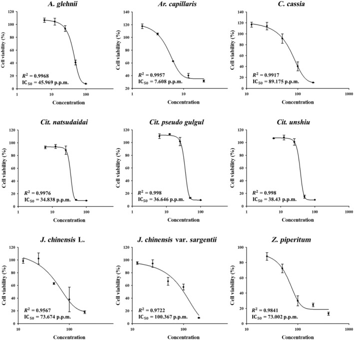
IC50 values of essential oils extracted from nine plants for skin dermal fibroblasts. Cells were treated with the nine essential oils in a wide range of concentrations, and cell viabilities were determined using the MTT assay (n = 3). Results are expressed as means ± SDs.
ABTS radical scavenging abilities of the essential oils
We evaluated the antioxidant characteristics of the essential oils using the ABTS assay. Essential oils were used at the same concentration as those used for the MTT assay to validate that plant essential oils at the test concentrations established in the MTT assay exhibited antioxidant efficacy. Seven of the nine essential oils showed at least 10% radical scavenging activity at the highest concentrations; the essential oils of Cit. natsudaidai and Cit. pseudo gulgul were the two exceptions (Fig. 2). However, to assess more accurately the antioxidant effects of essential oils on cells, we performed MTT assay under H2O2‐induced oxidative stress in NHDFs.
Fig. 2.
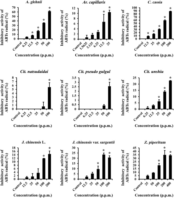
Antioxidant effects of essential oils in the absence of cells. The ABTS assay was used to assess the radical scavenging activity of the nine essential oils in a wide range of concentrations (n = 3). Results are expressed as means ± SDs. Statistical analysis: t‐test; *P < 0.05 significant difference compared to the control group.
Protective effects of essential oils against H2O2‐induced oxidative stress
We used the MTT assay to examine the protective effects of essential oils against H2O2‐induced oxidative stress. NHDFs were treated with the nine essential oils at their highest non‐toxic concentrations (Table 2). One hundred and fifty micromolar H2O2 decreased skin fibroblast viability to 60% (Fig. 3). The essential oils extracted from A. glehnii, C. cassia, Cit. unshiu, J. chinensis L., and J. chinensis var. sargentii significantly recovered cell viability to 72–106%. However, essential oils extracted from Ar. capillaris, Cit. natsudaidai, Cit. pseudo gulgul, and Z. piperitum had no protective effect. Therefore, we further investigated the effects of these five essential oils.
Fig. 3.
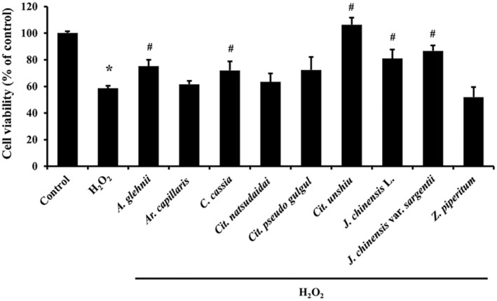
Antioxidant effects of essential oils on skin dermal fibroblasts. The protective effect of NHDFs on H2O2‐induced oxidative stress was investigated using the MTT assay (n = 3). Results are expressed as means ± SDs. Statistical analysis: t‐test; *P < 0.05 significantly different versus control group not treated with H2O2. # P < 0.05 significantly different with H2O2 treated group.
Effects of essential oils on NHDFs proliferation
The BrdU assay was used to estimate the proliferation of skin fibroblasts. NHDFs were treated with test concentrations of the essential oils of A. glehnii, C. cassia, Cit. unshiu, J. chinensis L., and J. chinensis var. sargentii. Essential oils extracted from Cit. unshiu, J. chinensis L., and J. chinensis var. sargentii significantly increased NHDFs proliferation versus controls (Fig. 4). The proliferation rate increased in the order J. chinensis var. sargentii, J. chinensis L., Cit. unshiu. However, A. glehnii and C. cassia did not regulate cell proliferation.
Fig. 4.
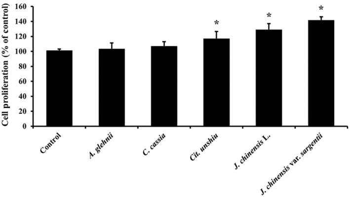
Effects of the five essential oils on skin fibroblast proliferation. The proliferations of NHDFs treated with the five essential oils were estimated using the BrdU assay (n = 3). Results are expressed as means ± SDs. Statistical analysis: t‐test; *P < 0.05 significant difference compared to the control.
Effects of the essential oils on migration of NHDFs
A scratch migration assay was performed to determine whether the essential oils affect cell migration. The essential oils extracted from A. glehnii and C. cassia did not regulate migration rate compared to controls at both 24 and 48 h. However, the essential oils of C. unchiu, J. chinensis L. and J. chinensis var. sargentii increased migration rate significantly compared to controls at both 24 h (about 68–80%) and 48 h (about 88–96%) (Fig. 5A,B).
Fig. 5.
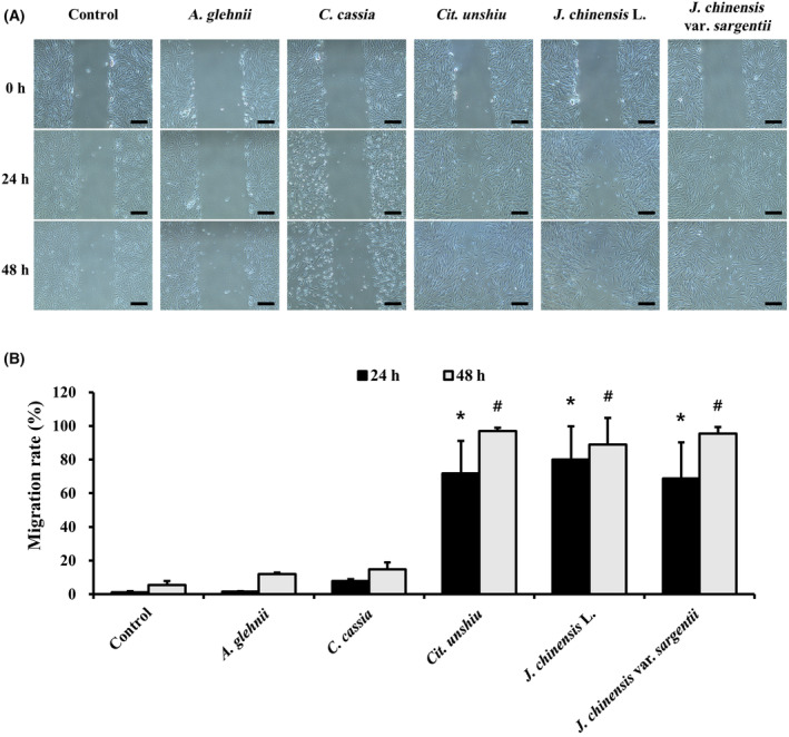
Cell migration of NHDFs treated with the five essential oils were analyzed using the scratch migration assay (A). Scale bars represent 200 μm. Migration rates are presented as graphs (B) (n = 3). Results are expressed as means ± SDs. Statistical analysis: t‐test; *P < 0.05 significant difference compared to the control (24 h). # P < 0.05 significant difference compared to the control (48 h).
Effects of the essential oils on ECM protein expression in skin fibroblasts
Extracellular matrix protein levels were measured by western blot (Fig. 6A). The essential oils extracted from J. chinensis L. and J. chinensis var. sargentii significantly increased collagen 1 expression (Fig. 6B). The essential oils extracted from A. glehnii, C. cassia, Cit. unshiu, and J. chinensis L. increased expression of collagen 3 non‐significantly, but J. chinensis var. sargentii upregulated the expression of collagen 3 significantly (Fig. 6C). The expression of elastin was increased non‐significantly by essential oils extracted from A. glehnii, C. cassia, Cit. unshiu, J. chinensis L., and J. chinensis var. sargentii (Fig. 6D).
Fig. 6.
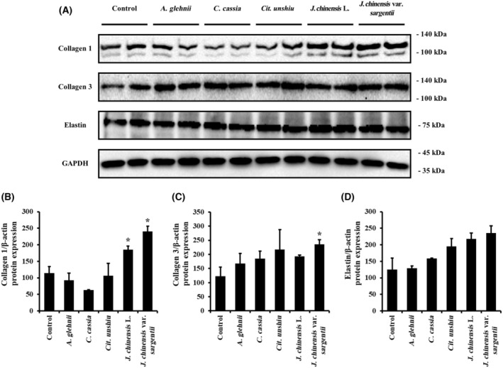
Translational levels of ECM proteins in NHDFs treated with the five essential oils. The protein expressions of ECM proteins were measured by western blot to evaluate protein production by fibroblasts treated with the five essential oils (A). The expression levels of (B) collagen 1, (C) collagen3, and (D) elastin are represented as graphs normalized versus GAPDH levels (n = 3). Results are expressed as means ± SDs. Statistical analysis: t‐test; *P < 0.05 significantly different versus controls.
Effects of the essential oils on 3D‐cultured skin fibroblasts
Normal human dermal fibroblasts were cultured in a 3D environment using a collagen lattice to mimic the natural skin environment, and contractile abilities were assessed (Fig. 7A). The control group exhibited ~23% contraction of the collagen lattice, while the positive control group treated with 5% FBS showed a substantial contraction of ~76% (Fig. 7B). Essential oils extracted from A. glehnii, C. cassia, and Cit. unshiu did not induce significant shrinkage of the collagen lattice compared to controls. However, the essential oils of J. chinensis L. and J. chinensis var. sargentii induced a contraction of ~45%, indicating a significant increase in contractile activity versus controls.
Fig. 7.
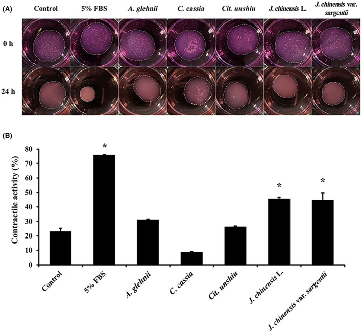
Gel contraction of 3D cultured NHDFs treated with essential oils (A). Contractile ratios are presented as histograms (B) (n = 3). Results are expressed as means ± SDs. Statistical analysis: t‐test; *P < 0.05 significant difference compared to the control.
Discussion
Skin elasticity is an indicator of healthy skin and has a significant esthetic effect [29]. Skin elasticity is an emotionally important factor because unhealthy skin, which loses elasticity, can cause psychological distress [30]. Consequently, the medical and cosmetics industry continues to focus on developing healthier, environmentally friendly ingredients with minimal side effects [31, 32]. As a result, there is an increasing need to explore the possible utilization of medicinal plant resources and investigate their antioxidant properties and their impacts on extracellular matrix proteins associated with skin elasticity [33]. In this study, we evaluated the effects of essential oils extracted from nine medicinal plants on skin elasticity using skin fibroblasts.
Antioxidant activity is closely linked to the ability to remove ROS, which includes free radicals such as and OH· and non‐radicals like H2O2 and 1O2 [34]. While ROS is necessary for normal cellular functions, excessive accumulation of ROS can lead to oxidative stress due to an imbalance between ROS production and antioxidant defense mechanisms. Oxidative stress can damage DNA, proteins, and lipids and contribute to the transformation of normal cells into cancer cells by causing mutations in key genes [35]. Furthermore, oxidative stress leads to the oxidation of DNA and mitochondria and subsequent cellular damage and cell death [36]. In our ABTS assay, we used a spectrophotometric method to assess antioxidant activity and found that seven of the nine plant essential oils exhibited at least 10% ABTS radical scavenging activity at the highest concentration tested [37]. Notably, A. glehnii, C. cassia, Cit. unshiu, and J. chinensis var. sargentii significantly scavenged radicals at concentrations below their IC50 values. In addition, the radical removal rate was increased in the order C. cassia, A. glehnii, Cit. unshiu, J. chinensis var. sargentii, and J. chinensis L. at the maximum non‐toxic concentration. Furthermore, A. glehnii, C. cassia, Cit. unshiu, J. chinensis L., and J. chinensis var. sargentii significantly inhibited the death of skin fibroblasts induced by H2O2, a representative ROS that induces oxidative stress leading to cell death [22, 36]. Therefore, these five plant essential oils demonstrated antioxidant properties irrespective of cellular activity.
Oxidative stress can impair cell viability, proliferation, and protein synthesis, and thus, intracellular antioxidant ability is beneficial as it acts to prevent cellular damage [38, 39]. Cit. unshiu, J. chinensis L. and J. chinensis var. sargentii significantly increased NHDFs proliferation and migration. The skin fibroblasts play a crucial role in maintaining skin elasticity by producing ECM proteins [40, 41]. When we examined ECM contents after treating NHDFs with essential oils, J. chinensis L. and J. chinensis var. sargentii were found to promote ECM protein production. In particular, collagen 1, collagen 3, and elastin were highly upregulated by the essential oils. Collagen is the most abundant ECM protein and plays an essential role in maintaining skin elasticity by preserving skin structure and enhancing physiological functions [42]. Also, elastin contributes to the unique elasticity of various connective tissues, plays a vital role in skin homeostasis, and is involved in dermal elasticity [43]. When NHDFs were cultured in a 3D environment, J. chinensis L. and J. chinensis var. sargentii significantly enhanced contractile activity [44]. Therefore, our findings show that J. chinensis L. and J. chinensis var. sargentii exhibit radical scavenging activity, act as antioxidants in NHDFs, and promote the survival, proliferation, and migration of these cells. Moreover, the activation of skin fibroblasts leads to the production of collagen and elastin, which are essential components of the ECM and increase the contractile activity of skin cells.
In this study, we did not confirm the compositional analysis of each plant essential oil, but we suggest that polyphenols or ascorbic acid may be the active ingredients in the oils. Polyphenols are a type of aromatic alcohol compounds found in plants. It has been reported that it prevents aging through its antioxidant effect, has the ability to protect DNA from damage due to exposure to active oxygen and has excellent functions to protect cellular proteins and enzymes, thereby lowering the risk of various diseases [45, 46]. Ascorbic acid is a water‐soluble vitamin found in plants, including citrus fruits. It is a powerful antioxidant that neutralizes free radicals and is reported to play an important role in collagen formation and is used as an ingredient in anti‐aging cosmetics [47].
Consequently, of the nine plant essential oils investigated in this study, those of J. chinensis L. and J. chinensis var. sargentii demonstrated medicinal potential as substances that enhance skin health and elasticity. These findings suggest that the essential oils of J. chinensis L. and J. chinensis var. sargentii could be used as novel ingredients that improve skin health and elasticity. Most commercially existing antioxidants are organic synthetic substances that could irritate the skin or have stability problems, while plant essential oils are mild and nature friendly. Therefore, these plant essential oils are expected to replace organic synthetic substances as natural antioxidants.
Conflict of interest
The authors declare no conflict of interest.
Author contributions
DSK, MJK, and B‐SA designed the experiments and wrote the manuscript. DSK, MJK, M‐JP, B‐JA, and S‐MA performed the experiments and analyzed the data. W‐JY and B‐SA confirmed the authenticity of all the raw data. M‐JP and B‐SA analyzed the data and revised the manuscript. All authors have read and approved the final manuscript.
Acknowledgements
The present study was performed with the support of the National Institute of Forest Science (Project no. FP0702‐2016‐03‐2020) and was partially supported by the BK21 FOUR Program (grant no. F20YY8109033) through the National Research Foundation of Korea (NRF), funded by the Ministry of Education, Korea.
Da Som Kim and Min Jae Kim contributed equally to this study.
Data accessibility
The datasets used and/or analyzed during the current study are available from the corresponding author on reasonable request.
References
- 1. Millikan LE (2001) Cosmetology, cosmetics, cosmeceuticals: definitions and regulations. Clin Dermatol 19, 371–374. [DOI] [PubMed] [Google Scholar]
- 2. Kusumawati I and Indrayanto G (2013) Natural antioxidants in cosmetics. Stud Nat Prod Chem 40, 485–505. [Google Scholar]
- 3. Masaki H (2010) Role of antioxidants in the skin: anti‐aging effects. J Dermatol Sci 58, 85–90. [DOI] [PubMed] [Google Scholar]
- 4. Merecz‐Sadowska A, Sitarek P, Kucharska E, Kowalczyk T, Zajdel K, Cegliński T and Zajdel R (2021) Antioxidant properties of plant‐derived phenolic compounds and their effect on skin fibroblast cells. Antioxidants 10, 726. [DOI] [PMC free article] [PubMed] [Google Scholar]
- 5. Baumann L (2007) Skin ageing and its treatment. J Pathol 211, 241–251. [DOI] [PubMed] [Google Scholar]
- 6. Tzaphlidou M (2004) The role of collagen and elastin in aged skin: an image processing approach. Micron 35, 173–177. [DOI] [PubMed] [Google Scholar]
- 7. Rossetti D, Kielmanowicz M, Vigodman S, Hu Y, Chen N, Nkengne A, Oddos T, Fischer D, Seiberg M and Lin C (2011) A novel anti‐ageing mechanism for retinol: induction of dermal elastin synthesis and elastin fibre formation. Int J Cosmet Sci 33, 62–69. [DOI] [PubMed] [Google Scholar]
- 8. Silva S, Ferreira M, Oliveira A, Magalhães C, Sousa M, Pinto M, Sousa Lobo J and Almeida I (2019) Evolution of the use of antioxidants in anti‐ageing cosmetics. Int J Cosmet Sci 41, 378–386. [DOI] [PubMed] [Google Scholar]
- 9. Ding Y, Pyo S and Müller R (2017) smartLipids® as third solid lipid nanoparticle generation–stabilization of retinol for dermal application. Pharmazie 72, 728–735. [DOI] [PubMed] [Google Scholar]
- 10. Zhao W, Yang A, Wang J, Huang D, Deng Y, Zhang X, Qu Q, Ma W, Xiong R and Zhu M (2022) Potential application of natural bioactive compounds as skin‐whitening agents: a review. J Cosmet Dermatol 21, 6669–6687. [DOI] [PubMed] [Google Scholar]
- 11. Stallings AF and Lupo MP (2009) Practical uses of botanicals in skin care. J Clin Aesthet Dermatol 2, 36. [PMC free article] [PubMed] [Google Scholar]
- 12. Chinou I (2008) Primary and secondary metabolites and their biological activity. Chromatogr Sci Ser 99, 59. [Google Scholar]
- 13. Lourith N and Kanlayavattanakul M (2009) Natural surfactants used in cosmetics: glycolipids. Int J Cosmet Sci 31, 255–261. [DOI] [PubMed] [Google Scholar]
- 14. Emerald M, Emerald A, Emerald L and Kumar V (2016) Perspective of natural products in skincare. Pharm Pharmacol Int J 4, 1–3. [Google Scholar]
- 15. Guzmán E and Lucia A (2021) Essential oils and their individual components in cosmetic products. Cosmetics 8, 114. [Google Scholar]
- 16. Falleh H, Jemaa MB, Saada M and Ksouri R (2020) Essential oils: a promising eco‐friendly food preservative. Food Chem 330, 127268. [DOI] [PubMed] [Google Scholar]
- 17. Kwon OS, Jung SH and Yang BS (2013) Topical administration of manuka oil prevents UV‐B irradiation‐induced cutaneous photoaging in mice. Evid Based Complement Alternat Med 2013, 930857. [DOI] [PMC free article] [PubMed] [Google Scholar]
- 18. Feng J, Du Z, Zhang L, Luo W, Zheng Y, Chen D, Pan W, Yang Z, Lin L and Xi L (2018) Chemical composition and skin protective effects of essential oil obtained from ginger (Zingiber officinale Roscoe). J Essent Oil Bear Plants 21, 1542–1549. [Google Scholar]
- 19. Ahn C, Yoo Y‐M, Park M‐J, Ham Y, Yang J and Jeung E‐B (2021) Cytotoxic evaluation of the essential oils from Korean native plant on human skin and lung cells. J Korean Wood Sci Technol 49, 371–383. [Google Scholar]
- 20. Kačániová M, Galovičová L, Valková V, Tvrdá E, Terentjeva M, Žiarovská J, Kunová S, Savitskaya T, Grinshpan D and Štefániková J (2021) Antimicrobial and antioxidant activities of Cinnamomum cassia essential oil and its application in food preservation. Open Chem 19, 214–227. [Google Scholar]
- 21. Tanaka T, Sugiura H, Inaba R, Nishikawa A, Murakami A, Koshimizu K and Ohigashi H (1999) Immunomodulatory action of citrus auraptene on macrophage functions and cytokine production of lymphocytes in female BALB/c mice. Carcinogenesis 20, 1471–1476. [DOI] [PubMed] [Google Scholar]
- 22. Kim MJ, Mohamed EA, Kim DS, Park M‐J, Ahn B‐J, Jeung E‐B and An B‐S (2022) Inhibitory effects and underlying mechanisms of Artemisia capillaris essential oil on melanogenesis in the B16F10 cell line. Mol Med Rep 25, 1–12. [DOI] [PMC free article] [PubMed] [Google Scholar]
- 23. Lee S‐G, Oh S‐C and Jang J‐S (2015) Antioxidant activities of Citrus unshiu extracts obtained from different solvents. Korean J Food Nutr 28, 458–464. [Google Scholar]
- 24. Park S‐A, Jegal J, Chung KW, Jung HJ, Noh SG, Chung HY, Ahn J, Kim J and Yang MH (2018) Isolation of tyrosinase and melanogenesis inhibitory flavonoids from Juniperus chinensis fruits. Biosci Biotechnol Biochem 82, 2041–2048. [DOI] [PubMed] [Google Scholar]
- 25. Yamazaki E, Inagaki M, Kurita O and Inoue T (2007) Antioxidant activity of Japanese pepper (Zanthoxylum piperitum DC.) fruit. Food Chem 100, 171–177. [Google Scholar]
- 26. Kim DS, Lee H, Kim MJ, Seong K‐Y, Jeong JS, Kim SY, Jung E‐M, Yang SY and An B‐S (2022) Dissolving biopolymer microneedle patches for the improvement of skin elasticity. J Ind Eng Chem 111, 200–210. [Google Scholar]
- 27. Kim MJ, Seong K‐Y, Jeong JS, Kim SY, Lee S, Yang SY and An B‐S (2022) Minoxidil‐loaded hyaluronic acid dissolving microneedles to alleviate hair loss in an alopecia animal model. Acta Biomater 143, 189–202. [DOI] [PubMed] [Google Scholar]
- 28. An B‐S, Ahn H‐J, Kang H‐S, Jung E‐M, Yang H, Hong E‐J and Jeung E‐B (2013) Effects of estrogen and estrogenic compounds, 4‐tert‐octylphenol, and bisphenol A on the uterine contraction and contraction‐associated proteins in rats. Mol Cell Endocrinol 375, 27–34. [DOI] [PubMed] [Google Scholar]
- 29. Baumann L, Bernstein EF, Weiss AS, Bates D, Humphrey S, Silberberg M and Daniels R. Clinical relevance of elastin in the structure and function of skin. Paper Presented at: Aesthetic Surgery Journal Open Forum 2021. [DOI] [PMC free article] [PubMed]
- 30. Wang Y‐N, Fang H and Zhu W‐F (2009) Survey on skin aging status and related influential factors in Southeast China. J Zhejiang Univ Sci B 10, 57–66. [DOI] [PMC free article] [PubMed] [Google Scholar]
- 31. Kerdudo A, Burger P, Merck F, Dingas A, Rolland Y, Michel T and Fernandez X (2016) Development of a natural ingredient–natural preservative: a case study. C R Chim 19, 1077–1089. [Google Scholar]
- 32. Amberg N and Fogarassy C (2019) Green consumer behavior in the cosmetics market. Resources 8, 137. [Google Scholar]
- 33. Ribeiro AS, Estanqueiro M, Oliveira MB and Sousa Lobo JM (2015) Main benefits and applicability of plant extracts in skin care products. Cosmetics 2, 48–65. [Google Scholar]
- 34. Das K and Roychoudhury A (2014) Reactive oxygen species (ROS) and response of antioxidants as ROS‐scavengers during environmental stress in plants. Front Environ Sci 2, 53. [Google Scholar]
- 35. Sammar M, Abu‐Farich B, Rayan I, Falah M and Rayan A (2019) Correlation between cytotoxicity in cancer cells and free radical‐scavenging activity: in vitro evaluation of 57 medicinal and edible plant extracts. Oncol Lett 18, 6563–6571. [DOI] [PMC free article] [PubMed] [Google Scholar]
- 36. Poljšak B and Dahmane R (2012) Free radicals and extrinsic skin aging. Dermatol Res Pract 2012, 135206. [DOI] [PMC free article] [PubMed] [Google Scholar]
- 37. Miliauskas G, Venskutonis P and Van Beek T (2004) Screening of radical scavenging activity of some medicinal and aromatic plant extracts. Food Chem 85, 231–237. [Google Scholar]
- 38. Nishiyama Y, Allakhverdiev SI and Murata N (2011) Protein synthesis is the primary target of reactive oxygen species in the photoinhibition of photosystem II. Physiol Plant 142, 35–46. [DOI] [PubMed] [Google Scholar]
- 39. Birben E, Sahiner UM, Sackesen C, Erzurum S and Kalayci O (2012) Oxidative stress and antioxidant defense. World Allergy Organ J 5, 9–19. [DOI] [PMC free article] [PubMed] [Google Scholar]
- 40. McCabe MC, Hill RC, Calderone K, Cui Y, Yan Y, Quan T, Fisher GJ and Hansen KC (2020) Alterations in extracellular matrix composition during aging and photoaging of the skin. Matrix Biol Plus 8, 100041. [DOI] [PMC free article] [PubMed] [Google Scholar]
- 41. Tracy LE, Minasian RA and Caterson E (2016) Extracellular matrix and dermal fibroblast function in the healing wound. Adv Wound Care 5, 119–136. [DOI] [PMC free article] [PubMed] [Google Scholar]
- 42. Bolke L, Schlippe G, Gerß J and Voss W (2019) A collagen supplement improves skin hydration, elasticity, roughness, and density: results of a randomized, placebo‐controlled, blind study. Nutrients 11, 2494. [DOI] [PMC free article] [PubMed] [Google Scholar]
- 43. Weihermann A, Lorencini M, Brohem C and De Carvalho C (2017) Elastin structure and its involvement in skin photoageing. Int J Cosmet Sci 39, 241–247. [DOI] [PubMed] [Google Scholar]
- 44. Fujimura T, Moriwaki S, Imokawa G and Takema Y (2007) Crucial role of fibroblast integrins α2 and β1 in maintaining the structural and mechanical properties of the skin. J Dermatol Sci 45, 45–53. [DOI] [PubMed] [Google Scholar]
- 45. Panche AN, Diwan AD and Chandra SR (2016) Flavonoids: an overview. J Nutr Sci 5, e47. [DOI] [PMC free article] [PubMed] [Google Scholar]
- 46. Shen N, Wang T, Gan Q, Liu S, Wang L and Jin B (2022) Plant flavonoids: classification, distribution, biosynthesis, and antioxidant activity. Food Chem 383, 132531. [DOI] [PubMed] [Google Scholar]
- 47. Akram NA, Shafiq F and Ashraf M (2017) Ascorbic acid‐a potential oxidant scavenger and its role in plant development and abiotic stress tolerance. Front Plant Sci 8, 613. [DOI] [PMC free article] [PubMed] [Google Scholar]
Associated Data
This section collects any data citations, data availability statements, or supplementary materials included in this article.
Data Availability Statement
The datasets used and/or analyzed during the current study are available from the corresponding author on reasonable request.