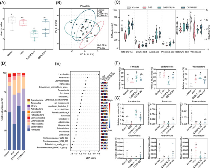
Figure 3.

Influence of Latilactobacillus sakei on gut microbiota in colitis mice. (A) α‐Diversity of gut microbiota in colitis mice. (B) β‐Diversity of the gut microbiota in colitis mice. (C) Effects of L. sakei on SCFAs content in colitis mice. (D, F) Effects of L. sakei on the phylum level of the gut microbiota in colitis mice. (E) LEfSe difference of gut microbiota in colitis mice after intervention by L. sakei. (G) The relative abundance of different bacterial species after L. sakei intervention. Different superscript lowercase letters (a–c) in the graph indicate significant differences between groups (p < 0.05) within the row by the Kruskal–Wallis test (Control, n = 7; DSS, n = 5; CCFM1267, n = 6; QJSNT1L10, n = 7). DSS, dextran sulfate sodium; LDA, linear discriminant analysis; PC, principal component; PCA, principal component analysis; SCFA, short‐chain fatty acid.
