Significance
We demonstrated that IL7 increases translation in T cells, increasing the expression of protein from delivered mRNA. We utilized LNP conjugated with a CD5 antibody (tLNP) carrying mRNA to transfect T cells to express the reporter protein mCherry and investigated methods to enhance protein expression after tLNP treatment. Culturing T cells with IL7 prior to tLNP exposure increased mCherry expression in T cells in vitro. In vivo, pretreating mice with systemic IL7 before tLNP injection increased the number of mCherry+ T cells in the spleen and lymph nodes. RNA sequencing showed the upregulation of pathways associated with translation when CD8 were cultured with IL7. These results point to unique ways to enhance tLNP-mediated in vivo engineering of T cells.
Keywords: IL7, translation, lipid nanoparticles
Abstract
The use of lipid nanoparticles (LNP) to encapsulate and deliver mRNA has become an important therapeutic advance. In addition to vaccines, LNP-mRNA can be used in many other applications. For example, targeting the LNP with anti-CD5 antibodies (CD5/tLNP) can allow for efficient delivery of mRNA payloads to T cells to express protein. As the percentage of protein expressing T cells induced by an intravenous injection of CD5/tLNP is relatively low (4-20%), our goal was to find ways to increase mRNA-induced translation efficiency. We showed that T cell activation using an anti-CD3 antibody improved protein expression after CD5/tLNP transfection in vitro but not in vivo. T cell health and activation can be increased with cytokines, therefore, using mCherry mRNA as a reporter, we found that culturing either mouse or human T cells with the cytokine IL7 significantly improved protein expression of delivered mRNA in both CD4+ and CD8+ T cells in vitro. By pre-treating mice with systemic IL7 followed by tLNP administration, we observed significantly increased mCherry protein expression by T cells in vivo. Transcriptomic analysis of mouse T cells treated with IL7 in vitro revealed enhanced genomic pathways associated with protein translation. Improved translational ability was demonstrated by showing increased levels of protein expression after electroporation with mCherry mRNA in T cells cultured in the presence of IL7, but not with IL2 or IL15. These data show that IL7 selectively increases protein translation in T cells, and this property can be used to improve expression of tLNP-delivered mRNA in vivo.
The development of mRNA lipid nanoparticles (LNP-mRNAs) that can be directly injected into patients has been an important therapeutic advance with a wide range of potential uses, as demonstrated by the extremely successful COVID-19 vaccines. The LNP used in the COVID-19 vaccines were not targeted and after intramuscular injection were taken up largely by phagocytic cells (dendritic cells and macrophages). However, other uses of LNP require uptake by specific cell types in specific organs in situ. One approach to realize this specificity requirement has been the generation of customized lipid formulations that have allowed some degree of organ-specific delivery (1, 2). Targeted LNP (tLNP) provide another useful strategy to effectively deliver RNA to particular cell types based on the expression of cell surface markers. These tLNP are coated with a targeting antibody allowing for the delivery of the mRNA cargo to specific cells of interest. Upon binding of the antibody to its respective target on the cell surface, the LNP is internalized and trafficked to the endolysosomal compartment from where the mRNA is released into the cytoplasm and the cargo mRNA is translated into protein.
We have performed proof-of-concept studies delivering mRNA to different cell types, such as endothelial cells, T cells, and hematopoietic stem cells (3–6). T cells are an attractive cell type for tLNP as they can carry an array of cargoes including siRNA, genome editors, activating cytokines, tumor microenvironment activators, checkpoint inhibitor antibodies, bispecific T cell–activating antibodies, and chimeric antigen receptors (CARs) (7–9). Previous studies have used this technology to induce the expression of marker proteins like mCherry (10) as well as functional proteins such as a genome editor [cre recombinase (4)]. CAR mRNAs are an especially interesting T cell cargo in that in situ delivery could bypass the need for ex vivo transduction using lentivirus or retroviral systems (which have insertional mutagenesis risks), reduce the labor intensiveness, shorten the lengthy ex vivo CAR production process, and alleviate the need for lymphodepletion prior to CAR infusion (11, 12). Recently, our group has shown that CD5-targeted LNP (CD5/tLNP) could express an mRNA encoding a CAR targeting fibroblast activation (FAP CAR) protein and that T cells expressing this CAR were able to reverse experimental cardiac fibrosis (3). This study demonstrated the feasibility of generating functional CAR T cells in situ using tLNPs.
LNP efficiency in delivering functional mRNA payload may be significantly limited by uptake in the endolysosomes, payload release into the cytoplasm aided by the ionizable lipid within the LNP matrix, and/or translation of the mRNA to protein—all influenced by the metabolic activation state of the cells (13). Hence, since tLNP-mRNA is a recently developed technology, there has been limited investigation into adjunct strategies that might enhance their ability to express proteins in vivo. It is unknown what “threshold/level” of transfected cells is required for therapeutic effects in both preclinical and clinical contexts. For example, 20% of T cells expressing a FAPCAR was sufficient to reduce heart fibrosis in a murine model (3), but a higher level of expression may be required for a solid tumor or other applications, especially since the mRNA is transient. Hence, the identification of ways to improve protein expression levels could be vital for the effectiveness and success of in situ cellular engineering with tLNP-mRNA.
To study this question, we used tLNP conjugated with a CD5 targeting antibody to transfect CD4+ and CD8+ T cells. CD5 is a marker expressed highly on the surface of T cells in both mice and humans and at low levels on B cells, NK cells, and myeloid cells (14, 15). These tLNP carried mRNA coding for the model reporter protein mCherry, allowing us to easily measure the protein expression efficiency with flow cytometry.
We found that activating T cells by stimulating the TCR using αCD3, with or without CD28 costimulation, improved tLNP-mediated protein expression of mRNA cargo in vitro, but not in vivo. Treating T cells with the common gamma chain cytokine IL7 improved tLNP-mediated protein expression in both human and mouse T cells in vitro and was effective at increasing the total number of CD4+ and CD8+ T cells expressing the reporter protein in vivo. When CD8+ T cells were cultured with IL7 (compared to either IL2 or IL15), we observed an enrichment of translation-related pathways and genes and a parallel increase in protein expression after electroporation of mRNA. Our data thus suggest that treatment with IL7 increases the ability of T cells to express protein after delivery of mRNA by tLNP. These findings may be translatable to clinically viable approaches to enable increased mRNA-based engineering of T cells in vivo, that may be necessary in disease indications with higher therapeutic efficacy bar.
Results
T Cell Activation Augments Protein Expression in T Cells Transfected with CD5/tLNP Containing mCherry mRNA.
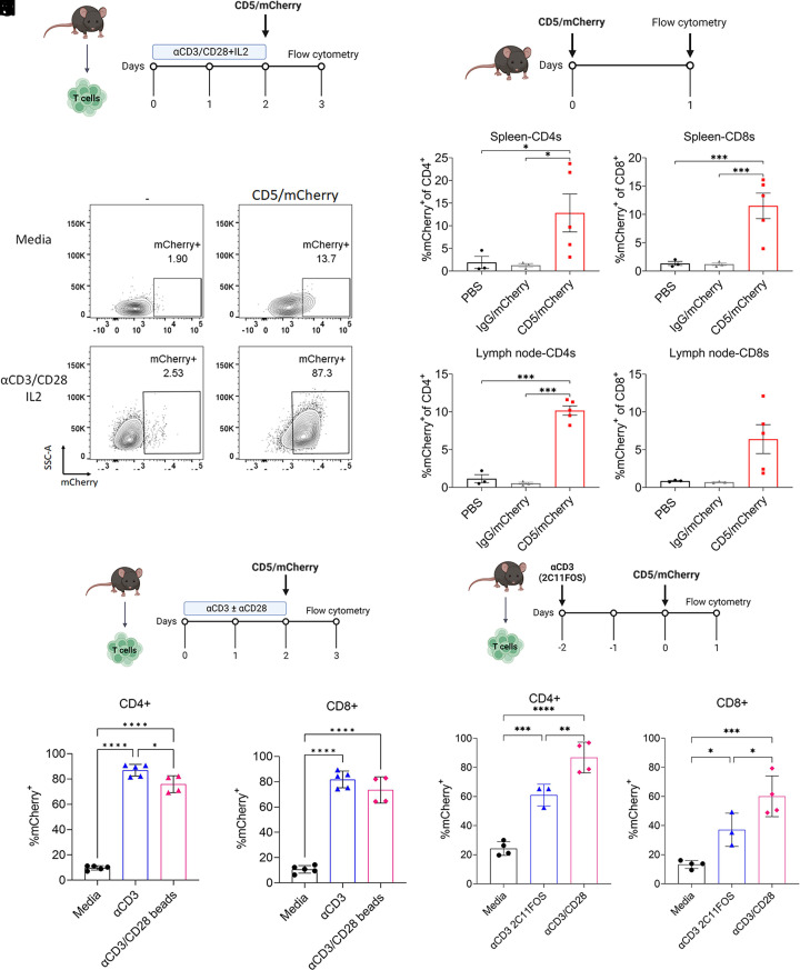
We initially defined the efficiency of protein expression of anti-CD5 directed tLNP carrying mRNA coding for the reporter protein mCherry (referred to as CD5/LNP-mCherry) in either “resting” (inactivated) or activated mouse T cells. T cells were isolated from the spleens of C57BL/6 mice and cultured in T cell media with or without the addition of αCD3/CD28 beads and recombinant murine IL2 (Fig. 1A). After 48 h, the beads were removed, 1 µg of CD5/LNP-mCherry was added per million cells, and expression of mCherry was measured 24 h later using flow cytometry. The proportion of mCherry-expressing T cells was low in the resting T cells, with only ~12% of T cells expressing mCherry (Figs. 1B and 2B). After activation with αCD3/CD28 beads, however, the expression rate improved dramatically, with 75 to 80% of T cells expressing mCherry (Figs. 1B and 2B—P < 0.001). These findings suggest that metabolically active T cells are more amenable to tLNP-mRNA-mediated engineering.
Fig. 1.
Activating T cells using anti-CD3 antibodies improves tLNP-induced mCherry expression rates in vitro but not in vivo. (A) Experimental design for in vitro T cell activation. T cells were magnetically isolated from the spleens of C57BL/6 mice, activated with αCD3/CD28 beads, and supplemented with IL2. Forty-eight hours later, beads were removed, 1 µg of tLNP was added per million cells, and flow cytometry was performed 24 h later. (B) Representative flow plots of nonactivated (media) and activated T cells treated with mCherry tLNP. (C) Experimental design for the in vivo experiment. Mice were given 10 µg of IgG or anti-CD5-targeted LNP i.v, with spleen and lymph nodes collected 24 h later. (D and E) Percent mCherry+ CD4+ (D) or CD8+ (E) T cells in the spleen. (F and G) Percent mCherry+ CD4+ (F) or CD8+ (G) T cells in the lymph node. (H) Experimental design. T cells were isolated and cultured with 1 µg/mL αCD3 or αCD3/CD28 beads for 48 h. One microgram of CD5/mCherry tLNP was added per million cells and incubated for an additional 24 h before flow cytometry was performed (I and J) Percentage of mCherry+ CD4+ (I) or CD8+ (J) T cells. (K) Experimental design. T cells were isolated and cultured with or without 10 µg/mL αCD3 2C11FOS for 48 h while bead-activated T cells were used as a positive control. One microgram of CD5-mCherry tLNP was added per million cells and incubated for an additional 24 h before flow cytometry was performed (L and M) Percentage of mCherry+ CD4+ (L) or CD8+ (M) T cells after tLNP transfection. One-way ANOVA with Sidak’s test was used for multiple comparisons. *P < 0.05, **P < 0.01, ***P < 0.001, and ****P < 0.0001.
Fig. 2.
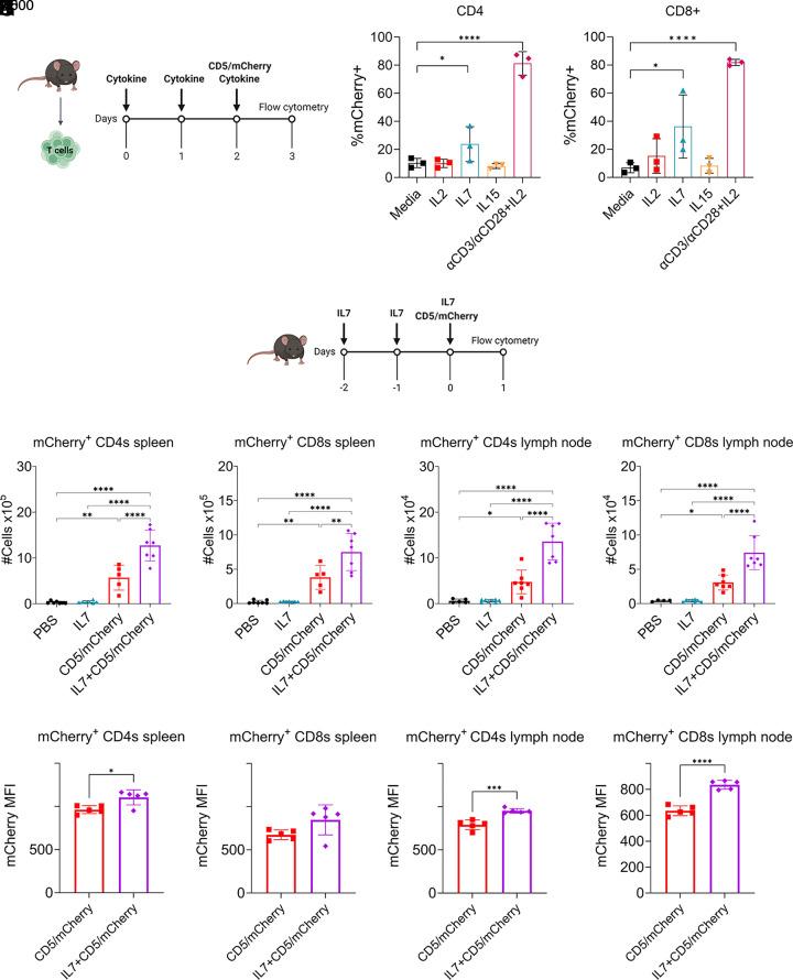
IL7 enhances CD5/mCherry tLNP-induced protein expression in vitro and in vivo. (A) Experimental design for in vitro cytokine treatment. T cells were isolated and cultured with either IL2, IL7, or IL15. Cytokines were refreshed daily, and tLNPs were added on day 2. (B and C) Proportion of mCherry+ CD4+ (B) and CD8+ (C) T cells in vitro in the presence of IL2, IL7, or IL15. Media-treated and activated T cells were used as controls. (D) Experimental design for in vivo experiments. C57BL/5 mice were injected i.p with 5 µg of recombinant murine IL7 daily for 3 d. On the third day, mice received 10 µg CD5-mCherry tLNPs i.v. Twenty-four hours after tLNP treatment, spleens and lymph nodes were collected for flow cytometry. (E and F) Total number of CD4+ (E) and CD8+ (F) T cells in the spleen expressing mCherry. (G and H) Total number of CD4+ (G) and CD8+ (H) T cells in the lymph node expressing mCherry. (I and J) Median fluorescent intensity (MFI) of mCherry of the mCherry+ CD4+ (I) and CD8+ (J) T cells in the spleen. (K and L) MFI of mCherry of the mCherry+ CD4+ (K) and CD8+ (L) T cells in the spleen. One-way ANOVA with Sidak’s test was used for multiple comparisons. *P < 0.05, **P < 0.01, ***P < 0.001, and ****P < 0.0001.
tLNP Transfect in 5 to 15% of T Cells In Vivo in Naive Mice.
We next tested our targeted LNP in vivo in naive mice. We administered either IgG/LNP-mCherry or CD5/LNP-mCherry i.v to mice and performed flow cytometry 24 h later (Fig. 1C). We found that between 5 and 15% of CD4+ and CD8+ T cells in the spleen (Fig. 1 D and E) and lymph nodes (Fig. 1 F and G) expressed mCherry. In contrast, no mCherry expression was seen in T cells after injection of LNP conjugated with IgG, indicating that there was minimal nonspecific uptake of the tLNP by T cells.
CD3 Stimulation Is Required for Effective Protein Expression from tLNP in T Cells In Vitro but Is Ineffective In Vivo.
Although we observed 5 to 15% of T cells expressed mCherry protein after i.v injection of tLNP, it is unknown whether this expression level for a relatively short period of time would be sufficient in a therapeutic context where the mRNA cargo encodes for a CAR, cytokine, or another protein. As these mice were naive and otherwise “healthy” with their T cells in a low activation state, i.e., resting, we hypothesized that tLNP-mediated protein expression could be increased by also activating the T cells in vivo.
Since activation beads stimulate both the CD3/TCR and CD28 pathways, we next investigated whether both CD3 and CD28 stimulation were required for the T cells to effectively express protein after tLNP treatment and whether antibody-mediated stimulation was equivalent to the coated beads. T cells were thus isolated from the spleens of mice and activated for 48 h using plate-bound anti-CD3 or anti-CD3/CD28 beads (Fig. 1H). CD5/LNP-mCherry were then added and incubated for 24 h. Activating T cells with the αCD3 resulted in 87% of CD4+ and 82% of CD8+ T cells expressing mCherry (Fig. 1 I and H).
Although CD3 stimulation substantially improved protein expression induced by tLNP in vitro, the clinical use of full-length anti-CD3 antibodies is associated with immunosuppression and severe side effects, such as the systemic release of cytokines due to cross-linking between T cells and Fc receptors on antigen-presenting cells (16, 17). However, an Fc silent anti-CD3 antibody, Teplizumab, was recently approved for use in type 1 diabetes (18) and was well tolerated. Therefore, we next investigated whether a murine equivalent non-Fc receptor binding form of anti-CD3 (2C11FOS) (17, 19, 20) had a similar effect on increasing tLNP efficacy.
We repeated the in vitro experiments following a similar protocol, incubating the mouse T cells with 2C11FOS for 48 h before adding CD5/LNP-mCherry (Fig. 1K). The antibody did not activate the T cells, as measured by CD25, PD-1, or CD69 expression (SI Appendix, Fig. S1). However, 2C11FOS did increase the tLNP-induced mCherry expression in CD4+ (Fig. 1L) and CD8+ (Fig. 1M) T cells, from 24% to 61% (P = 0.0008) and 13% to 37% (P = 0.049) expressing mCherry, respectively. We thus tested the antibody in vivo, administering it as one large dose (100 µg) systemically with tLNP given 24 h later (SI Appendix, Fig. S2A). Although the antibody caused some activation of the T cells, resulting in a small but insignificant increase in CD69, ICOS, and PD-1 (SI Appendix, Fig. S1B), the addition of the anti-CD3 antibody did not increase the number or the percentage of mCherry+ T cells in the spleen (SI Appendix, Fig. S2 B and C). In contrast, 2C11FOS decreased the proportion of mCherry-expressing cells when compared to tLNP alone from 30% to 15% mCherry+ CD4+ T cells (P < 0.0001) and from 27% to 10% mCherry+ CD8+ T cells (P < 0.0001, SI Appendix, Fig. S2 D and E).
In summary, although both full-length and non-FcR binding anti-CD3 antibodies improved the protein expression of tLNP delivered mRNA in vitro, unexpectedly, this strategy was ineffective in vivo.
Pretreating T Cells with IL7 Increases tLNP Transfection Rates Both In Vitro and In Vivo.
Since tLNP-induced protein expression was not increased in vivo using anti-CD3 antibodies, we hypothesized that an alternative form of activation, treatment with common gamma chain cytokines, could increase protein expression. We thus compared three cytokines, IL2, IL7, and IL15, which have a variety of reported effects on CD8 (vs. CD4) T cells in vitro and have been used successfully in the clinic or in clinical trials.
T cells were isolated from the spleens of mice and cultured in the presence of anti-CD3/CD28 beads, recombinant murine IL2, IL7, or IL15 for 48 h, then transfected with CD5/LNP-mCherry, and incubated for an additional 24 h (Fig. 2A). Consistent with prior results, 81% and 84% of anti-CD3/CD28 bead-activated CD4+ (Fig. 2B) and CD8+ T cells (Fig. 2C), respectively, expressed mCherry, while 10% (P < 0.0001) of resting T cells CD4+ and 7% of resting CD8+ (P < 0.0001) expressed mCherry. IL2 and IL15 had no effect on tLNP-induced expression of mCherry+, similar to those cultured without cytokines. However, IL7 significantly increased the expression rate compared to resting T cells, increasing the proportion of mCherry-expressing CD4+ T cells severalfold to 24% (P = 0.012) and to 36% (P = 0.002) within the CD8+ subset (Fig. 2 B and C).
We next investigated whether IL7 increased tLNP-mediated protein expression in vivo. To prime the T cells, recombinant murine IL7 was given daily by intraperitoneal injection for 3 d, with CD5/LNP-mCherry dosed on the third day (Fig. 2D). Spleens and lymph nodes were collected 24 h after tLNP injection and analyzed. IL7 pretreatment significantly increased mCherry expression. As IL7 has reported effects on increasing T cell proliferation, we quantified the total number of T cells expressing mCherry in the spleen and lymph nodes. IL7 did not cause a change in the proportion of CD4+ or CD8+ T cells of CD45+ leukocytes (SI Appendix, Fig. S3A). However, there was a significant increase in the number of T cells expressing mCherry, with the total number of mCherry+ CD4+ and CD8+ T cells increasing in both the spleen (CD4+ P < 0.0001, CD8+ P = 0.004, Fig. 2 E and F) and lymph node (CD4+ P < 0.0001, CD8+ P < 0.0001, Fig. 2 G and H). This represents an approximately twofold increase in mCherry-expressing T cells in the spleen and a threefold and twofold increase in mCherry+ in CD4+ and CD8+ T cells in the lymph node, respectively. The proportion of mCherry+ CD4+ and mCherry+CD8+ T cells also increased (SI Appendix, Fig. S3B). Additionally, the average level of mCherry+ protein expression per cell was increased with IL7 pretreatment. The MFI of mCherry in mCherry+CD4+ T cells in the spleen (Fig. 2I, P = 0.012), mCherry+CD4+ T cells in the lymph node (Fig. 2K, P = 0.0004), and mCherry+CD8+ T cells in the lymph node (Fig. 2L, P = <0.0001) were all significantly increased when the CD5/mCherry tLNPs were combined with IL7. There was a trend toward increased mCherry expression in CD8+ T cells in the spleen (Fig. 2J, P = 0.067).
We also tested the effects of IL15, another common gamma chain cytokine, given before CD5-mCherry tLNP injection and found that it did not significantly increase the in vivo protein expression rate (SI Appendix, Fig. S4).
Since activating T cells in vitro increased tLNP-induced protein expression, we looked at whether IL7 treatment increased the expression of activation markers on T cells in vivo. IL7 did not increase CD69 expression on CD4 and CD8 T cells in the spleen and lymph nodes (SI Appendix, Fig. S3C). There was a trend toward increased ICOS expression in mice treated with IL7 alone (SI Appendix, Fig. S3D). Last, we were interested in whether IL7 increases CD5 expression on T cells, resulting in an increase of the target on the surface for the tLNP to recognize, bind, and then be endocytosed. We found that CD5 expression was similar between PBS- and IL7-treated mice (SI Appendix, Fig. S3E), showing that increased availability of the tLNP target was not responsible for the increased protein expression. Altogether, these results show that a clinically applicable gamma-chain receptor cytokine IL7 may enable both in vivo and ex vivo T cell engineering with tLNP, but the mechanism may not include receptor expression upregulation.
IL7-Treated T Cells Selectively Up-Regulate Pathways Associated with Protein Translation.
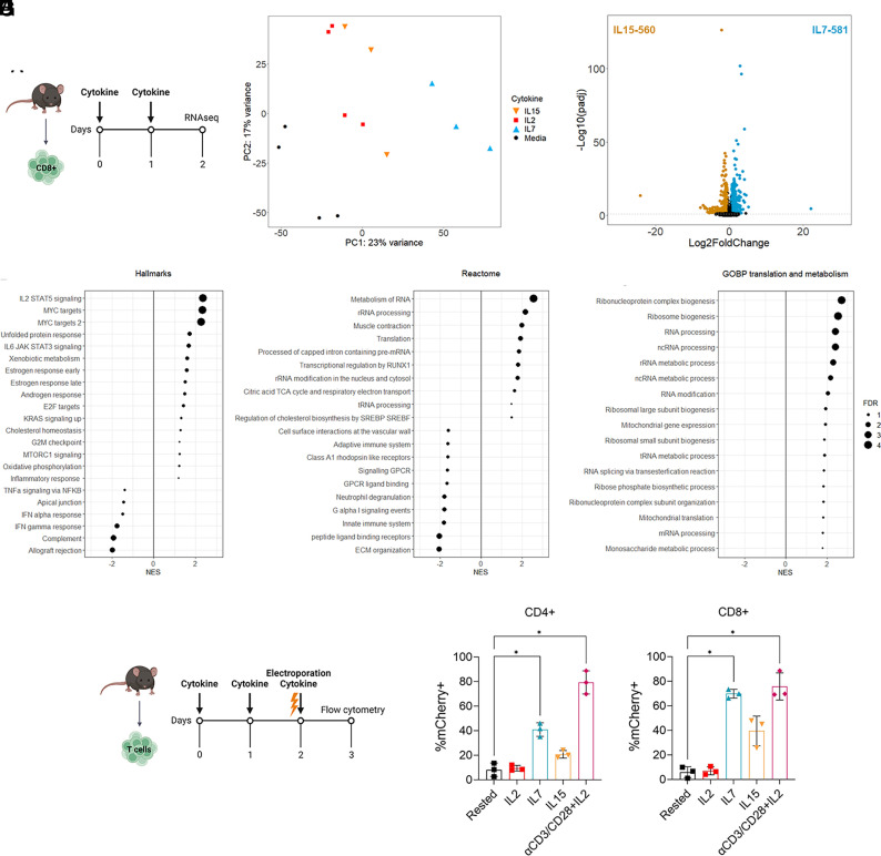
To identify the mechanism(s) behind the effect of IL7 on tLNP-mediated payload protein expression, we performed bulk RNA sequencing on purified CD8+ T cells cultured in T cell media alone or in the presence of IL2, IL7, or IL15 for 48 h (Fig. 3A). Principal component analysis (PCA) showed a clear separation between IL7-treated cells and cells treated with either IL2 or IL15 (Fig. 3B), indicating that they have a distinct transcriptomic profile. T cells cultured in the absence of cytokines were separated clearly from the cytokine-treated T cells. As cells cultured in media alone or with IL2 had poor viability (SI Appendix, Fig. S5A), we were concerned that the presence of dead cells in these samples could potentially interfere within the sequencing data. Therefore, we focused our primary transcriptomic analyses on comparing cells cultured with IL7 to those cultured with IL15, which had a similar viability, expansion rate, and CD5 expression (SI Appendix, Fig. S5 A-C), but different abilities to augment protein expression after tLNP transfection.
Fig. 3.
IL7 increases specific changes in genes related to protein translation in T cells and supports increased protein production after mRNA electroporation. (A) Experimental design. CD8+ T cells were isolated from the spleens of C57BL/6 mice and cultured with T cell media alone or supplemented with IL2, IL7, or IL15. After 48 h, T cells were collected and sent for bulk RNA sequencing. (B) PCA of cytokine-treated T cells. Counts were VST normalized. (C) Volcano plot showing the differentially expressed genes between IL7- and IL15-treated CD8 T cells. Genes with a positive log2fold change are up-regulated with IL7 treatment compared to IL15, while a negative log2fold change indicates upregulation with IL15 treatment compared to IL7. (D–F) Gene set enrichment analysis using the list of differentially expressed genes between IL7- and IL15-treated cells using the Hallmarks (D), Reactome (E), or Gene Ontology Biological Processes (GOBP) (F) databases. Gene sets associated with translation and metabolism are shown from the GOBP analysis. Size of point indicates the FDR (−log10Padj), with a positive NES indicating enrichment in IL7-treated cells and a negative NES indicating enrichment in IL15-treated cells. (G) Experimental design for electroporation experiment. T cells were isolated from the spleen C57BL/6 mice and either activated using CD3/CD28 beads or cultured in T cell media supplemented with IL2, IL7, or IL15. After 48 h T cells were electroporated with 2 µg of mCherry mRNA per 1 million cells. mCherry expression was measured 24 h later. (H and I) Proportion of mCherry+ CD4+ (H) or CD8+ (I) T cells after electroporation with mCherry mRNA. One-way ANOVA with Sidak’s test was used for multiple comparisons. *P < 0.05, **P < 0.01, ***P < 0.001, and ****P < 0.0001.
Gene expression analysis identified 1,141 differentially expressed genes (581 up and 560 down) between IL7- and IL15-treated CD8+ T cells (Fig. 3C). We next performed gene set enrichment analysis (21) using this list of differentially expressed genes against the Hallmarks (22), Reactome, and Gene Ontology databases. IL7 treatment was associated with the upregulation of “IL2 STAT5 signaling” and “MYC associated” pathways, while inflammatory and type I and II IFN-related genes were up-regulated with IL15 (Fig. 3D). Consistent with the results obtained from the Hallmarks gene set collection, IL15-treated CD8+ T cells up-regulated Reactome pathways associated with the immune system and interferons compared to IL7 (Fig. 3E).
Interestingly, pathways associated with protein translation, RNA processing, and cell metabolism were selectively enriched in IL7-treated T cells (Fig. 3 E and F). Similar results were obtained when T cells cultured in IL7 were compared with IL2 treatment, showing up-regulated pathways associated with cell division while IL7 up-regulating pathways relating to translation, MYC, and IL2 STAT5 signaling (SI Appendix, Fig. S5). Altogether, these results point to specific T cell metabolic activities that are upmodulated by IL7, including translational activity, that may enable tLNP mediated engineering.
IL7-Treated T Cells Express Higher Levels of mCherry After mRNA Electroporation.
Our genomic analysis suggested a potential mechanism for the increased tLNP protein expression efficiency observed after IL7 treatment could be an increase in translation efficiency of the T cells. To directly assess the effects of IL7 on protein translation, we electroporated mCherry mRNA into T cells cultured in T cell media alone or supplemented with IL2, IL7, or IL15 (Fig. 3 G–I). Since the transfection of T cells by tLNP involves the binding of the tLNP to their cognate target on the surface of the cell, the endocytosis of the tLNP and its processing and degradation to release the mRNA into the cytosol for translation into a functional protein (3), the removal of the LNP component by directly electroporating the mRNA into the cell allowed us to specifically focus the translation aspect of this process.
As in previous experiments, T cells were isolated from the spleens of mice and cultured either in T cell media alone or supplemented with IL2, IL7, or IL15 (Fig. 3G). Bead-activated T cells were used as a control as they readily express electroporated mRNA. Consistent with our earlier studies using tLNP, a low proportion of rested T cells expressed mCherry after electroporation (CD4+ 8%, CD8+ 6%) while up to 80% of CD4+ (P = 0.024) and 76% of CD8+ (P = 0.035) expressed mCherry after activation with αCD3/CD28 beads (Fig. 3 H and I). IL2 treatment had little effect on increasing the rate of mCherry expression (CD4+ P = 0.991; CD8+ P = 0.999), and IL15 led to an increase in expression rate in CD4+ T cells to 21% and to 40% in CD8+ T cells; however, these changes did not reach statistical significance (CD4+, P = 0.114, CD8+, P = 0.234). In contrast, IL7 had a dramatic effect on electroporation efficiency, increasing the proportion of mCherry+ in CD4+ T cells fivefold compared to resting T cells, from 8% to 40% (P = 0.025, Fig. 3H). This was even more pronounced in CD8+ T cells with a nearly 12-fold increase from 6% to 70% (P = 0.017), with mCherry+ cells nearly equivalent to those activated with αCD3/CD28 beads (Fig. 3I).
These data support our transcriptomic analyses, indicating that IL7 increased translation of electroporated mRNA and supporting the hypothesis that this cytokine enables tLNP-mRNA engineering of T cells through increasing cells’ translational capabilities.
IL7 Pretreatment of Human T Cells Improves the Transfection Efficiency of CD5/LNP-mCherry In Vitro.
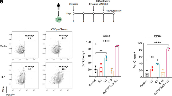
We next sought to confirm whether IL7—a clinically applicable cytokine—has the same effect at increasing tLNP-induced protein expression in human T cells. T cells were isolated from PBMCs and either activated using anti-CD3/CD28 beads and IL2 or cultured in T cell media alone or supplemented with recombinant human IL2, IL7, or IL15 (with the cytokines replenished every 48 h). After 72 h in culture, the T cells were treated with CD5/mCherry-LNP, and mCherry expression and analyzed 24 h later (Fig. 4A). 20% and 11% of rested CD4+ and CD8+ T cells, respectively, expressed mCherry. Consistent with the studies using mouse T cells, αCD3/CD28 bead-activated human T cells express high levels of mCherry (Fig. 4 B–D), which increased to 89% (P < 0.0001, Fig. 4C) and 83% (P = 0.0001, Fig. 4D), respectively, after activation. IL2 and IL15 had no significant effect on the amount of tLNP-induced protein expression of either CD4+ or CD8+ T cells. However, similar to our mouse studies, IL7 had a strong effect in increasing the proportion of mCherry+ T cells after CD5-mCherry tLNP transfection. Within the CD4+ T cells, the expression rate increased from 20% in resting cells to 54% (P = 0.001, Fig. 4C), and for CD8+ T cells, the increase was of a similar magnitude from 11% to 40% after IL7 treatment (P = 0.0248, Fig. 4D).
Fig. 4.
IL7 pretreatment of human T cells improves CD5-mCherry tLNP-induced protein expression rates in vitro. (A) Experimental design for tLNP transfection. Human T cells were isolated from PBMC and either activated using anti-CD3/CD28 beads with IL2 or cultured in T cell media supplemented with IL2, IL7, or IL15 and replenished after 48 h. After 72 h, T cells were transfected with 0.6 µg of CD5-LNP-mCherry per 2 × 10⁵ cells. mCherry expression was measured 24 h later. (B) Representative flow plots of rested (Media) and IL7-cultured T cells transfected with CD5-LNP mCherry. Data are from two donors across three experiments. (C and D) Proportion of mCherry+ CD4+ (B) or CD8+ (C) T cells after transfection with mCherry mRNA. One-way ANOVA with Sidak’s test was used for multiple comparisons. *P < 0.05, **P < 0.01, ***P < 0.001, and ****P < 0.0001.
Last, we screened a larger panel of human cytokines for their ability to increase of tLNP-induced protein expression of mCherry. Unlike IL7, we saw no increased expression after treatment with IL2, IL4, IL7, IL4+IL-7, IL15, and IL7+IL15 (SI Appendix, Fig. S6 A and B) or after treatment with IL18, TNFα, IL6, IL9, IL12, IL21, IL27, and IL33 (SI Appendix, Fig. S6 C and D).
In summary, IL7 increases tLNP-induced protein expression in human T cells, with an increased proportion of T cells expressing mCherry when cultured with IL7, an effect not seen with IL2, IL15, or other common gamma chain cytokines.
Discussion
tLNP encapsulating mRNA are a relatively newly developed technology, and there has thus been limited investigation into methods to optimize their ability to enhance protein expression from their mRNA cargo, especially in vivo. Studies that have injected tLNP into mice have shown a range of protein expression levels in T cells from 4 to 8% targeting CD3 (10) and up to 20% when targeting CD4 or CD5 (3, 4). Similarly, we found that mCherry expression ranged from 5 to 15% in the spleen after using CD5/tLNP. It is unknown what threshold/level of protein expression by re-engineered cells is required for therapeutic effects in both preclinical and clinical contexts as the therapeutic efficacy bar to achieve may vary widely. Hence, identification of ways to improve payload protein expression levels could be vital for the effectiveness and success of in situ tLNP-driven cell engineering, especially in indications with high therapeutic efficacy bar.
We found that in vitro TCR stimulation using plate-bound anti-CD3 antibodies, with or without the presence of CD28 costimulation, increased the level of protein expression induced by CD5/LNP-mCherry in vitro. Therefore, we were interested whether this could be translated into an in vivo setting. Since systemic administration of full-length anti-CD3 antibodies (such as OKT3) leads to severe side effects in patients due to an increase in T cell proliferation and the release of inflammatory cytokines (16, 23–26), we instead used antibodies with a modified Fc portion. These antibodies have reduced binding affinity to the Fc receptors (FcR) on antigen-presenting cells. This reduces cross-linking and decreases the magnitude of T cell activation through the CD3/TCR complex on T cells, reducing cytokine release and therefore side effects (27, 28). Therefore, we studied the non-FcR binding antibody 2C11FOS (17, 19), the murine equivalent of teplizumab which has been recently approved for use in diabetes (18). We hypothesized that partial stimulation of T cells using the non-FcR binding anti-CD3 would lead to partial stimulation through the TCR and increase tLNP protein expression without adverse side effects. However, in contrast to the in vitro results, pretreatment with 2C11FOS before CD5/mCherry tLNP administration surprisingly led to a decrease in the proportion of mCherry-expressing T cells. Although this finding is quite interesting, given this lack of effect, we did not conduct additional studies, so can only speculate based on the literature. One reason as to the differences observed in vitro and in vivo relate to the degree of cross-linking of the antibody that occurred. The ex vivo anti-CD3 data shown In Fig. 1 utilized plate-bound anti-CD3 antibodies. Binding the antibodies to the plate augments cross-linking and activates the T cells to a much larger degree than adding soluble antibody and thus triggers TCR signaling with many of the accompanying changes, such as increased translation. Administration of the 2C11FOS antibody (which has impaired Fc activity) largely prevents cross-linking in vivo. The modified non-cross-linked anti-CD3 antibody, which is known to be immunosuppressive, is thought to exert its effects via internalization of the TCR, partial activation of the TCR pathway leading to anergy and exhaustion, and an eventual reduction in the number of T cells. These events do not appear to acutely activate the translational pathway. To increase protein translation in T cells in vivo, both CD3 and CD28 stimulation may be required. However, administration of anti-CD28 antibodies is not feasible clinically due to the severe side effects associated with treatment (29), and we did not explore this combination in our study.
Accordingly, we explored another potentially clinically translatable approach to “activate” T cells using three common γ-chain cytokines. IL2 acts to stimulate the proliferation, activation, and effector function of T cells, along with promoting the survival and differentiation of memory T cells (30). IL7 is critical for T cell development in the thymus, T cell maturation and differentiation (31), promotes the survival of T cells in the periphery, maintains T cell homeostasis, and can enhance T cell production by CD4+ and CD8+ T cells (32). IL15 has similar effects, promoting the survival and proliferation of T cells, development of memory T cells, and enhances production of cytokines by T cells and directs cytotoxic activity of CD8+ T cells (33). Interestingly, our experiments showed that IL7 was much more effective than IL2, IL15, and all of the other common γ chain cytokines, as well as IL6, IL27, IL33, and TNFα (in human T cells) in increasing protein expression levels after tLNP administration in mice in vivo and in isolated mouse and human T cells in vitro.
Clinically, repeated doses of IL7 have been shown to be well tolerated and can induce T cell expansion with little toxicity (34–36). CYT107, a glycosylated recombinant IL7 has been widely used and has similar immunological effects (37, 38). As IL7 causes the expansion of both CD4+ and CD8+ T cells, this adds an additional benefit to combining IL7 with tLNP in a clinical setting as it could not only augment protein expression, but also increase the total number of T cells, some of which would be transfected with the mRNA cargo. Thus, the use of preconditioning with a short course of IL7 before tLNP injection may be a translationally viable and useful approach. This is a paradigm shift from lymphodepletion preconditioning widely used in context of ex vivo engineered CAR T cells and presents several advantages including avoidance of hematological toxicity and preservation or possibility to co-opt an intact endogenous immune system.
For effective tLNP-mediated transfection, the antibody-targeted particles must bind to their cognate ligands on the surface of the cell of interest, be endocytosed, processed and then release the mRNA cargo into the cytoplasm where it is then translated into a functional protein (3, 4). To explore possible mechanisms for this selective IL7 effect, we conducted transcriptomic studies comparing the gene changes seen in resting cells cultured in media alone or in media supplemented with IL2, IL7, or IL-15. We focused primarily on the comparison between IL7- and IL15-treated cells because the cell viability was much higher under these conditions than the cells treated with IL2 or cultured in media alone. However, we also compared IL7 to IL2 and the results were consistent with comparing IL7 to IL15. Our data were rather striking in showing that IL7-treated T cells were enriched for pathways associated with MYC and a large variety of ribosomal and translation-associated gene sets, whereas in the IL15-treated cells, pathways involved in the innate immune system, interferon responses, and G-protein coupled receptor signaling were enriched. The functional significance of these changes on protein translation was demonstrated by showing that culturing T cells with IL7 before mRNA electroporation led to an increase in the mRNA-encoded protein expressed, in this case mCherry, when compared to IL2, IL15, or T cells cultured in media alone in the absence of cytokines. These data thus show that a major mechanism behind the increase in tLNP-induced protein expression by IL7 is likely enhancing translation of administered mRNA in T cells. We did not explore other mechanisms in detail, however, after treatment with IL7, we saw no difference in CD5 expression (which could have potentially increased the availability of the tLNP target), nor the enrichment of endocytosis-related genes.
The effects of TCR stimulation and T cell activation through anti-CD3/CD28 antibodies on both mRNA production and translation have been previously reported (39, 40). Stimulation of the TCR leads to a 30- to 100-fold increase in protein production and a 1,000-fold increase in the number of T cells actively producing protein (41). Activating T cells leads to an increase in tRNA writer proteins which enhances translation efficiency (39). This then leads to rapid and enhanced translation and synthesis of key functional proteins to promote T cell expansion and proliferation, one important example being MYC (a protein that we also saw up-regulated by IL7). The marked increase in protein translation of mCherry mRNA in anti-CD3/CD28 activated T cells after electroporation is very consistent with these findings, indicating that the increased protein expression after tLNP treatment in activated T cells is in part associated with an increase in translational capacity. Our data indicate that IL7 has a similar effect.
To our knowledge, the ability of IL7 to augment protein translation in T cells has not been reported, although a recent paper provides some indirect evidence by showing that the amount of monosome-associated ribosomal protein mRNA was positively correlated with the expression of CD127 (an IL7 receptor) when T cells were not stimulated with antigen (42). However, it has been reported that culturing human T cells in IL7 increases the transduction of the cells by lentiviral and HIV-derived vectors compared to untreated cells (43, 44). Given our findings, it would be interesting to try to determine whether these effects may be at least partially due to increased mRNA translation versus increased proliferation or other virally related mechanisms.
Although this study showed that IL7 could increase tLNP-induced expression of a well-described marker mRNA, mCherry, it will be of interest to extend these findings to actual therapeutic cargos such as CARs, cytokines, or bispecific antibodies. Preliminary data from our lab, in fact, indicate that IL7 can increase expression levels of the fibroblast activation protein targeted CAR that we showed had in situ efficacy in cardiac fibrosis (3) (manuscript in preparation).
In conclusion, we show that IL7, a clinically utilizable cytokine, can be used in vitro and in vivo to improve the protein expression of tLNP delivered mRNA. Importantly, we show that one mechanism behind this finding is an increase in the translational capacity of T cells by IL7. We thus propose that this property can be used to improve delivered mRNA translation in vivo after tLNP administration.
Materials and Methods
Mouse T Cell Isolation.
To isolate mouse T cells for in vitro experiments, spleens were collected from C57BL/6 mice and pushed through a 70-μm filter into serum-free RPMI media. Cells were collected via centrifugation; the supernatant was removed and resuspended in 2 mL Pharm Lyse (BD Biosciences) per spleen for red blood cell lysis. After incubation at RT for 5 min, an equal volume of PBS+2%FBS was added, and samples were centrifuged again, with the supernatant removed. The splenocytes were resuspended in PBS+2%FBS, filtered to remove large clumps, and centrifuged again, with the supernatant removed. To isolate mouse T cells, the EasySepTM Mouse T cell Isolation kit (STEMCELL Technologies) was used following the supplied protocol. After isolation, the T cells were resuspended at 1 × 10⁶ cells/mL in mouse T cell media (RPMI 1640, 10% FBS, 10 mM sodium pyruvate, 1% Pen/Strep, and 50 µM 2-mercaptoethanol).
Mouse T Cell Activation and Cytokine Treatments.
To activate the mouse T cells, the cells were cultured with a 2:1 ratio of αCD3/CD28 beads (Thermo Fisher) to T cells for 48 h in mouse T cell media supplemented with 50 IU/mL recombinant murine IL2 (Gibco). The T cells were then separated from the beads using a magnet, washed, and then resuspended at 1 × 10⁶ cells/mL for future experiments.
For activation using CD3 or CD28 antibodies, 1 µg/mL of anti-CD3 (BD Biosciences) was added to a 24-well plate and incubated for 2 h at 37 °C to bind to the plastic surface. The anti-CD3 solution was aspirated, and 1 × 10⁶ T cells in media containing 5 µg/mL of anti-CD28 antibody (BD Biosciences) were added to the plate and incubated for 48 h. The T cells were then removed, washed, and resuspended at 1 × 10⁶/mL. One microgram of tLNP was added per million cells and incubated for 24 h before analysis using flow cytometry. For T cell stimulation using the non-Fc receptor binding anti-CD3 antibody 2C11FOS (19, 20) (the generous gift of Dr. Jeffery Bluestone), the antibody was added at 10 µg/mL to the T cells 48 h prior to the addition of tLNPs.
For in vitro cytokine experiments, mouse T cells were cultured at 1 × 10⁶ cells/mL for 48 h in T cell media alone or supplemented with 50 IU/mL recombinant mouse IL2, 10 ng/mL recombinant murine IL7 (PeproTech), or 10 ng/mL I recombinant murine IL15 (PreproTech).
Mouse T Cell tLNP Transfection and Flow Cytometry Analysis.
After 48 h, T cells were collected and resuspended at 1 × 10 6 cells/mL in mouse T cell media. One microgram of CD5/LNP-mCherry tLNP was added, and the T cells were cultured for a further 24 h. For flow cytometry analysis, the T cells were collected, washed in PBS, and stained for dead cell exclusion for 15 min in the dark at RT (live/dead Aqua, Thermo Fisher). The cells were then washed with 2% PBS/FCS and labeled using anti-CD3, anti-CD4, and anti-CD8 (all BioLegend, further information in SI Appendix, Table S1). After 30 min of incubation at RT, cells were washed with 2% PBS/FCS prior to flow cytometry analysis. Data were acquired using the BD LSRFortessa or Beckman Coulter CytoFLEX and analyzed using FlowJo (BD Biosciences).
Human T Cell Isolation.
Human PBMCs were isolated from freshly acquired leukopaks from deidentified healthy donors. The PBMCs were subsequently used to isolate T cells via a negative selection immunomagnetic cell separation method using the EasySepTM Human T Cell Isolation kit (STEMCELL Technologies) and cryopreserved until needed.
Human T Cell Activation and Cytokine Treatments.
To activate the human T cells, the cells were thawed and cultured at 1 × 10⁶ cells/mL with a 2:1 ratio of αCD3/CD28 beads (Thermo Fisher) to T cells for 72 h in T cell media supplemented with 100 IU/mL recombinant human IL2 (R&D Systems). The T cells were then separated from the beads using a magnet, washed, and then resuspended at 1 × 10⁶ cells/mL for future experiments.
For in vitro cytokine experiments, human T cells were cultured at 1 × 10⁶ cells/mL for 72 h in T cell media alone or supplemented with 100 IU/mL recombinant human IL2, 15 ng/mL recombinant human IL7 (R&D Systems), or 20 ng/mL I recombinant human IL15 (R&D Systems). The following cytokines (all from R&D systems) were studied at the following concentrations (based on the literature): IL18 (20 and 100 ng/mL), IL6 (10 ng/mL), IL9 (10 ng/mL), IL12 (15 ng/mL), IL21 (15 ng/mL), IL27 (15 ng/mL), IL33 (10 ng/mL), and TNFα (10 ng/mL). At 48 h, the T cells cultured in the various cytokines were collected, counted, and resuspended at 1 × 10⁶ cells/mL in fresh media with fresh cytokines.
Human T Cell tLNP Transfection and Flow Cytometry Analysis.
At 72 h, the cells were collected, counted, and resuspended at 2 × 10⁶ cells/mL in fresh media with fresh cytokines and plated in u-bottom 96-well plates in triplicates at 2 × 10⁵ cells/well. The cells were immediately transfected with 600 ng Capstan CD5/LNP-mCherry for 1 h and washed with 1x PBS twice to remove excess tLNP and minimize nonspecific uptake. The transfected cells were then incubated as before for a further 23 h, for a total of 24 h posttransfection.
At 24 h posttransfection, the cells were washed and stained with a viability dye (Zombie Aqua, BioLegend) and labeled with anti-CD3 (pan-T cell marker, BioLegend), anti-CD4 (BD Biosciences), and anti-CD8 (BioLegend) antibodies. The labeled cells were then analyzed on a flow cytometer (Agilent NovoCyte Quanteon, running NovoExpress version 1.6). The data were then analyzed using FlowJo version 10.8.1, and a gating strategy was established to exclude dead cells using the viability dye, followed by gating on CD3+ cells (pan-T cell), and further gating on the two T cell subpopulations CD4+ and CD8+ cells. Last, the expression of mCherry was recorded as a percentage of cells expressing the protein on the gated CD4+ cells and CD8+ cells for each cell culture condition.
mRNA Synthesis.
The DNA coding sequence for mCherry fluorescent protein was sourced from SnapGene (www.snapgene.com/resources) software and then codon optimized. The mCherry sequence was then cloned into an in vitro transcribed mRNA (IVT-mRNA) production template plasmid carrying a T7 promoter, 5′ and 3′ UTR elements, Kozak consensus sequence, and 101 poly(A) tail. DNA synthesis, cloning, and industrial grade endotoxin-free plasmid preparation service was provided by GenScript. IVT-mRNA (in vitro transcribed mRNA) was synthesized on linearized plasmid using the T7 MEGAScript (Thermo Fisher). Nucleoside-modified N1-Methylpseudouridine-5’-Triphosphate (TriLink, N-1081) was incorporated into the IVT-mRNA reaction. 5′ Capping of the IVT-mRNA was included in the reaction using the trinucleotide cap1 analog, CleanCap® Reagent AG (3′ OMe) (TriLink, N-7413). Single-stranded IVT-mRNA was purified by cellulose purification, as previously described (45). The mRNA was analyzed by agarose gel electrophoresis and was stored at −20 °C.
LNP Encapsulation and Antibody Conjugation.
Cellulose-purified m1Ψ-containing mRNAs were encapsulated in LNP using a self-assembly process as previously described (46); briefly, an ethanolic lipid mixture of ionizable cationic lipid, phosphatidylcholine, cholesterol, and polyethylene glycol-lipid was rapidly mixed with an aqueous solution containing the mRNA at acidic pH. The RNA-loaded particles were characterized by dynamic light scattering using a Zetasizer Nano ZS (Malvern Instruments) and a Quant-iT Ribogreen assay (Thermo Fisher). The mean hydrodynamic diameter of these LNP-mRNAs was approximately 80 nm with a polydispersity index of 0.02 to 0.06 and an encapsulation efficiency of ~95%. Unless otherwise mentioned, the ionizable cationic lipid and LNP composition used here are described in US patent US10,221,127 and are proprietary to Acuitas Therapeutics.
To prepare tLNP, LNP-mRNA were conjugated with purified rat anti-mouse CD5, clone 53-7.3 (BioLegend) or mouse anti-human CD5, clone UCHT2 (BioLegend), and control isotype-matched IgG using N-succinimidyl S-acetylthioacetate (SATA)–maleimide chemistry, as described previously (3, 6). Briefly, LNP was modified with maleimide functioning groups (DSPE-PEG-mal) by a postinsertion technique. The antibody was functionalized with SATA (Thermo Fisher) to introduce sulfhydryl groups allowing conjugation to maleimide. SATA was deprotected using 0.5 M hydroxylamine followed by removal of the unreacted components by Zeba spin desalting columns (Thermo Fisher). The reactive sulfhydryl group on the antibody was then conjugated to maleimide moieties using thioether conjugation chemistry. tLNP were purified using Sepharose CL-4B gel filtration columns (MilliporeSigma). mRNA content was calculated by performing a modified Quant-iT RiboGreen RNA assay (Thermo Fisher). Finally, tLNP preparations were kept at 4 °C and were used within three days of preparation.
Mouse Treatments.
For in vivo experiments, mice were dosed with 2.5 to 10 µg of CD5/LNP-mCherry or IgG/LNP-mCherry in 100 µL of PBS via intravenous injection (i.v). PBS alone was used as a negative control. 2C11FOS was dosed at 100 µg per mouse i.p in PBS. IL7 or IL15 (PeproTech) was dosed at 5 µg in 100 µL PBS via i.p injection, daily for 3 d.
Generation of Single-Cell Suspensions and Flow Cytometry of Spleens and Lymph Nodes.
For flow cytometry, spleens and lymph nodes were collected and pushed through a 70-μm filter into a single-cell suspension. For spleen samples, red blood cells were lysed with Pharm Lyse (BD Biosciences) at RT for 5 min. Cells were collected via centrifugation and resuspended in PBS for staining. Samples were stained for dead cell exclusion (FVD-ef780 or ZombieUV) for 15 min in the dark at RT. Antibodies for surface staining were suspended in PBS+2%FCS and incubated for 30 min at RT. Samples were washed 2x in PBS+2%FCS and stored at RT. Data were acquired using the BD LSRFortessa or Beckman Coulter CytoFLEX and analyzed using FlowJo (BD Biosciences). Antibodies are listed in SI Appendix, Table S1.
RNA Sequencing.
To obtain samples for sequencing, spleens from four C57/BL6 mice were processed into a single-cell suspension as described above. CD8+ T cells were isolated (CD8+ T cell isolation kit, Miltenyi) and cultured with IL2, IL7, or IL15 for 48 h. Dead cells were removed using the EasySepTM Dead cell removal kit (STEMCELL Technologies), and samples were stored at −80 °C. RNA extraction, library preparation, and sequencing were performed by AZENTA life sciences using the Illumina platform (150 bp paired-end reads, 350 M reads per lane).
Data were aligned using Kalisto (47), and differential expression analysis was performed using DESeq2 (48) with counts prefiltered for low expression. Differentially expressed genes (DEGS) were calculated for each combination of groups. An FDR of <0.05 (Benjamini–Hochberg method, B-H) and log2fold change >1.5 were considered significant. A list of DEG is found in SI Appendix, Supplementary File 1. For PCA, data were transformed using the variance-stabilizing transformation from the DEseq2 package. GSEA (21) was performed using lists of DEG. The GSEA list of hallmark gene sets (22), the Reactome database and the Gene Ontology database were used for analysis and 10,000 permutations were run with all other settings were as default. Gene sets with an FDR<0.25 were considered significant.
Mouse T Cell Electroporation.
To electroporate mouse T cells, T cells were isolated from the spleens of C57BL/6 mice as above and either activated with αCD3/CD28 beads or cultured with IL2, IL7, or IL15 as described above. Cells were collected, washed twice in PBS, and resuspended at 1 × 10⁶/mL in Opti-MEM (Gibco). Two micrograms of mCherry mRNA was added per million cells and electroporated (BTX ECM 830 square wave electroporation system) for one 300 V pulse for 5 ms in cuvettes kept on ice. After electroporation, the samples were placed back on ice and quickly transferred into T cell media and cultured at 1 × 10⁶ cells/mL. Expression of mCherry was measured 24 h later by flow cytometry.
Statistics.
Statistical details for each experiment can be found in the respective figure legends. A one-way ANOVA with Sidak’s test for multiple comparisons as performed on all flow cytometry data in Graphpad Prism V8 and significance was defined as P < 0.05. Statistical analysis of RNAseq or microarray data was analyzed as described in the respective methods. Multiple replicates were performed to assess variability.
Supplementary Material
Appendix 01 (PDF)
Dataset S01 (XLSX)
Acknowledgments
We thank Shaun Grosskurth for assistance with the bioinformatics analysis. Components of figures were created with BioRender.com.
Author contributions
C.M.T., H.A., C.H.J., S.M.A., and H.P. designed research; C.M.T., B.A.S., T.E.P., P.A., K.K., E.N.-O., J.S., N.C., and H.P. performed research; B.M. and Y.T. contributed new reagents/analytic tools; C.M.T., A.B., D.W., S.M.A., and H.P. analyzed data; and C.M.T., H.A., D.W., C.H.J., S.M.A., and H.P. wrote the paper.
Competing interests
B.A.S. and A.B. are employees of Capstan Therapeutics. C.H.J., H.A., D.W., S.M.A., and H.P. are scientific founders and hold equity in Capstan Therapeutics. Authors have patent filings in the field of RNA therapeutics.
Footnotes
Reviewers: Y.D., Icahn School of Medicine at Mount Sinai; and J.O., National Institute of Allergy and Infectious Diseases.
Contributor Information
Carl H. June, Email: cjune@upenn.edu.
Hamideh Parhiz, Email: hamideh.parhiz@pennmedicine.upenn.edu.
Data, Materials, and Software Availability
All study data are included in the article and/or SI Appendix. Data has been deposited with accession number GSE259434 (49).
Supporting Information
References
- 1.Agrawal S., Garg A., Varshney V., Recent updates on applications of lipid-based nanoparticles for site-specific drug delivery. Pharm. Nanotechnol. 10, 24–41 (2022). [DOI] [PubMed] [Google Scholar]
- 2.Godbout K., Tremblay J. P., Delivery of RNAs to specific organs by lipid nanoparticles for gene therapy. Pharmaceutics 14, 2129 (2022). [DOI] [PMC free article] [PubMed] [Google Scholar]
- 3.Rurik J. G., et al. , CAR T cells produced in vivo to treat cardiac injury. Science 375, 91–96 (2022). [DOI] [PMC free article] [PubMed] [Google Scholar]
- 4.Tombácz I., et al. , Highly efficient CD4+ T cell targeting and genetic recombination using engineered CD4+ cell-homing mRNA-LNPs. Mol. Ther. J. Am. Soc. Gene Ther. 29, 3293–3304 (2021). [DOI] [PMC free article] [PubMed] [Google Scholar]
- 5.Breda L., et al. , In vivo hematopoietic stem cell modification by mRNA delivery. Science 381, 436–443 (2023). [DOI] [PMC free article] [PubMed] [Google Scholar]
- 6.Parhiz H., et al. , PECAM-1 directed re-targeting of exogenous mRNA providing two orders of magnitude enhancement of vascular delivery and expression in lungs independent of apolipoprotein E-mediated uptake. J. Control. Release 291, 106–115 (2018). [DOI] [PMC free article] [PubMed] [Google Scholar]
- 7.Billingsley M. M., et al. , Ionizable lipid nanoparticle-mediated mRNA delivery for human CAR T cell engineering. Nano Lett. 20, 1578–1589 (2020). [DOI] [PMC free article] [PubMed] [Google Scholar]
- 8.Jeong M., Lee Y., Park J., Jung H., Lee H., Lipid nanoparticles (LNPs) for in vivo RNA delivery and their breakthrough technology for future applications. Adv. Drug Deliv. Rev. 200, 114990 (2023), 10.1016/j.addr.2023.114990. [DOI] [PubMed] [Google Scholar]
- 9.Kiaie S. H., et al. , Recent advances in mRNA-LNP therapeutics: Immunological and pharmacological aspects. J. Nanobiotechnol. 20, 276 (2022). [DOI] [PMC free article] [PubMed] [Google Scholar]
- 10.Kheirolomoom A., et al. , In situ T-cell transfection by anti-CD3-conjugated lipid nanoparticles leads to T-cell activation, migration, and phenotypic shift. Biomaterials 281, 121339 (2022). [DOI] [PMC free article] [PubMed] [Google Scholar]
- 11.June C. H., Sadelain M., Chimeric antigen receptor therapy. N. Engl. J. Med. 379, 64–73 (2018). [DOI] [PMC free article] [PubMed] [Google Scholar]
- 12.Vormittag P., Gunn R., Ghorashian S., Veraitch F. S., A guide to manufacturing CAR T cell therapies. Curr. Opin. Biotechnol. 53, 164–181 (2018). [DOI] [PubMed] [Google Scholar]
- 13.Hou X., Zaks T., Langer R., Dong Y., Lipid nanoparticles for mRNA delivery. Nat. Rev. Mater. 6, 1078–1094 (2021). [DOI] [PMC free article] [PubMed] [Google Scholar]
- 14.Wada T., Downregulation of CD5 and dysregulated CD8+ T-cell activation Pediatr. Int. 60, 776–780 (2018). [DOI] [PubMed] [Google Scholar]
- 15.Dalloul A., CD5: A safeguard against autoimmunity and a shield for cancer cells. Autoimmun. Rev. 8, 349–353 (2009). [DOI] [PubMed] [Google Scholar]
- 16.Kuhn C., Weiner H. L., Therapeutic anti-CD3 monoclonal antibodies: From bench to bedside. Immunotherapy 8, 889–906 (2016). [DOI] [PubMed] [Google Scholar]
- 17.Alegre M. L., et al. , An anti-murine CD3 monoclonal antibody with a low affinity for Fc gamma receptors suppresses transplantation responses while minimizing acute toxicity and immunogenicity. J. Immunol. Baltim. Md 1950, 1544–1555 (1995). [PubMed] [Google Scholar]
- 18.Keam S. J., Teplizumab: First approval. Drugs 83, 439–445 (2023). [DOI] [PubMed] [Google Scholar]
- 19.Smith J. A., Tso J. Y., Clark M. R., Cole M. S., Bluestone J. A., Nonmitogenic anti-CD3 monoclonal antibodies deliver a partial T cell receptor signal and induce clonal anergy. J. Exp. Med. 185, 1413–1422 (1997). [DOI] [PMC free article] [PubMed] [Google Scholar]
- 20.Kostelny S. A., Cole M. S., Tso J. Y., Formation of a bispecific antibody by the use of leucine zippers. J. Immunol. Baltim. Md 1950, 1547–1553 (1992). [PubMed] [Google Scholar]
- 21.Subramanian A., Kuehn H., Gould J., Tamayo P., Mesirov J. P., GSEA-P: A desktop application for gene set enrichment analysis. Bioinformatics 23, 3251–3253 (2007). [DOI] [PubMed] [Google Scholar]
- 22.Liberzon A., et al. , The molecular signatures database hallmark gene set collection. Cell Syst. 1, 417–425 (2015). [DOI] [PMC free article] [PubMed] [Google Scholar]
- 23.Wong J. T., Colvin R. B., Selective reduction and proliferation of the CD4+ and CD8+ T cell subsets with bispecific monoclonal antibodies: Evidence for inter-T cell-mediated cytolysis, Clin. Immunol. Immunopathol. 58, 236–250 (1991). [DOI] [PubMed] [Google Scholar]
- 24.Loubaki L., Tremblay T., Bazin R., In vivo depletion of leukocytes and platelets following injection of T cell-specific antibodies into mice. J. Immunol. Methods 393, 38–44 (2013). [DOI] [PubMed] [Google Scholar]
- 25.Wesselborg S., Janssen O., Kabelitz D., Induction of activation-driven death (apoptosis) in activated but not resting peripheral blood T cells. J. Immunol. Baltim. Md 1950, 4338–4345 (1993). [PubMed] [Google Scholar]
- 26.Ferran C., et al. , Cytokine-related syndrome following injection of anti-CD3 monoclonal antibody: Further evidence for transient in vivo T cell activation. Eur. J. Immunol. 20, 509–515 (1990). [DOI] [PubMed] [Google Scholar]
- 27.Chatenoud L., CD3-specific antibody-induced active tolerance: From bench to bedside. Nat. Rev. Immunol. 3, 123–132 (2003). [DOI] [PubMed] [Google Scholar]
- 28.Perruche S., et al. , CD3-specific antibody-induced immune tolerance involves transforming growth factor-beta from phagocytes digesting apoptotic T cells. Nat. Med. 14, 528–535 (2008). [DOI] [PubMed] [Google Scholar]
- 29.Attarwala H., TGN1412: From discovery to disaster. J. Young Pharm. 2, 332–336 (2010). [DOI] [PMC free article] [PubMed] [Google Scholar]
- 30.Abbas A. K., Trotta E., Simeonov D. R., Marson A., Bluestone J. A., Revisiting IL-2: Biology and therapeutic prospects. Sci. Immunol. 3, eaat1482 (2018). [DOI] [PubMed] [Google Scholar]
- 31.Winer H., et al. , IL-7: Comprehensive review. Cytokine 160, 156049 (2022). [DOI] [PubMed] [Google Scholar]
- 32.Mackall C. L., Fry T. J., Gress R. E., Harnessing the biology of IL-7 for therapeutic application. Nat. Rev. Immunol. 11, 330–342 (2011). [DOI] [PMC free article] [PubMed] [Google Scholar]
- 33.Lin J.-X., Leonard W. J., The common cytokine receptor γ chain family of cytokines. Cold Spring Harb. Perspect. Biol. 10, a028449 (2018). [DOI] [PMC free article] [PubMed] [Google Scholar]
- 34.Barata J. T., Durum S. K., Seddon B., Flip the coin: IL-7 and IL-7R in health and disease. Nat. Immunol. 20, 1584–1593 (2019). [DOI] [PubMed] [Google Scholar]
- 35.Sportès C., et al. , Administration of rhIL-7 in humans increases in vivo TCR repertoire diversity by preferential expansion of naive T cell subsets. J. Exp. Med. 205, 1701–1714 (2008). [DOI] [PMC free article] [PubMed] [Google Scholar]
- 36.Rosenberg S. A., et al. , IL-7 administration to humans leads to expansion of CD8+ and CD4+ cells but a relative decrease of CD4+ T-regulatory cells. J. Immunother. 29, 313 (2006). [DOI] [PMC free article] [PubMed] [Google Scholar]
- 37.Sheikh V., et al. , Administration of interleukin-7 increases CD4 T cells in idiopathic CD4 lymphocytopenia. Blood 127, 977–988 (2016). [DOI] [PMC free article] [PubMed] [Google Scholar]
- 38.Francois B., et al. , Interleukin-7 restores lymphocytes in septic shock: The IRIS-7 randomized clinical trial. JCI Insight 3, e98960 (2018). [DOI] [PMC free article] [PubMed] [Google Scholar]
- 39.Liu Y., et al. , tRNA-m1A modification promotes T cell expansion via efficient MYC protein synthesis. Nat. Immunol. 23, 1433–1444 (2022). [DOI] [PubMed] [Google Scholar]
- 40.Asmal M., et al. , Production of ribosome components in effector CD4+ T cells is accelerated by TCR stimulation and coordinated by ERK-MAPK. Immunity 19, 535–548 (2003). [DOI] [PubMed] [Google Scholar]
- 41.Scheu S., et al. , Activation of the integrated stress response during T helper cell differentiation. Nat. Immunol. 7, 644–651 (2006). [DOI] [PubMed] [Google Scholar]
- 42.Araki K., et al. , Translation is actively regulated during the differentiation of CD8+ effector T cells. Nat. Immunol. 18, 1046–1057 (2017). [DOI] [PMC free article] [PubMed] [Google Scholar]
- 43.Verhoeyen E., Costa C., Cosset F.-L., “Lentiviral vector gene transfer into human T cells” in Genetic Modification of Hematopoietic Stem Cells: Methods and Protocols, Baum C., Ed. (Humana Press, 2009), pp. 97–114. 10.1007/978-1-59745-409-4_8. [DOI] [PubMed] [Google Scholar]
- 44.Dardalhon V., et al. , IL-7 differentially regulates cell cycle progression and HIV-1-based vector infection in neonatal and adult CD4+ T cells. Proc. Natl. Acad. Sci. U.S.A. 98, 9277–9282 (2001). [DOI] [PMC free article] [PubMed] [Google Scholar]
- 45.Baiersdörfer M., et al. , A Facile Method for the Removal of dsRNA Contaminant from In Vitro-Transcribed mRNA. Mol. Ther. Nucleic Acids 15, 26–35 (2019). [DOI] [PMC free article] [PubMed] [Google Scholar]
- 46.Maier M. A., et al. , Biodegradable lipids enabling rapidly eliminated lipid nanoparticles for systemic delivery of RNAi therapeutics. Mol. Ther. J. Am. Soc. Gene Ther. 21, 1570–1578 (2013). [DOI] [PMC free article] [PubMed] [Google Scholar]
- 47.Bray N. L., Pimentel H., Melsted P., Pachter L., Near-optimal probabilistic RNA-seq quantification. Nat. Biotechnol. 34, 525–527 (2016). [DOI] [PubMed] [Google Scholar]
- 48.Love M. I., Huber W., Anders S., Moderated estimation of fold change and dispersion for RNA-seq data with DESeq2. Genome Biol. 15, 550 (2014). [DOI] [PMC free article] [PubMed] [Google Scholar]
- 49.Tilsed C. M., Albelda S. M., Parhiz H., RNA sequencing of isolated mouse CD8 T cells treated with IL2, IL7 or IL15. Gene Expression Omnibus. https://www.ncbi.nlm.nih.gov/geo/query/acc.cgi?acc=GSE259434. Deposited 28 February 2024. [Google Scholar]
Associated Data
This section collects any data citations, data availability statements, or supplementary materials included in this article.
Supplementary Materials
Appendix 01 (PDF)
Dataset S01 (XLSX)
Data Availability Statement
All study data are included in the article and/or SI Appendix. Data has been deposited with accession number GSE259434 (49).


