There is significant interest in the development of 212Pb-PSMA–based targeted α-therapy for patients with metastatic castration-resistant prostate cancer. A previous phantom study has shown that 212Pb SPECT is feasible by imaging the 238.6 keV and 75 to 91 keV γ-emissions produced after the β-decay of 212Pb to its α-emitting progeny (1).
Here we present—to the best of our knowledge—the first human 212Pb SPECT/CT images published to date. They were acquired after administration of 60 MBq of 212Pb-ADVC001 to a 73-y-old man with metastatic castration-resistant prostate cancer. This study was approved by the local institutional review board. Imaging was at 1.5, 5, 20, and 28 h after infusion. Two simultaneous triple-energy window acquisitions (78 keV ± 20% with 20% scatter [31% abundance] and 239 keV ± 10% with 10% scatter [43% abundance] were obtained using a Siemens Intevo Bold (high-energy collimators at 30 s per view for 120 views per rotation at 2 bed positions with noncircular orbits; total time, 60 min). Each energy window was reconstructed independently, and the resulting images were summed with removal of Compton-based orbit artifacts.
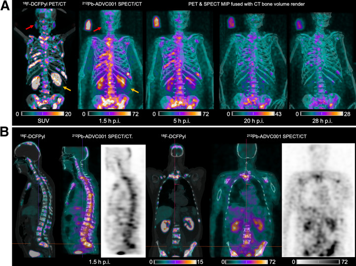
Representative 212Pb SPECT/CT images (Fig. 1) showed rapid tumor uptake of 212Pb-ADVC001 highly concordant with tumor burden delineated on the pretreatment 18F-DCFPyl PET/CT images. Images acquired after 20 h showed persistent tumor uptake despite low counts due to 212Pb decay (10.6 h half-life).
FIGURE 1.
(A) 18F-DCFPyl PET/CT and 212Pb SPECT/CT images showing concordant tumor biodistribution with low salivary gland uptake (red arrow) and rapid kidney clearance of 60 MBq of 212Pb-ADVC001 (structure of 212Pb-ADVC001 available as supplemental material at http://jnm.snmjournals.org). A 3-MBq standard solution (100 mL) was included. (B) Sagittal and coronal images at 1.5 h after injection (p.i.). MIP = maximum-intensity projection.
212Pb is a challenging isotope to image because of the high-energy γ-rays from the lead progeny generating Compton scatter from the patient and collimator (1). Our approach of summing images reconstructed from both energy windows shows the feasibility and benefit of 212Pb SPECT/CT imaging in providing postinfusion radiopharmaceutical biodistribution and patient-specific dosimetry for clinical development of 212Pb-targeted α-therapy.
DISCLOSURE
This work was financially supported by AdvanCell through the TheraPb clinical trial (NCT05720130). Kevin Kuan, Stephen Taylor, William Tieu, Thomas Kryza, Stephen Rose, and Simon Puttick are AdvanCell employees. Danielle Meyrick and Boon Quan Lee are AdvanCell consultants. No other potential conflict of interest relevant to this article was reported.
ACKNOWLEDGMENT
We acknowledge the clinical staff at the Princess Alexandra Hospital.
REFERENCE
- 1. Kvassheim M, Revheim MER, Stokke C. Quantitative SPECT/CT imaging of lead-212: a phantom study. EJNMMI Phys. 2022;9:52. [DOI] [PMC free article] [PubMed] [Google Scholar]


