Abstract
Lung cancer is the leading cause of cancer-related morbidity and mortality worldwide. The initial treatment of lung cancer depends on the definition of the tumor type and its staging. The most common treatment is chemotherapy, and the first-line treatment is a combination of carboplatin and paclitaxel. Although this treatment has good efficacy, there is a high prevalence of adverse events, particularly hematological reactions. Studies on new biomarkers related to these adverse events, such as circulating microRNAs (miRNAs/miRs), are important for optimizing the quality of life of patients. miRNAs have high stability in several biological fluids and they have specific expressions in different tissues or pathologies. Thus, the present study aimed to assess the relationship between circulating miRNAs and adverse hematologic reactions caused by treatment with carboplatin + paclitaxel in patients with lung cancer. Blood was collected from patients before and 15 days after chemotherapy for hematological adverse reaction analysis, microarray and quantitative (q)PCR validation. Adverse reactions were classified according to the Common Terminology Criteria for Adverse Events v4.0. Microarray analysis was performed using plasma from six patients without anemia and six patients with anemia, and nine miRNAs were differentially expressed. miR-1273g-3p, miR-3613-5p and miR-455-3p, identified using microarray, were assessed using qPCR in 20 patients without anemia and 26 patients with anemia. Bioinformatic analyses of miR-455-3p were performed using miRWalk, the Database for Annotation, Visualization and Integrated Discovery and GeneMania software. Microarray analysis of patients with and without anemia revealed nine significant differentially-expressed plasma miRNAs among these patients. Of these, miR-1273g-3p, miR-3613-5p and miR-455-3p were chosen for further assessment. Only miR-455-3p demonstrated a significant reduction in expression (P=0.04) between the groups before chemotherapy with carboplatin + paclitaxel. Bioinformatics analysis of miR-455-3p revealed a relationship between this miRNA and the hematopoietic pathway, particularly with respect to the RUNX family transcription factor 1 (RUNX1) and TAL bHLH transcription factor 1, erythroid differentiation factor (TAL1) genes. The most prevalent adverse reactions in patients with lung cancer treated with carboplatin + paclitaxel were hematological, particularly anemia. This adverse reaction, caused by dysfunction of the hematopoietic system, may be explained by a possible association between the important genes in this system, RUNX1 and TAL1, and hsa-miR-455-3p.
Keywords: carboplatin, paclitaxel, hematological adverse reactions, microRNAs, lung cancer
Introduction
Lung cancer is the leading cause of cancer-related morbidity and mortality worldwide, with ~2.2 million new cases and accounting for ~1.8 million deaths in 2020 (1). Lung cancer is classified into two main groups according to the histological type: i) Small cell lung cancer (SCLC) and ii) non-(N)SCLC (2). NSCLC is the most prevalent worldwide histological type, accounting for ~85% of lung cancer cases (2). The most common subtypes are adenocarcinoma (40%), squamous cell carcinoma (25–30%) and large cell carcinoma (5–10%) (2). In most cases, patients with lung cancer are in an advanced or metastatic stage at the time of diagnosis (3).
The treatment strategy has evolved from empirical chemotherapy to a personalized approach based on improved histological and molecular characterization of NSCLC primary tumors (4). Currently, the management of patients with NSCLC consists of the determination of epidermal growth factor receptor (EGFR), anaplastic lymphoma kinase, ROS proto-oncogene 1 and BRAF status, as they receive US Food and Drug Administration- and European Medicines Agency-approved targeted therapies (5). In the same way, several new drugs have been developed with an immunotherapy focus on immune checkpoint inhibitors. The most significant clinical improvements have occurred with monoclonal antibodies against programmed cell death protein 1 and programmed cell death protein ligand 1 (6). However, despite the great advances made in targeted therapies and immunotherapy, platinum-based chemotherapy treatments continue to be used in different situations and are still used as indicated therapies in international guidelines (7,8).
Platinum-based chemotherapy has proven efficacy; however, it is associated with a high prevalence of adverse events, particularly hematological effects, which affect the quality of life of patients (9,10). One of the most common hematological effects caused by platinum-based chemotherapy is anemia, a condition that develops when the blood produces a lower-than-normal amount of healthy red blood cells. Platinum-based chemotherapy can cause anemia, as these drugs can damage the kidneys, leading to less erythropoietin production. As a result, the bone marrow gets less of a stimulus to make new red blood cells, so it produces fewer of them (11,12). Thus, studies on new biomarkers that can identify adverse reactions to treatment and their mechanisms are increasingly important, and the study of microRNAs (miRNAs/miRs) is widely used nowadays.
miRNAs are small single-chain molecules containing ~22 noncoding nucleotides capable of regulating gene expression (13–15). They are highly stable in extracellular fluids (16) and are specifically expressed in different tissues or pathological states; therefore, they have been widely studied as possible diagnostic, therapeutic and prognostic biomarkers (17). A review of miRNAs in lung cancer revealed numerous studies (228 studies) on miRNAs as diagnostic biomarkers and their associations with treatment response (18). The review discussed the possible miRNAs that may influence treatment, particularly immunotherapy. However, for platinum-based chemotherapy, the review included only four studies on cisplatin and one on carboplatin (18). Furthermore, the studies on miRNAs associated with cisplatin treatment did not assess the inherent toxicities of these treatments (18).
Prognostic biomarkers should aid in the decision to justify highly toxic chemotherapy. Our research group previously performed a scope review that assessed the possible associations between adverse reactions to chemotherapy and circulating miRNAs in patients with NSCLC; however, only one article was identified that met the inclusion criteria, which indicated a substantial lack of data in this area (19). To the best of our knowledge, the present study is the first to evaluate plasma miRNAs as possible biomarkers of adverse hematological reactions in patients with NSCLC.
Methods
Patient selection and hematological adverse reactions
This study was conducted at the Clinical Oncology Department of the Hospital de Clínicas, Universidade de Campinas (HC-UNICAMP; Campinas, Brazil), a large tertiary teaching hospital, from February 2018 to December 2020. The present study was a case-control study. The inclusion criteria were as follows: Patients aged 18–80 years with primary NSCLC treated with carboplatin + paclitaxel (6 cycles every 21 days) were recruited. Patients were excluded if they had a second primary tumor, declined to participate at any time during the course of the study or did not provide blood samples for the study.
Blood was collected in tubes containing EDTA as an anticoagulant and centrifuged at 700 × g for 10 min at 4°C to separate the plasma. The plasma was aliquoted and stored in a freezer at −80°C until RNA extraction was performed. Blood samples were collected before and 15 days after the administration of carboplatin + paclitaxel. The present study was performed only in the first cycle of chemotherapy to assess differences in the miRNA expression of patients with and without chemotherapy. In addition, after the first cycle, there may be a considerable reduction in patients due to reasons such as the decrease in Karnofsky Performance Status (20), adverse events, deaths and others, which would have made statistical analysis unfeasible.
Hematological adverse reactions were classified by levels of hemoglobin, neutrophils, leukocytes, lymphocytes and platelets, according to the Common Toxicity Criteria for Adverse Events, version 4 (21). Subsequently, the patients were divided into a case group (without anemia) and a control group (with anemia).
Microarray of miRNAs
miRNAs were extracted from the plasma samples collected from 6 patients with any grade of anemia and 6 patients with grade 0 anemia (n=6) (Tables IA and IIA), using the miRNeasy Serum/Plasma Kit (cat. no. 217184; Qiagen, Inc.). These patients were selected consecutively when a sufficient number of patients with and without anemia had been reached to perform the microarray analysis (6 with anemia and 6 without anemia). After miRNA extraction, microarray analysis was performed. The samples went through processes of marking, insertion in the chips, hybridization for 18 h, washing and scanning. All procedures were performed using the Affymetrix multi-user platform® (Applied Biosystems™; Thermo Fisher Scientific, Inc.), which includes the FlashTag™ Biotin HSR RNA Labeling Kit, Genechip® miRNA 4.0, GeneChip Hybridization, Wash and Stain Kit and GeneChip Hybridization Control Kit (cat. nos. 902445, 900720 and 900454, respectively; Applied Biosystems™; Thermo Fisher Scientific, Inc.).
Table I.
Patient and clinical characteristics of patients with non-small cell lung cancer treated with carboplatin + paclitaxel.
| A, Microarray | |||
|---|---|---|---|
|
| |||
| Parameter | Non-hematological adverse reaction (n=6) | Hematological adverse reaction (n=6) | P-value |
| Patient and clinical characteristics | |||
| Age, years | 62.00±3.57 | 65.33±5.31 | 0.3300a |
| Sex, n (%) | 0.9999b | ||
| Male | 2 (33.33) | 1 (16.67) | |
| Female | 4 (66.67) | 5 (83.33) | |
| Ethnicity | - | ||
| Caucasian | 6 (100.00) | 5 (83.33) | |
| Non-caucasian | 0 (0.00) | 1 (16.67) | |
| Smoker | 0.9999b | ||
| Never | 2 (33.33) | 3 (50.00) | |
| Light | 0 (0.00) | 0 (0.00) | |
| Moderate | 0 (0.00) | 0 (0.00) | |
| Heavy | 4 (66.67) | 3 (50.00) | |
| Drinker | 0.9999b | ||
| Abstainer | 4 (66.67) | 4 (66.67) | |
| Light | 0 (0.00) | 1 (16.67) | |
| Moderate | 1 (16.67) | 0 (0.00) | |
| Heavy | 0 (0.00) | 1 (16.67) | |
| Very heavy | 1 (16.67) | 0 (0.00) | |
| KPS | 0.5900b | ||
| 100 | 5 (83.33) | 4 (66.67) | |
| 90 | 1 (16.67) | 2 (33.33) | |
| 80 | 0 (0.00) | 0 (0.00) | |
| 70 | 0 (0.00) | 0 (0.00) | |
| 60 | 0 (0.00) | 0 (0.00) | |
| Histological type | 0.9999b | ||
| Adenocarcinoma | 3 (50.00) | 4 (66.67) | |
| Squamous cell carcinoma | 3 (50.00) | 2 (33.33) | |
| Stage | - | ||
| I | 0 (0.00) | 0 (0.00) | |
| II | 0 (0.00) | 1 (16.67) | |
| III | 1 (16.67) | 0 (0.00) | |
| IV | 3 (50.00) | 3 (50.00) | |
| N/A | 2 (33.33) | 2 (33.33) | |
|
| |||
| B, Validation | |||
|
| |||
| Parameter | Non-hematological adverse reaction (n=20) | Hematological adverse reaction (n=26) | P-value |
|
| |||
| Patient and clinical characteristics | |||
| Age, years | 61.13±5.25 | 65.38±6.91 | 0.0400a |
| Sex | 0.7900b | ||
| Male | 10 (50.00) | 12 (46.15) | |
| Female | 10 (50.00) | 14 (53.85) | |
| Ethnicity | 0.4700b | ||
| Caucasian | 15 (75.00) | 22 (84.60) | |
| Non-caucasian | 5 (25.00) | 4 (15.40) | |
| Smoker | 0.4000b | ||
| Never | 5 (25.00) | 6 (23.10) | |
| Light | 7 (35.00) | 0 (0.00) | |
| Moderate | 8 (40.00) | 5 (19.20) | |
| Heavy | 0 (0.00) | 15 (57.70) | |
| Drinker | 0.8700b | ||
| Abstainer | 7 (35.00) | 10 (38.45) | |
| Light | 7 (35.00) | 8 (30.75) | |
| Moderate | 3 (15.00) | 2 (7.70) | |
| Heavy | 3 (15.00) | 4 (15.40) | |
| Very heavy | 0 (0.00) | 2 (7.70) | |
| KPS | 0.6400b | ||
| 100 | 18 (90.00) | 22 (84.60) | |
| 90 | 1 (5.00) | 3 (11.55) | |
| 80 | 0 (0.00) | 1 (3.85) | |
| 70 | 0 (0.00) | 0 (0.00) | |
| 60 | 1 (5.00) | 0 (0.00) | |
| Histological type | 0.2000b | ||
| Adenocarcinoma | 15 (75.00) | 16 (61.54) | |
| Squamous cell carcinoma | 5 (25.00) | 10 (38.46) | |
| Stage | - | ||
| I | 0 (0.00) | 0 (0.00) | |
| II | 0 (0.00) | 2 (7.69) | |
| III | 5 (25.00) | 8 (30.77) | |
| IV | 10 (50.00) | 11(42.30) | |
| N/A | 5 (25.00) | 5 (19.23) | |
Data are presented as the mean ± standard deviation or n (%). KPS, Karnofsky performance status; N/A, not available. Some P-values are not discriminated because there is no distribution of variables in the different categories.
Mann-Whitney test,
Fisher's exact test.
Table II.
Hematological adverse reactions parameters of patients with non-small cell lung cancer treated with carboplatin + paclitaxel.
| A, Microarray | |||
|---|---|---|---|
|
| |||
| Parameter | Non-hematological adverse reaction (n=6) | Hematological adverse reaction (n=6) | P-value |
| Hematological adverse reaction parameters | |||
| Anemia grade | 0.0100 | ||
| 0 | 6 (100.00) | 0 (0.00) | |
| 1 | 0 (0.00) | 6 (100.00) | |
| 2 | 0 (0.00) | 0 (0.00) | |
| 3 | 0 (0.00) | 0 (0.00) | |
| Leukopenia grade | - | ||
| 0 | 6 (100.00) | 4 (66.67) | |
| 1 | 0 (0.00) | 1 (16.67) | |
| 2 | 0 (0.00) | 1 (16.67) | |
| 3 | 0 (0.00) | 0 (0.00) | |
| Neutropenia grade | - | ||
| 0 | 6 (100.00) | 4 (66.67) | |
| 1 | 0 (0.00) | 1 (16.67) | |
| 2 | 0 (0.00) | 1 (16.67) | |
| 3 | 0 (0.00) | 0 (0.00) | |
| Lymphopenia grade | - | ||
| 0 | 6 (100.00) | 5 (83.33) | |
| 1 | 0 (0.00) | 1 (16.67) | |
| 2 | 0 (0.00) | 0 (0.00) | |
| 3 | 0 (0.00) | 0 (0.00) | |
| Thrombocytopenia grade | - | ||
| 0 | 6 (100.00) | 4 (66.67) | |
| 1 | 0 (0.00) | 2 (33.33) | |
| 2 | 0 (0.00) | 0 (0.00) | |
| 3 | 0 (0.00) | 0 (0.00) | |
|
| |||
| B, Validation | |||
|
| |||
| Parameter | Non-hematological adverse reaction (n=20) | Hematological adverse reaction (n=26) | P-value |
|
| |||
| Hematological adverse reactions parameters | |||
| Anemia grade | <0.0001 | ||
| 0 | 20 (100.00) | 0 (0.00) | |
| 1 | 0 (0.00) | 24 (92.31) | |
| 2 | 0 (0.00) | 2 (7.69) | |
| 3 | 0 (0.00) | 0 (0.00) | |
| Leukopenia grade | 0.0800 | ||
| 0 | 18 (90.00) | 19 (73.07) | |
| 1 | 2 (10.00) | 2 (7.69) | |
| 2 | 0 (0.00) | 3 (11.55) | |
| 3 | 0 (0.00) | 2 (7.69) | |
| Neutropenia grade | 0.1200 | ||
| 0 | 20 (100.00) | 21 (80.77) | |
| 1 | 0 (0.00) | 4 (15.38) | |
| 2 | 0 (0.00) | 1 (3.85) | |
| 3 | 0 (0.00) | 0 (0.00) | |
| Lymphopenia grade | - | ||
| 0 | 20 (100.00) | 26 (100.00) | |
| 1 | 0 (0.00) | 0 (0.00) | |
| 2 | 0 (0.00) | 0 (0.00) | |
| 3 | 0 (0.00) | 0 (0.00) | |
| Thrombocytopenia grade | 0.2100 | ||
| 0 | 19 (95.00) | 20 (76.92) | |
| 1 | 1 (5.00) | 6 (23.08) | |
| 2 | 0 (0.00) | 0 (0.00) | |
| 3 | 0 (0.00) | 0 (0.00) | |
Data are presented as the mean ± standard deviation or n (%). Some P-values are not discriminated because there is no distribution of variables in the different categories. Fisher's exact test was used.
Validation of selected miRNAs
miR-1273g-3p, miR-3613-5p and miR-455-3p were selected for validation in a larger cohort of patients, as they are genes that are most related to the hematopoiesis pathway, according to mirPath v.3 (22). miRNAs were extracted from samples collected from 50 patients with and without anemia using the miRNeasy Serum/Plasma Kit (cat. no. 217184; Qiagen, Inc.). After extracting the miRNAs from all plasma samples collected at baseline and on day 15 from all patients included in the study, cDNA synthesis was performed using the TaqMan™ Advanced miRNA cDNA Synthesis Kit (cat. no. A28007; Applied Biosystems; Thermo Fisher Scientific, Inc.) and quantitative (q)PCR using TaqMan Advanced miRNA Assays (cat. no. A25576; Applied Biosystems; Thermo Fisher Scientific, Inc.). In addition, qPCR of the endogenous control, miR-98-3p, was performed for normalization, which was selected as an endogenous control as its expression was stable, according to the microarray results of the present study. Furthermore, miR-39 was selected as an exogenous control to ensure the quality of the technique, and samples with expression levels above two standard deviations (SDs) were excluded from the analysis. The thermocycling conditions were as follows: hold at 95°C for 20 sec followed by 70 cycles at 95°C for 15 sec and then at 60°C for 60 sec. The sequences of the TaqMan probes used were are follows: miR-1273g3p (assay ID no. 475626_mat; cat. no. 4440886), 5′-ACCACUGCACUCCAGCCUGAG-3′; miR-3613-5p (assay ID no. 479424_mir; cat. no. A25576), 5′-UGUUGUACUUUUUUUUUUGUUC-3′; miR-455-3p (assay ID no. 478112_mir; cat. no. A25576), 5′-GCAGUCCAUGGGCAUAUACAC-3′; miR-98-3p (assay ID no. 479223_mir; cat. no. A25576), 5′-CUAUACAACUUACUACUUUCCC-3′; and miR-39 (assay ID no. 478293_mir; cat. no. A25576), 5′-UCACCGGGUGUAAAUCAGCUUG-3′. Thermo Fisher Scientific, Inc. confirms that for the miR-1273g-3p sequence, the aforementioned ‘assay detects a miRNA target from an earlier version of miRbase that has been obsoleted in miRbase v.22. This assay is still available for purchase for continuity of studies’. At the time that the present study was performed, this probe matched the Homo sapiens version of its gene target. A C. elegans probe was used for the miR-39 sequence, as it was an exogenous process quality control. The miRNA qPCR results were analyzed using Rotor-Gene™ Q Series software 2.3.5 (23). Each miRNA had its expression evaluated and relative expressions were quantified using the 2−ΔΔCq method (24). The aforementioned experiments were performed using samples from the patients before and 15 days after chemotherapy.
Bioinformatics analysis
miR-455-3p was selected for bioinformatics analysis. Predicted miRNA target genes were identified using the miRWalk platform 2.0 (25), which provides a list of predicted miRNA target genes according to 12 different algorithms. These genes were selected for unsupervised enrichment analysis using the Database for Annotation, Visualization and Integrated Discovery Functional Annotation Bioinformatics Microarray Analysis software 6.8 (26) to identify the main canonical signaling pathways involving differentially-expressed miRNAs. To visualize the networks in which the target genes were involved, all predicted genes in the matrix were analyzed using GeneMania 3.5.2 (27). The hematopoietic pathway was selected for further evaluation.
Statistical analysis
The frequencies of clinical/demographic data and degrees of adverse events related to adverse reactions are presented as n (%) and descriptive measures are presented as the mean ± SD. The expression of miRNAs was compared between the different groups (with vs. without hematological adverse reactions) at baseline and day 15 using the Mann-Whitney U-test, and between the same group during the time (D0 vs. D15 with hematological adverse reactions and D0 vs. D15 without hematological adverse reactions), using the paired t-test. For comparisons with P<0.05 receiver operating characteristic (ROC) curves were produced. For the enrichment analysis of the predicted target genes regulated by miR-455-3p, Fisher's exact test was performed. The significance level adopted for the present study was 5%. P<0.05 was considered to indicate a statistically significant difference.
Results
Patient characteristics and toxicities
For the array, a total of 6 patients with any grade of anemia and 6 patients with grade 0 anemia (n=6) (Tables IA and IIA) were included; these patients were representative of the whole cohort, since the age of the groups was similar and the majority of patients were Caucasians and women when comparing patients selected for the array and the larger cohort (Table I). For validation, a total of 50 patients were included in the present study. These patients were 50% male with a mean age of 63.5 years; the majority of the patients were Caucasians and heavy smokers, with adenocarcinoma stage IV. For the validation step, 4 patients were excluded; thus, 46 subjects were used.
Hematological adverse reactions were prevalent, with anemia observed in 26 patients (52.0%). The clinical characteristics and hematological adverse reaction parameters of patients whose miRNA samples were analyzed using microarray and patients included in the validation step are presented in Table I.
Microarray results
A total of nine miRNAs were significantly differentially expressed between patients without and with anemia (hsa-miR-6779-5p, hsa-miR-3940-5p, hsa-miR-3656, hsa-miR-8072, hsa-miR-1273g-3p, hsa-miR-6869-5p, hsa-miR-6794-5p, hsa-miR-455-3p and hsa-miR-3613-5p), in which there was a fold change (FC) >1.5 or FC<-1.5, and P<0.05 (Table III).
Table III.
Plasma miRNAs with changes in gene expression level between patients without (n=6) vs. with (n=6) anemia in which P<0.05 and FC>1.5 or <-1.5.
| Differentially-expressed miRNA | FC | P-value |
|---|---|---|
| hsa-miR-6779-5p | 1.57 | 0.027 |
| hsa-miR-3940-5p | −1.64 | 0.039 |
| hsa-miR-3656 | −1.52 | 0.012 |
| hsa-miR-8072 | −1.51 | 0.037 |
| hsa-miR-1273g-3pa | −2.92 | 0.004 |
| hsa-miR-6869-5p | −1.99 | 0.040 |
| hsa-miR-6794-5p | 1.55 | 0.004 |
| hsa-miR-455-3pa | 1.55 | 0.040 |
| hsa-miR-3613-5pa | 2.46 | 0.030 |
miRNAs chosen for validation using quantitative PCR. FR, fold regulation; miRNA/miR, microRNA.
Validation of the miRNAs
A total of four miRNAs were chosen for expression measurement: miR-1273g-3p, miR-3613-5p and miR-455-3p as targets, and miR-98-3p as the endogenous control. The targets were chosen using bioinformatics analysis, with all nine miRNAs observed in the microarray. This analysis demonstrated that the three miRNAs were notably associated with more genes in the hematopoietic pathway compared with the other six miRNAs differentially expressed on the microarray analysis. The endogenous control was chosen from the microarray analysis, where miR-38-3p demonstrated markedly stable expression between the two groups (FC=−1.04; P=0.97) (Table SI).
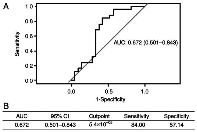
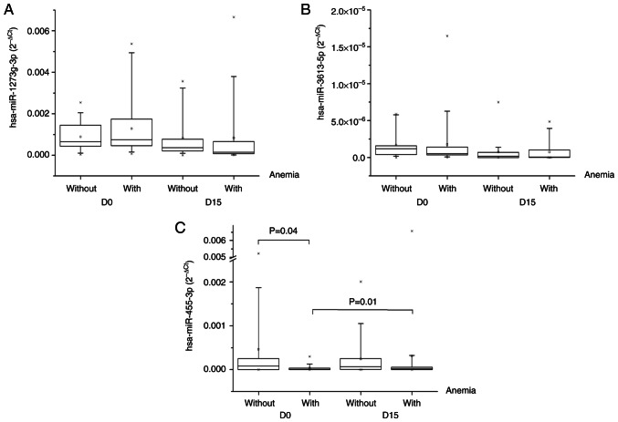
In total, 50 patients were enrolled for the validation; however, four patients were excluded, as miR-39 (quality control) expression was higher than two SDs. Thus, the expression of the three miRNAs from 46 patients was assessed. The expression of miR1273g-3p and miR-3613-5p did not significantly differ between the groups (Fig. 1A and B), whereas miR-455-3p expression was significantly lower before carboplatin + paclitaxel administration in patients with anemia than in those without (P=0.04; Fig. 1C). There was no significant difference in the expression of miR1273g-3p and miR-3613-5p between the same group during the time (D0 vs. D15 with hematological adverse reactions and D0 vs. D15 without hematological adverse reactions), whereas miR-455-3p expression significantly increased between baseline and day 15 for the group with anemia (P=0.01). Furthermore, in the predictive performance analysis, miR-455-3p had an area under the curve (AUC) of 0.672, sensitivity of 84.00% and specificity of 57.14% (Fig. 2).
Figure 1.
Expression of miRNAs. Expression of (A) miR-1273g-3p, (B) miR-3613-5p and (C) miR-455-3p in the non-anemia and anemia group before and 15 days after carboplatin + paclitaxel administration. miRNA/miR, microRNA; D0, day 0; D15, day 15.
Figure 2.

Predictive performance of miR-455-3p. (A) Receiver operating characteristic curve of baseline miR-455-3p expression. (B) AUC, CI, cutpoint, sensitivity and specificity of miR-455-3p expression before carboplatin + paclitaxel administration as prognostic markers of carboplatin + paclitaxel-induced anemia. miR, microRNA; AUC, area under the curve; CI, confidence interval.
Bioinformatics analysis
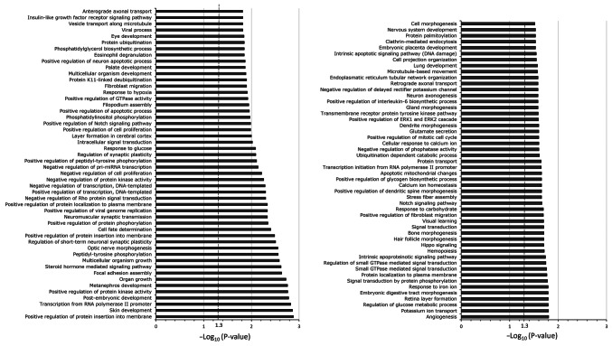
miR-455-3p was selected for bioinformatics analysis. Gene set enrichment analysis was performed using 9,589 predicted target genes of miR-455-3p. The top 100 canonical signaling pathways of the predicted target genes of miR-455-3p are presented in Fig. 3. Enrichment of the signaling pathways involved in carboplatin- and paclitaxel-induced adverse hematological reactions was observed through the hematopoietic signaling pathway; this is represented by the hemopoiesis pathway.
Figure 3.
Enrichment analysis of the predicted target genes regulated by miR-455-3p. The top 100 canonical signaling pathways are presented (left, 1–50; right, 51–100). Enrichment analysis was performed using the Database for Annotation, Visualization and Integrated Discovery Functional Annotation Bioinformatics Microarray Analysis software. The dashed line represents -log(P-value)=1.3 or P=0.05 (Fisher's exact test).
Discussion
The present study aimed to identify biomarkers of adverse hematological reactions induced by chemotherapy with carboplatin + paclitaxel in patients with NSCLC. Differential expression of miRNAs was assessed before and 15 days after treatment.
Microarray analysis of plasma samples from patients with and without anemia demonstrated the differential expression of nine plasma miRNAs between the two groups of patients. Among the nine differentially-expressed miRNAs, miR-1273g-3p, miR3613-5p and miR-455-3p were selected for further assessment.
In the validation cohort, miR-455-3p demonstrated a statistically significant differential expression between the groups, showing lower expression in the group with anemia than in the group without anemia before chemotherapy with carboplatin + paclitaxel (P=0.04). An ROC curve was produced to evaluate the accuracy of predicting anemia. The AUC and specificity were 0.672 and 57%, respectively, with a good sensitivity of ~84%. This indicated the potential of miR-455-3p as a biomarker for predicting anemia after treatment with carboplatin + paclitaxel.
However, although the aforementioned findings are still results, they encourage future studies to include a larger number of patients and functional analyses of this miRNA. To the best of our knowledge, there is currently no study that demonstrates the cost effectiveness of using this protocol before a chemotherapy regimen to predict anemia, and this could be the next step of the present work. Thus, if more studies are performed to confirm the predictive role of miR-455-3p and its cost effectiveness, this will be clinically valuable and plasma quantification for patients prior to chemotherapy with carboplatin + paclitaxel will be useful, as it allows for the identification of patients with a prior risk of developing anemia and the opportunity of making a better choice of treatment for them.
It is important to highlight that miR-455-3p serves a well-established role as a tumor suppressor in different types of cancer (28,29). Numerous studies have reported that a lower expression of miR-455-3p resulted in a worse prognosis for lung cancer (30), breast cancer (31), osteosarcoma (32), prostate cancer (28) and esophageal cancer (29). Therefore, these findings, along with those of the present study, indicate that miR-455-3p may serve as a biomarker of cancer prognosis in the future, as well as a predictor of adverse events related to hematological adverse reactions when the treatment of choice is carboplatin + paclitaxel.
The in-silico analysis of miR-455-3p in the present study demonstrated enrichment of a canonical pathway (hemopoiesis) of high importance for hematological adverse reactions induced by carboplatin + paclitaxel through the hematopoiesis pathway. Among the genes important for this pathway are runt-related transcription factor 1 (RUNX1) and T-cell acute lymphocytic leukemia protein 1 (TAL1). RUNX1 is a transcription factor that serves an essential role in hematopoiesis. It is indispensable for the formation of hematopoietic stem cells, which are precursors of all blood cells, and for the maturation of T and B lymphocytes (33–35). However, it is also responsible for the negative regulation of hematopoietic stem cells and myeloid precursor cells (36). Therefore, the results of the present study indicate that, due to the decreased expression of miR-455-3p, there was an increase in the expression of RUNX1. This would lead to increased negative regulation of hematopoietic stem cells and myeloid precursor cells, and consequently, a lower production of blood cells in general, particularly those of the myeloid series. Furthermore, similar to RUNX1, TAL1 is an important transcription factor in hematopoiesis. It is responsible for regulating different processes, with an emphasis on the maturation of lymphoid series cells (36,37). Reynaud et al (38) reported that increased expression of TAL1 reduced the differentiation of B lymphocytes by up to 40%, and the production of B lymphocytes and macrophages by ~20%. This indicates that the results of the present study agree with the observation that a reduction in miR-455-3p leads to an increase in TAL1 expression, consequently leading to a reduction in lymphocytes.
Therefore, the present study demonstrated that miR-455-3p has important potential as a predictor of hematological adverse reactions during treatment with carboplatin + paclitaxel, which may explain the fact that this miRNA interacts with important genes involved in the hematopoietic pathway, such as RUNX1 and TAL1. However, although promising, this is still initial evidence and further investigation is needed so that in the future, a possible clinical implementation for these findings can be considered.
There are certain limitations to the analyses presented. The present study comprised 50 patients, of whom 2 experienced grade ≥2 anemia. This small population with an absence of severe anemia (grade ≥2) may have generated bias in the results. However, even with this population, significant differences were observed.
In conclusion, this present study showed that patients with lung cancer who were treated with carboplatin and paclitaxel are mostly male, with a mean age of 63.50 years, retired, with low education, married, severe smokers, with a Karnofsky performance status of 100%, who are not eligible for surgical resection before chemotherapy and who are not eligible for histopathological diagnosis of adenocarcinoma and stage 4. Among the adverse reactions evaluated, the high prevalence of anemia above the degree of 0 was highlighted. The microarray of patients with and without hematologic adverse reactions found 9 plasma miRNAs differently expressed among these patients. Of these, 3 were chosen for validation: miR-1273g-3p, miR-3613-5p and miR-455-3p. Only miR-455-3p showed a significant reduction in expression (P=0.04) before carboplatin + paclitaxel administration in patients with anemia than in those without anemia. Therefore, this miRNA has potential as a predictor of carboplatin and paclitaxel-induced haematological adverse reactions and should be evaluated in a larger number of patients, as well as in functional studies. Although chemotherapy based on the combination of carboplatin and paclitaxel has been used in the treatment of lung cancer for numerous years, hematological adverse reactions are an obstacle to the efficiency of treatment and, consequently, to patients' quality of life. The present study highlighted the significance of hematological adverse reactions induced by carboplatin and paclitaxel and the importance of identifying new biomarkers for them. In addition to identifying miR-455-3p as a potential predictor of hematological adverse reactions, information was provided for further studies aimed at identifying new biomarkers and important collaboration for a better understanding of the etiology of hematological adverse reactions induced by carboplatin and paclitaxel.
Supplementary Material
Acknowledgements
Not applicable.
Funding Statement
The present work was supported by funding from the Coordenação de Aperfeiçoamento de Pessoal de Nível Superior-Brasil (CAPES)-finance code 001, Fundação de Amparo à Pesquisa do Estado de São Paulo – Brasil (FAPESP)-finance code 2022/09438-8 and the Pharmaceutical Security Nucleus Project, object of Agreement no. 895688/2019, the result of a partnership between the Ministry of Justice and Public Security of Brazil, through the Fund for the Defense of Diffuse Rights and the State University of Campinas.
Availability of data and materials
The array data were uploaded to ArrayExpress with accession no. E-MTAB-13518. The data generated in the present study may be requested from the corresponding author.
Authors' contributions
PM and ECP conceived the present study. PENSV designed the study and performed the experiments. PENSV and PM confirm the authenticity of all the raw data. CSS, LZ, ASB and HMH selected patients based on the study inclusion criteria, characterized the type, stage of the tumor and directed treatment, and reviewed the description and interpretation of clinical data MWPJr supported on patients' inclusion flow, helping us to reach the patients that met the inclusion criteria, also on all the demographics and clinical data analysis and interpretation of clinical data. MVG helped with the bioinformatical analysis. All authors have read and approved the final manuscript.
Ethics approval and consent to participate
All procedures were authorized by the Research Ethics Committee of the State University of Campinas (Campinas, Brazil; approval no. 83196318.8.0000.5404). All patients signed a consent form approved by the Research Ethics Committee of the State University of Campinas, where they authorized the use of the obtained data.
Patient consent for publication
Not applicable.
Competing interests
The authors declare that they have no competing interests.
References
- 1.Sung H, Ferlay J, Siegel RL, Laversanne M, Soerjomataram I, Jemal A, Bray F. Global cancer statistics 2020: GLOBOCAN estimates of incidence and mortality worldwide for 36 cancers in 185 countries. CA Cancer J Clin. 2021;71:209–249. doi: 10.3322/caac.21660. [DOI] [PubMed] [Google Scholar]
- 2.Duma N, Santana-Davila R, Molina JR. Non-Small cell lung cancer: Epidemiology, screening, diagnosis, and treatment. Mayo Clin Proc. 2019;94:1623–1640. doi: 10.1016/j.mayocp.2019.01.013. [DOI] [PubMed] [Google Scholar]
- 3.Sateia HF, Choi Y, Stewart RW, Peairs KS. Screening for lung cancer. Semin.Oncol. 2017;44:74–82. doi: 10.1053/j.seminoncol.2017.02.003. [DOI] [PubMed] [Google Scholar]
- 4.Shroff GS, de Groot PM, Papadimitrakopoulou VA, Truong MT, Carter BW. Targeted therapy and immunotherapy in the treatment of non-small cell lung cancer. Radiol Clin North Am. 2018;56:485–495. doi: 10.1016/j.rcl.2018.01.012. [DOI] [PubMed] [Google Scholar]
- 5.López-Castro R, García-Peña T, Mielgo-Rubio X, Riudavets M, Teixidó C, Vilariño N, Couñago F, Mezquita L. Targeting molecular alterations in non-small-cell lung cancer: What's next? Per Med. 2022;19:341–359. doi: 10.2217/pme-2021-0059. [DOI] [PubMed] [Google Scholar]
- 6.Luo W, Wang Z, Tian P, Li W. Safety and tolerability of PD-1/PD-L1 inhibitors in the treatment of non-small cell lung cancer: A meta-analysis of randomized controlled trials. J Cancer Res Clin Oncol. 2018;144:1851–1859. doi: 10.1007/s00432-018-2707-4. [DOI] [PubMed] [Google Scholar]
- 7.Planchard D, Popat S, Kerr K, Novello S, Smit EF, Faivre-Finn C, Mok TS, Reck M, Van Schil PE, Hellmann MD, et al. Metastatic non-small cell lung cancer: ESMO Clinical Practice Guidelines for diagnosis, treatment and follow-up. Ann Oncol. 2018;29((Suppl 4)):iv192–iv237. doi: 10.1093/annonc/mdy275. [DOI] [PubMed] [Google Scholar]
- 8.Ganti AKP, Loo BW, Bassetti M, Blakely C, Chiang A, D'Amico TA, D'Avella C, Dowlati A, Downey RJ, Edelman M, et al. Small cell lung cancer, version 2.2022, NCCN clinical practice guidelines in oncology. J Natl Compr Canc Netw. 2021;19:1441–1464. doi: 10.6004/jnccn.2021.0058. [DOI] [PMC free article] [PubMed] [Google Scholar]
- 9.Cepeda V, Fuertes MA, Castilla J, Alonso C, Quevedo C, Pérez JM. Biochemical mechanisms of cisplatin cytotoxicity. Anticancer Agents Med Chem. 2007;7:3–18. doi: 10.2174/187152007779314044. [DOI] [PubMed] [Google Scholar]
- 10.Capriotti K, Capriotti JA, Lessin S, Wu S, Goldfarb S, Belum VR, Lacouture ME. The risk of nail changes with taxane chemotherapy: A systematic review of the literature and meta-analysis. Br J Dermatol. 2015;173:842–845. doi: 10.1111/bjd.13743. [DOI] [PubMed] [Google Scholar]
- 11.NIH National Heart, Lung and Blood Institute, corp-author. What is anemia? https://www.nhlbi.nih.gov/health/anemia. [ October 4; 2023 ]; [Google Scholar]
- 12.Rodgers DM, III, Becker PS, Blinder M, Cella D, Chanan-Khan A, Cleeland C, Coccia PF, Djulbegovic B, Gilreath JA, Kraut EH, et al. Cancer- and chemotherapy-induced anemia. J Natl Compr Canc Netw. 2012;10:628–653. doi: 10.6004/jnccn.2012.0044. [DOI] [PubMed] [Google Scholar]
- 13.Lagos-Quintana M, Rauhut R, Lendeckel W, Tuschl T. Identification of novel genes coding for small expressed RNAs. Science. 2001;294:853–858. doi: 10.1126/science.1064921. [DOI] [PubMed] [Google Scholar]
- 14.Lau NC, Lim LP, Weinstein EG, Bartel DP. An abundant class of tiny RNAs with probable regulatory roles in Caenorhabditis elegans. Science. 2001;294:858–862. doi: 10.1126/science.1065062. [DOI] [PubMed] [Google Scholar]
- 15.Lee RC, Ambros V. An extensive class of small RNAs in Caenorhabditis elegans. Science. 2001;294:862–864. doi: 10.1126/science.1065329. [DOI] [PubMed] [Google Scholar]
- 16.Weber JA, Baxter DH, Zhang S, Huang DY, Huang KH, Lee MJ, Galas DJ, Wang K. The microRNA spectrum in 12 body fluids. Clin Chem. 2010;56:1733–1741. doi: 10.1373/clinchem.2010.147405. [DOI] [PMC free article] [PubMed] [Google Scholar]
- 17.Pereira TC. Sociedade Brasileira de Genética; Ribeirão Preto, Brazil: 2015. Introduction to the world of microRNAs; p. 44. (In Portuguese) [Google Scholar]
- 18.Zhong S, Golpon H, Zardo P, Borlak J. miRNAs in lung cancer. A systematic review identifies predictive and prognostic miRNA candidates for precision medicine in lung cancer. Transl Res. 2021;230:164–196. doi: 10.1016/j.trsl.2020.11.012. [DOI] [PubMed] [Google Scholar]
- 19.Vasconcelos PE, Visacri MB, Pincinato EC, Torso NG, Seguin CS, Zambon L, Barbeiro AS, Junior MW, Moriel P. miRNAs as biomarkers of adverse drug reactions to platinum-based agents in patients with non-small-cell lung cancer. Biomark Med. 2021;15:1067–1069. doi: 10.2217/bmm-2021-0443. [DOI] [PubMed] [Google Scholar]
- 20.Karnofsky DA, Burchenal JH. MacLeod CM. Evaluation of chemotherapeutic agents. Columbia University Press; New York, USA: 1946. The clinical evaluation of chemotherapeutic agents; p. 196. [Google Scholar]
- 21.U.S Department of Health and Human Services, corp-author. U.S Department of Health and Human Services, National Institutes of Health, National Cancer Institute; 2010. [ March 11; 2024 ]. Common Terminology Criteria for Adverse Events (CTCAE) Version 4.0. [Google Scholar]
- 22.Vlachos IS, Zagganas K, Paraskevopoulou MD, Georgakilas G, Karagkouni D, Vergoulis T, Dalamagas T, Hatzigeorgiou AG. DIANA-miRPath v3. 0: deciphering microRNA function with experimental support. Nucleic Acids Res. 2015;43:W460–W466. doi: 10.1093/nar/gkv403. [DOI] [PMC free article] [PubMed] [Google Scholar]
- 23.Rotor-Gene Q 2.3.5-Windows platforms. https://www.qiagen.com/us/resources/resourcedetail?id=9d8bda8e-1fd7-4519-a1ff-b60bba526b57&lang=en. [ January 30; 2024 ]; [Google Scholar]
- 24.Schmittgen TD, Livak KJ. Analyzing real-time PCR data by the comparative C(T) method. Nat Protoc. 2008;3:1101–1108. doi: 10.1038/nprot.2008.73. [DOI] [PubMed] [Google Scholar]
- 25.Dweep H, Gretz N. MiRWalk2.0: A comprehensive atlas of microRNA-target interactions. Nat Methods. 2015;12:697. doi: 10.1038/nmeth.3485. [DOI] [PubMed] [Google Scholar]
- 26.Huang da W, Sherman BT, Lempicki RA. Systematic and integrative analysis of large gene lists using DAVID Bioinformatics Resources. Nature Protoc. 2009;4:44–57. doi: 10.1038/nprot.2008.211. [DOI] [PubMed] [Google Scholar]
- 27.Warde-Farley D, Donaldson SL, Comes O, Zuberi K, Badrawi R, Chao P, Franz M, Grouios C, Kazi F, Lopes CT, et al. The GeneMANIA prediction server: Biological network integration for gene prioritization and predicting gene function. Nucleic Acids Res. 2010;38:W214–W220. doi: 10.1093/nar/gkq537. [DOI] [PMC free article] [PubMed] [Google Scholar]
- 28.Zhao Y, Yan M, Yun Y, Zhang J, Zhang R, Li Y, Wu X, Liu Q, Miao W, Jiang H. MicroRNA-455-3p functions as a tumor suppressor by targeting eIF4E in prostate cancer. Oncol Rep. 2017;37:2449–2458. doi: 10.3892/or.2017.5502. [DOI] [PubMed] [Google Scholar]
- 29.Yang H, Wei YN, Zhou J, Hao TT, Liu XL. MiR-455-3p acts as a prognostic marker and inhibits the proliferation and invasion of esophageal squamous cell carcinoma by targeting FAM83F. Eur Rev Med Pharmacol Sci. 2017;21:3200–3206. [PubMed] [Google Scholar]
- 30.Gao X, Zhao H, Diao C, Wang X, Xie Y, Liu Y, Han J, Zhang M. miR-455-3p serves as prognostic factor and regulates the proliferation and migration of non-small cell lung cancer through targeting HOXB5. Biochem Biophys Res Commun. 2018;495:1074–1080. doi: 10.1016/j.bbrc.2017.11.123. [DOI] [PubMed] [Google Scholar]
- 31.Guo J, Liu C, Wang W, Liu Y, He H, Chen C, Xiang R, Luo Y. Identification of serum miR-1915-3p and miR-455-3p as biomarkers for breast cancer. PLoS One. 2018;13:e0200716. doi: 10.1371/journal.pone.0200716. [DOI] [PMC free article] [PubMed] [Google Scholar]
- 32.Yi X, Wang Y, Xu S. MiR-455-3p downregulation facilitates cell proliferation and invasion and predicts poor prognosis of osteosarcoma. J Orthop Surg Res. 2020;15:454. doi: 10.1186/s13018-020-01967-1. [DOI] [PMC free article] [PubMed] [Google Scholar]
- 33.Lam K, Zhang DE. RUNX1 and RUNX1-ETO: Roles in hematopoiesis and leukemogenesis. Front Biosci (Landmark Ed) 2012;17:1120–1139. doi: 10.2741/3977. [DOI] [PMC free article] [PubMed] [Google Scholar]
- 34.Okuda T, Nishimura M, Nakao M, Fujita Y. RUNX1/AML1: A central player in hematopoiesis. Int J Hematol. 2001;74:252–257. doi: 10.1007/BF02982057. [DOI] [PubMed] [Google Scholar]
- 35.Ichikawa M, Yoshimi A, Nakagawa M, Nishimoto N, Watanabe-Okochi N, Kurokawa M. A role for RUNX1 in hematopoiesis and myeloid leukemia. Int J Hematol. 2013;97:726–734. doi: 10.1007/s12185-013-1347-3. [DOI] [PubMed] [Google Scholar]
- 36.Lécuyer E, Hoang T. SCL: From the origin of hematopoiesis to stem cells and leukemia. Exp Hematol. 2004;32:11–24. doi: 10.1016/j.exphem.2003.10.010. [DOI] [PubMed] [Google Scholar]
- 37.Vagapova ER, Spirin PV, Lebedev TD, Prassolov VS. The Role of TAL1 in hematopoiesis and leukemogenesis. Acta Naturae. 2018;10:15–23. doi: 10.32607/20758251-2018-10-1-15-23. [DOI] [PMC free article] [PubMed] [Google Scholar]
- 38.Reynaud D, Ravet E, Titeux M, Mazurier F, Rénia L, Dubart-Kupperschmitt A, Roméo PH, Pflumio F. SCL/TAL1 expression level regulates human hematopoietic stem cell self-renewal and engraftment. Blood. 2005;106:2318–2328. doi: 10.1182/blood-2005-02-0557. [DOI] [PubMed] [Google Scholar]
Associated Data
This section collects any data citations, data availability statements, or supplementary materials included in this article.
Supplementary Materials
Data Availability Statement
The array data were uploaded to ArrayExpress with accession no. E-MTAB-13518. The data generated in the present study may be requested from the corresponding author.


