Abstract
Attachment of Sindbis virus to the cell surface glycosaminoglycan heparan sulfate (HS) and the selection of this phenotype by cell culture adaptation were investigated. Virus (TR339) was derived from a cDNA clone representing the consensus sequence of strain AR339 (K. L. McKnight, D. A. Simpson, S. C. Lin, T. A. Knott, J. M. Polo, D. F. Pence, D. B. Johannsen, H. W. Heidner, N. L. Davis, and R. E. Johnston, J. Virol. 70:1981–1989, 1996) and from mutant clones containing either one or two dominant cell culture adaptations in the E2 structural glycoprotein (Arg instead of Ser at E2 position 1 [designated TRSB]) or this mutation plus Arg for Ser at E2 114 [designated TRSB-R114]). The consensus virus, TR339, bound to baby hamster kidney (BHK) cells very poorly. The mutation in TRSB increased binding 10- to 50-fold, and the additional mutation in TRSB-R114 increased binding 3- to 5-fold over TRSB. The magnitude of binding was positively correlated with the degree of cell culture adaptation and with attenuation of these viruses in neonatal mice. HS was identified as the attachment receptor for the mutant viruses by the following experimental results. (i) Low concentrations of soluble heparin inhibited plaque formation on and binding of mutant viruses to BHK cells by >95%. In contrast, TR339 showed minimal inhibition at high concentrations. (ii) Binding and infectivity of TRSB-R114 was sensitive to digestion of cell surface HS with heparinase III, and TRSB was sensitive to both heparinase I and heparinase III. TR339 infectivity was only slightly affected by either digestion. (iii) Radiolabeled TRSB and TRSB-R114 attached efficiently to heparin-agarose beads in binding assays, while TR339 showed virtually no binding. (iv) Binding and infectivity of TRSB and TRSB-R114, but not TR339, were greatly reduced on Chinese hamster ovary cells deficient in HS specifically or all glycosaminoglycans. (v) High-multiplicity-of-infection passage of TR339 on BHK cell cultures resulted in rapid coselection of high-affinity binding to BHK cells and attachment to heparin-agarose beads. Sequencing of the passaged virus population revealed a mutation from Glu to Lys at E2 70, a mutation common to many laboratory strains of Sindbis virus. These results suggest that TR339, the most virulent virus tested, attaches to cells through a low-affinity, primarily HS-independent mechanism. Adaptive mutations, selected during cell culture growth of Sindbis virus, enhance binding and infectivity by allowing the virus to attach by an alternative mechanism that is dependent on the presence of cell surface HS.
Alphaviruses have a broad host range in nature, replicating in mammalian, avian, arthropod, and amphibian species, and are capable of infecting a wide variety of cultured cells (60, 61). Because of their broad host range, it has been suggested that alphaviruses use a ubiquitously expressed molecule for attachment to cells (27, 60). Attachment is thought to be mediated primarily by the virus E2 structural glycoprotein (reviewed in reference 61). Attached Sindbis virus is uniformly distributed over fixed cell surfaces, occupying 104 to 105 receptor sites (similar to results with Semliki Forest virus [SFV] [21]), and treatment of attached virus with high-ionic-strength buffer results in the elution of most bound virus (5, 50). These results suggest that a charge interaction might play a significant role in alphavirus attachment. Indeed, several studies have suggested that sulfated polyanions, such as cell surface glycosaminoglycans (GAGs), are involved in binding of alphaviruses to cells. Schleupner et al. (57) implicated polyanionic polysaccharide in inactivation of a small-plaque variant of Sindbis virus. Symington and Schlesinger (63) found that pretreatment of mouse plasmacytoma cells with heparin resulted in a large increase in Sindbis virus binding. Mastromarino et al. (39) were able to partially compete Sindbis virus infectivity with soluble heparin and several other sulfated polyanions and diminish infectivity by digestion of cell surface heparan sulfate (HS) with heparinase, suggesting that a sulfated molecule may be a component of the receptor site. Polyanionic compounds which block human immunodeficiency virus (HIV) attachment also inhibit infectivity of alphaviruses including SFV and Sindbis virus (58).
Although a single cellular receptor has not been unambiguously identified for any alphavirus (27, 46; reviewed in reference 60), studies using methods such as chemical cross-linking (37), copurification of virus-receptor complexes (16), enzymatic inactivation of cellular receptors (59, 69), analysis of anti-idiotype antibodies (69, 71), and analysis of antireceptor antibodies (36, 70) have indicated that attachment is mediated through virus interaction with a cell surface protein. However, putative receptors of different molecular weights have been identified in different laboratories and with different cell types (16, 36, 37, 69–71). Given the number of different cell surface structures shown to interact with alphaviruses, several investigators have suggested that multiple receptors may exist, in some instances on the same cell type (59, 69, 70). A significant complicating factor is that single amino acid differences in the structural glycoproteins of different strains may profoundly affect virus attachment-entry processes. As few studies have compared attachment differences between different alphavirus strains on a single cell type, it is possible that differences observed in attachment-entry mechanisms may result, at least in part, from the use of different Sindbis virus laboratory strains.
Adaptation of Sindbis virus to growth in tissue culture or in animals has generated virus mutants that can be used to evaluate strain-specific differences in receptor usage. Single amino acid changes in the E2 glycoprotein coordinately alter virus attachment-entry of cultured cells and pathogenesis in vivo. Mutations from Ser to Arg at E2 position 114 (11), Glu to Lys at E2 70 (40, 54), and Gln to His at E2 55 (67) have been associated with rapid penetration of BHK cells. The mutations selected in vitro (E2 Lys 70 and E2 Arg 114) are attenuating in neonatal mice (11, 40, 66), while the in vivo-selected E2 His 55 mutation enhances virulence in older mice (68). A correlation between in vitro selection for mutations of certain E2 residues with rapid penetration of BHK cells and attenuation of virulence in vivo also has been shown for other alphaviruses: S.A.AR86 (54), Venezuelan equine encephalitis virus (VEE) (13), and SFV (22). However, it remains unknown whether penetration mutants use the same attachment-entry mechanism as the parental strains from which they were derived. Also, the effects of preexisting cell culture-adaptive mutations in parental strains used as genetic background for mutant selections have not been evaluated. Together, alphavirus receptor-entry and mutant selection studies have indicated that while a protein receptor likely plays an important role in the infection process, charge interactions, perhaps with GAG molecules, may also contribute to alphavirus attachment.
We have investigated strain-specific differences in Sindbis virus attachment in a comparative study of virus generated from cDNA clones of a Sindbis virus AR339 ancestral consensus sequence (TR339) and two cell culture-adapted mutants. The cloned consensus sequence eliminates several cell culture-adaptive mutations present in sequenced laboratory strains of AR339 (40). In our studies, virus differing from the consensus sequence by either one or two dominant adaptive mutations exhibited greatly increased binding to BHK cells that was dependent upon the presence of cell surface HS. Binding of TR339 was only slightly dependent upon HS. The HS-dependent phenotype could be selected rapidly upon passage of TR339 in BHK cells and was correlated with attenuation of virus disease in neonatal mice.
(Portions of this work were presented at the 1997 meeting of the American Society for Virology.)
MATERIALS AND METHODS
Cell culture.
Baby hamster kidney (BHK-21) (ATCC CCL-10), Swiss 3T3 (ATCC CCL-92), L929 (ATCC CCL-1), and Neuro-2A (ATCC CCL-131) cells were maintained in alpha minimal essential medium supplemented with 10% donor calf serum, 10% tryptose phosphate broth, 0.29 mg of l-glutamine per ml, 100 U of penicillin per ml, and 0.05 mg of streptomycin per ml. Chinese hamster ovary (CHO-K1) (ATCC CRL-61) and CHO mutant (psgA-745 [ATCC CRL-2242] and pgsD-677 [ATCC CRL-2244]) cells were maintained in Ham’s F-12 medium supplemented with 10% fetal bovine serum. Primary mouse and chicken embryo fibroblast (MEF and CEF, respectively) cultures were prepared by homogenization of 12- to 15-day (MEF) or 9- to 10-day (CEF) gestation embryos followed by incubation in trypsin for cell dissociation. Dissociated homogenates were then strained to remove remaining tissue, concentrated through centrifugation, and plated. Cells were grown to confluence and either used directly in assays or used after one passage. Primary cells were maintained in Dulbecco minimal essential medium–F-12 supplemented with 10% fetal bovine serum, 100 U of penicillin per ml, 0.05 mg of streptomycin per ml, and 0.05 mg of gentamicin per ml. All cell culture reagents were obtained from Gibco.
Viruses.
All viruses were generated from cDNA clones. The construction of pTRSB, pE2S1, and pTRSB-R114 (the “p” prefix designates the plasmid form of the clone) have been described previously (24, 40, 65). pToto 1101 (51), pToto 50 (51), pTE5′2J (23), and pTE3′2J (23) were kindly provided by Charles Rice, Washington University, St. Louis, Mo. pTR339 was constructed in the background of pE2S1 by introducing the consensus nsP3 Arg 528 through exchange of an SpeI-to-HpaI fragment from pTE5′2J and exchange of a BssHII-to-XhoI fragment from a pToto 1101 clone previously mutagenized to return E1 237 to the consensus sequence residue (E1 Ala 237). Virus stocks were generated by in vitro transcription of linearized plasmid DNA followed by electroporation into BHK cells. To minimize introduction of cell culture-adaptive mutations, supernatants from electroporated cultures were clarified by centrifugation and used directly as viral stocks. Stocks were titered by plaque assay on BHK cells.
RP.
Replicon particles (RP), similar to those described by Bredenbeek et al. (6), which contained a Sindbis virus genome with the structural protein genes replaced by the gene for green fluorescent protein (GFP) were constructed, with this genome packaged by using various viral E2 glycoproteins. Replicon helper constructs expressing the capsid and glycoprotein genes but lacking most nonstructural protein sequences and RNA packaging signals were constructed from plasmids pTRSB, pE2S1, pTR339, and pTRSB-R114 by digestion with SmaI and HpaI, followed by religation (removing nucleotides 767 to 6991 of the viral genome). GFP-expressing replicon genomes were constructed by introducing the mut2 GFP gene (kindly provided by Stanley Falkow, Stanford University) into the Sindbis virus double 26S promoter vectors pTE5′2J and pTE3′2J. The AatII-to-HpaI restriction fragment of pTE5′2JGFP was then introduced into pTRSB. This construct contains a second copy of the 26S subgenomic promoter and the GFP gene upstream of the authentic 26S promoter but is otherwise isogenic with pTRSB (pTRSB5′GFP). The BsiWI-to-XhoI fragment of pTE3′2JGFP was likewise introduced into pTRSB, creating a downstream double promoter vector (pTRSB3′GFP) with the second 26S promoter and GFP gene just downstream of the E1 gene. The E1 237 coding difference between pTE3′2J and pTRSB was corrected by restriction fragment exchange. PCR primers then were designed to amplify the GFP gene and surrounding sequences from the downstream GFP vector, preserving the XhoI site and nontranslated region at the 3′ end of the virus genome and introducing an XbaI site at the 5′ end of the GFP gene. The XbaI-to-XhoI fragment of pTRSB5′GFP was removed (deleting all of the structural genes) and replaced with the PCR fragment, thereby generating a replicon genome that would produce nonstructural proteins and drive GFP expression in infected cells from the 26S viral promoter. RP stocks were generated by electroporation of BHK cells with a mixture of GFP replicon and structural protein helper RNAs. Replicon stocks were titered by fluorescent center assay on BHK cells. All genetic manipulations described in this and other sections were confirmed by DNA sequencing.
Fluorescent center assay.
RP were diluted to a concentration of 100 to 200 BHK infectious units per 50 μl in virus diluent-binding buffer (VB) (phosphate-buffered saline, 1% donor calf serum). Cells were seeded in 12- or 24-well plates and infected in 50 μl of VB for 60 min at 37°C. Monolayers were washed twice with VB and incubated in growth medium for 8 to 12 h postinfection. Growth medium was then removed and monolayers were fixed with 2% paraformaldehyde. Individual infected cells expressing GFP were visualized on an Olympus fluorescence microscope with a fluorescein isothiocyanate filter block.
Plaque reduction assays.
Virus was diluted to ∼100 BHK PFU per 200 μl in VB. Cells were infected in 12-well or 60-mm2 plates for 60 min at 37°C. Depending upon the experiment, plates were either washed two times with 1 ml of VB prior to overlay or were overlaid directly with an 0.5% immunodiffusion agarose-growth medium solution. After 24 h, plates were stained with neutral red and plaques were enumerated. For competition experiments with heparin, dextran sulfate, chondroitin sulfate A (CS-A), chondroitin sulfate B (CS-B), or bovine serum albumin (BSA) (all obtained from Sigma), competitor was added to the diluted virus 30 min prior to infection and the mixture was incubated on ice. For heparinase I, heparinase III, and chondroitinase ABC (all obtained from Sigma) digestions, cell monolayers were incubated with the enzyme for 1 h at 37°C, followed by two washes with VB prior to infection.
Virulence of viruses in mice.
Neonatal (12- to 24-h-old) CD-1 (Charles River) mice were infected subcutaneously with 1,000 BHK PFU of each virus in 50 μl of VB. Virus doses were confirmed by plaque assay of the inoculum on BHK cells. Mice were observed at 12- or 24-h intervals to determine average survival times (AST) and percent mortality.
Virus purification.
Radiolabeled virus was prepared by infection of BHK cells with stock virus as described previously (26) with the following modifications. Virus-infected monolayers were incubated in medium containing 10 μCi of [35S]methionine (Amersham) per ml. Clarified virus preparations were purified on discontinuous 20 to 60% (wt/wt) sucrose gradients in TNE (0.05 M Tris-HCl [pH 7.2], 0.1 M NaCl, 0.001 M EDTA) buffer, followed by continuous 20 to 60% sucrose gradients. For final concentration, virus was pelleted through 20% sucrose and resuspended in VB. BHK-specific infectivity (PFU per count per minute [cpm]) was calculated for each radiolabeled virus preparation by plaque assay on BHK cells.
Virus attachment assay.
Due to high background binding of some virus mutants to the surfaces of tissue culture plates, cells were dissociated from plates with enzyme-free cell dissociation buffer (Gibco) and binding assays were conducted in suspension. Cells were washed three times with VB prior to reaction with virus. Fifty microliters of cells (∼106) were added to Eppendorf tubes followed by 50 μl of purified virus (ranging from 5 × 10⁴ to 1 × 10⁵ cpm, equivalent to ∼5 × 10⁹ to ∼1 × 10¹⁰ particles per reaction), each diluted in VB. Mixtures were incubated at 4°C for 60 min with gentle agitation. Cells then were washed three times with 1 ml of VB, and pellets were resuspended in 100 μl of 0.6% Nonidet P-40 in TNE. Radioactivity associated with cells was enumerated by scintillation counting of 50 μl of the suspension. Control reactions with no added cells were performed for each binding experiment, and cpm adherent to reaction tubes were subtracted from total cpm bound. Binding reactions were performed in duplicate or triplicate, and all experiments were repeated at least twice. For heparin competition assays, heparin was added to diluted virus 30 min prior to mixing with cells and incubated at 4°C. For binding to heparin-agarose beads and BSA-agarose beads (Sigma), 1 ml of beads was washed with VB three times, followed by resuspension in 1 ml of VB. Fifty microliters of beads was then added to 50 μl of diluted virus exactly as in the cell binding assays. Incubation, washes, resuspension, and enumeration of bead-associated radioactivity also were exactly as with cell binding assays. For GAG precursor addition experiments with psgA-745 cells, monolayers (pgsD-745 and CHO-K1) were incubated for 48 h in complete medium supplemented with 1 mM p-nitrophenyl-β-d-galactoxylopyranoside (Sigma) followed by processing for binding assays as described above.
BHK-passaged virus.
BHK cell monolayers were infected with TR339 at a multiplicity of infection of ∼1, followed by incubation at 37°C for ∼20 h. Supernatants were harvested and clarified as with viral stocks and then titered and used for infection of a new BHK monolayer. This was repeated until five passages had been achieved. For radiolabeling of BHK-passaged virus, clarified supernatants of passage 2 virus were used to infect BHK cell monolayers followed by processing, as described above. This resulted in production of radiolabeled virus particles with a passage level equivalent to passage 3. Virus genome RNA was harvested directly from the third BHK cell passage supernatant by extraction with RNAzol (Bio 101), followed by isopropanol precipitation. RNA was then subjected to reverse transcriptase PCR with E2 and E1 glycoprotein-specific primers on a Perkin-Elmer Cetus thermocycler. The cDNA was then amplified by PCR with the same primer sets. Amplified cDNA was sequenced at the University of North Carolina at Chapel Hill Automated DNA Sequencing Facility on a model 373A DNA sequencer (Applied Biosystems) with the Taq DyeDeoxy Terminator Cycle Sequencing Kit (Applied Biosystems).
RESULTS
A cDNA clone (designated pTR339) of the ancestral Sindbis virus AR339 sequence (40) has been constructed. This sequence differs from the clone of our biological laboratory strain of AR339 (pTRSB) at three coding positions: nsP3 528, E2 1, and E1 72 (Table 1). The biological isolate which was the source for pTRSB was derived by single-step selection for growth in BHK cells (40). An Arg-for-Ser substitution at E2 114, conferring rapid penetration of BHK cells in comparison with TRSB and significantly attenuating virulence, was selected in the TRSB background and incorporated into pTRSB to give pTRSB-R114 (11, 65). We have used TR339, TRSB, and TRSB-R114 in mouse virulence, cell binding, and infectivity assays to determine if adaptation to BHK cells is correlated with altered cell attachment and changes in virulence for neonatal mice.
TABLE 1.
Amino acid differences of viruses used in the present studies
| Virusa | Amino acid position in glycoprotein sequence
|
|||
|---|---|---|---|---|
| nsP3 528 | E2 1 | E2 114 | E1 72 | |
| TR339 | Arg | Ser | Ser | Ala |
| E2S1 | Gln | Ser | Ser | Val |
| TRSB | Gln | Arg | Ser | Val |
| TRSB-R114 | Gln | Arg | Arg | Val |
| E2S1-R114 | Gln | Ser | Arg | Val |
Viruses were derived from cDNA clones that are isogenic except for the indicated loci.
Virulence of viruses for neonatal mice.
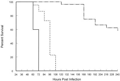
Twelve- to 24-h-old CD-1 mice were inoculated subcutaneously with each virus and monitored for mortality at 12-h intervals (Fig. 1). Infection with TR339 resulted in 100% mortality, with an AST of 1.8 ± 0.34 (mean ± standard deviation) days. TRSB infection also resulted in 100% mortality, but the AST was extended significantly (3.2 ± 0.46 days [two-tailed Student’s t test with unequal variance; P, <0.001]). TRSB-R114 caused only ∼50% mortality, with a considerably extended survival time (7.9 ± 2.5 days). These data indicate that strains which diverge at only one or two amino acids from the AR339 consensus sequence can exhibit significant attenuation of disease in neonatal mice.
FIG. 1.
Survival of neonatal CD-1 mice infected with TR339 (solid line; n, 30; AST, 1.8 ± 0.34 days), TRSB (evenly dashed line; n, 30; AST, 3.2 ± 0.46 days), or TRSB-R114 (unevenly dashed line; n, 25; AST, 7.9 ± 2.5 days). Mice were inoculated subcutaneously with 1,000 BHK PFU of virus in 50 μl of diluent.
Virus binding to and infectivity for BHK cells.
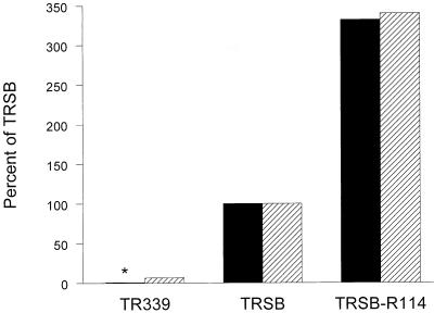
BHK cell binding assays revealed a dramatic difference in attachment efficiencies of TR339 and the cell culture-adapted mutants (Fig. 2). In multiple experiments, only 0.05 to 0.5% of added TR339 bound to BHK cells, while the bound proportion of added TRSB and TRSB-R114 increased to 5 to 10% and 20 to 30%, respectively. These results suggest a strong correlation between BHK cell-adaptive mutations and attachment efficiency and an inverse correlation with virulence in mice. We have also found this correlation with other AR339 strains (33). For TR339, the percent cpm bound was much lower than previously reported for AR339 laboratory strains (59, 67, 69, 70). Preliminary assays indicated that the binding of TR339 and TRSB to BHK cells was not saturable. However, TRSB-R114 exhibited saturable binding (data not shown).
FIG. 2.
Relative binding and specific infectivity of TR339, TRSB, and TRSB-R114 for BHK cells. For binding assay (solid bars), 105 cpm of each radiolabeled virus was added to 106 BHK cells in suspension and incubated at 4°C for 60 min. After three washes with VB, radioactivity associated with cells was quantitated. For specific infectivity (hatched bars), radiolabeled virus was diluted and assayed for plaque formation on BHK cell monolayers. Specific infectivity (PFU/cpm) was then calculated for each virus. Values are presented as percentages of values for TRSB (100%). ∗, binding of TR339 was <1% of that of TRSB in this assay.
Specific infectivity of radiolabeled virus (PFU/cpm) was closely correlated with relative binding to BHK cells (Fig. 2). Covariation of binding and infectivity suggests that binding measured in these assays reflects an interaction that leads directly to infection of cells and that the increase in infectivity of TRSB compared with TR339, and the additional increase of TRSB-R114 compared with TRSB, results from increased efficiency of attachment to cells. Binding reactions with primary CEF, primary MEF, murine L929 fibroblasts, murine Swiss 3T3 fibroblasts, and murine Neuro-2A neuroblastoma cells revealed the same patterns of binding efficiency as with BHK cells: TRSB-R114 > TRSB > TR339 (data not shown). The absolute amount of binding for each virus varied between cell types; however, TR339 binding was never more than 1% of added cpm.
Binding and BHK cell infectivity of a virus with Ser at E2 1, but the TRSB residues at nsP3 528 and E1 72 (E2S1) (Table 1), were indistinguishable from TR339 (data not shown). This demonstrates that of the three TRSB mutations relative to the consensus sequence (Table 1), the E2 Arg 1 mutation alone accounts for the increased binding and infectivity.
We also placed the E2 Arg 114 mutation in the E2 Ser 1 background (E2S1-R114) (Table 1). The resultant virus exhibited >90% of the binding efficiency of TRSB-R114 (data not shown). This suggests that the E2 Arg 114 mutation by itself confers a very large increase in attachment ability and that the E2 Arg 1 and E2 Arg 114 mutations can independently increase cell binding.
Heparin competition of binding and infectivity.
As described above, several previous Sindbis virus studies indicated that a charge-based interaction, perhaps involving a cell surface sulfated polysaccharide, might be important to binding. In preliminary assays, we found that the binding and infectivity of TR339 were insensitive to ionic strength, whereas TRSB and TRSB-R114 binding and infectivity were significantly reduced by NaCl concentrations above 175 mM (data not shown). This indicated that alteration of cell surface charge could affect the binding of BHK cell-adapted Sindbis virus strains.
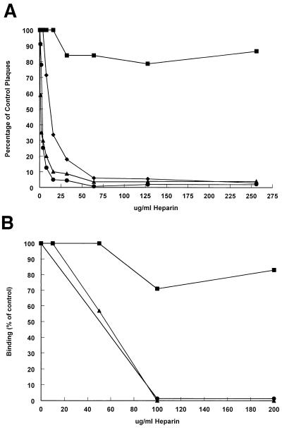
We next evaluated the ability of the sulfated polysaccharide heparin to block plaque formation by these viruses (Fig. 3A). Virus derived from pToto 50 (cDNA clone of Sindbis virus strain HRsp [51]) also was included to evaluate the effects of heparin on the infectivity of an extensively passaged AR339 strain. Heparin is commonly used as an analog of cellular HS in receptor-ligand interaction assays, since ligand interactions with heparin and analogous HS structures have little qualitative difference (31). TR339 showed only limited (∼25%) competition at heparin concentrations ranging from <50 μg/ml to >10 mg/ml. In sharp contrast, soluble heparin effectively competed infectivity of TRSB and TRSB-R114 at low concentrations (50% plaque reduction endpoints of 1.1 and 3.3 μg/ml, respectively [Fig. 3A]). Plaque formation by Toto 50 also was >95% competed; however, the 50% plaque reduction endpoint was at a 5- to 10-fold-higher heparin concentration than for TRSB and TRSB-R114. In control competition experiments, BSA slightly increased plaque numbers with increasing concentrations, while the GAG molecule CS-A had no detectable effect on infectivity of any of the viruses. CS-B (dermatan sulfate), which is structurally similar to HS, and dextran sulfate, a highly sulfated molecule similar to heparin, inhibited virus plaque formation only at high concentrations (>1 mg/ml [data not shown]).
FIG. 3.
Soluble heparin competition of TR339, TRSB, TRSB-R114, and Toto 50. (A) Reduction in plaque formation of TR339 (squares), TRSB (circles), TRSB-R114 (triangles), and Toto 50 (diamonds) on BHK cell monolayers with increasing concentrations of heparin. Viruses were diluted to 100 to 200 PFU/200 μl and reacted with heparin for 30 min at 4°C, followed by infection of BHK cell monolayers in the presence of heparin. (B) Reduction in binding of TR339 (squares), TRSB (circles), and TRSB-R114 (triangles) to BHK cells with increasing concentrations of heparin. Viruses (105 cpm/reaction) were reacted with heparin as described above, followed by binding assay in the presence of heparin. Results are averages of two or three reactions at each concentration.
It is of interest that maximum competition of TR339 was achieved at <50 μg of heparin per ml, with no further increase in competition observed at 10 mg/ml. This result may indicate the presence of two populations of virus in the TR339 stock. Production of virus stocks by electroporation of in vitro transcripts may involve limited selection for growth on BHK cells (see below). If electroporation efficiency is less than 100%, then virus progeny from successfully electroporated cells will be amplified in cells that received no viral transcripts initially.
Binding assays showed a similar pattern, in that TRSB and TRSB-R114 were competed in binding by soluble heparin while TR339 showed resistance similar to that exhibited for plaque formation (Fig. 3B). However, concentrations required to inhibit binding of the viruses were significantly greater than for plaque reduction, likely reflecting the much higher concentration of virus used in binding assays. Consistent with plaque formation assays, BSA increased binding slightly and CS-A had no effect. These results indicate that the block in generating TRSB or TRSB-R114 plaques in the presence of heparin is at the level of attachment to cells.
Enzymatic digestion of cell surface HS.
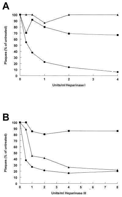
Heparinase digestion of cells is commonly used to examine the effect of removal of HS on ligand binding to cell surface receptors (53). Virus infectivity and binding were evaluated after digestion of BHK cells with heparinase I (which digests highly sulfated heparin-like structures on HS [15]) or heparinase III (which digests both HS and heparin [15]). Digestion of BHK cells with increasing concentrations of either heparinase I or III resulted in a maximum reduction of TR339 infectivity of only ∼25% regardless of the enzyme used, consistent with the heparin competition experiments (Fig. 4). However, a dose-dependent inhibition of TRSB plaque-forming ability was observed with both heparinase I and heparinase III. TRSB-R114 was unaffected by heparinase I digestion; however, this virus was nearly as sensitive as TRSB to heparinase III.
FIG. 4.
Effect of heparinase digestion on plaque-forming efficiency of TR339, TRSB, and TRSB-R114 on BHK cells. (A) BHK cell monolayers were digested with increasing concentrations of heparinase I, followed by three rounds of washing with virus buffer and infection with 100 to 200 PFU of TR339 (squares), TRSB (circles), or TRSB-R114 (triangles). (B) BHK cell monolayers were digested with increasing concentrations of heparinase III, washed, and infected as above. Data are averages of two or three assays at each concentration.
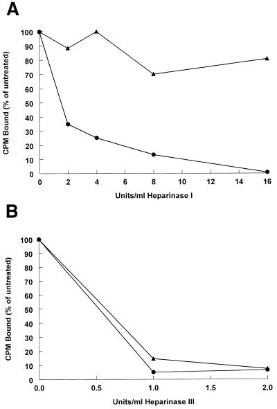
Binding experiments were consistent with efficiency of plaque formation. Binding of TRSB was sensitive to both heparinase I and heparinase III, while TRSB-R114 was sensitive only to heparinase III (Fig. 5). In control experiments, binding and infectivity of all viruses were reduced significantly less (maximum of ∼30% reduction) by digestion of cells with chondroitinase ABC (data not shown). These heparinase experiments suggest that TRSB and TRSB-R114 interact with different types of HS structures. TRSB-R114 binding may be dependent on a structural characteristic of HS or the context of HS displayed on cell surfaces, while TRSB may bind promiscuously to HS structures. The fact that TRSB-R114 binding is saturable, while that of TRSB is not, also suggests that these viruses attach to different HS structures.
FIG. 5.
Effect of heparinase digestion on binding of TRSB (circles) and TRSB-R114 (triangles) to BHK cells. (A) BHK cell monolayers were digested with increasing concentrations of heparinase I, followed by three rounds of washing with VB, suspension, and use in binding assays (5 × 10⁴ cpm of radiolabeled virus per reaction). (B) BHK cell monolayers were digested with increasing concentrations of heparinase III and processed as above. Data are averages of two or three binding reactions at each concentration. In these assays, binding of TR339 to digested or undigested cells was not above background cpm measured in cell-free control reactions.
Genetic alteration of cell surface GAGs.
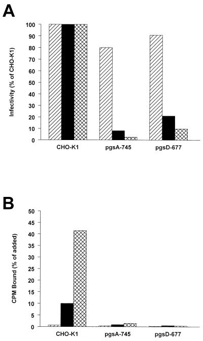
Binding and infectivity of viruses also were evaluated on wild-type CHO-K1 cells and CHO mutants defective in GAG and HS synthesis. The mutant CHO cell line pgsA-745 is deficient in xylosyltransferase (18), an enzyme of a pathway common to all GAG biosynthesis, and hence is GAG deficient. The mutant line pgsD-677 is deficient in N-acetylglucosaminyl- and glucuronosyltransferase activities (interrupting HS synthesis) and overproduces CS (35). Due to extremely variable plaque sizes of Sindbis virus on the different CHO cell lines, an RP-based fluorescent infectious center assay was used to determine infectivity on these cells. Cell monolayers were infected with RP assembled with structural proteins of either TR339, TRSB, or TRSB-R114 and containing a defective genome that directed the expression of GFP within infected cells. Since structural protein genes were deleted from the RP genomes, virus spread beyond the initially infected cells was prevented. Therefore, relative RP infectivity, determined by the TR339, TRSB, and TRSB-R114 glycoproteins in their respective envelopes, could be measured by enumeration of single infected cells by fluorescence microscopy.
Consistent with the role of GAGs suggested by the competition and enzymatic digestion assays, the infectivity of TRSB and TRSB-R114 RP was greatly reduced on the GAG-deficient cells (by 90 and >95%, respectively [Fig. 6A]). Reminiscent of heparinase digestion of BHK cells, TR339 RP exhibited only ∼30% reduction in infection efficiency. Infectivity of mutant RP on the HS-deficient pgsD-677 cells was greatly reduced compared to wild-type cells but was not reduced as much as with GAG-deficient cells (Fig. 6A). However, this difference between the two cell lines appears to be unrelated to cell binding (see below).
FIG. 6.
Infectivity of TR339, TRSB, and TRSB-R114 GFP-expressing RP and binding of analogous viruses to wild-type CHO-K1 cells and CHO cell mutants deficient in all GAGs (pgsD-745) or HS (pgsD-677). (A) Monolayers of each cell type were infected (60 min) with 100 to 200 BHK GFP-infectious units followed by three washes with VB, medium replacement, and incubation for 8 to 12 h. Cells expressing GFP were enumerated by fluorescence microscopy. Data are averages of four infections of TR339 (hatched bars), TRSB (solid bars), and TRSB-R114 (cross-hatched bars). (B) Suspension binding assays (105 cpm added per reaction) were completed as described for BHK cells. Results for TR339 (hatched bars), TRSB (solid bars), and TRSB-R114 (cross-hatched bars) are presented as percent cpm bound and represent averages of two or three reactions.
In close correlation with infectivity, binding of TRSB and TRSB-R114 to the GAG-deficient pgsA-745 cells was reduced by >90 and >95%, respectively, compared with CHO-K1 cells (Fig. 6B). Binding of the mutant viruses to the HS-deficient pgsD-677 cells was similarly reduced; however, as indicated above, all RP were slightly more infectious for these cells, suggesting that overproduction of CS may increase infection efficiency at a postbinding step of virus entry. In addition, binding to the mutant pgsD-677 cells was consistently lower than to pgsA-745 cells, suggesting either that the pgsA-745 cells produce a small amount of HS or that the overproduction of CS on pgsD-677 cells inhibits virus binding. In the absence of GAGs or HS, the amount of binding of TRSB and TRSB-R114 was reduced to the level of TR339 binding. This indicates that interaction with HS accounts for most, if not all, of the increased binding and infectivity of TRSB and TRSB-R114 on normal cells relative to TR339.
The specific effect of HS-GAG deficiency on binding to pgsA-745 cells was further shown by restoring GAG precursors to these cells. The pgsA-745 cells cannot transfer xylose precursors to core proteins to initiate GAG chain synthesis (18). The addition of xyloside precursors increases production of GAGs in these cells to a level similar to precursor-treated CHO-K1 cells and restores binding of the HS binding-dependent basic fibroblast growth factor (17). However, GAGs produced under these conditions are not covalently attached to core proteins and may adhere to cell surfaces as free GAG chains (49). Incubation of pgsA-745 cells in 1 mM p-nitrophenyl-β-d-galactoxylopyranoside for 48 h resulted in the restoration of TRSB binding to >90% of TRSB binding to similarly treated CHO-K1 cells. However, TRSB-R114 binding was restored only to ∼20% of TRSB-R114 binding to similarly treated CHO-K1 cells (data not shown). This result, in combination with the heparinase digestion and binding saturation studies, suggests that TRSB binds promiscuously to HS molecules on cell surfaces. In contrast, the majority of TRSB-R114 binding may be dependent on a subset of HS, perhaps requiring attachment of these molecules to proteoglycan core proteins. Separate control experiments demonstrated that, like BHK cells, binding and infectivity of TRSB and TRSB-R114 (but not TR339) on wild-type CHO-K1 cells were efficiently competed with soluble heparin. In addition, CHO-K1 cells showed reductions in binding and infectivity similar to BHK cells after digestion with heparinases (data not shown).
Direct virus binding to an HS analog.
To determine whether TRSB and TRSB-R114 bind directly to HS structures, heparin-agarose and BSA-agarose beads were used as cell surrogates in suspension binding assays. Results shown in Table 2 demonstrate that both mutant viruses bound efficiently to heparin-agarose beads, while TR339 did not. In contrast to cell binding assays, the beads bound ∼70% of TRSB and TRSB-R114 radioactivity added. The actual binding in these assays may have been even greater, since there was some unavoidable loss of beads during wash steps. None of the viruses bound appreciably to BSA-agarose beads (Table 2), and binding of TRSB and TRSB-R114 to the heparin-agarose beads could be competed with soluble heparin but not soluble BSA or CS-A (data not shown). These results taken together strongly indicate that attachment to heparin-agarose beads results from a specific virus-heparin interaction. The E2S1-R114 virus (Table 1) also attached efficiently to heparin-agarose beads (Table 2), suggesting that the E2 Arg 114 mutation defines a separate locus (compared with E2 1) capable of independently mediating HS attachment.
TABLE 2.
Binding of viruses to heparin-agarose and BSA-agarose beads
| Virus | % cpm bound
|
|
|---|---|---|
| Heparin-agarose | BSA-agarose | |
| TR339 | 4.5 | 0.35 |
| TRSB | 70.5 | 0.50 |
| TRSB-R114 | 68.1 | 0.31 |
| E2S1-R114 | 74.0 | 0.76 |
Passage of TR339 in BHK cells coselects for increased cell binding and heparin interaction.
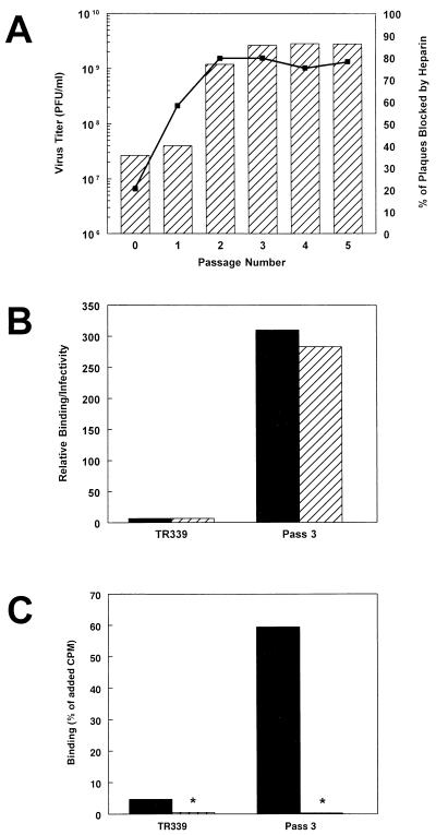
Mutation to efficient HS attachment is the result of active selection pressure during Sindbis virus adaptation to BHK cell growth. As with the other experiments reported here, the TR339 stock (passage 0) consisted of culture fluids from cells electroporated with in vitro transcripts of a full-length cDNA clone. TR339 was passaged five times in BHK cells, followed by assay of the attachment phenotype of virus obtained from each passage. Figure 7A shows increasing virus titer and susceptibility to competition with soluble heparin (200 μg/ml) for virus from passages 1 through 5. Between passage 1 and passage 3, virus titer increased ∼100-fold, concomitant with an increase in susceptibility to heparin competition of from ∼20 to >80%. No further increase in either phenotype was noted after passage 3. For this reason, virus equivalent to passage 3 was used for subsequent analysis. Radiolabeled virus binding assays indicated that one consequence of the adaptation of TR339 to BHK cells was a large increase in binding to and specific infectivity for BHK cells (Fig. 7B). In fact, binding and infectivity of the passage 3 virus was significantly greater than TRSB and similar to TRSB-R114 (Fig. 2 and 7B). The passage 3 virus also attached efficiently to heparin-agarose beads, indicating that heparin binding and cell binding were covariant phenotypes (Fig. 7C). The E1 and E2 glycoproteins of the passage 3 virus population were sequenced, and a single coding change from Glu to Lys at E2 70 was found. This mutation was observed previously in our laboratory during BHK cell passage experiments (40, 54). It also is a constituent of most Sindbis virus AR339 laboratory strains, including Toto 50, which is effectively competed by soluble heparin. Similar to the effect of E2 Arg 114, E2 Lys 70 confers rapid penetration of BHK cells when introduced into the TRSB (E2 Arg 1) background and significantly attenuates virulence in neonatal mice (40).
FIG. 7.
Changes in titer, heparin competition, BHK cell binding, and heparin-agarose bead binding phenotypes of TR339 after passage in BHK cells. (A) BHK cell monolayers were infected at a multiplicity of infection of 1 followed by incubation for 20 h, harvesting of supernatants, and infection of new monolayers. This cycle was repeated for a total of five passages. Hatched bars (left y axis) indicate BHK cell titers for each of the five passages. The solid line (right y axis) indicates change in inhibition of BHK cell plaque-forming efficiency due to competition with soluble heparin (200 μg/ml). (B) Increases in BHK cell binding (solid bars) and specific infectivity (hatched bars) of virus derived from the third passage, compared to TR339 stock virus. (C) Increase in heparin-agarose bead binding (solid bars) but not BSA-agarose bead binding (hatched bars) of third-passage virus, compared to TR339 stock virus. ∗, binding of either virus to BSA-agarose beads was <1% of added cpm.
DISCUSSION
HS as a Sindbis virus attachment receptor.
HS can serve as an attachment receptor for Sindbis virus but contributes significantly to binding and infection only by cell culture-adapted strains. While infection with the AR339 consensus sequence virus, TR339, was predominantly HS independent, four other cell culture-adapted strains, each with mutations in the E2 gene, attached to cells through an HS-dependent mechanism. These findings suggest that Sindbis virus rapidly mutates to utilize HS during cell culture adaptation and that binding to cell surface HS plays little or no role in interactions between natural Sindbis virus isolates and cells in vivo. Our AR339 laboratory strain, TRSB, acquired a mutation conferring HS attachment (Ser to Arg at E2 1) during single-step BHK cell adaptation prior to cDNA cloning (40). Attachment of TRSB was significantly increased relative to TR339 and was sensitive to both heparinase I and heparinase III but was not saturable. When TRSB was subjected to selection for rapid penetration of BHK cells, an additional mutation conferring HS attachment arose at E2 114 (Ser to Arg [11, 47]). Attachment of this virus showed high affinity, was saturable, and was sensitive only to heparinase III. The effect of E2 Arg 1 was independent of the residue at E1 72, as shown in control binding and heparin competition experiments using E2S1 (Table 1). Additionally, passage of TR339 in BHK cells selected for a Glu-to-Lys mutation at E2 70 that conferred high-affinity attachment to BHK cells and to heparin-agarose beads.
While it has been known for some time that herpesviruses, including herpes simplex virus (73), human cytomegalovirus (10), bovine herpesvirus (45), and pseudorabies virus (41), attach to HS during in vitro infection, it has only recently become apparent that a number of other viruses can use this molecule for cell attachment. Recent publications have implicated HS in the in vitro attachment of type O foot-and-mouth disease virus (FMDV) (29, 55), HIV (48, 52), respiratory syncytial virus (34), porcine reproductive and respiratory syndrome virus (30), equine arteritis virus (1), Dengue virus (8), adeno-associated virus (62), and vaccinia virus (9).
In the herpes simplex virus system, a protein that can mediate entry, but not attachment, has been identified (43). This suggests that utilization of GAG molecules in virus infection can result in a two-step entry process in which interaction with the GAG molecule increases the concentration of the virus ligand in the vicinity of its entry receptor, thereby enhancing the overall efficiency of infection. That this model may be generally applicable is supported by the studies of Wickham et al. (72), who found that an HS binding sequence introduced into the tip of the adenovirus fiber protein greatly increased infectivity of the virus for cell types that did not express a high-affinity receptor. Our data do not clearly indicate whether infection of HS-dependent Sindbis virus occurs in one or several steps. It is clear that all of the viruses tested maintained a low level of infectivity for cells lacking HS. This suggests that infection can proceed without efficient binding and may reflect the presence of an additional receptor(s).
Our results closely parallel those of Sa-Carvalho et al. (55) with type O FMDV. In their studies, passage in culture was associated with changes to positive amino acids at several positions in the capsid protein and acquisition of heparin-HS attachment. The coselection for rapid penetration of BHK cells, mutation in E2, and ability to attach to HS analogs also has been documented for VEE in our laboratory (2). Thus, adaptation to HS attachment may represent a common cell culture-adaptive mechanism among alphaviruses. Our results and those of Sa-Carvalho et al. may suggest further that adaptation to HS binding in cultured cells could occur in multiple virus families. Therefore, studies of both virus attachment to cultured cells and virus pathogenesis in vivo could be compromised by the presence of such adaptive mutations embedded in the genetic background of “wild-type” laboratory strains or cloned viruses derived from them.
The relationship of our results to previous studies that have identified different proteins as putative attachment receptors for Sindbis virus on different cells (e.g., references 16, 37, and 69–71) is not clear. Cell surface chains of HS are generally attached to proteoglycan core proteins (31). Moreover, HS exhibits structural heterogeneity between cell types and is presented in different contexts on different cell types (e.g., through attachment to different core proteins or in different protein-proteoglycan-GAG complexes [4, 28, 31]). The heterogeneous expression of HS and GAG chains on different cell types raises the possibility for virus interaction with different HS-protein structures on different cells and that cell-type-specific GAG or proteoglycan core protein expression could regulate virus binding (3, 56). It is unknown whether any of the putative Sindbis virus receptor proteins are full-time or facultative proteoglycans or whether any of these are intimately complexed with proteoglycan-HS chains on cell surfaces. However, a major conclusion from the results presented here is that single amino acid differences between AR339 strains can dramatically alter cell interaction properties. Therefore, it is quite possible that unidentified structural glycoprotein differences between Sindbis virus laboratory strains account for the conflicting results from different laboratories.
Domains involved in HS attachment.
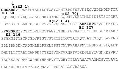
As defined by attachment to BHK cells and heparin-agarose beads, mutation to a positive charge at E2 1, E2 70, or E2 114 can independently increase attachment affinity, suggesting that these mutations may define three HS interaction domains on the glycoprotein spike. Most protein-GAG binding is mediated by electrostatic interaction between clusters of basic amino acids arranged in a three-dimensional array on the ligand and a concentrated negative charge on the sulfated polysaccharide chain (reviewed in reference 31). In an analysis similar to that undertaken by Flynn and Ryan with pseudorabies virus protein gC (20), we have scanned the PE2 and E1 glycoproteins for sequences that conform to the XBBXBX and XBBBXXBBX (B, basic; X, any amino acid) heparin interaction consensus motifs identified by Cardin and Weintraub (7). The activity of these consensus sequences is not dependent upon orientation (20). While no close matches were found in E1, three close matches were found in PE2 (Fig. 8).
FIG. 8.
Identification of heparin interaction consensus sequences (from reference 7) in the PE2 glycoprotein. Matches (XBBXBX or XBXBBX) are indicated in boldface type within the glycoprotein sequence (residue numbers of the amino-terminal consensus match residues are in boldface type below the sequence). E2 1, E2 70, and E2 114 mutations are indicated in boldface type above the glycoprotein sequence. The site of furin protease cleavage of PE2 is indicated by the arrow. Sequence matches adjacent to the transmembrane domain have not been included.
One exact match comprised the XBXBBX furin protease cleavage site of PE2 (14, 42; reviewed in reference 32), which is immediately adjacent to the E2 Arg 1 mutation present in both TRSB and TRSB-R114. Heidner et al. (25) have shown that substitution of Arg, Ile, Val, or Asn for Ser at E2 1 reduces PE2 cleavage efficiency, resulting in mature virus particles that contain various amounts of PE2 and its associated furin cleavage-heparin binding site. Appropriate mutagenesis experiments are currently in progress to test the hypothesis that BHK cell binding is increased as a function of PE2-furin cleavage site content in virions. Interestingly, the portion of the HIV V3 loop thought to interact with HS (52) contains a cluster of positively charged amino acid residues comprising an occult furin protease cleavage site that has been proposed to be cleaved during HIV entry of cells (44).
The positively charged mutations at E2 70 and E2 114 were not part of, or adjacent to, heparin interaction sequences. Positive charge mutations in the E2 glycoprotein could increase HS binding (i) by participating in conformationally determined HS binding sites similar to those proposed for type O strains of FMDV (55); (ii) by decreasing the efficiency of PE2 cleavage during virus maturation, thereby increasing the virion content of uncleaved PE2 containing the heparin interaction consensus motif; or (iii) by altering the conformation of virus glycoprotein spikes, resulting in exposure of preexisting HS interaction motifs.
In contrast to TRSB, the majority of binding of TRSB-R114 (mediated by E2 Arg 114) involves interaction with a specific HS structure, as shown by binding saturation, xyloside precursor reconstitution, and heparinase digestion experiments. This is consistent with studies indicating that protein-HS interactions can show significant HS sequence and sulfation specificity (e.g., references 19 and 38). Further study will be required to elucidate specific HS structures involved in interaction with different Sindbis virus mutants.
Previous studies had indicated that virus attachment to cells was not increased by the E2 Arg 114 mutation (47). Our results may differ from those for two reasons. First, the criterion for attachment in assays conducted by Olmsted et al. involved resistance to removal by washing of plaque-forming virus bound to cell monolayers, as opposed to the binding of radiolabeled virus particles to cell suspensions, as reported here. We have found that while these viruses exhibit large differences in the amount of virus that attaches to cells, once virus particles are attached they appear to be equally resistant to dislodging by washing (data not shown). Secondly, viruses in the previous assays were purified on potassium tartrate gradients. We have found that this purification technique reduces the infectivity and binding of TRSB-R114 preparations severalfold, perhaps due to the detrimental effects of high ionic strength on virus particles. This phenomenon also has been observed with a VEE E2 glycoprotein mutant (12).
Relationship of HS attachment to neonatal mouse virulence.
Perhaps most interesting is that the primarily HS-independent interaction of TR339 with cells is not saturable and is barely measurable in a variety of cell binding assays. This result is independent of cell type or temperature, is obtained when binding assays are done with cell suspensions or monolayers, and is consistent in both positive and subtractive binding assays (data not shown). Nevertheless, TR339 is the most virulent of the tested viruses in vivo (these studies; also references 33 and 65), suggesting that a weak, primarily HS-independent attachment may represent an important component of alphavirus entry in nature. TR339, TRSB, and TRSB-R114 exhibit indistinguishable tissue tropisms in neonatal mice, differing only by rate and extent of spread (33, 65). Therefore, the high-affinity attachment of Sindbis virus laboratory strains, as typically measured in in vitro binding assays, may simply be a consequence of adaptation to cultured cells and does not determine tissue tropism in neonatal mice. However, this assertion is tempered by the possibility that virus attachment in the context of an intact organismal host may be very different from that measured in cell binding assays. For instance, in a number of virus systems, particles may require activation by host factors for full infectivity in vivo (reviewed in reference 32).
In separate studies (33, 65), it has been shown that attenuation of TRSB and TRSB-R114 coincides with lower virus titers in sera and brains of infected mice. Therefore, we suggest that the HS attachment phenotype results in a general reduction in virus replication competence in vivo. A similar hypothesis was recently proposed by Sa-Carvalho et al. for FMDV (55), where tissue culture-adapted type O strains that had acquired the ability to attach to HS were greatly reduced in animal virulence. GAGs, and specifically HS molecules, comprise ubiquitous cell surface structures and also are abundant components of extracellular matrices and basal laminae (reviewed in references 4, 28, 31, and 64). Consequently, it is possible that HS attachment results in virus binding to unproductive receptor structures in vivo. In addition, HS attachment may affect virus replication competence by interfering with virus particle release from cells. We have observed that virus particle production from individual infected BHK cells is inversely correlated with HS attachment efficiency (data not shown). We are currently investigating whether a combination of inappropriate receptor targeting and reduced particle release may determine neonatal mouse virulence of Sindbis viruses that have adapted to attach to HS.
ACKNOWLEDGMENTS
We thank Peter Mason for several stimulating discussions. We also thank Nancy Davis for critical reading of the manuscript. Cherice Connor, Michael Hawley, and Travis Knott provided excellent technical assistance.
This work was supported by Public Health Service-NIH grant AI22186. W.B.K. was supported by an NIH Predoctoral Traineeship (T32 AI07419) and by the U.S. Army Research Office (DAAH04-95-1-0224).
REFERENCES
- 1.Asagoe T, Inaba Y, Jusa E R, Kouno M, Uwatoko K, Fukunaga Y. Effect of heparin on infection of cells by equine arteritis virus. J Vet Med Sci. 1997;59:727–728. doi: 10.1292/jvms.59.727. [DOI] [PubMed] [Google Scholar]
- 2.Bernard, K. A., and R. E. Johnston. Unpublished observations.
- 3.Bernfield, M., M. T. Hinkes, and R. L. Gallo. 1993. Developmental expression of the syndecans: possible function and regulation. Development 1993(Suppl.):205–212. [PubMed]
- 4.Bernfield M, Kokenyesi R, Kato M, Hinkes M T, Spring J, Gallo R L, Lose E J. Biology of the syndecans: a family of transmembrane heparan sulfate proteoglycans. Annu Rev Cell Biol. 1992;8:365–393. doi: 10.1146/annurev.cb.08.110192.002053. [DOI] [PubMed] [Google Scholar]
- 5.Birdwell C R, Strauss J H. Distribution of the receptor sites for Sindbis virus on the surface of chicken and BHK cells. J Virol. 1974;14:672–678. doi: 10.1128/jvi.14.3.672-678.1974. [DOI] [PMC free article] [PubMed] [Google Scholar]
- 6.Bredenbeek P J, Frolov I, Rice C M, Schlesinger S. Sindbis virus expression vectors: packaging of RNA replicons by using defective helper RNAs. J Virol. 1993;67:6439–6446. doi: 10.1128/jvi.67.11.6439-6446.1993. [DOI] [PMC free article] [PubMed] [Google Scholar]
- 7.Cardin A D, Weintraub H J R. Molecular modeling of protein-glycosaminoglycan interactions. Arteriosclerosis. 1989;9:21–32. doi: 10.1161/01.atv.9.1.21. [DOI] [PubMed] [Google Scholar]
- 8.Chen Y, Maguire T, Hileman R E, Fromm J R, Esko J D, Linhardt R J, Marks R M. Dengue virus infectivity depends on envelope protein binding to target cell heparan sulfate. Nat Med. 1997;3:866–871. doi: 10.1038/nm0897-866. [DOI] [PubMed] [Google Scholar]
- 9.Chung C-S, Hsaio J-C, Chang Y-S, Chang W. A27L protein-mediated vaccinia virus interaction with cell surface heparan sulfate. J Virol. 1998;72:1577–1585. doi: 10.1128/jvi.72.2.1577-1585.1998. [DOI] [PMC free article] [PubMed] [Google Scholar]
- 10.Compton T, Nowlin D M, Cooper N R. Initiation of human cytomegalovirus infection requires initial interaction with cell surface heparan sulfate. Virology. 1993;193:834–841. doi: 10.1006/viro.1993.1192. [DOI] [PubMed] [Google Scholar]
- 11.Davis N L, Fuller F J, Dougherty W G, Olmsted R A, Johnston R E. A single nucleotide change in the E2 glycoprotein gene of Sindbis virus affects penetration rate in cell culture and virulence in neonatal mice. Proc Natl Acad Sci USA. 1986;83:6771–6775. doi: 10.1073/pnas.83.18.6771. [DOI] [PMC free article] [PubMed] [Google Scholar]
- 12.Davis, N. L., and R. E. Johnston. Unpublished observations.
- 13.Davis N L, Powell N, Greenwald G F, Willis L V, Johnson B J B, Smith J F, Johnston R E. Attenuating mutations in the E2 glycoprotein gene of Venezuelan equine encephalitis virus: construction of single and multiple mutants in a full-length cDNA clone. Virology. 1991;183:20–31. doi: 10.1016/0042-6822(91)90114-q. [DOI] [PubMed] [Google Scholar]
- 14.de Curtis I, Simons K. Dissection of Semliki Forest virus glycoprotein delivery from the trans-Golgi network to the cell surface in permeablized BHK cells. Proc Natl Acad Sci USA. 1988;85:8052–8056. doi: 10.1073/pnas.85.21.8052. [DOI] [PMC free article] [PubMed] [Google Scholar]
- 15.Desai U R, Wang H-M, Linhardt R J. Specificity studies on the heparin lysases from Flavobacterium heparinum. Biochemistry. 1993;32:8140–8145. doi: 10.1021/bi00083a012. [DOI] [PubMed] [Google Scholar]
- 16.Duda E, Berencsi K. Sindbis virus receptor protein of BHK cells. Acta Virol. 1980;24:149–152. [PubMed] [Google Scholar]
- 17.Esko J D, Montgomery R I. Synthetic glycosides as primers of oligosaccharide biosynthesis and inhibitors of glycoprotein and proteoglycan assembly. In: Ausubel F M, Brent R, Kingston R, Moore D D, Seidman J G, Smith J A, Struhl K, Varki A, Coligan J, editors. Current protocols in molecular biology. New York, N.Y: Greene Publishing and Wiley-Interscience; 1995. pp. 17.11.1–17.11.6. [DOI] [PubMed] [Google Scholar]
- 18.Esko J D, Stewart T E, Taylor W H. Animal cell mutants defective in glycosaminoglycan biosynthesis. Proc Natl Acad Sci USA. 1985;82:3197–3201. doi: 10.1073/pnas.82.10.3197. [DOI] [PMC free article] [PubMed] [Google Scholar]
- 19.Feyzi E, Trybala E, Bergström T, Lindahl U, Spillman D. Structural requirement of heparan sulfate for interaction with herpes simplex virus type 1 virions and isolated glycoprotein C. J Biol Chem. 1997;272:24850–24857. doi: 10.1074/jbc.272.40.24850. [DOI] [PubMed] [Google Scholar]
- 20.Flynn S J, Ryan P. The receptor-binding domain of pseudorabies virus glycoprotein gC is composed of multiple discrete units that are functionally redundant. J Virol. 1996;70:1355–1364. doi: 10.1128/jvi.70.3.1355-1364.1996. [DOI] [PMC free article] [PubMed] [Google Scholar]
- 21.Fries E, Helenius A. Binding of Semliki Forest virus and its spike glycoproteins to cells. Eur J Biochem. 1979;97:213–220. doi: 10.1111/j.1432-1033.1979.tb13105.x. [DOI] [PubMed] [Google Scholar]
- 22.Glasgow G M, Sheahan B J, Atkins G J, Wahlberg J M, Salminen A, Liljestrom P. Two mutations in the envelope glycoprotein E2 of Semliki Forest virus affecting the maturation and entry patterns of the virus alter pathogenicity for mice. Virology. 1991;185:741–748. doi: 10.1016/0042-6822(91)90545-m. [DOI] [PubMed] [Google Scholar]
- 23.Hahn C S, Hahn Y S, Braciale T J, Rice C M. Infectious Sindbis virus transient expression vectors for studying antigen processing and presentation. Proc Natl Acad Sci USA. 1992;89:2679–2683. doi: 10.1073/pnas.89.7.2679. [DOI] [PMC free article] [PubMed] [Google Scholar]
- 24.Heidner H W, Johnston R E. The amino-terminal residue of Sindbis virus glycoprotein E2 influences virus maturation, specific infectivity for BHK cells, and virulence in mice. J Virol. 1994;68:8064–8070. doi: 10.1128/jvi.68.12.8064-8070.1994. [DOI] [PMC free article] [PubMed] [Google Scholar]
- 25.Heidner H W, Knott T A, Johnston R E. Differential processing of Sindbis virus glycoprotein PE2 in cultured vertebrate and arthropod cells. J Virol. 1996;70:2069–2073. doi: 10.1128/jvi.70.3.2069-2073.1996. [DOI] [PMC free article] [PubMed] [Google Scholar]
- 26.Heidner H W, McKnight K L, Davis N L, Johnston R E. Lethality of PE2 incorporation into Sindbis virus can be suppressed by second-site mutations in E3 and E2. J Virol. 1994;68:2683–2692. doi: 10.1128/jvi.68.4.2683-2692.1994. [DOI] [PMC free article] [PubMed] [Google Scholar]
- 27.Helenius A, Morein B, Fries E, Simons K, Robinson P, Schirrmacher V, Terhorst C, Strominger J L. Human (HLA-A and HLA-B) and murine (H-2K and H-2D) histocompatibility antigens are cell surface receptors for Semliki Forest virus. Proc Natl Acad Sci USA. 1978;75:3846–3850. doi: 10.1073/pnas.75.8.3846. [DOI] [PMC free article] [PubMed] [Google Scholar]
- 28.Jackson R L, Busch S J, Cardin A D. Glycosaminoglycans: molecular properties, protein interactions, and role in physiological processes. Physiol Rev. 1991;71:481–539. doi: 10.1152/physrev.1991.71.2.481. [DOI] [PubMed] [Google Scholar]
- 29.Jackson T, Ellard F M, Abu Ghazaleh R, Brookes S M, Blakemore W E, Corteyn A H, Stuart D I, Newman J W I, King A M Q. Efficient infection of cells in culture by type O foot-and-mouth disease virus requires binding to cell surface heparan sulfate. J Virol. 1996;70:5282–5287. doi: 10.1128/jvi.70.8.5282-5287.1996. [DOI] [PMC free article] [PubMed] [Google Scholar]
- 30.Jusa E R, Inaba Y, Kouno M, Hirose O. Effect of heparin on infection of cells by porcine reproductive and respiratory syndrome virus. Am J Vet Res. 1997;58:488–491. [PubMed] [Google Scholar]
- 31.Kjellen L, Lindahl U. Proteoglycans: structures and interactions. Annu Rev Biochem. 1991;60:443–475. doi: 10.1146/annurev.bi.60.070191.002303. [DOI] [PubMed] [Google Scholar]
- 32.Klenk H-D, Garten W. Activation cleavage of viral spike proteins by host proteases. In: Wimmer E, editor. Cellular receptors for animal viruses. Cold Spring Harbor, N.Y: Cold Spring Harbor Laboratory Press; 1994. [Google Scholar]
- 33.Klimstra, W. B., K. D. Ryman, K. A. Bernard, K. Nguyen, C. A. Biron, and R. E. Johnston. Unpublished observations.
- 34.Krusat T, Streckert H-J. Heparin-dependent attachment of respiratory syncytial virus (RSV) to host cells. Arch Virol. 1997;142:1247–1254. doi: 10.1007/s007050050156. [DOI] [PubMed] [Google Scholar]
- 35.Lidholt K, Weinke J L, Kiser C S, Lugemwa F N, Bame K J, Cheifetz S, Massagué J, Lindahl U, Esko J D. A single mutation affects both N-acetylglucosaminyltransferase and glucuronosyltransferase activities in a Chinese hamster ovary cell mutant defective in heparan sulfate biosynthesis. Proc Natl Acad Sci USA. 1992;89:2267–2271. doi: 10.1073/pnas.89.6.2267. [DOI] [PMC free article] [PubMed] [Google Scholar]
- 36.Ludwig G V, Kondig J P, Smith J F. A putative receptor for Venezuelan equine encephalitis virus from mosquito cells. J Virol. 1996;70:5592–5599. doi: 10.1128/jvi.70.8.5592-5599.1996. [DOI] [PMC free article] [PubMed] [Google Scholar]
- 37.Maassen J A, Terhorst C. Identification of a cell-surface protein involved in the binding site of Sindbis virus on human lymphoblastic cell lines using a heterobifunctional cross-linker. Eur J Biochem. 1981;115:153–158. doi: 10.1111/j.1432-1033.1981.tb06211.x. [DOI] [PubMed] [Google Scholar]
- 38.Marcum J A, Atha D H, Fritze L M S, Nawroth P, Stern D, Rosenberg R D. Cloned bovine aortic endothelial cells synthesize anticoagulantly active heparan sulfate proteoglycan. J Biol Chem. 1986;261:7507–7517. [PubMed] [Google Scholar]
- 39.Mastromarino P, Conti C, Petruzziello R, Lapadula R, Orsi N. Effect of polyions on the early events of Sindbis virus infection of Vero cells. Arch Virol. 1991;121:19–27. doi: 10.1007/BF01316741. [DOI] [PubMed] [Google Scholar]
- 40.McKnight K L, Simpson D A, Lin S C, Knott T A, Polo J M, Pence D F, Johannsen D B, Heidner H W, Davis N L, Johnston R E. Deduced consensus sequence of Sindbis virus strain AR339: mutations contained in laboratory strains which affect cell culture and in vivo phenotypes. J Virol. 1996;70:1981–1989. doi: 10.1128/jvi.70.3.1981-1989.1996. [DOI] [PMC free article] [PubMed] [Google Scholar]
- 41.Mettenleiter T C, Zsak L, Zuckerman F, Sugg N, Kern H, Ben-Porat T. Interaction of glycoprotein gIII with a cellular heparinlike substance mediates adsorption of pseudorabies virus. J Virol. 1990;64:278–286. doi: 10.1128/jvi.64.1.278-286.1990. [DOI] [PMC free article] [PubMed] [Google Scholar]
- 42.Moehring J M, Inocencio N M, Robertson B J, Moehring T J. Expression of mouse furin in a Chinese hamster cell resistant to Pseudomonas exotoxin A and viruses complements the genetic lesion. J Biol Chem. 1993;268:2590–2594. [PubMed] [Google Scholar]
- 43.Montgomery R I, Warner M S, Lum B J, Spear P G. Herpes simplex virus-1 entry into cells mediated by a novel member of the TNF/NGF receptor family. Cell. 1996;87:427–436. doi: 10.1016/s0092-8674(00)81363-x. [DOI] [PubMed] [Google Scholar]
- 44.Morikawa Y, Barsov E, Jones I. Legitimate and illegitimate cleavage of human immunodeficiency virus glycoproteins by furin. J Virol. 1993;67:3601–3604. doi: 10.1128/jvi.67.6.3601-3604.1993. [DOI] [PMC free article] [PubMed] [Google Scholar]
- 45.Okazaki K, Matsuzaki T, Sugahara Y, Okada J, Hasebe M, Iwamiura Y, Ohnishi M, Kanno T, Shimizu M, Honda E, Kono Y. BHV-1 adsorption is mediated by the interaction of glycoprotein gIII with heparinlike moiety on the cell surface. Virology. 1991;181:666–670. doi: 10.1016/0042-6822(91)90900-v. [DOI] [PubMed] [Google Scholar]
- 46.Oldstone M B, Tishon A, Dutko F J, Kennedy S I, Holland J J, Lampert P W. Does the major histocompatibility complex serve as a specific receptor for Semliki Forest virus? J Virol. 1980;34:256–265. doi: 10.1128/jvi.34.1.256-265.1980. [DOI] [PMC free article] [PubMed] [Google Scholar]
- 47.Olmsted R A, Baric R S, Sawyer B A, Johnston R E. Sindbis virus mutants selected for rapid growth in cell culture display attenuated virulence in animals. Science. 1984;225:424–427. doi: 10.1126/science.6204381. [DOI] [PubMed] [Google Scholar]
- 48.Patel M, Yanagishita M, Roderiquez G, Bou-Habib D C, Oravecz T, Hascall B C, Norcross M A. Cell-surface heparan sulfate proteoglycan mediates HIV-1 infection of T-cell lines. AIDS Res Hum Retroviruses. 1993;9:167–174. doi: 10.1089/aid.1993.9.167. [DOI] [PubMed] [Google Scholar]
- 49.Piepkorn M, Hovingh P, Linkers A. Glycosaminoglycan free chains: external plasma membrane components distinct from the membrane proteoglycans. J Biol Chem. 1989;264:8662–8669. [PubMed] [Google Scholar]
- 50.Pierce J S, Strauss E G, Strauss J H. Effect of ionic strength on the binding of Sindbis virus to chick cells. J Virol. 1974;13:1030–1036. doi: 10.1128/jvi.13.5.1030-1036.1974. [DOI] [PMC free article] [PubMed] [Google Scholar]
- 51.Rice C M, Levis R, Strauss J H, Huang H V. Production of infectious RNA transcripts from Sindbis virus cDNA clones: mapping of lethal mutations, rescue of a temperature-sensitive marker, and in vitro mutagenesis to generate defined mutants. J Virol. 1987;61:3809–3819. doi: 10.1128/jvi.61.12.3809-3819.1987. [DOI] [PMC free article] [PubMed] [Google Scholar]
- 52.Roderiquez G, Oravecz T, Yanagishita M, Bou-Habib D C, Mostowski H, Norcross M A. Mediation of human immunodeficiency virus type 1 binding by interaction of cell surface heparan sulfate proteoglycans with the V3 region of envelope gp120-gp41. J Virol. 1995;69:2233–2239. doi: 10.1128/jvi.69.4.2233-2239.1995. [DOI] [PMC free article] [PubMed] [Google Scholar]
- 53.Rostand K S, Esko J D. Microbial adherence to and invasion through proteoglycans. Infect Immun. 1997;65:1–8. doi: 10.1128/iai.65.1.1-8.1997. [DOI] [PMC free article] [PubMed] [Google Scholar]
- 54.Russell D L, Dalrymple J M, Johnston R E. Sindbis virus mutations which coordinately affect glycoprotein processing, penetration, and virulence in mice. J Virol. 1989;63:1619–1629. doi: 10.1128/jvi.63.4.1619-1629.1989. [DOI] [PMC free article] [PubMed] [Google Scholar]
- 55.Sa-Carvalho D, Reider E, Baxt B, Rodarte R, Tanuri A, Mason P W. Tissue culture adaptation of foot-and-mouth disease virus selects viruses that bind to heparin and are attenuated in cattle. J Virol. 1997;71:5115–5123. doi: 10.1128/jvi.71.7.5115-5123.1997. [DOI] [PMC free article] [PubMed] [Google Scholar]
- 56.San Antonio J D, Slover J, Lawler J, Karnovsky M J, Lander A D. Specificity in the interactions of extracellular matrix proteins with subpopulations of the glycosaminoglycan heparin. Biochemistry. 1993;32:4746–4755. doi: 10.1021/bi00069a008. [DOI] [PubMed] [Google Scholar]
- 57.Schleupner C J, Postic B, Armstrong J A, Atchison R W, Ho M. Two variants of Sindbis virus which differ in interferon induction and serum clearance. II. Virological characterizations. J Infect Dis. 1969;120:348–355. doi: 10.1093/infdis/120.3.348. [DOI] [PubMed] [Google Scholar]
- 58.Schols D, De Clercq E, Balzarini J, Baba M, Witvrouw M, Hosoya M, Andrei G, Snoeck R, Neyts J, Pauwels R, Nagy M, Gyorgyi-Edelenyi J, Machovich R, Horvath I, Low M, Gorog S. Sulphated polymers are potent and selective inhibitors of various enveloped viruses, including herpes simplex virus, vesicular stomatitis virus, respiratory syncytial virus, and toga-, arena-, and retroviruses. Antivir Chem Chemother. 1990;1:233–240. [Google Scholar]
- 59.Smith A L, Tignor G H. Host cell receptors for two strains of Sindbis virus. Arch Virol. 1980;66:11–26. doi: 10.1007/BF01315041. [DOI] [PubMed] [Google Scholar]
- 60.Strauss J H, Rumenapf T, Weir R C, Kuhn R J, Wang K-S, Strauss E G. Cellular receptors for alphaviruses. In: Wimmer E, editor. Cellular receptors for animal viruses. Plainview, N.Y: Cold Spring Harbor Laboratory Press; 1994. pp. 141–163. [Google Scholar]
- 61.Strauss J H, Strauss E G. The alphaviruses: gene expression, replication, and evolution. Microbiol Rev. 1994;58:491–562. doi: 10.1128/mr.58.3.491-562.1994. [DOI] [PMC free article] [PubMed] [Google Scholar]
- 62.Summerford C, Samulski R J. Membrane-associated heparan sulfate proteoglycan is a receptor for adeno-associated virus type 2 virions. J Virol. 1998;72:1438–1445. doi: 10.1128/jvi.72.2.1438-1445.1998. [DOI] [PMC free article] [PubMed] [Google Scholar]
- 63.Symington J, Schlesinger M J. Characterization of a Sindbis virus variant with altered host range. Arch Virol. 1978;58:127–136. doi: 10.1007/BF01315405. [DOI] [PubMed] [Google Scholar]
- 64.Timpl R. Proteoglycans of basement membranes. Experientia. 1993;49:417–428. doi: 10.1007/BF01923586. [DOI] [PubMed] [Google Scholar]
- 65.Trgovcich J, Aronson J F, Johnston R E. Fatal Sindbis virus infection of neonatal mice in the absence of encephalitis. Virology. 1996;224:73–83. doi: 10.1006/viro.1996.0508. [DOI] [PubMed] [Google Scholar]
- 66.Tucker P C, Griffin D E. Mechanism of altered Sindbis virus neurovirulence associated with a single-amino-acid change in the E2 glycoprotein. J Virol. 1991;65:1551–1557. doi: 10.1128/jvi.65.3.1551-1557.1991. [DOI] [PMC free article] [PubMed] [Google Scholar]
- 67.Tucker P C, Lee S H, Bui N, Martinie D, Griffin D E. Amino acid changes in the Sindbis virus E2 glycoprotein that increase neurovirulence improve entry into neuroblastoma cells. J Virol. 1997;71:6106–6112. doi: 10.1128/jvi.71.8.6106-6112.1997. [DOI] [PMC free article] [PubMed] [Google Scholar]
- 68.Tucker P C, Strauss E G, Kuhn R J, Strauss J H, Griffin D E. Viral determinants of age-dependent virulence of Sindbis virus for mice. J Virol. 1993;67:4605–4610. doi: 10.1128/jvi.67.8.4605-4610.1993. [DOI] [PMC free article] [PubMed] [Google Scholar]
- 69.Ubol S, Griffin D E. Identification of a putative alphavirus receptor on mouse neural cells. J Virol. 1991;65:6913–6921. doi: 10.1128/jvi.65.12.6913-6921.1991. [DOI] [PMC free article] [PubMed] [Google Scholar]
- 70.Wang K-S, Kuhn R J, Strauss E G, Ou S, Strauss J H. High-affinity laminin receptor is a receptor for Sindbis virus in mammalian cells. J Virol. 1992;66:4992–5001. doi: 10.1128/jvi.66.8.4992-5001.1992. [DOI] [PMC free article] [PubMed] [Google Scholar]
- 71.Wang K S, Schmaljohn A L, Kuhn R J, Strauss J H. Antiidiotypic antibodies as probes for the Sindbis virus receptor. Virology. 1991;181:694–702. doi: 10.1016/0042-6822(91)90903-o. [DOI] [PubMed] [Google Scholar]
- 72.Wickham T J, Roelvink P W, Brough D E, Kovesdi I. Adenovirus targeted to heparan-containing receptors increases its gene delivery efficiency to multiple cell types. Nat Biotechnol. 1996;14:1570–1573. doi: 10.1038/nbt1196-1570. [DOI] [PubMed] [Google Scholar]
- 73.WuDunn D, Spear P G. Initial interaction of herpes simplex virus with cells is binding to heparan sulfate. J Virol. 1989;63:52–58. doi: 10.1128/jvi.63.1.52-58.1989. [DOI] [PMC free article] [PubMed] [Google Scholar]


