Abstract
Background:
In the clinic, practitioners encounter many patients with an abnormal pattern of dense punctate magnetic resonance imaging (MRI) signal in the basal ganglia, a phenomenon known as "cheese sign". This sign is reported as common in cerebrovascular diseases, dementia, and old age. Recently, cheese sign has been speculated to consist of dense perivascular space (PVS). This study aimed to assess the lesion types of cheese sign and analyze the correlation between this sign and vascular disease risk factors.
Methods:
A total of 812 patients from Peking Union Medical College Hospital (PUMCH) dementia cohort were enrolled. We analyzed the relationship between cheese sign and vascular risk. For assessing cheese sign and defining its degree, the abnormal punctate signals were classified into basal ganglia hyperintensity (BGH), PVS, lacunae/infarctions and microbleeds, and counted separately. Each type of lesion was rated on a four-level scale, and then the sum was calculated; this total was defined as the cheese sign score. Fazekas and Age-Related White Matter Changes (ARWMC) scores were used to evaluate the paraventricular, deep, and subcortical gray/white matter hyperintensities.
Results:
A total of 118 patients (14.5%) in this dementia cohort were found to have cheese sign. Age (odds ratio [OR]: 1.090, 95% confidence interval [CI]: 1.064–1.120, P <0.001), hypertension (OR: 1.828, 95% CI: 1.123–2.983, P = 0.014), and stroke (OR: 1.901, 95% CI: 1.092–3.259, P = 0.025) were risk factors for cheese sign. There was no significant relationship between diabetes, hyperlipidemia, and cheese sign. The main components of cheese sign were BGH, PVS, and lacunae/infarction. The proportion of PVS increased with cheese sign severity.
Conclusions:
The risk factors for cheese sign were hypertension, age, and stroke. Cheese sign consists of BGH, PVS, and lacunae/infarction.
Keywords: Dementia, Cerebrovascular disease, Cheese sign, Dense punctate lesions, Basal ganglia, Risk factor, White matter
Introduction
Cribriform changes in the brain have always been a focus of neuroimaging and pathology research. Lesions that appear on head magnetic resonance imaging (MRI) as dense punctate regions of high signal in the basal ganglia are common in patients with cerebral small vessel disease and neurodegenerative disease;[1] these lesions are similar to cribriform changes. We defined dense punctate lesions in the basal ganglia as cheese sign.
The recognition of cheese sign can be traced back to 1842. The French doctor Durand Fardel first proposed multiple small ethmoid cavities (état criblé) in the brain.[2] Advances in imaging technology provide new insights into the structural and functional aspects of cheese sign. The term "dense perivascular space" is widely used to describe the imaging feature, which is thought to be associated with cerebrovascular risk.
However, relevant pathological studies suggest that there are several pathological changes and components.[2–4] The punctate lesions in the basal ganglia have multiple signal intensities in different sequences. This study aims to explore the properties of cheese sign and to analyze the relationship of vascular risk factors with the presence and degree of cheese sign.
Methods
Subjects
The study was a retrospective imaging study based on Peking Union Medical College Hospital (PUMCH) dementia cohort, which consisted of patients who visited a specialized clinic for dementia and leukoencephalopathy at Peking Union Medical College Hospital from 2007 to 2019. All patients met the fifth edition of the Diagnostic and Statistical Manual of Mental Disorders (DSM-V) dementia diagnostic criteria.[5] The cohort included patients with dementia due to various etiologies, including Alzheimer's disease, frontotemporal lobar degeneration, Parkinsonism, cerebral small vessel disease (CSVD), and leukoencephalopathy.
Baseline information was collected from each subject, including gender, age, handedness, years of education, and medical history. Any history of hypertension, diabetes, hyperlipidemia, and/or cerebrovascular events was recorded.
All procedures performed in the studies involving human participants were in accordance with the ethical standards of the institutional and/or national research committee and with the 1964 Helsinki Declaration and its later amendments or comparable ethical standards. Written informed consent was obtained from all the patients and their families. The study was approved by Peking Union Medical College Hospital Ethics Committee (No. JS-2810).
MR imaging data
MR imaging was performed for each subject using a 3-T MRI scanner (Avanto; Siemens, Erlangen, Germany). Sequences included axial T1-weighted, T2-weighted, fluid-attenuated inversion recovery (FLAIR), susceptibility-weighted imaging (SWI)/T2*-weighted, and diffusion-weighted imaging (DWI) sequences.
Cheese sign in basal ganglia
We defined cheese sign as dense punctate T2 hyperintensities in basal ganglia with at least 10 punctate hyperintensities on one side. All patients' head MRI data were read by experts who did not know the patients' clinical history. Based on the signal of different sequences, lesions in the basal ganglia, including perivascular space (PVS),[6] basal ganglia hyperintensity (BGH), lacunae,[6] and infarction, were counted and scored by semiquantitative methods.
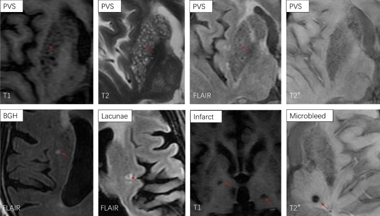
The definitions for the imaging markers included in the study were as follows [Figure 1 and Supplementary Table 1, http://links.lww.com/CM9/B646]: (1) BGH: small round or oval with a diameter of <5 mm, typically punctate T2/FLAIR-hyperintense lesions, isointense or slightly hypointense on T1 and isointense on other sequences. Similar to white matter hyperintensity, partly detected not only in white matter of internal and external capsule but also in the area of grey matter nuclei. (2) PVS: MRI signal intensity similar to that of cerebrospinal fluid, hypointensity on T1, hyperintensity on T2, hypointensity on FLAIR, and isointensity on DWI and T2*/SWI. The spaces were round or oval with a diameter of <3 mm, and sometimes, they appeared linear when imaged parallel to the vessel. Size was not strongly emphasized in this study.[6] (3) Lacunae: round or oval, with a diameter of 3–15 mm. MRI showed T1 hypointensity, T2 hyperintensity and FLAIR hypointensity always with a rim of hyperintensity. Sometimes, the central lumen fluid was not suppressed on FLAIR, and the lesions may show hyperintensity. However, on T1 and T2, it still showed a signal intensity similar to that of cerebrospinal fluid.[6] (4) Microbleeds (MBs): small circular hypointense lesions visible on T2*/SWI. They were generally isointense on T1, T2, FLAIR, and DWI, and they typically measured 2–5 mm in diameter.[6] (5) Infarct: irregular lesions that were hypointense on T1 and hyperintense on T2 and FLAIR.
Figure 1.
Imaging of punctate lesions in the basal ganglia. The figure shows examples for each type of lesion. PVS (red arrows) showed hypointensity on T1 and FLAIR, hyperintensity on T2. BGH (red arrow) was hyperintensity on FLAIR. Lacunae (red arrow) showed FLAIR hypointensity with a rim of hyperintensity. Infarct (red arrows) was irregular hypointense on T1. Microbleed (red arrow) was small circular hypointense lesion on T2*. BGH: Basal ganglia hyperintensity; FLAIR:Fluid-attenuated inversion recovery; PVS: Perivascular space.
Rating of lesions
We designed a new rating scale to evaluate the degree and distribution of different lesions in basal ganglion. The degrees of punctate lesions were rated on a 5-point scale. Ratings were performed on MR images.
BGH of the bilateral caudate nucleus head and lenticular nucleus (total of four regions) was counted separately. PVS was rated in the left and right basal ganglia. As a similar vascular mechanism, lacunae and infarcts were counted together in the bilateral basal ganglia.
The scores of each kind of lesion in each counting site were defined as follows: degree 1 when the number of lesions was <5; degree 2 when the number of lesions ≥5 and ≤10; degree 3 when the number of lesions >10 but ≤20; and degree 4 when the number of lesions >20. The total of left/right basal ganglia lesions was the sum of scores of ipsilateral BGH, PVS, and lacunae/infarct.
The definitions and operation instructions of rating scores (0–4) were shown in Supplementary Table 2, http://links.lww.com/CM9/B646.
Other MR imaging parameters
Microbleeds were counted in the bilateral cortex/gray matter, subcortical white matter, basal ganglia, bilateral internal capsule/external capsule, thalamus, brainstem, and cerebellum. Furthermore, the number of microbleeds in the cortex, deep and infratentorial regions was obtained.
Paraventricular and deep white matter hyperintensities were assessed with the Fazekas score.[7] The degree of paraventricular white matter hyperintensity was scored as follows: 0 = absence; 1 = cap-shaped or pencil-thin lining; 2 = smooth halo; 3 = irregular white matter hyperintensities extending into the deep white matter. Deep white matter lesions were scored as follows: 0 = absence; 1 = punctate foci; 2 = foci beginning to become confluent; 3 = large confluent areas.
The subcortical and subcortical white matter lesions in each region (frontal, parietal, temporal, and occipital lobe) were evaluated using the Age-Related White Matter Changes (ARWMC) score[8]: 0 point, no lesions; 1 point, with punctate or single plaque lesions; 2 points, lesions beginning to become confluent; 3 points, diffuse lesions involving the entire region, with or without the involvement of U-shaped fibers.
Definition of groups
We classified the patients with cheese sign into three groups according to the score for significant lateral basal ganglia (the sum of BGH and PVS in the caudate nucleus head and lenticular nucleus, along with lacunae and infarction in the basal ganglia), with a total score of <4 being classified as mild, 4–7 as moderate, and ≥8 as severe. Figure 2 showed typical images of different degrees of cheese sign.
Figure 2.
Different degrees of cheese sign in the basal ganglia. (A) Mild cheese sign, with a score of <4. (B) Moderate cheese sign, with a score of 4–7. (C) Severe cheese sign, with a score ≥8.
Statistical analysis
Statistical analysis and graphs were completed using GraphPad Prism 8 software (https://www.graphpad.com/). Measurement data were described as the mean ± standard deviation, and count data were described as numbers (n) and percentages (%). Comparisons between the cheese sign-negative and cheese sign-positive groups were completed by the Mann–Whitney test, Fisher's exact test and chi-squared analysis. Multiple logistic regression was used for risk factor analysis.
One-way analysis of variance (ANOVA) was used for comparisons between groups with normally distributed measurement data. Kruskal–Wallis test was used in multiple groups with the skew distribution. Tukey's test was used for comparisons between groups in post hoc analyses. Correlation analysis was performed by calculating Spearman's rank correlation coefficient. P <0.05 was considered to be statistically significant.
Results
Demographics of the population and comparison between cheese sign–negative and cheese sign–positive individuals
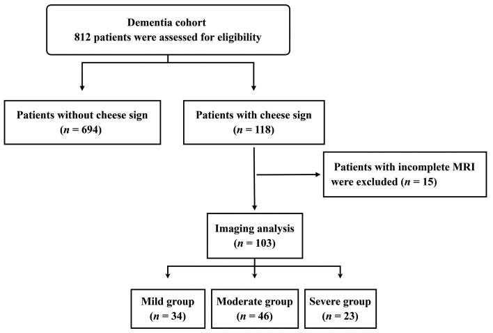
A total of 812 patients in the cohort completed 3.0 T head MRI examination, and 118 (14.5%) patients had cheese sign (positive) in the basal ganglia [Figure 3].
Figure 3.
Flow chart of the patients' enrolment and allocation. 812 patients with dementia were included, and divided into two groups. Patients with at least 10 punctate T2 hyperintensities in one basal ganglia were divided into cheese sign group, which were further divided into mild, moderate and severe group according to the score for significant lateral basal ganglia.
The cheese sign–positive group was older and had a higher proportion of males than the negative group. A significant difference was found between groups for hypertension (χ2 = 14.20,P <0.001) and stroke (χ2 = 15.00, P <0.001). There was no statistically significant difference in diabetes (χ2 = 0.107,P = 0.743) or hyperlipidemia (χ2 = 0.082, P = 0.774) [Supplementary Table 3, http://links.lww.com/CM9/B646]. The number of cerebrovascular risk factors was not related to the presence or absence of cheese sign (χ2 = 1.795, P = 0.408). Logistic regression analysis of risk factors showed correlations between age (OR: 1.090, 95% CI: 1.064–1.120, P <0.001), hypertension (OR: 1.828, 95% CI: 1.123–2.983, P = 0.014), cerebrovascular events (OR: 1.901, 95% CI: 1.092–3.259,P = 0.025), and cheese sign [Table 1].
Table 1.
Forest plot of risk and vascular factors in patients with cheese sign vs. those without cheese sign. Multiple logistic regression analysis (left) and forest plot (right). CI: Confidence interval; OR: Odds ratio.
| Risk factor | OR | 95% CI | Z | P |
|---|---|---|---|---|
| Age | 1.090 | 1.064–1.120 | 6.597 | <0.001 |
| Female sex | 1.429 | 0.904–2.268 | 1.543 | 0.123 |
| Hypertension | 1.828 | 1.123–2.983 | 2.460 | 0.014 |
| Diabetes | 0.682 | 0.366–1.215 | 1.255 | 0.210 |
| Hyperlipidemia | 0.832 | 0.466–1.439 | 0.633 | 0.527 |
| Stroke | 1.901 | 1.092–3.259 | 2.238 | 0.025 |
Assessment of cheese sign
Images of 103 patients with complete MR sequences were used to analyze the lesions of cheese sign. Among them, 65 were male (63.1%) and 38 were female (36.9%), with an average age of 75.03 ± 7.71 years (male: average age 73.60 ± 6.79, female: average age 77.47 ± 8.62) [Supplementary Table 4, http://links.lww.com/CM9/B646].
Punctate lesions were classified based on different signal intensities on MRI. Statistical analysis showed that 62 cases (60.2%) had BGH, PVS, and lacunae/infarcts in the basal ganglia, and 36 cases (34.9%) had BGH and PVS; four cases (3.9%) had BGH and lacunae/infarcts; and one case (1.0%) had only BGH. Among the punctate hyperintensities in the basal ganglia of the total population, BGH was the main component, accounting for 62.82 ± 15.03 %; PVS was the second largest component, accounting for 28.77 ± 14.05 %; and lacunae accounted for 8.41 ± 8.00 %. In addition, we found some subjects with microbleeds in the basal ganglia, which appear isointense or hypointense on T2-WI.
Comparison of the mild, moderate, and severe groups
The patients with cheese sign and with complete MR sequences were divided into three groups: mild (34/103, 33.0%), moderate (46/103, 44.7%), and severe (23/103, 22.3%) [Figure 3]. The average ages of the three groups were 73.79 ± 9.02 years in the mild group, 74.48 ± 6.68 years in the moderate group, and 77.96 ± 7.08 years in the severe group. There was no significant difference in age among the three groups (F = 2.268, P = 0.109) [Supplementary Table 5, http://links.lww.com/CM9/B646].
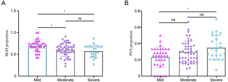
We performed a statistical comparison of lesion types in cheese sign among these groups. BGH was the main component in the three groups (69.50 ± 13.56 % in the mild group, 60.62 ± 15.10 % in the moderate group, and 57.35 ± 13.95 % in the severe group). The average rating of BGH was 4.54 ± 1.21 in the mild group, 6.54 ± 2.23 in the moderate group and 9.43 ± 2.29 in the severe group. The proportion of BGH was associated with the severity of cheese sign (F = 5.894, P = 0.004) [Figure 4A, Supplementary Table 6, http://links.lww.com/CM9/B646]. There were significant differences among the mild group, the moderate group, and the severe group (mild vs. moderate: P = 0.020, mild vs. severe: P = 0.006), but there was no significant difference between the moderate and severe groups (P = 0.647). [Figure 4A]
Figure 4.
The ratio of foci of BGH and PVS in the mild, moderate, and severe groups: (A) the proportion of BGH in three groups, (B) the proportion of PVS in three groups. BGH: Basal ganglia hyperintensity; PVS: Perivascular space.*: P <0.05; †: P <0.01; ns: P >0.05;
The average rating of PVS was 1.46 ± 0.94 in the mild group, 3.13 ± 1.50 in the moderate group and 6.17 ± 2.29 in the severe group. The proportion of the PVS increased with a higher level of cheese sign (23.29 ± 12.64 % in the mild group, 29.71 ± 13.30 % in the moderate group, 34.97 ± 15.02 % in the severe group, F = 5.347, P = 0.006) [Supplementary Table 6, http://links.lww.com/CM9/B646]. The difference was mainly between the mild and severe groups (P = 0.005) [Figure 4B]. The proportion of lacunae/infarct was similar in all three groups and there was no statistically significant difference (7.21 ± 10.71 % in the mild group, 9.67 ± 7.08 % in the moderate group, and 7.68 ± 4.03 % in the severe group, H = 5.051, P = 0.080) [Supplementary Table 6, http://links.lww.com/CM9/B646]. There were no statistically significant differences for either group in the number of microbleeds in the basal ganglia.
Correlation between cheese sign and other imaging findings
There was no significant difference in the number of cortical, deep, and infratentorial microbleeds among the three groups (cortical: H = 0.183, P = 0.913, deep: H = 0.729, P = 0.695, infratentorial: H = 1.844, P = 0.398). There was correlation between the number of cerebral infarctions and the severity of cheese sign (H = 8.318, P = 0.016) [Supplementary Table 5, http://links.lww.com/CM9/B646].
For cerebral white matter lesions, the paraventricular Fazekas scores of 13/34 (38.2%) of cases in the mild group were degrees 2 and 3, while 19/46 (41.3%) in the moderate group and 10/23 (43.5%) in the severe group scored 2 or 3. In the severe group, 18/23 (78.2%) of the deep white matter abnormalities were fusion lesions or large confluent areas (degrees 2 and 3). However, statistical analysis showed that there was no significant difference in paraventricular or deep white matter abnormalities among the three groups (χ2 = 0.166, P-para = 0.921; χ2 = 3.338, P-deep = 0.188). In addition, there was no significant difference in cortical and subcortical white matter abnormalities (ARWMC score) among the three groups (H = 2.918, P = 0.233) [Supplementary Table 5, http://links.lww.com/CM9/B646].
We further compared the correlation between the components of cheese sign, microbleeds, and white matter lesions. The analysis showed that BGH, PVS, lacunae/infarct in the basal ganglia, and the number of total cerebral microbleeds were related to the Fazekas score [Supplementary Table 7, http://links.lww.com/CM9/B646].
Discussion
The purpose of this study was to explore the risk factors and components of the cheese sign, that is, the dense T2 punctate abnormal signals in the basal ganglia. The earliest description of this sign came from some pathological studies in the 19th century.[2–4] There were some similar and easily confused terms, including état criblé and cribriform changes. This finding occurred mostly in the basal ganglia and centrum semiovale. Cheese sign was always considered to be dense PVS. More recent studies have focused on the imaging feature and function of PVS and its correlation with clinical disorders.[9–11] However, there is no comprehensive study on the composition of cheese sign, ignoring the various lesions more than the PVS.
In this study, the punctate lesions in the basal ganglia were classified, summarized, and analyzed based on different sequences. This study explored the lesion composition of cheese sign, which laid a foundation for future studies on the relationship between cheese sign and degenerative diseases as well as cerebrovascular diseases.
The study population was from PUMCH dementia cohort. A total of 812 patients underwent 3.0 T MRI examination, of which 118 (14.5%) had cheese sign in the basal ganglia. Referring to the classification of PVS in the basal ganglia, the PVS of the patients with cheese sign included in this study is usually of grade 3–4.[12] A previous multicenter imaging study of elderly individuals reported that the incidence rate of dense PVS in the basal ganglia was 11.0%.[12] The study population was from a dementia and leukoencephalopathy clinic, and the enrolled patients were diagnosed with dementia and generally older. The disease spectrum contained but was not limited to Alzheimer's disease, frontotemporal lobar degeneration, Parkinson's disease with dementia, and vascular cognitive impairment. This explained the higher detection rate of cheese sign in this study.
We investigated the association of age, sex, vascular risk factors, stroke, and cheese sign. The result showed that cheese sign was associated with age, hypertension, and stroke, which was consistent with previous studies.[13,14] Surprisingly, there was no association between diabetes or hyperlipidemia and cheese sign. Cheese sign might be a promising marker of cerebrovascular events and underlying vascular damage for further studies of cardiovascular disease.[15,16] Age had little influence on the imaging features. This might be due to the old age of our cohort.
The cheese sign was first proposed by the French doctor Durand Fardel in 1842.[2] It was reported that there were multiple ethmoid foci resembling small cavities specifically in the white matter of the brain. These numerous, small, round, well-defined cavities were often around the blood vessels. It was believed that the cribriform appearance was caused by vascular congestion, accompanied by an abnormal small artery wall and perivascular inflammatory cell infiltration. From 1919 to 1920, Vogt summarized the similar phenomena of globus pallidus and striatum with "status de integrationis",[2] which was mainly characterized by lacunar, accompanied by the expansion of Virchow–Robin spaces. At the same time, we could see the loss of the myelin sheath (sparse neurons and myelinated fibers). In 2002, Japanese researchers proposed that the pathology of état criblé was mainly PVS, sometimes including white matter loosening and occasionally small infarcts and gliosis.[4]
Previous pathological and imaging studies suggested that the pathological mechanism and components of brain cribriform changes were complex. Based on previous work, we hypothesized that the components of the cheese sign include BGH, PVS, and lacunae. The lesions of cheese sign included BGH, PVS, occasional lacunae, and microbleeds in the SWI partially. Among them, BGHs were the main components, accounting for approximately 57.11–69.96%. There was punctate hyperintensity on T2/FLAIR and isointensity or slightly hypointensity on T1. The proportion of PVS was second. Lacunae and infarcts were also found in the basal ganglia of some patients with cheese sign. This finding was not consistent with previous studies reporting that the cribriform changes were mainly PVS. It was not appropriate to equate these cribriform changes with dense PVS.
We compared the proportion of each component in the three groups. There were significant differences in the proportions of BGH and PVS between mild and moderate/severe groups. Interestingly, the results revealed that the ratio of PVS gradually increased as the cheese sign was aggravated, suggesting that PVS might play a key role in the underlying mechanism. As a part of system for drainage of waste metabolites in the brain, PVS influence the pathogenesis of common cerebrovascular, neuroinflammatory, and neurodegenerative disorders.[17,18] The association of PVS and vascular dementia was detected.[19] In addition, some studies confirmed that oxidative stress[20] and inflammatory[21,22] played a key role in stroke and vascular dementia, and it provided a direction to explore the mechanism of PVS. We also explored the correlation of cheese sign with other imaging lesions (white matter lesions and microbleeds), and there were no positive findings.
A key strength of the study was the comprehensive research of cheese sign formation. In addition, the population was from the dementia cohort, providing a typical view of cheese sign in dementia. However, there were also several limitations. First, the selection of the population was limited in patients mostly with dementia, and there was a lack of normal controls. Second, the study sample size was limited, and it was from a single center. Third, the imaging marker was counted by semiquantitative ratings.
Notwithstanding these limitations, we conclude that the cheese sign mainly consists of foci of hyperintensity in T2/FLAIR, described as BGH and PVS. Age, hypertension, and stroke are the risk factors of the cheese sign. Our future research will aim to explore cheese sign in different diseases, such as Alzheimer's disease, vascular dementia, and hereditary cerebral small vessel disease. In addition, high-field MRI, advanced neuroimaging quantification,[23] and more autopsy studies can prompt the determination of the underlying pathological mechanism.
Funding
This work was supported by grants from National Key Research and Development Program of China (Nos. 2020YFA0804500 and 2020YFA0804501), CAMS Innovation Fund for Medical Sciences (CIFMS) (Nos. 2021-I2M-1-020 and 2020-I2M-C&T-B-010), National Natural Science Foundation of China (Nos. 81550021 and 30470618), and Science Innovation 2030–Brain Science and Brain-Inspired Intelligence Technology Major Project (No. 2021ZD0201106).
Conflicts of interest
None.
Supplementary Material
Footnotes
How to cite this article: Huang XY, Hou B, Wang J, Li J, Shang L, Mao CH, Dong LL, Liu CY, Feng F, Gao J, Peng B. Assessment of cheese sign and its association with vascular risk factors: Data from PUMCH dementia cohort. Chin Med J 2024;137:830–836. doi: 10.1097/CM9.0000000000002785
References
- 1.Banerjee G Kim HJ Fox Z Jäger HR Wilson D Charidimou A, et al. MRI-visible perivascular space location is associated with Alzheimer's disease independently of amyloid burden. Brain 2017;140: 1107–1116. doi: 10.1093/brain/awx003. [DOI] [PubMed] [Google Scholar]
- 2.Roman GC. On the history of lacunes, etat criblé, and the white matter lesions of vascular dementia. Cerebrovasc Dis 2002;13(Suppl 2): 1–6. doi: 10.1159/000049142. [DOI] [PubMed] [Google Scholar]
- 3.Poirier J, Derouesné C. The concept of cerebral lacunae from 1838 to the present (in French). Rev Neurol (Paris) 1985;141: 3–17. [PubMed] [Google Scholar]
- 4.Udaka F, Sawada H, Kameyama M. White matter lesions and dementia: MRI-pathological correlation. Ann N Y Acad Sci 2002;977: 411–415. doi: 10.1111/j.1749-6632.2002.tb04845.x. [DOI] [PubMed] [Google Scholar]
- 5.American Psychiatric Association . Diagnostic and statistical manual of mental disorders (DSM-5®) [M]. American Psychiatric Publishing, Arlington, 2013. [Google Scholar]
- 6.Wardlaw JM Smith EE Biessels GJ Cordonnier C Fazekas F Frayne R, et al. Neuroimaging standards for research into small vessel disease and its contribution to ageing and neurodegeneration. Lancet Neurol 2013;12: 822–838. doi: 10.1016/S1474-4422(13)70124-8. [DOI] [PMC free article] [PubMed] [Google Scholar]
- 7.Fazekas F Barkhof F Wahlund LO Pantoni L Erkinjuntti T Scheltens P, et al. CT and MRI rating of white matter lesions. Cerebrovasc Dis 2002;13: 31–36. doi: 10.1159/000049147. [DOI] [PubMed] [Google Scholar]
- 8.Wahlund LO Barkhof F Fazekas F Bronge L Augustin M Sjögren M, et al. A new rating scale for age-related white matter changes applicable to MRI and CT. Stroke 2001;32: 1318–1322. doi: 10.1161/01.str.32.6.1318. [DOI] [PubMed] [Google Scholar]
- 9.Chen W, Song X, Zhang Y. Alzheimer's Disease Neuroimaging Initiative. Assessment of the Virchow-Robin Spaces in Alzheimer disease, mild cognitive impairment, and normal aging, using high-field MR imaging. AJNR Am J Neuroradiol 2011;32: 1490–1495. doi: 10.3174/ajnr.A2541. [DOI] [PMC free article] [PubMed] [Google Scholar]
- 10.Gertje EC, van Westen D, Panizo C, Mattsson-Carlgren N, Hansson O. Association of enlarged perivascular spaces and measures of small vessel and Alzheimer disease. Neurology 2021;96: e193–e202. doi: 10.1212/WNL.0000000000011046. [DOI] [PubMed] [Google Scholar]
- 11.Liu S Hou B You H Zhang Y Zhu Y Ma C, et al. The association between perivascular spaces and cerebral blood flow, brain volume, and cardiovascular risk. Front Aging Neurosci 2021;13: 599724. doi: 10.3389/fnagi.2021.599724. [DOI] [PMC free article] [PubMed] [Google Scholar]
- 12.Zhu YC Dufouil C Mazoyer B Soumaré A Ricolfi F Tzourio C, et al. Frequency and location of dilated Virchow-Robin spaces in elderly people: A population-based 3D MR imaging study. AJNR Am J Neuroradiol 2011;32: 709–713. doi: 10.3174/ajnr.A2366. [DOI] [PMC free article] [PubMed] [Google Scholar]
- 13.Francis F, Ballerini L, Wardlaw JM. Perivascular spaces and their associations with risk factors, clinical disorders and neuroimaging features: A systematic review and meta-analysis. Int J Stroke 2019;14: 359–371. doi: 10.1177/1747493019830321. [DOI] [PubMed] [Google Scholar]
- 14.Liu Y, Dong YH, Lyu PY, Chen WH, Li R. Hypertension-induced cerebral small vessel disease leading to cognitive impairment. Chin Med J 2018;131: 615–619. doi: 10.4103/0366-6999.226069. [DOI] [PMC free article] [PubMed] [Google Scholar]
- 15.Pan Y Mu Y Liu ZS Zhang YC He JQ Yu XP, et al. Impact of prior cerebrovascular events on patients with unprotected left main coronary artery disease treated with coronary artery bypass grafting or percutaneous coronary intervention. Chin Med J 2021;134: 1988–1990. doi: 10.1097/CM9.0000000000001645. [DOI] [PMC free article] [PubMed] [Google Scholar]
- 16.Yang MH Li B Yao DC Zhou Y Zhang WC Wang G, et al. Safety of early surgery for geriatric hip fracture patients taking clopidogrel: A retrospective case-control study of 120 patients in China. Chin Med J 2021;134: 1720–1725. doi: 10.1097/CM9.0000000000001668. [DOI] [PMC free article] [PubMed] [Google Scholar]
- 17.Wardlaw JM Benveniste H Nedergaard M Zlokovic BV Mestre H Lee H, et al. Perivascular spaces in the brain: Anatomy, physiology and pathology. Nat Rev Neurol 2020;16: 137–153. doi: 10.1038/s41582-020-0312-z. [DOI] [PubMed] [Google Scholar]
- 18.Lau AYL Ip BYM Ko H Lam BYK Shi L Ma KKY, et al. Pandemic of the aging society – sporadic cerebral small vessel disease. Chin Med J 2021;134: 143–150. doi: 10.1097/CM9.0000000000001320. [DOI] [PMC free article] [PubMed] [Google Scholar]
- 19.Ding J Sigurðsson S Jónsson PV Eiriksdottir G Charidimou A Lopez OL, et al. Large perivascular spaces visible on magnetic resonance imaging, cerebral small vessel disease progression, and risk of dementia: The age, gene/environment susceptibility-Reykjavik study. JAMA Neurol 2017;74: 1105–1112. doi: 10.1001/jamaneurol.2017.1397. [DOI] [PMC free article] [PubMed] [Google Scholar]
- 20.Xu Y, Wang K, Wang Q, Ma Y, Liu X. The antioxidant enzyme PON1: A potential prognostic predictor of acute ischemic stroke. Oxid Med Cell Longev 2021;2021: 6677111. doi: 10.1155/2021/6677111. [DOI] [PMC free article] [PubMed] [Google Scholar]
- 21.Wang Q, Wang K, Ma Y, Li S, Xu Y. Serum galectin-3 as a potential predictive biomarker is associated with poststroke cognitive impairment. Oxid Med Cell Longev 2021;2021: 5827812. doi: 10.1155/2021/5827812. [DOI] [PMC free article] [PubMed] [Google Scholar]
- 22.Wang X Wang Q Wang K Ni Q Li H Su Z, et al. Is immune suppression involved in the ischemic stroke? A study based on computational biology. Front Aging Neurosci 2022;14: 830494. doi: 10.3389/fnagi.2022.830494. [DOI] [PMC free article] [PubMed] [Google Scholar]
- 23.Zhao L, Lee A, Fan YH, Mok VCT, Shi L. Magnetic resonance imaging manifestations of cerebral small vessel disease: Automated quantification and clinical application. Chin Med J 2020;134: 151–160. doi: 10.1097/CM9.0000000000001299. [DOI] [PMC free article] [PubMed] [Google Scholar]


