Abstract
Most chemicals expressed in mammalian cells have complex delivery and transport mechanisms to get to the right intracellular sites. One of these mechanisms transports most transmembrane proteins, as well as almost all secreted proteins, from the endoplasmic reticulum, where they are formed, to their final location. Nearly all eukaryotic cells have a membrane trafficking mechanism that is both a prominent and critical component. This system, which consists of dynamically coupled compartments, supports the export and uptake of extracellular material, remodeling and signaling at the cellular interface, intracellular alignment, and maintenance of internal compartmentalization (organelles). In animal cells, this system enables both regular cellular activities and specialized tasks, such as neuronal transmission and hormone control. Human diseases, including neurodegenerative diseases, such as Alzheimer's disease, heart disease, and cancer, are associated with the dysfunction or dysregulation of the membrane trafficking system. Treatment and cure of human diseases depends on understanding the cellular and molecular principles underlying membrane trafficking pathways. A single gene mutation or mutations that result in impaired membrane trafficking cause a range of clinical disorders that are the result of changes in cellular homeostasis. Other eukaryotic organisms with significant economic and agricultural value, such as plants and fungi, also depend on the membrane trafficking system for their survival. In this review, we focused on the major human diseases associated with the process of membrane trafficking, providing a broad overview of membrane trafficking.
Keywords: Endocytosis, Exocytosis, Membrane proteins, Membrane trafficking, Vesicular transport
1. Introduction
The size, shape, and molecular nature of numerous cell organelles, including the plasma membrane, are regulated by membrane transport. Membrane transport secretes thousands of cargo species, including hormones, growth factors, antibodies, matrix and serum proteins, digestive enzymes, and more. It is also possible to transport many types of macromolecules, including lipids and complex carbohydrates ( 1 ). The development of various homeostatic and adaptive mechanisms is highly dependent on the endomembranous system, which is crucial for the interaction of the cell with the environment. A variety of organelles, such as the endoplasmic reticulum (ER), the Golgi complex, and the endolysosomal stations, as well as the underlying molecular machinery, thought to consist of about 2,000 proteins, are required for the functioning of the system ( 2 ). The three main types of membrane transport are the secretory pathway, the vacuolar transport pathway, and the endocytic pathway. In contrast to the vacuolar transport pathway, which transports newly formed proteins to the vacuole, the secretory pathway transports proteins from the ER to the plasma membrane (PM) and/or to the extracellular environment. Several sequential processes are involved in each transport pathway, including leakage of transport vesicles from the donor membrane, transport, targeting, anchoring, fusion, and recycling of transport vesicles to target organelles. Anterograde transport, which begins at the ER and moves to target organelles, is referred to as forward-directed transport, whereas retrograde transport, which is the opposite pathway, is referred to as backward-directed transport. The two categories of small GTPases known to be involved in a single transport process are Rab GTPases and ADP ribosylation factor (ARF)/secretion-associated and Ras-related 1 (SAR1) proteins. Rab controls other trafficking processes, such as alignment, anchoring, and docking of transport vesicles to the target membrane, while ARF/SAR1 facilitates the budding of transport vesicles ( 3 ). The most numerous group of small GTPases is the Rab family. By alternating between a GDP-bound (inactive) and a GTP-bound (active) state, these tiny GTPases serve as molecular switches. These GTPases are activated by certain particular guanine nucleotide exchange factors (GEFs), whereas GTPase-activating proteins (GAPs), which regulate the transition between GDP- and GTP-bound states, are responsible for their inactivation ( 3 ).
Many Rab effectors bind to specific Rabs in the GTP-bound active state in yeast and mammalian systems and mediate the binding of transport vesicles to target membranes through contact with Rab GTPases. Tethering factors are proteins with large coiled coils and protein complexes called tethering complexes. Different tethering complexes mediate each step of the tethering process. For example, these complexes include the exocyst, HOPS-CORVET (lysosome/vacuole tethering), and the conserved oligomeric Golgi (COG), which is vital for retrograde transport within the Golgi (which is important for the final step of the secretory pathway). Despite the fact that plants still contain certain filamentous coiled-coil proteins, there are also homologous genes encoding these tethering complex proteins ( 4 ).
After the tethering factors bind the transport vesicles together, the soluble N-ethylmaleimide-sensitive factor attachment protein receptors (SNAREs) cause membrane fusion. The two main classes of SNARE proteins include Q- and R-SNAREs, which are well-known. The Q-SNAREs are further subdivided into Qa, Qb-, and Qc-SNAREs. While Q-SNAREs are typically found on target membranes, R-SNAREs are typically found on transport vesicles. In a given arrangement, these four SNARE protein subclasses form a compact complex that results in two membrane layers coming into close contact with each other, eventually causing the two membranes to fuse ( 3 ).
It is gradually becoming clear that membrane transport is intimately linked to numerous other cellular processes, including quality control in ER, cytoskeletal dynamics, receptor signaling, apoptosis, and mitosis, as the mechanisms and regulatory processes of membrane transport come into sharper focus. The interaction between these processes is an exciting development in the field and represents another challenge to better understand the molecular details involved in this regulation. This review focuses on a general overview of membrane trafficking and highlights the major human diseases associated with the mechanism of membrane trafficking.
2. Materials and Methods
2.1. Major pathways of membrane trafficking
2.1.1. Secretory/ intracellular membrane trafficking
This is the mechanism by which a cell secretes large molecules. A membrane-enclosed intracellular vesicle must first fuse with the PM before it can be opened and its contents shuttled out. This is how lipids and proteins enter the intended compartments. It is the path that newly formed components take on their way from the ER, the compartment of synthesis, through other organelles, the plasma membrane, and into the extracellular medium. In constitutive secretion, which occurs in all cells, lipids and proteins are formed, transported, and secreted without interruption. Regulated secretion occurs only in response to a specific signal, such as the supply of certain ions (calcium), or by the contact of a hormone with its receptor. Once produced, the substances subjected to controlled secretion are stored in spherical membrane structures called vesicles or secretory granules (depending on their size) until the trigger for secretion appears. Regulated secretion occurs in neurons, macrophages, some types of white blood cells, and endocrine tissues (glands that produce hormones) ( 5 ).
2.2 Endocytic pathway
This is the transport pathway for soluble and membrane proteins into the cell. This pathway includes phagocytosis, receptor-mediated internalization, pinocytosis, caveolae-mediated internalization, and receptor-mediated internalization. In receptor-mediated internalization, exogenous molecules bind to a receptor, usually located at the PM or in some cases stored in intracellular compartments just below the cell surface, and are then rapidly and synchronously taken up with an incoming specific signal ( 5 ). Pinocytosis is the mechanism used for continuous turnover of the PM and involves the internalization of fluids. Most vesicles used in caveolae-mediated internalization contain the protein caveolin. Large particles, including viruses, bacteria, parasites, and intracellular inactive complexes, are taken up internally during phagocytosis, a specialized form of endocytosis. In particular, neutrophils and macrophages are the only cell types that contain it.
2.3 Recycling pathway
Some membrane elements are internalized; however, when they are released, the burden of the compound is shifted back to the PM where they can once again do their job. Most membrane receptors use this pathway, which combines the endocytic pathway (internalization) and the secretory pathway (back to the cell surface). There is a good balance between secretory and endocytic trafficking in terms of intracellular membrane content. Any deviation from this balance leads to abnormalities that affect the cell's ability to survive.
2.4 Secretory pathway
The secretory pathway serves to transport newly formed proteins from ER to their final destination, such as the PM and extracellular space. Coat protein complex II (COP II)-coated vesicles are commonly used to envelop and transport properly folded proteins from the ER, whether they are soluble or membrane-bound. Two processes that support cargo movement through the Golgi apparatus are cisternal maturation and/or vesicle transfer between cisternae. The trans-Golgi network (TGN), which acts as a sorting station in a number of transport pathways, then transports cargo to the PM or releases it into the extracellular space. According to Watanabe et al. ( 6 ), the secretory pathway is critical for the secretion of proteins related to pathogenesis, some of which have antibacterial properties, and for the transmission of functional pattern recognition receptors (PRRs) to the PM ( 7 ).
2.4.1 Secretion/exocytosis steps
Vesicle trafficking, tethering, docking, priming, and fusion are the basic steps of exocytosis.
(i) Vesicle trafficking refers to the movement of a vesicle over a relatively short distance.
(ii) Vesicle tethering distinguishes between more stable packing interactions and early, loose binding of vesicles to their target.
(iii) Vesicle docking involves keeping two membranes close enough to be separated by a bilayer.
(iv) Vesicle priming is a series of molecular rearrangements, ATP-dependent protein changes, and lipid alterations that occur after the initial docking of a synaptic vesicle.
(v) Vesicle fusion is stimulated by SNARE proteins. Large macromolecules are released into the extracellular space by the process of fusion of the vesicle membrane with the target membrane.
2.4.2 Endoplasmic Reticulum
Protruding from the nuclear membrane is a network of active pathways and tubes called the ER, which is involved in a variety of cellular processes, including protein synthesis, folding and transport; lipid and carbohydrate synthesis and metabolism; and intracellular calcium storage ( 8 ). Before proteins are released for secretion or transport to other organelles via the Golgi apparatus, they are examined to ensure they reach their properly folded state in the ER. This serves as a critical step in quality control, which is reinforced by a critical inspection system called ER quality control (ERQC). Disulfide bond development, folding, glycosylation, certain proteolytic cleavages, assembly of multimeric proteins, and formation of a tertiary structure are some of the post-translational modifications that occur in the ER. During transport into the ER, some proteins can fold rapidly into their native structure, while other proteins require more help from the chaperones and folding enzymes of the ER. There are three folding processes that have been found to control the quality of the endoplasmic reticulum: control, calnexin, and calreticulin, which work specifically in glycoprotein synthesis; a pathway dependent on disulfide isomerases and oxidoreductases, which help rearrange disulfide bonds in developing proteins; and a pathway mediated by chaperones, heat shock proteins, which bind directly to client proteins and help with their folding ( 9 ). The freshly formed proteins are isolated from the existing ER proteins and transported to the Golgi apparatus after appropriate folding, modification, and oligomer formation. This occurs at ER exit sites, also known as transition sites ER, which are specialized regions associated with outgoing vesicles and free of ribosomes ( 10 ). Studies have shown that proteins are selectively recruited to vesicles by interacting with transmembrane cargo receptors, such as the lectin ER-Golgi intermediate compartment (GIC)-53 and members of the p24 family, which interact with the components of the vesicle envelope of the COPII either indirectly (for soluble proteins) or directly (for transmembrane proteins) ( 11 ).
ERQC clients are essentially proteins that enter the ER. The proteasomal degradation pathway, known as ER-associated degradation (ERAD), is used to degrade misfolded proteins that do not pass through the ERQC and are translocated from the ER lumen to the cytoplasm ( 12 ). The various steps of ERAD include the identification of ERAD substrates, retrotranslocation to the cytosol, polyubiquitination, and 26S proteasome degradation. A thorough mutational analysis of the PM-localized protein revealed that the molecular mechanism of ERAD is generally conserved in yeast and animals. This mechanism is also used, at least in part, by plants ( 12 ). HMG-CoA reductase degradation ligase, Doa10, and RING-finger protein with membrane anchor 1 are ER membrane-bound ubiquitin ligases that catalyze the ubiquitination of substrates for degradation ( 13 ).
2.4.3 Endoplasmic reticulum-Golgi intermediate compartment
In intermediate vesicles or vesicles containing complexes of coat proteins, such as COPI or COPII, protein cargo is transported between ER and Golgi. In the ER, ERGIC, and Golgi apparatus, COP complexes are necessary for vesicle production. Bidirectional transport occurs between ER and Golgi, and COPI, assembled by the action of GTPase ARF1, mediates retrograde transport ( 14 ). Membrane-bound and soluble protein cargo are more specifically captured, packaged, transported, and delivered to an acceptor compartment using COP recruitment to membranes. Anterograde (forward) transport vesicles begin to develop when COPII is recruited to sites on the smooth ER. From ER, COPII vesicles migrate to the ERGIC or vesicular tubular clusters. COPI-coated vesicles are thought to facilitate anterograde transfer from the ERGIC to the cis-Golgi apparatus ( 15 ). The ERGIC is thought to serve as a temporary sorting station, between ER and the cis-Golgi apparatus. In addition, it acts as another quality control checkpoint where misfolded proteins can be returned to the ER for further folding or destruction. Palmer and Stephens ( 16 ) have made specific suggestions about the function of COPII in the transfer from ER to ERGIC. According to a new concept, the main purpose of COPII vesicles is to maintain the ER-output domains rather than to serve as important cargo carriers. Furthermore, research has shown that ER-to-Golgi transport carriers that are microtubule-dependent tubular intermediates are genuine ( 17 ).
2.4.4 Golgi apparatus
The flattened, membrane-bound cisternae that make up the Golgi apparatus are arranged in an exceptionally orderly row and are usually close together. The ERGIC and the TGN, which are the cargo entry and exit sites for cargo, are networks of interconnected cisternae and tubular structures that are located on the cis and trans sides of these stacks. The Golgi apparatus is the site where most posttranslational change occur ( 18 ). In the Golgi stack, mannose is removed and galactose and N-acetylglucosamine are added. Phosphorylation of the lysosomal proteins occurs here. The added N-acetyl neuraminic acid sorts the proteins at the TGN. In addition, proteins from the ER are returned from the Golgi apparatus. Numerous different proteins, including resident proteins and newly formed transit proteins, form the highly active organelle known as the Golgi apparatus. Although its structural arrangement differs to some extent between different cell types and organisms, it still serves as a processing center in the secretory pathway ( 19 ).
Glycosylation of proteins, which can result in a variety of N-linked glycans, is one of the most complicated and important processing functions of the Golgi. As cargo molecules, such as N-glycans, move through the Golgi cisternae, resident glycosyltransferases sequentially modify the carbohydrate side chains of these molecules. The concentrations of glycosyltransferases vary along the Golgi stack, with the cis and medial cisternae containing the bulk of the enzymes that act in the early phase of glycan biosynthesis, whereas the trans-Golgi and TGN harbor the enzymes that act in the later phase of the biosynthetic pathway ( 20 ). There are alternative theories about how transport occurs in the Golgi stacks of mammalian cells, including the idea that direct tubular connections may provide vectorial transport between the compartments of the secretory pathway. Mironov et al. have demonstrated the formation of intracisternal tubular junctions during secretory transport using electron tomography of single nocodazole-induced Golgi stacks ( 21 ).
2.4.5 Trans-Golgi Network
The TGN is located at the exit side of the Golgi stacks. As a result of the secretory function of the cells and the types of post-Golgi intermediates that the cells form, it also varies morphologically between different species and cell types ( 22 ). Similar to the other components of the Golgi stack, TGN is also involved in processing cargo molecules. Several proprotein convertases, including furin, and a glycosyltransferase called 2,6-sialyltransferase are present in it ( 23 ). For the purpose of transport to their final location, mature cargo proteins are sorted by the TGN, which functionally separates them from the Golgi apparatus. In the TGN, the various cargo proteins and lipids are organized and packaged into membrane carriers before being transported by various pathways to different PM domains, endosomes, lysosomes (via late endosomes), secretory granules, or earlier Golgi cisternae, and possibly directly to the ER. The TGN receives cargo from a variety of sources and is essential for the recycling process of numerous endosomal and PM proteins ( 24 ). The TGN is the site where the secretory and endocytic pathways converge.
2.5 Endosomes and lysosomes
The numerous processes performed by the endosomal and lysosomal systems include the uptake of extracellular molecules and ligands; the internalization of PM proteins and lipids; the recycling of proteins into the Golgi apparatus, TGN, and the plasma membrane; the regulation of cell signaling pathways; and the degradation of proteins from the secretory and endocytic pathways ( 25 ). Early/sorting endosomes, recycling endosomes, multivesicular bodies (MVBs), late endosomes, and lysosomes are just some of the vesicular organelles that make up the endocytic pathway. This system must be controlled by complicated molecular sorting processes and transport channels that are poorly understood. Endocytosed material typically enters early/sorting endosomes in mammals before maturing into late endosomes and lysosomes. Endosomes function as an important sorting compartment, similar to the TGN. Early/sorting endosomes are where much of the sorting and recycling occurs, and from there, proteins can either return rapidly to the PM or more slowly via recycling endosomes ( 25 ). Tubular carriers, because of their large surface-to-volume ratio, are thought to mediate the mass flow of membrane proteins from early/sorting endosomes to recycling endosomes, displacing most of the soluble cargo and any membrane proteins that have been preferentially retained and pass to late endosomes and lysosomes ( 25 ).
Some receptor proteins that are not recycled, such as the epidermal growth factor receptor, are endocytosed proteins that have been marked for degradation by ubiquitylation. These proteins are internalized into late endosomes to form MVBs, where they are degraded in lysosomes. The MVBs may occasionally undergo a different fate by binding to the PM and ejecting the intralumenal vesicles into the extracellular domain ( 26 ). Exosomes, which are secreted vesicles, were first identified as a mechanism for externalizing inactive membrane proteins during reticulocyte development ( 27 ). Exosome secretion has been linked to a unique intercellular communication mechanism that is essential for controlling immune responses and may also be useful in viral infections. This process has been demonstrated in a variety of cell types ( 26 ).
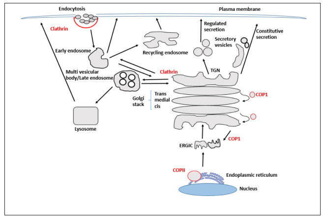
Because of the presence of additional compartments, including the recycling endosome, traffic across endosomal compartments, sorting, and transport, the endosomal system is more complex than previously thought. Rouille et al. ( 28 ) claim that material that needs to be degraded is relocated from the late endosome to the lytic environment of the lysosome, most likely as a result of the fusion of the two compartments into a hybrid organelle ( 29 ). This hypothesis states that lysosomal enzymes interacting with material endocytosed after fusion with the late endosome use the lysosome as a storage granule. The hybridized late endosome-lysosome organelle degrades the proteins. Lysosomes could therefore regenerate from these hybrid organelles by concentrating lysosomal enzymes, precisely removing endocytic proteins, and then cleaving a new lysosome compartment ( 29 ). An overview of the transport to the lysosome is shown in figure 1.
Figure 1.
Overview of trafficking. Proteins that are intended for secretion, including newly generated membrane and soluble proteins, are translocated into the endoplasmic reticulum (ER), where they are bundled into COPII - coated vesicles and fuse to form the ER-Golgi intermediate compartment (ERGIC). The cis-Golgi network is created when many ERGICs combine (CGN). ER proteins are recycled to and from the ERGIC and Golgi stack via COPI-coated vesicles. At the trans-Golgi network, antegrade cargo is sorted by destination as it passes through the Golgi stack (TGN). Transporting cargo to various locations requires various kinds of coated vesicles and tubulovesicular transporters. The early endosome receives proteins that have been endocytosed at the plasma membrane. Proteins from the early endosome can travel to the TGN, the late endosome, or the cell surface for recycling
2.5.1 Fates of endocytosed material
(i) Recycling is the process of releasing material back into the cell through the apical PM after it has been transported to an early endosome.
(ii) Degradation is the process by which chemicals are transported from the apical PM to the lysosome, where they are eliminated.
(iii) Transcytosis: the passage of substances across an early endosome as they enter the cell through the apical PM and exit through the basolateral plasma membrane.
2.6 Molecular processes underlying vesicular transport
The following processes contribute to the mechanism of vesicular transport:
(i) Cargo is sorted at the donor membrane, and a vesicle is formed by cleavage from the plasma membrane.
(ii) The vesicle migrates along microtubules in search of receptors in the cytosol.
(iii) Tethering or docking of the vehicle to the acceptor membrane occurs by identification between specific molecules on the vesicle and specific receptor molecules on the receptor membrane when the vesicle reaches the receptor membrane.
(iv). The vesicle is then opened and released into the lumen of the acceptor organelle by the fusion of the vesicle membrane and the receptor membrane.
Membrane carriers from one compartment (the donor membrane) must bud and fuse with another compartment (the acceptor membrane) to transport proteins and lipids within eukaryotic cells. Membrane-bound vesicles are required for several transport steps in the secretory and endocytic pathways. The mechanisms of sheath formation, vesicle budding, and fusion are similar, although a different protein is used for each step. A small GTPase that attaches to the donor membrane in the GTP-bound state conditions the membrane to curl up and form a vesicle ( 30 ). Additional proteins are sometimes required for the final cleavage of the vesicle from the donor membrane. GTP or ATP hydrolysis occurs as soon as the vesicle detaches from the membrane due to dissociation of the envelope. Interactions involving a variety of proteins associated with both the vesicle and the target membrane result in docking and fusion at the target membrane. The force for membrane fusion is provided by specialized integral membrane proteins that bring the membranes together.
2.6.1 Envelope proteins and vesicle biogenesis
To maintain organelle integrity due to the constant transport of lipids and proteins, it is critical that anterograde transport be selective or that it be balanced by selective recycling of proteins and lipids in retrograde transport vesicles. Dynamic concentrations at exit sites, relationships with charge receptors, or most likely connections with other cargo molecules could lead to selective cargo packaging ( 31 ). Unlike vesicle shells, which can bind only to specific organelles, many cargo molecules traverse multiple organelles. There are three different vesicle sheaths: the COPI, COPII, and clathrin sheaths. Cargo is carried from one membrane compartment to another along a specific pathway or combination of pathways by the action of each coat.
2.6.2 COP I
COPI-coated vesicles act as a medium for intra-Golgi transport and a retrograde pathway from the Golgi to the ER. Retrograde transport is essential for the rescue of ER-resident proteins that have escaped, coat and SNARE proteins that have entered the ERGIC and Golgi from COPII vesicles, or defectively modified glycosylation enzymes ( 32 ). COPI consists of the small GTPase ARF1 and the heptameric coatomer complex. ARF is a major G protein that is critical for membrane recruitment of COPI coats and the clathrin coat adaptors in addition to other important roles, such as binding to and activating the lipid metabolizing enzymes phosphatidylinositol phosphate kinase and phospholipase D ( 33 ). Guanine nucleotide exchange factors of the Sec7 family exchange GDP for GTP to activate the dormant ARF1-GDP ( 34 ). This is thought to occur after ARF1-GDP is attracted to sites where cargo concentrations are concentrated by binding to p24 family proteins, which are likely cargo receptor proteins ( 11 ). When ARF1-GTP binds to the Golgi and ERGIC membranes, it recruits the coatomer complex, which deforms the membrane as it polymerizes on the membrane surface to begin the synthesis of the COPI envelopes ( 35 ).
2.6.3 COP II
Like COPI, COPII consists of a small GTPase called SAR1P that binds to the donor membrane in its active state, the GTP-bound state, and recruits soluble envelope complexes, such as Sec23/24 and Sec13/31, to the membrane to initiate membrane deformation. The transmembrane protein Sec12p serves as a GEF for SAR1P by delivering SAR1GDP to the membranes of ER and exchanging it for GTP ( 36 ). COPII is known to play a critical role in ER-to-Golgi transit. COPII-coated vesicles bud at the exit sites of the ER and deliver their contents to the Golgi via the ERGIC. Interactions between the cytoplasmic tails of transmembrane cargo proteins and COPII components during vesicle formation determine cargo selection. SAR1P and the Sec23/24 complex form the membrane-proximal part of the COPII envelope, whereas the membrane-remote part consists of the Sec13/31 complex. It has been shown that Sec16p is a potential scaffold protein that connects to ER membranes and forms vesicles when it interacts with Sec23p, Sec24p, and Sec31p ( 37 ).
2.6.4 Clathri
Clathrin-coated vesicles are thought to support protein transport from the TGN to endosomes as well as from the PM to endosomes. The light and heavy chains of clathrin combine to form a structure called a triskelion, which forms a polygonal lattice or cage-like structure by self-polymerization and causes the membrane to invaginate ( 38 ). Clathrin is frequently linked to membranes via the adaptor protein (AP) complex, a heterotetrameric complex ( 39 ). Adaptins are the collective name for the four subunits of AP. In addition, studies have shown that different adaptins can combine to form different types of adaptor proteins. As a bridge that connects clathrin to membranes, adaptor proteins interact with both membrane cargo and clathrin. Different adaptor proteins bind to different membranes in response to interaction with a particular cargo ( 39 ). The attachment of the clustered cargo to the adaptors forms a clathrin-coated pit that is the basis for membrane deformation and invagination ( 38 ). The GTPase dynamin is known to be involved in the cleavage of the vesicle from the donor membrane. GTP hydrolysis contributes to dynamin's ability to self-assemble around the neck of the invaginated membrane ( 40 ).
2.6.5 Adaptor-protein complexes
Adaptor protein complexes of four different types (AP1, AP2, AP3, and AP4) have been discovered. When AP1 binds to membranes, it activates the GTPase ARF1, which has been shown to be associated with TGN and endosomal membranes (ARF1). Retrograde transport of receptors back to TGN for further rounds of transport appears to be mediated by AP1 ( 41 ). At the plasma membrane, specific phosphoinositides interact with AP2 vesicles and bind to the internalization signals of various PM receptors in association with the PM to transport PM receptors to endosomes ( 39 ). AP3 is mainly found in TGN and endosomes. It helps to direct certain proteins to lysosomes or other similar compartments, such as melanosomes ( 42 ). Studies have shown that lysosomal storage abnormalities are caused by significant mutations in AP3 subunits in yeast, drosophila, mice, and humans ( 43 ). The neurological abnormalities observed in mice and flies with these mutations suggest that AP3 is also involved in the synthesis of synaptic vesicles ( 43 ). The interaction of AP4 with clathrin has not yet been demonstrated, although it has been detected in some organisms and appears to be involved in TGN-endosome trafficking ( 41 ).
2.7 Docking and fusion of vesicles
Careful packaging of cargo into vesicles, appropriate targeting, and fusion of vesicles with acceptor membranes are necessary for maintaining the integrity of organelles. Vesicle docking and fusion involve three basic processes. These include tethering of the vesicle to the target membrane (using tethering complexes), interaction with the target membrane (using compatible fusion markers, such as SNAREs), and docking of the vesicle to the target membrane and fusion of the membrane as a result of the tight complex formed by the fusion markers. The donor membrane receives the fusion markers from the vesicle in preparation for further fusion events.
2.7.1 SNAREs
Biochemical and genetic studies of the elements involved in membrane fusion have led to the discovery of SNAREs. They are a family of integral membrane proteins present on both the target and vesicle membranes. They are membrane fusion indicators that are critical for both the identification of compatible membranes and their fusion ( 44 ). N-ethylmaleimide-responsive ATPase and soluble NSF attachment protein (SNAP), an interacting protein, are two of its components. Research has shown that the SNARE complexes or SNAREpins are extremely stable four-helix bundles that form when the cytoplasmic coiled-coil domains of SNAREs join. The formation of this complex enables the close approach of membranes, leading to membrane fusion either by SNARE association ( 44 ) or via downstream effectors ( 45 ). Because interacting SNAREs are present on both the target membrane (t-SNARE) and vesicle membranes (v-SNARE), SNAREs have unique cellular localizations. The V- and t-SNARE are released after fusion by the ATPase NSF. The original membrane compartments accommodate the recycled v-SNAREs. Purified SNAREs incorporated into liposomes were used to establish a high degree of specificity in v-SNARE-t-SNARE interactions. Small GTPases, Rabs, and their effector molecules were found to be critical for membrane fusion ( 46 ).
2.8 Role of Rabs in membrane trafficking
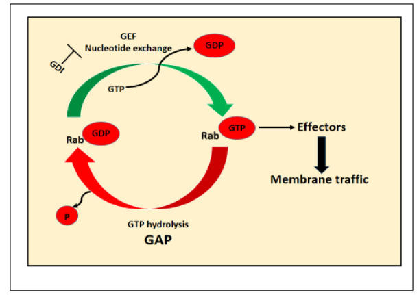
Rabs proteins are important vesicle docking and fusion regulators and form the largest branch of the Ras-GTPase superfamily. Numerous mammalian Rab proteins are expressed in only a few tissues and types of differentiated cells, where they participate in specific transport pathways. By switching between the GTP-bound and GDP-bound states, Rab GTPases act as molecular switches. Responsible for this switch are GEFs, which cause the binding of GTP, and GAPs, which accelerate the conversion of GTP to GDP, as shown in figure 2. In addition, Rabs undergo a membrane insertion and extraction cycle that is somewhat related to the nucleotide cycle. According to Kinsella and O'Mahony ( 47 ), membrane insertion requires the irreversible modification of two carboxyl-terminal cysteines with isoprenyl lipid units (geranylgeranyl). The proteins GDP dissociation inhibitor (GDI) and GDI-displacement factor (GDF) regulate the binding of Rab-GDP to membranes, which in turn limits Rab activity. Rab-GDP is removed from the membrane by GDI and thus remains inactive. In contrast, GDF replaces GDI and attracts Rab to membranes. The proteins that control Rab activation receive signals from upstream effectors, linking transport events to internal and external events ( 46 ). Each of the four primary phases of membrane transport-vesicle binding, vesicle promotion, vesicle binding, and fusion of the vesicle membrane with that of the target compartment is regulated by Rab through the guanine nucleotide-dependent switching mechanism common to the superfamily. Many different effector molecules work together to bind to specific Rabs in their GTP-bound state and perform different tasks. Two different types of proteins, Tethers and Rabs GTPases, control the orientation of vesicles. Rabs (downstream effectors) recruit or activate tethering proteins, such as long filamentous proteins and large multiprotein complexes. Vesicles and the membrane compartment are connected by a tethering protein. They play a role both in producing the envelope that forms the transport vesicles and in directing these vesicles to the appropriate membrane. Through interactions with the cytoskeleton, Rabs have also been shown to play a role in vesicle transport ( 48 ).
Figure 2.
Rab GTPase Cycle. Using signaling cascades to control the direction and selectivity of endosomal trafficking, small GTPases function as master regulators and molecular switches in a GTP-dependent manner
2.9 PI3-kinases
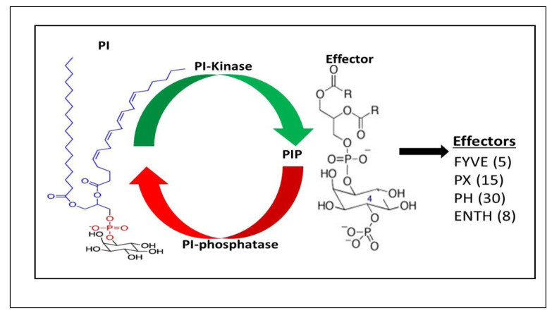
Cargo selection, vesicle formation, movement, and membrane fusion are just some of the intracellular trafficking events regulated by PI3-kinases. Phagocytosis, macropinocytosis, and controlled exocytosis are examples of receptor-induced trafficking processes promoted by class I and class II PI 3kinases. Class III has been conserved through evolution. Through the catalytic product PtdIns( 3 ) P and the domain containing the effectors of this lipid, the PI3kinase mediates most trafficking events. Most of these events can be considered constitutively occurring housekeeping tasks. Figure 3 shows the regulation of intracellular trafficking by the PI-3 kinase.
Figure 3.
Phosphoinositide cycle in cell signaling. Numerous intracellular trafficking activities, such as cargo choice, vesicle formation, vesicle mobility, and membrane fusion, are regulated by PI 3-kinases. It has been demonstrated that the catalytic byproduct of PI 3-kinases, PtdIns(2)P, contains a number of recognized protein effectors. These include the PX and PH domains, FYVE (conserved in Fab1, YOTB, Vac1, and EEA1), and Fab1. It is believed that effector proteins' ability to bind to PI 3-kinases' lipid products is mediated by the FYVE, PX, and PH domains. PtdIns(2)P effectors that can interact with Rab GTPases regulate the docking and fusing of endocytic membranes by promoting the development of SNARE complexes, which bind two membranes tightly by forming energetically advantageous tetrahelical bundles
2.10 Tethers
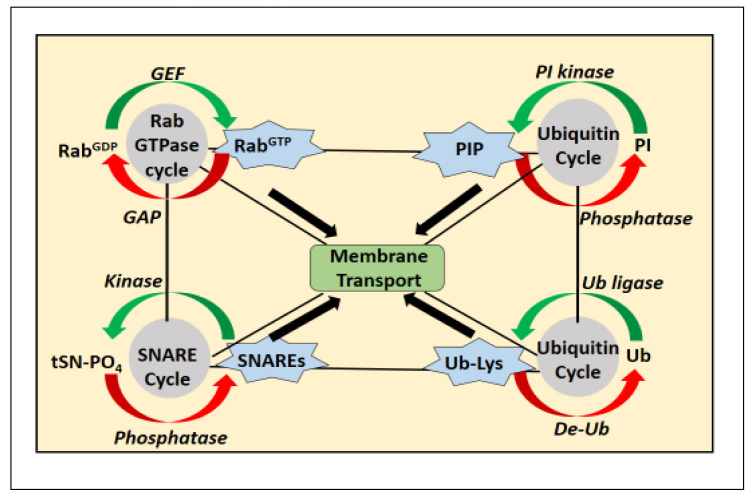
In vesicle binding, tethers are thought to be the primary effectors of Rab function. They are usually large multimeric complexes or long coiled-coil proteins. Before the SNARE complex is assembled and fused, tethering molecules connect the vesicle and the target membrane through activated Rab proteins. Tethers have been identified for a few transport steps. These involve the three different tethers discovered in ER to Golgi traffic. These include the long coiled-coil molecule Uso1p as well as the Sec34p/Sec35p complex and TRAPP ( 49 ). Genetic interaction among the three tethers shows that they are all involved in the same transport step. Overexpression of the corresponding Rab or SNARE molecules can prevent mutations of the tethering complex, indicating that the tethers function prior to the assembly of the complex SNARE ( 50 ). The regulatory circuits of membrane transport are displayed in figure 4.
Figure 4.
Regulatory circles in membrane trafficking. Rab GTPases act as molecular switches that regulate membrane trafficking events in a GTP-dependent manner. When certain cytoplasmic proteins are recruited to endosomal membranes by membrane bound phosphoinositides, specific membrane fusion, fission, and cargo sorting can take place inside and between different vesicle compartments. Additionally, to facilitate transport and signaling, phosphoinositides influence the operation of membrane proteins like ion channels and transporters in a compartment-specific way. The assembly of soluble N-ethylmaleimide-sensitive factor attachment protein receptor (SNARE) complexes, which connect two membranes, facilitates intracellular membrane docking and fusion. One of the most typical types of post-translational protein modification is ubiquitination (also known as ubiquitylation). Numerous transmembrane proteins' location, abundance, and trafficking are all influenced by it, directly affecting how they carry out their physiological tasks. The ubiquitin-activating enzyme, which forms a thioester bond between the ubiquitin's glycine-76 and a cysteine residue in the E1, is responsible for activating ubiquitin. Following that, ubiquitin is transported to the cysteine residue of an enzyme that catalyzes ubiquitin conjugation. Ubiquitin is transferred from the charged E2 to an internal lysine residue in the substrate using a ubiquitin ligase in the last step. At one or more lysine residues, substrates might be poly- or mono-ubiquitinated. Through the activity of deubiquitinatinating enzymes, ubiquitin can also be taken out of ubiquitinated proteins
3. Results and Discussion
The discovery of genes associated with human diseases has increased dramatically in recent decades, and it has become clear that several of these genes are related to membrane traffic. Increased attention is now being paid to how disease manifestations can be mechanistically explained using a basic understating of the trafficking machinery, and how this recently acquired understanding of the molecular basis of genetic syndromes can be used to gain insight into the structure of trafficking processes. Gene alterations that affect how cargo proteins or components of the biosynthetic and trafficking machinery are produced lead to Mendelian diseases of membrane trafficking. Among these genes, those that produce cargo proteins are more common because of their greater number, the fact that many of these cargo proteins are tissue-specific, and the fact that many of them are not necessary for embryo survival ( 51 ). On the other hand, alterations in genes that produce proteins used in all forms of transport mechanisms tend to be lethal. However, a number of these mutations have been found to contribute to a variety of diseases, including Mendelian diseases. Plasticity of transport systems can partially compensate for some of these changes under ideal conditions.
For example, the transport and release of some apoptogenic proteins in response to various apoptotic stimuli is a point of no return for programmed cell death, an essential physiological process that plays a critical role in tissue homeostasis, differentiation, regulation of growth, development, and immune response, and elimination of redundant or abnormal cells ( 52 , 53 ). The water permeability and selectivity of aquaporins vary widely. Water transport mediated by aquaporins is regulated either by gating, conformational change, or by changing the aquaporin density in a given membrane. Leakage of some aquaporin-2 mutants into the ER with the aid of chemical chaperones due to a defect in the aquaporin-2 water channel results in nephrogenic diabetes insipidus ( 54 ). The transport of aquaporins is regulated at transcriptional and/or translational levels and involves the shuttling of aquaporins between intracellular storage vesicles and the target membrane ( 55 ).
Defective sodium transport at the cell surface and ER retention of sodium channel subunits lead to cardiac disease ( 56 ). Defective transport of chloride channels to the PM due to defects in the chloride channel and cystic fibrosis transmembrane conductance regulator leads to multiorgan disease, most commonly affecting the pancreas and lungs ( 57 ). Mutations in the sulfonylurea 1 subunit of the pancreatic ATP‐sensitive potassium channel (KATP) lead to its retention in the ER or Golgi and subsequently to congenital hyperinsulinism ( 58 ).
A list of monogenic disorders caused by mutations in genes encoding parts of the membrane transport machinery is generated by their underlying role in functionally related processes. The immune system appears to be highly influenced by membrane transport, which directly affects its ability to function. The immune system uses the endomembrane system to receive and integrate information from different cells of the organism as well as from pathogens, altering several cellular signaling pathways ( 59 ). Alteration of intracellular trafficking can lead to the development of a variety of immune system disorders, such as autoimmune diseases or immunodeficiency. Cellular trafficking is critical for the subsequent immunological response. Targeting and regulating cellular trafficking is important for the treatment of a number of immune-related diseases, including those termed immune-mediated diseases, as a growing body of research shows ( 60 ).
The specific disease resulting from a defect in the secretory pathway is known as cystic fibrosis, which results from a blockage at the exit of the ER due to protein misfolding. A disease resulting from a defect in the endocytic pathway is known as hypercholesterolemia. A mutation in the receptor for low-density lipoprotein prevents receptor-mediated endocytosis of low-density lipoprotein. Numerous other diseases, such as cancer, infections, and several hereditary abnormalities, are associated with the intracellular transport pathway.
Vesicles help cells take up and expel material in loose form as well as particles too large for transport proteins through the processes of endocytosis and exocytosis. Motor proteins and ATP are involved in the formation and movement of vesicles composed of membranes. During exocytosis, a vesicle fuses with the cell membrane and releases its contents into the environment, whereas during endocytosis, a vesicle is formed from the cell membrane, trapping substances near the cell surface and transporting them into the cell. Exocytosis and endocytosis constantly remove portions of the PM and replace them with membrane cycles. In the ER, new membrane lipids and proteins are produced, processed in the Golgi bodies, and then packaged into vesicles that attach to the plasma membrane. Exocytosis and endocytosis allow large packets of material and encapsulated cells to pass through the plasma membrane. These processes involve the entry and exit of membrane proteins and lipids from the plasma membrane. Using the guanine nucleotide-dependent switching mechanism common to the superfamily, Rabs control each of the four basic steps of membrane trafficking. Numerous intracellular trafficking activities, such as cargo selection, vesicle formation, vesicle motility, and membrane fusion, are regulated by PI3-kinases. The mechanism responsible for the correct localization of molecules within the cell is called membrane trafficking. It is the basic requirement for cells to maintain cellular homeostasis and meet certain particular requirements in information uptake and transmission. Numerous genetic disorders and other diseases are associated with alterations in membrane trafficking.
Acknowledgments
Authors' Contribution
AOA wrote and revised the content of the manuscript.
Ethics
Not applicable.
Conflict of Interest
The author declares no known competing interest.
References
- 1.Jackson CL, Walch L, Verbavatz JM. Lipids and Their Trafficking. An Integral Part of Cellular Organization.Dev.Cell. 2016;39:139–153. doi: 10.1016/j.devcel.2016.09.030. [DOI] [PubMed] [Google Scholar]
- 2.Gilchrist TA, Au CE, Hiding J. Quantitative proteomics analysis of the secretory pathway. Cell. 2006;127:1265–1281. doi: 10.1016/j.cell.2006.10.036. [DOI] [PubMed] [Google Scholar]
- 3.Saito C, Ueda T. Functions of RAB and SNARE proteins in plant life. Int. Rev. Cell Mol. Biol. 2009;274:183–233. doi: 10.1016/S1937-6448(08)02004-2. [DOI] [PubMed] [Google Scholar]
- 4.Fujimoto M, Ueda T. Conserved and plant-unique mechanisms regulating plant post-Golgi traffic. Front. Plant Sci. 2012;3:197. doi: 10.3389/fpls.2012.00197. [DOI] [PMC free article] [PubMed] [Google Scholar]
- 5.Schutze MP, Peterson PA, Jackson MR. An N-terminal double-arginine motif maintains type II membrane proteins in the endoplasmic reticulum. EMBO J. 1994;13:1696–1705. doi: 10.1002/j.1460-2075.1994.tb06434.x. [DOI] [PMC free article] [PubMed] [Google Scholar]
- 6.Watanabe S, Shimada TL, Hiruma K, Takano Y. Pathogen infection trial increases the secretion of proteins localized in the endoplasmic reticulum body of Arabidopsis. Plant Physiol. 2013;163:659–664. doi: 10.1104/pp.113.217364. [DOI] [PMC free article] [PubMed] [Google Scholar]
- 7.Beck M, Heard W, Mbengue M, Robatzek S. The Ins and OUTs of pattern recognition receptors at the cell surface. Curr. Opin. Plant Biol. 2012;15: 367–374. doi: 10.1016/j.pbi.2012.05.004. [DOI] [PubMed] [Google Scholar]
- 8.Schwarz DS, Blower MD. The endoplasmic reticulum: Structure, function and response to cellular signaling. Cell. Mol. Life Sci. 2016;73: 79–94. doi: 10.1007/s00018-015-2052-6. [DOI] [PMC free article] [PubMed] [Google Scholar]
- 9.Saijo Y. ER quality control of immune receptors and regulators in plants. Cell. Microbiol. 2010;12:716–724. doi: 10.1111/j.1462-5822.2010.01472.x. [DOI] [PubMed] [Google Scholar]
- 10.Hammond AT, Glick BS. Dynamics of transitional endoplasmic reticulum sites in vertebrate cells. Mol. Biol. 2000;11(9):3013–30. doi: 10.1091/mbc.11.9.3013. [DOI] [PMC free article] [PubMed] [Google Scholar]
- 11.Kaiser C. Thinking about p24 proteins and how transport vesicles select their cargo. Proc. Natl. Acad. Sci. USA. 2000;97(8):3783–3785. doi: 10.1073/pnas.97.8.3783. [DOI] [PMC free article] [PubMed] [Google Scholar]
- 12.Huttner S, Strasser R. Endoplasmic reticulum-associated degradation of glycoproteins in plants. Front. Plant Sci. 2012;3:67. doi: 10.3389/fpls.2012.00067. [DOI] [PMC free article] [PubMed] [Google Scholar]
- 13.Hirsch C, Gauss R, Horn SC, Neuber O, Sommer T. The ubiquitylation machinery of the endoplasmic reticulum. Nature. 2009;458:453–460. doi: 10.1038/nature07962. [DOI] [PubMed] [Google Scholar]
- 14.Brandizzi F, Barlowe C. Organization of the ER–Golgi interface for membrane traffic control. Nat. Rev. Mol. Cell Biol. 2013;14: 382–392. doi: 10.1038/nrm3588. [DOI] [PMC free article] [PubMed] [Google Scholar]
- 15.Scales SJ, Pepperkok R, Kreis TE. Visualization of ER‐to‐Golgi transport in living cells reveals a sequential mode of action for COPII and COPI. 1997:1137–1148. doi: 10.1016/s0092-8674(00)80379-7. [DOI] [PubMed] [Google Scholar]
- 16.Palmer KJ, Stephens DJ. Biogenesis of ER‐to‐Golgi transport carriers: Complex roles of COPII in ER export. Trends Cell Biol. 2004;14:57–61. doi: 10.1016/j.tcb.2003.12.001. [DOI] [PubMed] [Google Scholar]
- 17.Simpson, JC, Nilsson T, Pepperkok R. Biogenesis of tubular ER‐to‐Golgi transport intermediates. Mol. Biol. 2006:723–737. doi: 10.1091/mbc.E05-06-0580. [DOI] [PMC free article] [PubMed] [Google Scholar]
- 18.Huang S, Wang Y. Golgi structure formation, function, and post-translational modifications in mammalian cells. F1000Research. 2017;6: 2050. doi: 10.12688/f1000research.11900.1. [DOI] [PMC free article] [PubMed] [Google Scholar]
- 19.Altan‐Bonnet N, Sougrat R, Lippincott‐Schwartz J. Molecular basis for Golgi maintenance and biogenesis. Curr. Opin. Cell Biol. 2004:364–372. doi: 10.1016/j.ceb.2004.06.011. [DOI] [PubMed] [Google Scholar]
- 20.Rabouille C, Hui N, Hunte F, Kieckbusch R, Berger EG, Warren G, Nilsson T. Mapping the distribution of Golgi enzymes involved in the construction of complex oligosaccharides. J. Cell Sci. 1995;108(Pt. 4): 1617–1627. doi: 10.1242/jcs.108.4.1617. [DOI] [PubMed] [Google Scholar]
- 21.Trucco A, Polishchuk RS, Martella O, Di Pentima A, Fusella A, Di Giandomenico D, et al. Secretory traffic triggers the formation of tubular continuities across Golgi sub-compartments. Nat. Cell Biol.6. 2004:1071–1081. doi: 10.1038/ncb1180. [DOI] [PubMed] [Google Scholar]
- 22.Gu F, Crump CM, Thomas G. Trans‐Golgi network sorting. Cell. Mol. Life Sci.58. 2001:1067–1084. doi: 10.1007/PL00000922. [DOI] [PMC free article] [PubMed] [Google Scholar]
- 23.Thomas G. Furin at the cutting edge: From protein traffic to embryogenesis and disease. Nat.Rev.Mol.Cell Biol. 2002:753–766. doi: 10.1038/nrm934. [DOI] [PMC free article] [PubMed] [Google Scholar]
- 24.Medigeshi GR, Schu P. Characterization of the in vitro retrograde transport of MPR46. Traffic 4. 2003:802–811. doi: 10.1034/j.1600-0854.2003.00136.x. [DOI] [PubMed] [Google Scholar]
- 25.Maxfield FR, McGraw TE. Endocytic recycling. Nat Rev.Mol. 2004:121–132. doi: 10.1038/nrm1315. [DOI] [PubMed] [Google Scholar]
- 26.Fevrier B, Raposo G. Exosomes: Endosomal‐derived vesicles shipping extracellular messages. Curr.Opin. 2004;16(4):415–21. doi: 10.1016/j.ceb.2004.06.003. [DOI] [PubMed] [Google Scholar]
- 27.Johnstone RM, Mathew A, Mason AB, Teng K. Exosome formation during maturation of mammalian and avian reticulocytes: Evidence that exosome release is a major route for externalization of obsolete membrane proteins. J.Cell.Physiol. 1991;147: 27–36. doi: 10.1002/jcp.1041470105. [DOI] [PubMed] [Google Scholar]
- 28.Rouillé Y, Rohn W, Hoflack B. Targeting of lysosomal proteins. In Seminars in cell & developmental biology Academic Press. 2000;11(3): 165–171. doi: 10.1006/scdb.2000.0168. [DOI] [PubMed] [Google Scholar]
- 29.Luzio JP, Rous BA, Bright NA, Pryor PR, Mullock BM, Piper RC. Lysosome-endosome fusion and lysosome biogenesis. Journal of Cell Science. 2000;113(9):1515–24. doi: 10.1242/jcs.113.9.1515. [DOI] [PubMed] [Google Scholar]
- 30.Schekman R, Orci L. Coat proteins and vesicle budding. Science. 1996;271(5255):1526–1533. doi: 10.1126/science.271.5255.1526. [DOI] [PubMed] [Google Scholar]
- 31.Herrmann JM, Malkus P, Schekman R. Out of the ER-outfitters, escorts and guides. Trends Cell Biol. 1999;9(1):5–7. doi: 10.1016/s0962-8924(98)01414-7. [DOI] [PubMed] [Google Scholar]
- 32.Duden R. ER‐to‐Golgi transport: COP I and COP II function (Review) . Mol. Membr. 2003;20(3):197–207. doi: 10.1080/0968768031000122548. [DOI] [PubMed] [Google Scholar]
- 33.Nie Z, Hirsch DS, Randazzo PA. Arf and its many interactors. Curr. Opin. 2003;15(4):396–404. doi: 10.1016/s0955-0674(03)00071-1. [DOI] [PubMed] [Google Scholar]
- 34.Jackson CL, Casanova JE. Turning on ARF: The Sec7 family of guanine nucleotide‐exchange factors. 2000;10(2):60–7. doi: 10.1016/s0962-8924(99)01699-2. [DOI] [PubMed] [Google Scholar]
- 35.Spang A, Matsuoka K, Hamamoto S, Schekman R, Orci L. Coatomer, Arf1p, and nucleotide are required to bud coat protein complex I‐coated vesicles from large synthetic liposomes. Proc.Natl.Acad.Sci. 1998;95(19):11199–204. doi: 10.1073/pnas.95.19.11199. [DOI] [PMC free article] [PubMed] [Google Scholar]
- 36.Barlowe C, Schekman R. SEC12 encodes a guanine‐nucleotide‐exchange factor essential for transport vesicle budding from the ER. 1993;365(6444):347–9. doi: 10.1038/365347a0. [DOI] [PubMed] [Google Scholar]
- 37.Supek F, Madden DT, Hamamoto S, Orci L, Schekman R. Sec16p potentiates the action of COPII proteins to bud transport vesicles. J.Cell Biol. 2002;158(6):1029–1038. doi: 10.1083/jcb.200207053. [DOI] [PMC free article] [PubMed] [Google Scholar]
- 38.Marsh M, McMahon HT. The structural era of endocytosis. 1999;285(5425):215–20. doi: 10.1126/science.285.5425.215. [DOI] [PubMed] [Google Scholar]
- 39.Schmid S. Clathrin-coated vesicle formation and protein sorting: an integrated process. Annu.Rev. 1997;66:511–48. doi: 10.1146/annurev.biochem.66.1.511. [DOI] [PubMed] [Google Scholar]
- 40.Sweitzer SM, Hinshaw JE. Dynamin undergoes a GTP-dependent conformational change causing vesiculation. 1998;93(6):1021–9. doi: 10.1016/s0092-8674(00)81207-6. [DOI] [PubMed] [Google Scholar]
- 41.Robinson MS, Bonifacino JS. Adaptor-related proteins. Curr. Opin.Cell Biol. 2001;13(4):444–53. doi: 10.1016/s0955-0674(00)00235-0. [DOI] [PubMed] [Google Scholar]
- 42.Jackson T. Transport vesicles: Coats of many colours. Curr. Biol. 8. 1998;8(17):R609–12. doi: 10.1016/s0960-9822(98)70388-4. [DOI] [PubMed] [Google Scholar]
- 43.Rohn WM, Rouille Y, Waguri S, Hoflack B. Bi-directional trafficking between the trans-Golgi network and the endosomal/lysosomal system. J.CellSci.113. 2000;113(Pt12):2093–2101. doi: 10.1242/jcs.113.12.2093. [DOI] [PubMed] [Google Scholar]
- 44.Weber T, Zemelman B, McNew J, Westermann B, Gmachl M, Parlati F, Sollner T, Rothman J. SNAREpins: minimal machinery for membrane fusion. Cell. 1998; 92(6):759–72. doi: 10.1016/s0092-8674(00)81404-x. [DOI] [PubMed] [Google Scholar]
- 45.Ungermann C, Nichols B, Pelham H, Wickner W. A vacuolar v-t-SNARE complex, the predominant form in vivo and on isolated vacuoles and on isolated vacuoles, is disassembled and activated for docking and fusion. J.Cell Biol. 1998;40(1):61–9. doi: 10.1083/jcb.140.1.61. [DOI] [PMC free article] [PubMed] [Google Scholar]
- 46.Segev N. Ypt and Rab GTPases: insight into functions through novel interactions. Curr.Opin.Cell Biol. 2001;13(4):500–11. doi: 10.1016/s0955-0674(00)00242-8. [DOI] [PubMed] [Google Scholar]
- 47.Kinsella BT, O'Mahony DJ. (Lipid modification of G proteins. Trends in Cardiovascular Medicine. 1994;4(1): 27–34. doi: 10.1016/1050-1738(94)90022-1. [DOI] [PubMed] [Google Scholar]
- 48.Nielsen E, Severin F, Backer JM, Hyman AA, Zerial M. Rab5 regulates motility of early endosomes on microtubules. Nat.Cell Biol.1. 1999:376–382. doi: 10.1038/14075. [DOI] [PubMed] [Google Scholar]
- 49.Lowe, M. Membrane transport: tethers and TRAPPS. Current Biology. 2000;10:R407–R409. doi: 10.1016/s0960-9822(00)00505-4. [DOI] [PubMed] [Google Scholar]
- 50.Sapperstein SK, Lupashin VV, Schmitt HD, Waters MG. Assembly of the ER to Golgi SNARE complex requires Uso1p. J.Cell Biol. 1996;132(5):755–67. doi: 10.1083/jcb.132.5.755. [DOI] [PMC free article] [PubMed] [Google Scholar]
- 51.Rothman JE. The machinery and principles of vesicle transport in the cell. Nat Med. 2002;8(10):1059–1062. doi: 10.1038/nm770. [DOI] [PubMed] [Google Scholar]
- 52.Adeoye AO, Olanlokun JO, Tijani H, Lawal SO, Babarinde CO, Akinwole MT, Bewaji CO. Molecular docking analysis of apigenin and quercetin from ethylacetate fraction of Adansonia digitata with malaria-associated calcium transport protein: An in silico approach. Heliyon. 2019;5(9):e02248. doi: 10.1016/j.heliyon.2019.e02248. [DOI] [PMC free article] [PubMed] [Google Scholar]
- 53.Daniel OO, Adeoye AO, Ojowu J, Olorunsogo OO. Inhibition of liver mitochondrial membrane permeability transition pore opening by quercetin and vitamin E in streptozotocin-induced diabetic rats. Biochemical and Biophysical Research Communication. 2018;504(2018):460–469. doi: 10.1016/j.bbrc.2018.08.114. [DOI] [PubMed] [Google Scholar]
- 54.Tamarappoo BK, Yang B, Verkman AS. Misfolding of mutant aquaporin‐2 water channels in nephrogenic diabetes insipidus. J. Biol. 1999;274(49):34825–31. doi: 10.1074/jbc.274.49.34825. [DOI] [PubMed] [Google Scholar]
- 55.Adeoye AO, Odugbemi A, Ajewole TO. Structure and Function of Aquaporins: the Membrane Water Channel Proteins. Biointerface research in Applied Chemistry. 2022;12(1): 690–705. [Google Scholar]
- 56.Baroudi G, Napolitano C, Priori SG, Del Bufalo A, Chahine M. Loss of function associated with novel mutations of the SCN5A gene in patients with Brugada syndrome. Can.J. 2004;20(4):425–30. [PubMed] [Google Scholar]
- 57.Heda GD, Tanwani M, Marino CR. The delta F508 mutation shortens the biochemical half‐life of plasma membrane CFTR in polarized epithelial cells. Am. J.Physiol. 2001;280(1):C166–74. doi: 10.1152/ajpcell.2001.280.1.C166. [DOI] [PubMed] [Google Scholar]
- 58.Yan F, Lin CW, Weisiger E, Cartier EA, Taschenberger G, Shyng SL. Sulfonylureas correct trafficking defects of ATP‐sensitive potassium channels caused by mutations in the sulfonylurea receptor. J.Biol. 2004;279(12):11096–105. doi: 10.1074/jbc.M312810200. [DOI] [PubMed] [Google Scholar]
- 59.Taguchi T, Mukai K. Innate immunity signalling and membrane trafficking. Curr.Opin.Cell Biol. 2019;59:1–7. doi: 10.1016/j.ceb.2019.02.002. [DOI] [PubMed] [Google Scholar]
- 60.Hwang H, Kim H, Han G, Lee J, Kim K, Kwon I, et al. Extracellular Vesicles as Potential Therapeutics for Inflammatory Diseases. Int.J.Mol.Sci. 2021;22(11):5487. doi: 10.3390/ijms22115487. [DOI] [PMC free article] [PubMed] [Google Scholar]


