ABSTRACT
Background:
Natural compounds rich in secondary metabolites have gained attention as alternative therapies for wound healing due to their potential advantages over conventional treatments. Pleurotus ostreatus mushrooms have been identified for their wound-healing properties, including promoting neovascularization, epithelialization, and collagen synthesis.
Material and Methods:
This study aimed to investigate the wound-healing properties of different doses of topical Extract of Pleurotus ostreatus in albino mice using an excisional wound model. The experimental design involved administering various concentrations of the extract to evaluate its effects on wound closure and histopathological and immunohistochemical parameters.
Results:
The results revealed significant wound closure and improvements in histopathological and immunohistochemical parameters in mice treated with Pleurotus ostreatus extract. These findings suggest the potential of Pleurotus ostreatus extract as a viable wound healing agent.
Conclusion:
Pleurotus ostreatus extract demonstrates promising wound-healing capabilities, including promoting wound closure and enhancing histopathological and immunohistochemical parameters in albino mice. Further research is warranted to explore its full therapeutic potential and mechanism of action in wound healing.
KEYWORDS: Epidermal growth factor intensity, histopathological analysis, immunohistochemical, topical dosage form
INTRODUCTION
The skin, as the body’s largest protective barrier, heals wounds through three phases: inflammatory, proliferative, and remodeling. Successful healing relies on proper hemostasis, inflammation, and blood vessel formation. Natural compounds, especially from plants, are gaining attention for wound healing due to their antioxidant, anti-inflammatory, analgesic, and anti-bacterial properties.[1] Pleurotus ostreatus, the oyster mushroom, is rich in therapeutic potential, including anti-inflammatory and antioxidant properties. Also, Mushroom extracts have shown promising anti-elastase, anti-tyrosinase, anti-collagenase, and anti-hyaluronidase activities.[2] This study aims to explore Pleurotus ostreatus as a wound-healing agent in mice, examining wound contraction, histopathological parameters, and epidermal growth factor intensity.
MATERIALS AND METHOD
Chemicals: (mebo) ointment, formalin, (Vaseline), shaving cream, ketamine, xylazine chloride, EGF kit, hematoxylin, and eosin were used in the study.
Plant Material: Pleurotus ostreatus extract mushrooms, 5% and 10% ointments formulated with Vaseline.
Experimental Animals: 66 female mice for a wound in vivo study. The experiment was approved by the Institute Review Board (IRB) of the College of Medicine at Al-Nahrain University.
Acute Dermal Toxicity Test: Six mice were employed for this test and were divided into two groups: group (a) treated with an ointment of extract 10% p.ous and another group (b) treated with Vaseline (vehicle) monitoring for 14 days.
Experimental Animal Groups: 60 mice divided into six groups,
Excision Wound Model: Wounds were created, and treated for ten days, wound size was measured on the 5th and 10th day.
Histopathological Analysis: Skin samples were collected on the 10th day for evaluation.
Immunohistochemistry Analysis: EGF was tested using a kit, scored for expression.
Statistical Analysis: Data analyzed using Excel, P < 0.05 considered significant, P ≤ 0.001 highly significant.
RESULTS
1. Acute Dermal Toxicity: The maximum dose of 10% (w/w) mushroom extract was found to be safe in mice after 14 days of observation as shown in Figure 1.
Figure 1.
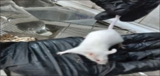
After 14-day monitoring
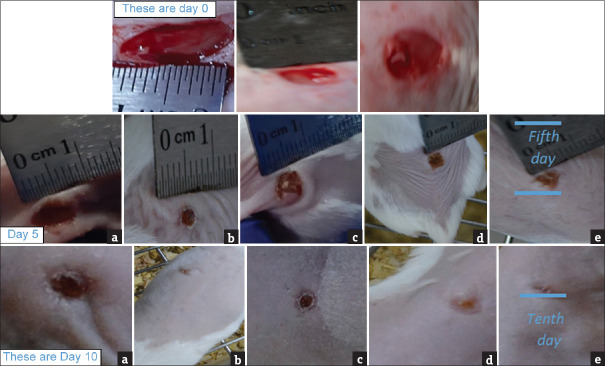
2. Wound Closure Measurement: The progress of wound healing induced by Pleurotus ostreatus extract at 5% and 10% w/w concentrations, along with induction untreated, mebo, and Vaseline bases groups, was evaluated. Table 1 and Figure 2
Table 1.
Comparison of wound size closure between untreated and all other studied treated groups by unpaired t-test
| Mean Percentage of Wound Closure | Untreated group | Vaseline base group | Mebo group | Ointment Extract 5% group | Ointment Extract 10% group |
|---|---|---|---|---|---|
| Fifth day | |||||
| Mean±SE | 28.87±2.41 | 36.67±0.88 | 58.00±1.15 | 50.70±2.01 | 54.60±1.45 |
| Pa | 0.039 | <0.001 | 0.002 | 0.001 | |
| Pb | <0.001 | 0.003 | <0.001 | ||
| Pc | 0.035 | 0.14 | |||
| Pd | 0.19 | ||||
| Tenth day | |||||
| Mean±SE | 64.40±2.01 | 74.43±2.41 | 94.20±0.61 | 89.67±2.03 | 95.67±0.88 |
| Pa | 0.033 | <0.001 | 0.001 | <0.001 | |
| Pb | 0.011 | 0.009 | 0.001 | ||
| Pc | 0.099 | 0.24 | |||
| Pd | 0.08 |
Figure 2.
Photographic representation of wound size closure in different groups of mice in day zero, 5, and 10 post-injury. (a) untreated, (b) mebo, (c) Vaseline (d) 5% ointment extract, (e) 10% ointment ex
On day zero, there were no significant differences in wound size closure between each group (P values of Pa, Pb, Pc, and Pd >0.05) [Table 1]. However, on day five, the extract of Pleurotus ostreatus at 5% and 10% w/w concentrations and mebo ointment significantly enhanced wound closure compared to other groups (P < 0.05). Mebo ointment also showed significant closure in the wound size compared to the 5% w/w extract treatment group (Pc = 0.035). By day ten, the extract at 10% w/w concentration induced a maximum healing ratio with 95% wound closure, outperforming the untreated and Vaseline base groups [Table 1].
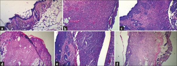
3. Histopathological Evaluation Scores: On day ten, the histopathological evaluation showed highly significant differences in neovascularization, rate of re-epithelization, and collagen synthesis between healthy and untreated groups.
The induction untreated group exhibited a significant decrease in these scores compared to the other treatment groups as shown in Figure 3
Figure 3.
Photographs of Histopathological Evaluation of wound tissues in excision model on the 10th day (1) the healthy group, which showed complete re-epithelization, (2) the induction group showed highly inflammatory cell infiltration, decreased collagen synthesis, moderate neovascularization and in-complete epithelization (3) mebo standard drug represents moderate collagen synthesis, complete re-epithelization and highly neovascularization, (d) Vaseline base showed mild collagen synthesis, in- complete re-epithelization, highly fibroblast infiltration and moderate neovascularization (e) ointment extract 5%group showed moderate collagen synthesis, complete re-epithelization and highly neovascularization (f) ointment extract 10%.showed complete re-epithelization, highly collagen synthesis and highly neovascularization on microscopical examination magnification (10x)
Microscopic Photographs: Figure 3 displays microscopic photographs of tissues from full-thickness excisional wounds in mice.
4. Immunohistochemistry - Epidermal Growth Factor Intensity: On day three, higher doses of 10% and (mebo, 5%) showed significantly higher EGF intensity compared to the untreated and Vaseline groups. However, there were no significant differences in EGF intensity between treatment groups on day three [Figure 4]. From days 3 to 10, EGF intensity was significantly lower in the induction untreated group compared to other groups. The highest EGF intensity was observed in the 10% extract group on day 10, with significant differences observed between the treatment groups and untreated and Vaseline groups.
Figure 4.
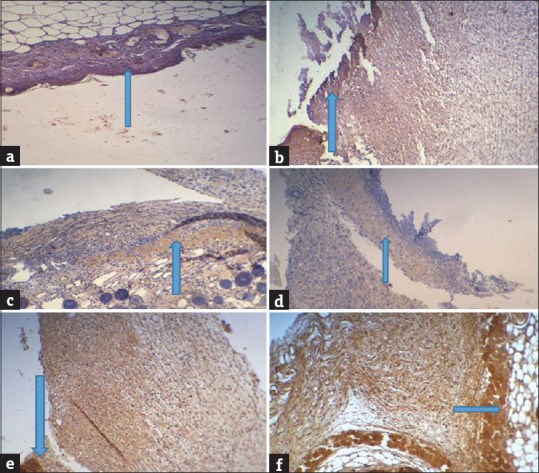
Intensity of EGF within extracellular dermis immunohistochemistry at day 3 (magnification10x), (blue arrow). (a) the healthy group has shown normal intensity, (b) the Induction untreated group has shown low intensity, (c) mebo standard group has shown moderate intensity; (d) the Vaseline base group with medium intensity, (e and f) Both extract treatment of doses (5%and 10%% w/w respectively) groups have shown moderate intensity
DISCUSSION
The study aimed to investigate the wound-healing potential of Pleurotus ostreatus extract due to its secondary metabolites, including tannins, terpenes, flavonoids, and saponins, known for their wound-healing properties.[3,4] showed that phenolic compound anti-oxidant activity also may aid in speeding up the healing process 46.
The methanol extract was selected as it contains various phytochemicals from the mushroom.[5]
Topical application of the hydroalcoholic extract significantly enhanced wound healing in mice due to its antibacterial, antioxidant, and anti-inflammatory activities.[6] Collagen synthesis was also increased, likely attributed to flavonoids and saponins in the extract.[7,8] The enhanced neovascularization observed in treated groups supports the extract’s ability to supply oxygen and nutrients for tissue regeneration.
And the ability of p.ostreatus to activate the EGF pathway causes the upregulation of proangiogenic factors like VEGF.[9]
Increasing re-epithelization in extract groups due to the ability of pleurotus extract to activate keratinocyte encouraging re-epithelialization.[10]
Immunohistochemistry revealed higher EGF intensity in p.ostreatus extract treated groups,[11] Noticing the relationship between increased (EGF) levels and healing rate.[12] Noticed that Extract of (P.ostrateus) promotes increased EGF expression in ulcer healing. This is in agreement with current findings
Reduced EGF intensity in the induction untreated group may explain the delayed wound healing observed in this group.[13]
Figure 5 shows the comparison of EGF immunohistochemical parameter intensity in (3rd and 10th) days between all studied groups.
Figure 5.
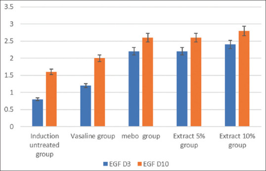
Comparison of EGF immunohistochemical parameter intensity in (3rd and 10th) day between all studied groups
CONCLUSION
The results demonstrate that the topical application of hydroalcoholic Pleurotus ostreatus extract promotes wound healing in mice. The extract’s antioxidant, antibacterial, and anti-inflammatory activities, along with its ability to enhance collagen synthesis and neovascularization, contribute to its wound-healing potential. The increased EGF intensity in treated groups further supports the extract’s role in tissue repair. These findings highlight the therapeutic potential of Pleurotus ostreatus extracts for wound healing applications.
Financial support and sponsorship
Nil.
Conflicts of interest
There are no conflicts of interest.
REFERENCES
- 1.Fox LT, Mazumder A, Dwivedi A, Gerber M, du Plessis J, Hamman JH. In Vitro wound healing and cytotoxic activity of the gel and whole-leaf materials from selected aloe species. J Ethnopharmacol. 2017;200:1–7. doi: 10.1016/j.jep.2017.02.017. [DOI] [PubMed] [Google Scholar]
- 2.Bahadori MB, Asghari B, Dinparast L, Selcuk Z, Sarikurkcu C, Abbas-Mohammadi A, et al. Salvia Nemorosa L.: A novel source of bioactive agents with functional connections. LWT. 2017;75:42–50. [Google Scholar]
- 3.Ogidi OI, Maureen L, Oguoma O, Adigwe PC, Anthony BB. phytochemical properties and in-vitro antimicrobial potency of wild edible mushrooms (pleurotus ostreatus) obtained from yenagoa, Nigeria. J Phytopharmacol. 2021;10:180–4. [Google Scholar]
- 4.Hassan RF, Kadhim HM. Exploring the role of phenolic extract as an ointment dosage form in inducing wound healing in mice. J Pharm Negat Results. 2022;13:186–93. [Google Scholar]
- 5.Dinssa B, Temesgen S, Mosisa W. Introduction and cultivation of P. ostreatu and L. edodes using sugar cane bagasse, leaves of Prosopis juliflora and waste paper at Oda Bultum University, Chiro, Ethiopia. Am J Agric For. 2020;8:40. [Google Scholar]
- 6.Shah A, Amini-Nik S. The role of phytochemicals in the inflammatory phase of wound healing. Int J Mol Sci. 2017;18:1068. doi: 10.3390/ijms18051068. [DOI] [PMC free article] [PubMed] [Google Scholar]
- 7.Dkhil MA, Diab MSM, Lokman MS, El-Sayed H, Bauomy AA, Al-Shaebi EM, et al. Nephroprotective effect of pleurotus ostreatus extract against cadmium chloride toxicity in rats. An Acad Bras Cienc. 2020;92:e20191121. doi: 10.1590/0001-3765202020191121. [DOI] [PubMed] [Google Scholar]
- 8.Yitayih LY, Jeevan TSMA, Bikilla SL, Demelash TD. Determination of the antibacterial and antioxidant activity of crude extract of bruceaantidysenterica leaves. Modern Chem. 2023;11:34–42. [Google Scholar]
- 9.Lee SH, Jeong D, Han YS, Baek MJ. Pivotal role of vascular endothelial growth factor pathway in tumor angiogenesis. Ann Surg Treat Res. 2015;89:1–8. doi: 10.4174/astr.2015.89.1.1. [DOI] [PMC free article] [PubMed] [Google Scholar]
- 10.Zulli F, Suter F, Biltz H, Nissen H. Improving skin function with CM-glucan, a biological response modifier from yeast. Int J Cosmet Sci. 1998;20:79–86. doi: 10.1046/j.1467-2494.1998.171740.x. [DOI] [PubMed] [Google Scholar]
- 11.Fang YF, Xu WL, Wang L, Lian QW, Qiu LF, Zhou H, et al. Effect of hydrotalcite on indometacin-induced gastric injury in rats. BioMed Res Int. 2019;2019:4605748. doi: 10.1155/2019/4605748. [DOI] [PMC free article] [PubMed] [Google Scholar]
- 12.Dilfy SH., Hanawi MJ, Al-bideri AW, Jalil AT. Determination of chemical composition of cultivated mushrooms in Iraq with spectrophotometrically and high performance liquid chromatographic. J Green Eng. 2020;10:6200–16. [Google Scholar]
- 13.Zhao R, Liang H, Clarke E, Jackson C, Xue M. Inflammation in chronic wounds. Int J Mol Sci. 2016;17:2085. doi: 10.3390/ijms17122085. [DOI] [PMC free article] [PubMed] [Google Scholar]


