Abstract
The sequence encoding the viral leader proteinase Npro was replaced by the murine ubiquitin gene in a full-length cDNA clone of the classical swine fever virus (CSFV) strain Alfort/187. The recombinant virus vA187-Ubi showed growth characteristics similar to those of the parent vA187-1 virus. At two occasions cells infected with vA187-Ubi exhibited a cytopathic effect and were found to contain a subgenomic viral RNA. This RNA lacked the same viral genes as the subgenomic RNA which has been found in all cytopathogenic CSFV strains analyzed so far, but it maintained the ubiquitin sequence.
The family Flaviviridae comprises three genera, the flaviviruses, the pestiviruses, and the hepatitis C viruses (17). While the overall genome organization is similar for all three genera, only the pestiviruses encode a nonstructural protein at the 5′ terminus of the open reading frame (11). This protein is an autoproteinase termed Npro (for N-terminal proteinase) which cleaves at its C terminus between amino acid residues Cys-168 and Ser-169 of the viral polyprotein, which are conserved among pestiviruses (15). As Npro precedes the nucleocapsid protein (C) in the viral polyprotein, the amino terminus of C is generated upon cleavage. Rümenapf and coauthors (14) have recently shown that Npro is a novel type of cysteine proteinase with no precedence in a viral system but which has some similarity to the subtilisin-like proteinases. An N-terminal autoproteinase is also encountered in aphthoviruses, which are positive-strand RNA viruses belonging to the family Picornaviridae (12). This proteinase, also referred to as leader proteinase, belongs to the papain family of proteinases (7). In addition to cleaving itself off the polyprotein, it causes the proteolytic degradation of the 220-kDa subunit of the eukaryotic initiation factor 4G and thus contributes to the shutoff of cap-dependent host cell protein synthesis (2). However, the aphthovirus leader proteinase gene is not required for viral replication in cell culture (8).
To explore the role of Npro in classical swine fever virus (CSFV) replication, we generated synthetic full-length viral RNA in which the coding sequence for Npro was replaced by the gene coding for the murine ubiquitin gene. In eukaryotic cells ubiquitin is cleaved immediately after its C-terminal glycine by cellular ubiquitin carboxyl-terminal hydrolase (UCH) (9). Thus, we expected that translation of viral RNA in which the ubiquitin gene is fused in frame to the gene encoding the viral nucleocapsid would allow the synthesis of authentic C. This approach has been used before to generate defined N termini of viral proteins in host cells (3). Ubiquitin-coding sequences have also been found as insertions in some cytopathogenic (cp) bovine viral diarrhea viruses (BVDV). Insertion of ubiquitin-coding sequences or of other cellular or viral sequences apparently occurs by nonhomologous recombination in animals persistently infected with noncytopathogenic (ncp) BVDV. In all cases analyzed so far, the ubiquitin gene is inserted at the identical position in the BVDV genome, providing a processing signal for cellular UCH which leads to the generation of NS3 protein. Independently of the type of recombination, the cytopathogenicity of BVDV always correlates with expression of NS3, whereas in ncp BVDV, NS3 is exclusively expressed as a fusion NS2-3 protein (for a review, see reference 5).
Construction of pA187-Ubi.
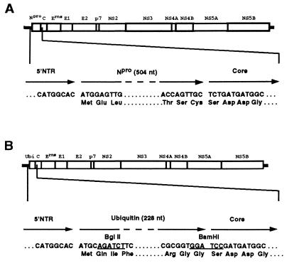
The murine ubiquitin gene was released from plasmid pTM3/HCV/Ubi-NS5B (kindly provided by C. Rice, Washington University School of Medicine, St. Louis, Mo.) by BglII-to-BamHI cleavage. This fragment comprising the complete ubiquitin gene (228 nucleotides [nt]) except the 7 5′-terminal nt including the AUG start codon was subcloned in a pBluescriptIISK(+)-derived plasmid carrying a BglII site to obtain pBS-Ubi. To engineer the ubiquitin gene into the CSFV Alfort/187 cDNA clone pA187-1, the respective viral DNA sequences were inserted upstream and downstream of the ubiquitin sequence in pBS-Ubi. Briefly, from plasmid pAT7G1 (13), structurally corresponding to pA187-1 but containing only the 5′-terminal 778 nt of the genome, a PCR fragment was synthesized by using a sense primer located upstream of the viral sequence in the backbone vector and antisense primer NTR-R2 (5′GAAGATCTGCATGTGCCATGTACAGCAGAG3′), which comprises the last 18 nt of the 5′ nontranslated region (5′ NTR) of the viral genome and the start codon as well as a BglII site (underlined) to allow in-frame fusion to the ubiquitin sequence. The PCR product was digested with BglII endonuclease, which cleaves 20 nt upstream of the viral cDNA insert in pAT7G1 as well as within the antisense primer NTR-R2, and the resulting BglII fragment was inserted into the respective site of pBS-Ubi to obtain pBS-NTR-Ubi. Plasmid pAT7G1P-4 (13) was used as a substrate to synthesize a PCR fragment for insertion downstream of the ubiquitin gene in pBS-NTR-Ubi. The sense primer P14-L2 (5′CGGGATCCGATGATGGCGCAAGTGGAAG3′) corresponds to the 5′-terminal sequence of the nucleocapsid gene starting with the codon TCC (in boldface type) and contains a BamHI site (underlined) for fusion to the ubiquitin gene. The antisense primer (MR1) corresponds to a sequence stretch around nt 2570 of the viral genome within the gene encoding envelope protein E2 and contains a BamHI site. The 1.7-kb PCR fragment was cleaved with BamHI and inserted into the respective site of pBS-NTR-Ubi. From the resulting plasmid, pBS-NTR-Ubi-C, the EagI-to-SpeI (nt 82 and 2420 of the Alfort/187 genome, respectively) fragment was released and used to replace the respective sequences in plasmid pA187-5′. This plasmid contains nt 1 to 6437 of the viral cDNA and was obtained by deletion of the viral sequences located downstream of the unique BamHI site at nt 6437 in plasmid pA187-1. Finally, the EagI (nt 82)-to-KpnI (nt 4453) fragment was released from the latter construct and used to replace the corresponding sequences in the full-length clone pA187-1 to obtain pA187-Ubi (Fig. 1).
FIG. 1.
Diagram of the wild-type CSFV vA187 genome (A) and of the vA187-Ubi genome (B). The 5′-terminal nontranslated region (5′NTR) and the coding region (boxed) of the genome are shown. Sequences surrounding the ends of the Npro (A) and the ubiquitin gene (B) are given as cDNA. Restriction sites related to the construction of vA187-Ubi are underlined.
Generation of infectious vA187-Ubi virus.
Linearization of pA187-Ubi DNA with SrfI, in vitro RNA transcription, and lipofection of the RNA into porcine SK-6 cells were performed exactly as described before (13). Two micrograms of the runoff RNA transcripts were transfected into SK-6 cells seeded the day before in six-well dishes at a concentration of 106 cells/well. Forty-eight hours after transfection, the culture medium was collected and the cells were tested for expression of viral envelope protein E2 by an indirect immunoperoxidase assay using monoclonal antibody HC/TC26 as described elsewhere (6). Several cell foci stained positive, indicating that the RNA was infectious. Supernatant was passaged on SK-6 cells and the virus, vA187-Ubi, released by two cycles of freezing and thawing 48 h after infection. Analysis of the viral RNA by reverse transcription (RT)-PCR after one and three virus passages confirmed the presence of the ubiquitin gene in the viral genome (not shown).
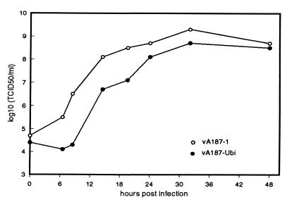
The growth kinetics of vA187-Ubi (passage-2 virus) and of the parent recombinant virus vA187-1 was compared by parallel infection of SK-6 cultures at a multiplicity of infection of 2. Virus was extracted at various times after infection by freezing and thawing of the cells and was titrated (Fig. 2). Both viruses grew to similar titers, 108.5 and 108.7 50% tissue culture infective doses/ml, respectively, but vA187-Ubi showed a prolonged lag phase compared to vA187-1 (titer, >2 log units lower at 8.5 h after infection).
FIG. 2.
Growth curves of wild-type vA187-1 and vA187-Ubi. SK-6 cells seeded in 25-cm2 flasks (2 × 10⁶ cells/flask) were infected at a multiplicity of infection of 2, either with wild-type vA187-1 or with vA187-Ubi. After 1 h at 37°C, cells were washed and incubated in fresh medium for up to 48 h. At different time points postinfection, the cultures were frozen and thawed twice and the virus contained in the cleared supernatants was titrated on SK-6 cells. TCID50, 50% tissue culture infective dose.
Subgenomic RNA and CPE.
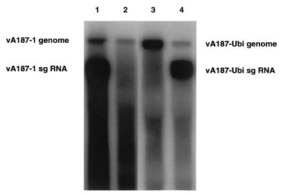
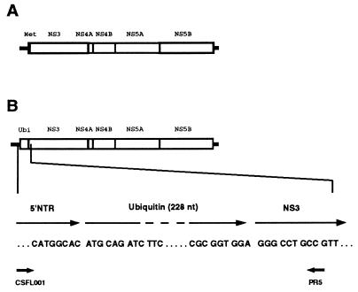
Recently we have described the spontaneous appearance of a defined subgenomic viral RNA in porcine kidney cells persistently infected with CSFV. This subgenomic RNA lacks 4,764 nt of the open reading frame of the viral genome, including the genes coding for all structural as well as for the nonstructural proteins Npro and NS2. Generation of the subgenomic RNA was always accompanied by a cytopathic effect (CPE) and occurred spontaneously. In one culture it was observed as early as after 8 passages of the infected cells, whereas in another culture the CPE occurred only after 94 cell passages. Furthermore, the subgenomic RNA was found to be packaged to give defective particles which induced a CPE when inoculated into noninfected cells (6). To determine if subgenomic viral RNA was also generated in cells persistently infected with a recombinant virus in which the Npro gene was replaced, we infected SK-6 cells with vA187-Ubi at a multiplicity of infection of 0.1 and passaged the cells twice weekly. After two cell passages a CPE was observed. Inoculation of noninfected SK-6 cells with supernatant from the latter culture again resulted in a CPE, indicating the presence of the expected cytopathogenic defective particles in the inoculum. Northern blot analysis (Fig. 3) of RNA obtained by Trizol (Gibco BRL) extraction (6) of the cells which showed a CPE confirmed the presence of a subgenomic viral RNA (lane 4). It had a size (∼8 kb) similar to that of subgenomic RNA derived from cells infected with cp vA187-1 (lane 1). To further characterize the subgenomic RNA, we performed RT-PCR using primers CSFL001 and PR5, corresponding to nt 1 to 21 and nt 5596 to 5577 of the Alfort/187 genome, respectively (Fig. 4). These primers were designed to obtain a PCR product containing the sequences flanking the expected deletion of the subgenomic RNA. A PCR fragment of ∼1 kb was obtained and sequenced directly (not shown). The sequence obtained across the deletion of the subgenomic RNA is shown in Fig. 4. In this RNA exactly the same region of the genome is deleted as in the subgenomic RNA of wild-type CSFV, namely, the complete viral coding sequence located upstream of the presumed NS3 gene. However, the vA187-Ubi-derived subgenomic RNA maintained the ubiquitin gene and consequently was approximately 200 bp longer than the former subgenomic RNA (compare lanes 1 and 4 in Fig. 3). We observed the generation of the same cp subgenomic RNA of vA187-Ubi at a second occasion. This occurred after seven virus passages during routine virus passage in SK-6 cells (not shown).
FIG. 3.
Northern blot analysis of vA187-Ubi RNA. RNA was extracted from SK6 cells infected either with ncp (lane 3) or cp (lane 4) vA187-Ubi. Virus-specific genomic and subgenomic RNA of positive polarity was detected by hybridization with a 32P-labelled riboprobe (6). RNAs extracted from SK6 cells freshly infected either with ncp (lane 2) or cp (lane 1) vA187-1 are shown as references. The sizes of the respective RNAs as deduced from our sequencing data (13) are 12,298 nt for wild-type genomic RNA (lanes 1 and 2), 7,534 nt for wild-type subgenomic RNA (lane 1), 12,022 nt for vA187-Ubi genomic RNA (lanes 3 and 4), and 7,762 nt for vA187-Ubi subgenomic RNA (lane 4).
FIG. 4.
Structure of the vA187-Ubi subgenomic RNA. The deletion of the subgenomic RNA was determined by RT-PCR amplification of RNA extracted from persistently infected cells which showed a CPE when primers PR5 and CSFL001 (indicated) were used followed by direct sequencing of the PCR fragment. The vA187-Ubi subgenomic RNA differs from the corresponding RNA derived from vA187-1 (A) only by the presence of the ubiquitin sequence (B).
Discussion.
The data presented here show that the Npro gene of CSFV can be replaced by the murine ubiquitin gene. The mutant virus vA187-Ubi proved to replicate in porcine kidney SK-6 cells to titers similar to those of the parent virus vA187-1. The only difference observed was a slight delay in the growth kinetics, which was most pronounced between 6 and 12 h after infection (Fig. 2). This could be due to different kinetics of the cleavage reaction. Whereas C-terminal autocatalytic cleavage of Npro is expected to occur cotranslationally, removal of the ubiquitin moiety from the polyprotein by cellular UCH might occur in a more delayed fashion. Our findings suggest that at least in cell culture the Npro gene product has no essential function besides autocatalytic cleavage at its C terminus to generate the N terminus of the viral nucleocapsid protein. An authentic N terminus of C could be crucial for virus replication, as we did not succeed in obtaining a viable virus after deletion in the viral RNA of the complete Npro gene except six nucleotides (AUGGGG) which added a methionine and a glycine residue to the amino terminus of C (data not shown). Whether Npro has any additional role in the CSFV life cycle or in the pathogenesis of classical swine fever remains to be investigated. Preliminary results obtained after intranasal infection of pigs suggest that vA187-Ubi is completely avirulent compared to parent vA187-1, which is a moderately virulent virus. Serum anti-CSFV antibodies were detected 22 days after infection with vA187-Ubi, indicating replication of the virus in the host animal. In contrast, pigs infected with wild type CSFV strain Alfort/187 regularly become seropositive between 10 and 14 days postinfection. These findings indicate that vA187-Ubi could be useful for further studies towards the development of a live viral vaccine. Interestingly, Brown and coworkers (1) have recently reported a leaderless foot-and-mouth disease virus which is avirulent yet induces an immune response in cattle. The fact that the ubiquitin gene could be inserted immediately downstream of the authentic AUG of the viral large open reading frame indicates that the internal ribosome entry site of CSFV (10) does not extend beyond the initiation codon. Further support for this is provided by the subgenomic RNA of CSFV, which is associated with cytopathogenicity of the respective virus stock (4, 6). This RNA seems to be translated very efficiently although it lacks the 4,764 nt following the AUG.
Meyers and Thiel (4) have described three independent cp CSFV isolates for which in addition to genomic RNA a subgenomic RNA carrying the same internal deletion was identified in infected cells. On several occasions we have observed the spontaneous generation of the identical subgenomic RNA in SK-6 cells persistently infected with CSFV strain Alfort/187 at the time when a CPE occurred (6). In 1 out of 10 cultures analyzed it was observed after as few as eight cell passages whereas three cultures did not experience a CPE even when passaged 100 times. Thus, in this experimental setup, generation of cp subgenomic RNA seems to be not only a random but also a rare event. Furthermore, we and others (4) have never detected subgenomic RNA and/or a CPE in cells acutely infected with naturally occurring CSFV strains or isolates. Interestingly, with the mutant virus vA187-Ubi we have observed a CPE and at the same time the generation of subgenomic RNA after seven virus passages and in infected SK-6 cells after as few as two cell passages. The subgenomic RNA had the same structure as the CSFV subgenomic RNA described before, but in both cases it contained the ubiquitin gene. Thus, the 3′ (but not the 5′) recombination site is the same as in all subgenomic RNAs of pestiviruses described so far (5, 6, 16), supporting the hypothesis that the ability of pestiviruses to exhibit a cp phenotype is connected with recombination at this specific site (5).
Acknowledgments
This work was supported by the Swiss National Science Foundation (grant 31-46933.96) and the Swiss Federal Veterinary Office.
We thank Christian Griot for continuous support and Christian Mittelholzer and Peter Stettler for helpful discussions. We acknowledge the technical assistance of M. Gerber, A. Bosshart, and S. Bossy.
REFERENCES
- 1.Brown C C, Piccone M E, Mason P W, McKenna T S-C, Grubman M J. Pathogenesis of wild-type and leaderless foot-and-mouth disease virus in cattle. J Virol. 1996;70:5638–5641. doi: 10.1128/jvi.70.8.5638-5641.1996. [DOI] [PMC free article] [PubMed] [Google Scholar]
- 2.Devaney M A, Vakharia V N, Lloyd R E, Ehrenfeld E, Grubman M J. Leader protein of foot-and-mouth disease virus is required for cleavage of the p220 component of the cap-binding protein complex. J Virol. 1988;62:4407–4409. doi: 10.1128/jvi.62.11.4407-4409.1988. [DOI] [PMC free article] [PubMed] [Google Scholar]
- 3.Lemm J A, Rümenapf T, Strauss E G, Strauss J H, Rice C M. Polypeptide requirements for assembly of functional Sindbis virus replication complexes: a model for the temporal regulation of minus- and plus-strand RNA synthesis. EMBO J. 1994;13:2925–2934. doi: 10.1002/j.1460-2075.1994.tb06587.x. [DOI] [PMC free article] [PubMed] [Google Scholar]
- 4.Meyers G, Thiel H-J. Cytopathogenicity of classical swine fever virus caused by defective interfering particles. J Virol. 1995;69:3683–3689. doi: 10.1128/jvi.69.6.3683-3689.1995. [DOI] [PMC free article] [PubMed] [Google Scholar]
- 5.Meyers G, Thiel H-J. Molecular characterization of pestiviruses. Adv Virus Res. 1996;47:53–118. doi: 10.1016/s0065-3527(08)60734-4. [DOI] [PubMed] [Google Scholar]
- 6.Mittelholzer C, Moser C, Tratschin J-D, Hofmann M A. Generation of cytopathogenic subgenomic RNA of classical swine fever virus in persistently infected porcine cell lines. Virus Res. 1997;51:125–137. doi: 10.1016/S0168-1702(97)00081-6. [DOI] [PMC free article] [PubMed] [Google Scholar]
- 7.Piccone M E, Zellner M, Kumosinski T F, Mason P W, Grubman M J. Identification of the active-site residues of the L proteinase of foot-and-mouth disease virus. J Virol. 1995;69:4950–4956. doi: 10.1128/jvi.69.8.4950-4956.1995. [DOI] [PMC free article] [PubMed] [Google Scholar]
- 8.Piccone M E, Rieder E, Mason P W, Grubman M J. The foot-and-mouth disease virus leader proteinase gene is not required for viral replication. J Virol. 1995;69:5376–5382. doi: 10.1128/jvi.69.9.5376-5382.1995. [DOI] [PMC free article] [PubMed] [Google Scholar]
- 9.Pickart C M, Rose I A. Ubiquitin carboxyl-terminal hydrolase acts on ubiquitin carboxyl-terminal amides. J Biol Chem. 1985;260:7903–7910. [PubMed] [Google Scholar]
- 10.Poole T L, Wang C Y, Popp R A, Potgieter L N D, Siddiqui A, Marc S. Pestivirus translation initiation occurs by internal ribosome entry. Virology. 1995;206:750–754. doi: 10.1016/s0042-6822(95)80003-4. [DOI] [PubMed] [Google Scholar]
- 11.Rice C M. Flaviviridae: the viruses and their replication. In: Fields B N, Knipe D M, Howley P M, editors. Fundamental virology. New York, N.Y: Raven Press, Ltd.; 1996. pp. 1059–1073. [Google Scholar]
- 12.Rueckert R R. Picornaviridae: the viruses and their replication. In: Fields B N, Knipe D M, Howley P M, editors. Fundamental virology. New York, N.Y: Raven Press, Ltd.; 1996. pp. 609–654. [Google Scholar]
- 13.Ruggli N, Tratschin J-D, Mittelholzer C, Hofmann M A. Nucleotide sequence of classical swine fever virus strain Alfort/187 and transcription of infectious RNA from stably cloned full-length cDNA. J Virol. 1996;70:3478–3487. doi: 10.1128/jvi.70.6.3478-3487.1996. [DOI] [PMC free article] [PubMed] [Google Scholar]
- 14.Rümenapf T, Stark R, Heimann M, Thiel H-J. N-terminal protease of pestiviruses: identification of putative catalytic residues by site-directed mutagenesis. J Virol. 1998;72:2544–2547. doi: 10.1128/jvi.72.3.2544-2547.1998. [DOI] [PMC free article] [PubMed] [Google Scholar]
- 15.Stark R, Meyers G, Rümenapf T, Thiel H-J. Processing of pestivirus polyprotein: cleavage site between autoprotease and nucleocapsid protein of classical swine fever virus. J Virol. 1993;67:7088–7095. doi: 10.1128/jvi.67.12.7088-7095.1993. [DOI] [PMC free article] [PubMed] [Google Scholar]
- 16.Tautz N, Thiel H-J, Dubovi E J, Meyers G. Pathogenesis of mucosal disease: a cytopathogenic pestivirus generated by an internal deletion. J Virol. 1994;68:3289–3297. doi: 10.1128/jvi.68.5.3289-3297.1994. [DOI] [PMC free article] [PubMed] [Google Scholar]
- 17.Wengler G, Bradley D W, Collett M S, Heinz F X, Schlesinger R W, Strauss J H. Family flaviviridae. In: Murphy F A, Fauquet C M, Bishop D H L, Ghabrial S A, Jarvis A W, Martelli G P, Mayo M A, Summers M D, editors. Classification and nomenclature of viruses. Berlin, Germany: Springer-Verlag; 1995. pp. 415–427. [Google Scholar]


