



Abstract
Background:
Allele-specific KRAS inhibitors are an emerging class of cancer therapies. KRAS-mutant (KRASMUT) non-small-cell lung cancers (NSCLCs) exhibit heterogeneous outcomes, driven by differences in underlying biology shaped by co-mutations. In contrast to KRASG12C NSCLC, KRASG12D NSCLC is associated with low/never-smoking status and is largely uncharacterized.
Patients and methods:
Clinicopathologic and genomic information were collected from patients with NSCLCs harboring a KRAS mutation at the Dana-Farber Cancer Institute (DFCI), Memorial Sloan Kettering Cancer Center, MD Anderson Cancer Center, and Imperial College of London. Multiplexed immunofluorescence for CK7, programmed cell death protein 1 (PD-1), programmed death-ligand 1 (PD-L1), Foxp3, and CD8 was carried out on a subset of samples with available tissue at the DFCI. Clinical outcomes to PD-(L)1 inhibition ± chemotherapy were analyzed according to KRAS mutation subtype.
Results:
Of 2327 patients with KRAS-mutated (KRASMUT) NSCLC, 15% (n = 354) harbored KRASG12D. Compared to KRASnon-G12D NSCLC, KRASG12D NSCLC had a lower pack-year (py) smoking history (median 22.5 py versus 30.0 py, P < 0.0001) and was enriched in never smokers (22% versus 5%, P < 0.0001). KRASG12D had lower PD-L1 tumor proportion score (TPS) (median 1% versus 5%, P < 0.01) and lower tumor mutation burden (TMB) compared to KRASnon-G12D (median 8.4 versus 9.9 mt/Mb, P < 0.0001). Of the samples which underwent multiplexed immunofluorescence, KRASG12D had lower intratumoral and total CD8+PD1+ T cells (P < 0.05). Among 850 patients with advanced KRASMUT NSCLC who received PD-(L)1-based therapies, KRASG12D was associated with a worse objective response rate (ORR) (15.8% versus 28.4%, P = 0.03), progression-free survival (PFS) [hazard ratio (HR) 1.51, 95% confidence interval (CI) 1.45–2.00, P = 0.003], and overall survival (OS; HR 1.45, 1.05–1.99, P = 0.02) to PD-(L)1 inhibition alone but not to chemo-immunotherapy combinations [ORR 30.6% versus 35.7%, P = 0.51; PFS HR 1.28 (95%CI 0.92–1.77), P = 0.13; OS HR 1.36 (95%CI 0.95–1.96), P = 0.09] compared to KRASnon-G12D.
Conclusions:
KRASG12D lung cancers harbor distinct clinical, genomic, and immunologic features compared to other KRAS-mutated lung cancers and worse outcomes to PD-(L)1 blockade. Drug development for KRASG12D lung cancers will have to take these differences into account.
Keywords: KRAS, G12D, PD-(L)1 blockade, NSCLC
INTRODUCTION
Building upon decades of work to target mutant KRAS,1 recently developed potent KRASG12C inhibitors have brought clinical benefit to patients with non-small-cell lung cancer (NSCLC).2,3 The registrational trial leading to regulatory approval of the direct KRASG12C inhibitor sotorasib in patients with NSCLC showed an objective response rate (ORR) of 36% and a median progression-free survival (PFS) of 6.8 months. The ability to target this ‘undruggable’ oncogene has ushered in a new era of mutant KRAS drug development. Several allele-specific KRAS inhibitors are currently under investigation for patients with KRASnon-G12C cancers, in particular, those targeting KRASG12D.4
While KRAS-mutated NSCLC is often considered as a singular subgroup in research studies, the biology of individual oncogenic KRAS drivers is context dependent.5 In isogenic cell line models, murine models, and retrospective clinical cohorts of patients with KRAS-mutant (KRASMUT) NSCLC, KRAS driver subtypes are thought to differ in downstream signaling dependencies,6 transcriptional phenotypes,7 and clinical features.8,9
KRASG12D is the most common mutant oncogenic form of KRAS in human cancers and represents 4% of NSCLC.10–12 This alteration is associated with a low/never-smoking status.13 The underlying missense mutation leading to KRASG12D is a transition mutation (c.35Guanine>Adenine, purine>purine), as opposed to a transversion mutation (purine>pyrimidine), the predominant form of nucleotide substitutions observed with tobacco exposure.14 However, the genomic, transcriptomic, and immunophenotypic profile of KRASG12D NSCLC has not been characterized. Similarly, whether KRASG12D NSCLC is associated with different outcomes to standard-of-care PD-(L)1 blockade therapy is unknown.
We carried out a comprehensive analysis of the clinicopathologic, genomic, and immunophenotypic correlates of KRASG12D NSCLC, and explored the efficacy of PD-(L)1 blockade therapy in patients with advanced KRASG12D NSCLC. Characterizing the unique features of KRASG12D, the dominant KRAS subtype in human cancers, is critical to personalized therapy development in the era of direct KRAS inhibition.
PATIENTS AND METHODS
Study population
Overview.
Our study population included patient-level data from KRAS-mutant NSCLC from four institutions: clinical, genomic, immunophenotypic, and outcome to PD-1 blockade from the Dana-Farber Cancer Institute (DFCI; n = 1982); clinical and outcome data from Memorial Sloan Kettering Cancer Center (MSKCC; n = 232); clinical data and outcome data from MD Anderson Cancer Center (MDACC; n = 58); and clinical data and outcome data from Imperial College NHS Trust (n = 55) (Figure 1A). Patients were identified via manual chart review by medical oncologists or trained abstractors at each participating center. Tumor histology was reviewed by a dedicated thoracic pathologist and standard immunohistochemistry [thyroid transcription factor (TTF)-1, p40, p63, napsin, cytokeratin 7] was carried out to confirm the epithelial lung origin of each tumor, as appropriate. Cancers of uncertain origin (e.g. TTF-1 and napsin negative) were further adjudicated as NSCLC by reviewing scan reports and clinic visit notes. Carcinomas of extrathoracic origin were excluded from this study.
Figure 1. Study cohorts and baseline clinicopathologic features of KRAS G12D and KRAS non-G12D NSCLC.
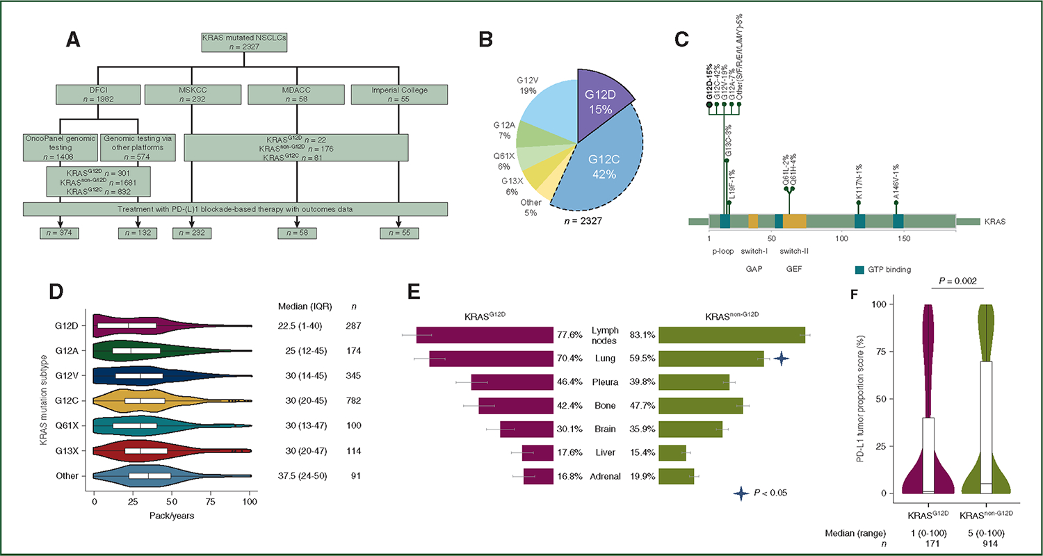
(A) The cohorts of patients included in this study. (B) The distribution of the most common KRAS mutations identified among NSCLCs at the DFCI, MSKCC, MDACC, and Imperial College (n = 2327). (C) Lollipop plot of the most common KRAS mutations identified at participating institutions (n = 2327). (D) The distribution of pack-years across the most common KRAS subtypes. (E) Distribution of metastatic sites in stage IV KRASG12D (n = 125) and KRASnon-G12D (n = 781) NSCLCs in the DFCI cohort. (F) PD-L1 TPS in KRASG12D NSCLC and KRASnon-G12D NSCLC.
DFCI, Dana-Farber Cancer Institute; MDACC, MD Anderson Cancer Center; MSKCC, Memorial Sloan Kettering Cancer Center; NSCLC, non-small-cell lung cancer; PD-L1, programmed death-ligand 1; TPS, tumor proportion score.
We also examined transcriptomic data from lung adenocarcinoma (LUAD) samples (n = 506) from the Cancer Genome Atlas (TCGA) (portal.gdc.cancer.gov).
Site-specific cohort overview
Dana-Farber Cancer Institute.
Consecutive patients at the DFCI who consented to institutional review board-approved protocols DF/HCC 02–180, 11–104, 13–364, and/or 17–000, and whose tumors underwent KRAS and EGFR driver mutation assessment from September 2013 to December 2021 were included in this study. KRAS and EGFR status was determined using one of the following methods in a total of 1982 patients: the in-house panel next-generation sequencing (NGS) assay OncoPanel15 which interrogates for 277 (v1, 4/2013–07/2014), 302 (v2, 07/2014–09/2016), and 447 (v3, 09/2016-ongoing) cancer-associated genes (n = 1408); a commercial plasma sequencing assay (Guardant360 ctDNA or FoundationOne Liquid CDx); or a commercial tissue sequencing assay (e.g. FoundationOne CDx) in the remaining cases. Baseline clinical and pathologic features to PD-(L)1 blockade-based therapies were abstracted using automated and manual review.
Imperial College London.
Consecutive patients who received PD-(L)1 blockade therapy (n = 55) at the Imperial College London from October 2016 to September 2021 and whose tumor underwent KRAS and EGFR assessment using a custom-built QIAseq-targeted DNA panel based on multiplex PCR-targeted enrichment technology were included. All patients consented to institutional review board-approved protocols.
MD Anderson Cancer Center.
Consecutive patients who received PD-(L)1 blockade therapy (n = 58) at the MD Anderson Cancer Center from October 2013 to September 2019 whose tumors underwent KRAS and EGFR assessment with the Oncomine Comprehensive Assay v3 were included. This panel NGS assay interrogates single-nucleotide variants (SNVs), copy number variants (CNVs), gene fusions, and insertion/deletion mutations (indels) from 161 unique cancer-associated genes. All patients consented to institutional review board-approved protocols.
Memorial Sloan Kettering Cancer Center.
Clinicopathologic, genomic, and clinical outcomes data from patients who received PD-(L)1 blockade therapy (n = 232) from October 2015 to December 2021 at the Memorial Sloan Kettering Cancer Center and whose tumor underwent KRAS and EGFR assessment with the MSK-IMPACT NGS platform were included. This assay detects SNVs, CNVs, gene fusions, and indels from 341 (v1), 410 (v2), and 468 (v3) unique cancer-associated genes.16
PD-L1 tumor proportion score assessment
The PD-L1 tumor proportion score (TPS) by immunohistochemistry was scored by trained pathologists using validated monoclonal anti-PD-L1 antibodies: E1L3N (Cell Signaling Technology, Danvers, MA), 22C3 (Dako North America Inc., Carpinteria, CA), and 28–8 (Epitomics Inc., Burlingame, CA).
Dana-Farber Cancer Institute site-specific data
Tumor genomic profiling and tumor mutational burden assessment.
Targeted exome NGS (Profile) was carried out using the validated OncoPanel assay in the Center for Cancer Genome Discovery at the DFCI for 277 (POPv1), 302 (POPv2), or 447 (POPv3) cancer-associated genes. Briefly, tumor DNA was prepared as previously published, hybridized to custom RNA bait sets (Agilent SureSelect™, San Diego, CA) and sequenced using Illumina HiSeq 2500 with 2 × 100 paired-end reads. Sequence reads were aligned to reference sequence b37 edition from the Human Genome Reference Consortium, using bwa, and further processed via Picard (version 1.90, http://broadinstitute.github.io/picard/) to remove duplicates and Genome Analysis Toolkit (GATK, version 1.6–5-g557da77) to carry out localized realignment around indel sites. SNVs were called using MuTect v1.1.46, and insertions and deletions were called using GATK Indelocator. Variants were filtered to remove potential germline variants as previously published and annotated using Oncotractor. To remove additional germline noise, variants that were annotated as benign/likely benign in ClinVar or were present at a population maximum allele frequency of 0.1% were excluded. Variants were retained in either case if they were annotated as confirmed somatic in at least two samples in COSMIC. CNVs and structural variants were called using the internally developed algorithms RobustCNV and BreaKmer5. For each gene, the absolute copy number (ACN) was estimated based on the tumor purity (P) and the weighted average of segmented log2 ratios across the gene (l) using the formula:
Tumor mutational burden (TMB), defined as the number of somatic mutations per megabase (Mb) of genome examined (including coding, base substitution, and indels), was determined using OncoPanel, as previously described.15
Co-occurrence and mutual exclusivity analysis
Co-occurrence and mutual exclusivity between groups were assessed among NSCLC samples which underwent comprehensive genomic profiling at the DFCI. Fisher’s exact test P values and conditional odds ratios were used to assess co-occurrence and mutual exclusivity for genes with at least 2% frequency in the relevant groups. Positive odds ratios represented the tendency to co-occur and negative odds ratios represented the tendency to mutual exclusivity. Correction for multiple comparisons was carried out using the Benjamini–Hochberg procedure using the qvalue package in R. Gene mutations included in this analysis included nonsense mutations, frameshift and in-frame insertion-deletion mutations (indels), splice site mutations, and missense mutations defined as oncogenic in the OncoKB repository. Missense mutations of unknown significance were filtered out.
Multiplexed immunofluorescence (ImmunoProfile)
Multiplexed immunofluorescence (mIF) was carried out on 140 samples from the DFCI by staining 5-μm formalin-fixed, paraffin-embedded whole tissue sections with standard, primary antibodies sequentially and paired with a unique fluorochrome followed by staining with nuclear counterstain/4′,6-diamidino-2-phenylindole (DAPI).17 All samples were stained for PD-L1 (clone E1L3N), PD-1 (clone EPR4877 [2]), CD8 (clone 4B11), FOXP3 (clone D608R), and cytokeratin (clone AE1/AE3). Each sample had a single slide stained and scanned at 20× resolution by a Vectra Polaris imaging platform. Regions of interest (ROIs) were defined for each image, and only these regions were used for quantitative image analysis. Within each ROI, InForm Image Analysis software (Perkin Elmer, Marlborough, MA/Akoya) was run to phenotype and score cells based on biomarker expression. A custom script quantified the number/percentage of cells which were positive for relevant biomarkers in specific tissue regions. Each ROI was divided into one or more of these defined regions: intra-tumoral (IT), which was defined as the region of the slide consisting of tumor beyond the tumor–stroma interface (TSI); TSI, which was defined as the region within 40 μm to either side of the defined border between tumor and stroma; and total (IT + TSI). Cell count was calculated per ROI and averaged (unweighted) across ROIs, reported as count per millimeter ± squared standard error. Tumor cells were defined as cells with DAPI + CK (AE1/AE3) staining. Non-tumor (inflammatory) cells were defined as cells without CK staining. Statistical significance of differential cell type enrichment between groups was estimated with the Wilcoxon rank sum test.
TCGA cohort-specific analysis
Gene expression analysis from the TCGA.
LUAD TCGA gene expression and somatic mutation data were downloaded from Firehose (TCGA Firehose Legacy version) and cBio-Portal (cbioportal.org), respectively. The lung squamous TCGA cohort was not explored, given the lack of KRAS mutations (n = 1/511, 0.2%). The RSEM V2 values were used to represent gene expression, and genes with counts < 10 were filtered out. Gene expression profiles were analyzed according to KRAS mutation status. Median expression within each group was used to estimate expression fold-change (FC) to minimize the possible impact of outlier samples. Gene differential expression analyses across KRAS allele subgroups were conducted using the R package DESeq2. P values were corrected for multiple hypothesis testing via false discovery rate (FDR) adjustment using the Benjamini–Hochberg procedure. FC threshold of an absolute value > 1.5 and FDR adjusted P value thresholds < 0.1 were utilized to identify differentially expressed genes. Pathway enrichment analyses were conducted separately for up- and down-regulated genes using Molecular Signatures Database (MSigDB).
Statistical analysis for all cohorts
Categorical and continuous variables were summarized descriptively using percentages and medians. The Wilcoxon rank sum test and Kruskal–Wallis test were used to examine differences between continuous variables, and Fisher’s exact test and chi-square test were used to compare associations between categorical variables, when appropriate. Kaplane–Meier methodology was used to estimate event-time distributions, and the Greenwood formula was used to estimate the standard errors of the estimates. Log-rank tests were used to test for differences in event-time distributions, and Cox proportional hazards models were fitted to obtain estimates of hazard ratios in univariable and multivariable models. ORR and PFS were determined using RECIST version 1.1 by a dedicated thoracic radiologist.18 PFS was defined as the time from the start of immunotherapy to the date of disease progression or death, whichever occurred first. Patients who were alive without disease progression were censored on the date of their last scan. Overall survival (OS) was defined as the time from the start of immunotherapy to death. Patients who were still alive on the datalockdate were censored at the date of last contact.
As PD-L1 TPS is one of the most relevant predictors for PD-(L)1 blockade based therapies efficacy, we used a multiple imputation approach using the R package MICE to address the potential selection bias arising from PD-L1 TPS missingness. Multivariable analysis results were pooled based on five repeated complete imputed datasets. All P values are two-sided with statistical significance defined as P < 0.05. Multiple comparison correction was carried out using the Benjamini–Hochberg method using the qvalue package in R. All statistical analyses were carried out using R version 3.6.3 (R Foundation for Statistical Computing, Vienna, Austria).
RESULTS
Characteristics of the entire cohort
We identified a total of 2327 patients with NSCLC harboring oncogenic KRAS mutations (Figure 1A). Overall, the median age was 66 years (range: 20–98 years), 65% (n = 1502/2327) were women, and 99% (n = 2293/2327) had non-squamous histology. Among those with known pack-year smoking data (n = 1993/2327), 15% had a <10 pack-year smoking history, and 55% had a ≥30 pack-year smoking history. Median PD-L1 TPS was 5% (range: 0%–100%) among 1175 assessable patients, and median TMB was 9.7 mutations/megabase (range: 1.2–56.3 mt/Mb, among 1426 assessable patients at DFCI). Baseline characteristics of patients included in this study are summarized in Supplementary Table S1, available at https://doi.org/10.1016/j.annonc.2022.07.005.
Baseline characteristics of patients with KRASG12D non-small-cell lung cancers
The KRASG12D NSCLC subtype comprised 15% of all KRAS NSCLC (n = 354/2327). Other common KRAS drivers (KRASnon-G12D) included: G12C (n = 969/2327, 42%), G12V (n = 438/2327, 19%), G12A (n = 175/2327, 7%), G13X (n = 144/2327, 6%), and Q61X (n = 130/2327, 6%) (Figure 1B and C). A similar distribution in KRAS mutation was reported in the subset of patients who received PD-(L)1-based therapies (Supplementary Figure S1A, available at https://doi.org/10.1016/j.annonc.2022.07.005). In comparing clinicopathologic features of KRASG12D with KRASnon-G12D, patients with KRASG12D were significantly more likely to be never smokers (22% versus 5%, P < 0.0001) and have less tobacco use (median 22.5 versus 30.0 pack-years, P < 0.0001) (Table 1, Figure 1D). In this largely US-based cohort, we also found differences in self-reported race, with Asians more likely to have a KRASG12D mutation compared to KRASnon-G12D (4% versus 1%). The distribution of metastatic sites at diagnosis was largely similar between KRASG12D and KRASnon-G12D NSCLC (n = 906/1160 stage IV cases with known sites of metastasis), except for a higher proportion of lung metastasis in KRASG12D NSCLC (P = 0.02, Figure 1E). Additionally, compared to KRASnon-G12D NSCLCs, KRASG12D was associated with significantly lower median PD-L1 TPS (1% versus 5%, P = 0.002) among 1175 cases which underwent PD-L1 assessment (Figure 1F). Other baseline clinicopathologic characteristics of patients with KRASG12D and KRASnon-G12D NSCLCs are shown in Table 1.
Table 1.
Baseline clinicopathologic characteristics of patients with KRASG12D versus KRASnon-G12D driven non-small-cell lung cancer
| Clinical characteristic, n (%) | KRASG12D n = 354 | KRASnonG12D a n = 1973 | P value |
|---|---|---|---|
|
| |||
| Age, median (range) | 66 (20–92) | 66 (28–98) | 0.53 |
| Sex | 0.76 | ||
| Male | 128 (36.2) | 697 (35.3) | |
| Female | 226 (63.8) | 1276 (64.7) | |
| Cigarette smoking status | <0.0001 | ||
| Current/former | 276 (78.0) | 1870 (95.2) | |
| Never | 78 (22.0) | 94 (4.8) | |
| Not assessed | 9 | ||
| Pack-years, median (range) | 22.5 (0–150) | 30.0 (0–200) | <0.0001 |
| Histology | 0.99 | ||
| Non-squamous | 349 (98.6) | 1944 (98.5) | |
| Squamous | 5 (1.4) | 29 (1.5) | |
| Stage at diagnosis | 0.31 | ||
| I | 85 (24.0) | 418 (21.2) | |
| II | 35 (9.9) | 156 (7.9) | |
| III | 46 (13.0) | 280 (14.2) | |
| IV | 188 (53.1) | 1119 (56.7) | |
| Race, self-reported | 0.006 | ||
| White | 272 (91.9) | 1551 (94.3) | |
| Black | 6 (2.0) | 50 (3.0) | |
| Asian | 11 (3.7) | 20 (1.2) | |
| Other | 7 (2.4) | 23 (1.4) | |
| Not available | 58 | 329 | |
| PD-L1 TPS | 0.006 | ||
| <1% | 80 (46.8) | 346 (34.5) | |
| 1–49% | 49 (28.7) | 324 (32.3) | |
| ≥50% | 42 (24.6) | 334 (33.3) | |
| Not assessed | 183 | 969 | |
| PD-L1 TPS, median (range) | 1 (0–100) | 5 (0–100) | 0.002 |
| TMB, median (range) | 8.4 (1.2–25.1) | 9.9 (1.2–56.3) | <0.0001 |
PD-L1 TPS, programmed death protein ligand 1 tumor proportion score using immunohistochemistry; TMB, tumor mutational burden.
Current/former, active smoking or quit within past year; Never, <100 lifetime cigarettes.
KRASnon-G12D variants include: G12C (49.1%), G12V (22.2%), G12A (8.9%), G13X (7.3%), Q61X (6.6%), and other (5.9%). P values compare KRASG12D and KRASnon-G12D groups.
Given that KRASG12C is the first KRAS-driven lung cancer with an approved direct targeted therapy, we also compared the clinicopathologic features of KRASG12D with KRASG12C and confirmed that patients with KRASG12D were more likely to be never smokers and have lower PD-L1 expression levels (Supplementary Table S2, available at https://doi.org/10.1016/j.annonc.2022.07.005, Supplementary Figure S1B, available at https://doi.org/10.1016/j.annonc.2022.07.005). No differences in the baseline sites of metastatic disease were identified between the two groups (Supplementary Figure S2A, available at https://doi.org/10.1016/j.annonc.2022.07.005). Baseline characteristics of patients with KRASG12D versus KRASG12C NSCLC are summarized in Supplementary Table S2, available at https://doi.org/10.1016/j.annonc.2022.07.005. To validate these findings in an independent cohort, we compared clinical features of 3949 NSCLCs from the AACR GENIE v11.0 registry. Patients with KRASG12D NSCLCs have similar age at diagnosis, sex, and histology compared to those with KRASnon-G12D (Supplementary Table S3, available at https://doi.org/10.1016/j.annonc.2022.07.005). Similar results were observed in comparing KRASG12D and KRASG12C NSCLCs (Supplementary Table S3, available at https://doi.org/10.1016/j.annonc.2022.07.005).
Genomic and transcriptomic features of KRASG12D non-small-cell lung cancers
We next examined whether genomic features of NSCLCs harboringKRASG12D mutations differed from other KRAS NSCLC subtypes. Of the original cohort, 1408 patients (65%) underwent large-panel NGS of tumor samples (OncoPanel), which examined somatic oncogenic mutations and copy number variation (n = 201 KRASG12D, n = 1207 KRASnon-G12D). Of these patients, 30% (418/1408) were stage I, 26% (373/1408) stage II-III, and 44% (617/1408) stage IV. The most common pathogenic mutations in all comers with KRAS-mutated NSCLC included TP53 (39%), STK11 (21%), CDKN2A (11%), ATM (9%), RBM10 (8%), and KEAP1 (7%) (Supplementary Figure S3, available at https://doi.org/10.1016/j.annonc.2022.07.005). The most commonly mutated genes in KRASG12D NSCLCs included TP53 (32%), CDKN2A (15%), STK11 (13%), NKX2–1 (10%), and RBM10 (6%) (Figure 2A).
Figure 2. Genomic profile of KRAS G12D NSCLC.
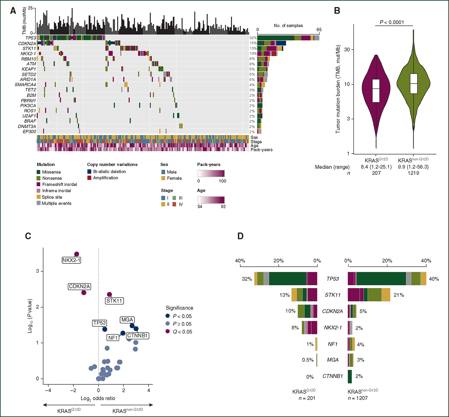
(A) Oncoprint of the top 20 genes altered in KRASG12D NSCLC. (B) Tumor mutational burden (TMB) in KRASG12D and KRASnon-G12D NSCLC. (C) Volcano plot showing oncogenic gene mutations-enriched KRASG12D versus KRASnon-G12D NSCLC. (D) Prevalence of oncogenic mutations significantly enriched in KRASG12D versus KRASnon-G12D NSCLC.
NSCLC, non-small-cell lung cancer.
To determine whether KRASG12D had unique mutational patterns, we next compared the genomic profile of KRASG12D and KRASnon-G12D NSCLC. The median TMB was significantly lower among KRASG12D compared to KRASnon-G12D NSCLC, likely reflective of the higher number of patients with a never/light smoking history among KRASG12D NSCLC (Figure 2B). Compared to KRASnon-G12D, KRASG12D NSCLCs were significantly enriched in mutations affecting NKX2–1 and CDKN2A (Q < 0.05) (Figure 2C). By contrast, loss-of-function mutations in STK11 were more likely to occur in KRASnon-G12D cases compared to KRASG12D cases (Figure 2C and D).
We next explored the transcriptomic profiles of KRASG12D (n = 18) and KRASnon-G12D (n = 124) tumors using TCGA LUAD RNA-seq dataset (54.2%, n = 274 stage I, 40.1%, n = 203 stage II-III, 5.3%, n = 27 stage IV). We identified different gene expression patterns between these two groups (Supplementary Figure S4, available at https://doi.org/10.1016/j.annonc.2022.07.005). Compared to KRASnon-G12D NSCLC, KRASG12D NSCLC showed a markedly different transcriptomic profile characterized by a significant reduction in the expression of genes involved in cellular senescence (q < 0.01), cAMP signaling (q = 0.01), DNA methylation (q < 0.01), and the interleukin-7 pathway (q < 0.01). A list of all differentially expressed pathways is shown in Supplementary Table S4, available at https://doi.org/10.1016/j.annonc.2022.07.005.
Similarly, compared to KRASG12C NSCLCs, KRASG12D NSCLC had significantly lower TMB (Supplementary Figure S2B, available at https://doi.org/10.1016/j.annonc.2022.07.005) and was significantly enriched in NKX2–1 and CDKN2A mutations (Supplementary Figure S2C and D, available at https://doi.org/10.1016/j.annonc.2022.07.005).
Immunophenotype of KRASG12D non-small-cell lung cancer
We next asked whether KRASG12D NSCLCs differed in immunophenotype compared to other KRASMUT NSCLCs, given their clinical and genomic differences. To address this question, we carried out multiplexed immunofluorescence (mIF) examining CD8, PD-1, Foxp3, and PD-L1 on tumor tissue from 140 patients with NSCLC harboring a KRAS mutation and sufficient sample to perform mIF from the DFCI cohort (KRASG12D NSCLC, n = 24; KRASnon-G12D, n = 116). Representative images from KRASG12D NSCLC and KRASnon-G12D (in this case KRASG12C) NSCLC are shown in Figure 3A and B, respectively. We were interested in the comparisons of average populations of tumor and non-tumor (e.g. immune) cells within the following regions from each patient sample: overall (total), within the tumor (intratumor), and within the TSI. TSI was defined as the region with in 40 μm of the border between the tumor and stroma (Supplementary Figure S5, available at https://doi.org/10.1016/j.annonc.2022.07.005). The clinicopathologic characteristics of these patients are included in Supplementary Table S5, available at https://doi.org/10.1016/j.annonc.2022.07.005.
Figure 3. Immunophenotypic characteristics of KRAS G12D NSCLC.
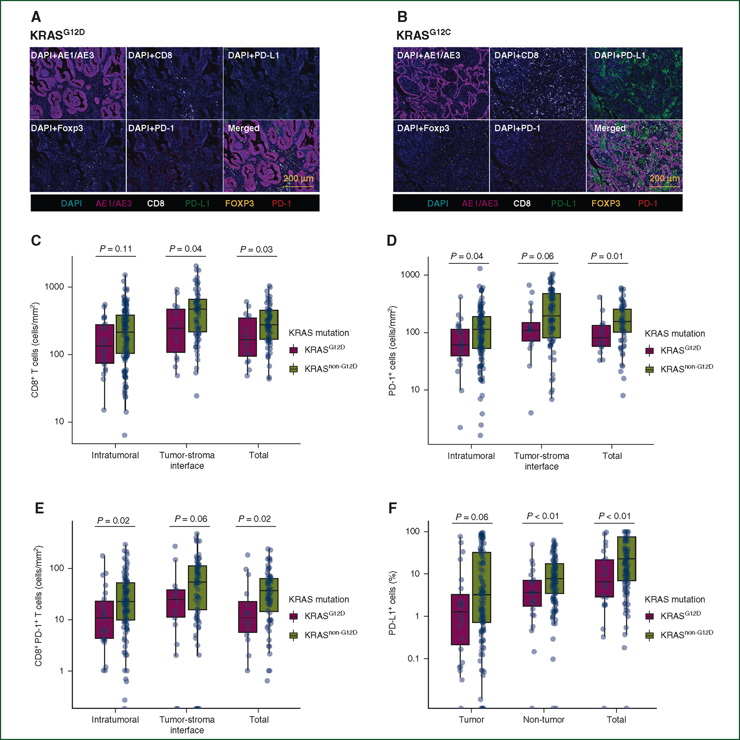
(A, B) Representative multiplex immunofluorescence of a KRASG12D and a KRASnon-G12D (in this case, KRASG12C) NSCLC sample. Each image shown represents 925 μm × 693 μm from a single 20× region of interest. See Methods for additional details. (C) CD8+ T cells (D) PD-1+ cells, and (E) CD8+PD-1+ T cells in KRASG12D NSCLC (N = 24) versus KRASnon-G12D NSCLC (N = 116). (F) Proportion of PD-L1+ tumor, non-tumor, and total cells in NSCLCs with KRASG12D versus KRASnon-G12D mutations. Tumor–stroma interface (TSI) CD8+, PD-1+, and CD8+PD-1+ cells were examined in 16 KRASG12D and 68 KRASnon-G12D samples in which TSI was included in the biopsy.
NSCLC, non-small-cell lung cancer; PD-1, programmed cell death protein 1; PD-L1, programmed death-ligand 1.
Importantly, compared to KRASnon-G12D, KRASG12D NSCLCs had significantly fewer CD8+ T cells at the TSI (median 244 versus 458 cells/mm2, P = 0.04) and in total (median 165 versus 276 cells/mm2, P = 0.03), fewer intratumoral (median 63 versus 111 cells/mm2, P = 0.04) and total PD-1+ cells (median 81 versus 153 cells/mm2, P = 0.01), and fewer intratumoral (median 11 versus 23 cells/mm2, P = 0.02) and total CD8+PD-1+ T cells (median 11 versus 36 cells/mm2, P = 0.02) (Figure3C-E). There was no difference in intratumoral, TSI, and total Foxp3+ cells between these groups (Supplementary Figure S6, available at https://doi.org/10.1016/j.annonc.2022.07.005). When we examined the proportion of PD-L1+ cells in this cohort, KRASG12D was associated with a significantly lower proportion of PD-L1+ non-tumor cells (median 3.4% versus 7.5%, P < 0.01) and PD-L1+ total cells (median 6.3% versus 21.7%, P < 0.01), compared with KRASnon-G12D (Figure 3F).
We next compared immunophenotype of KRASG12D (n = 24) and KRASG12C (n = 60) NSCLCs. Compared to KRASG12C NSCLC, KRASG12D NSCLC had significantly fewer CD8+, PD1+ non-tumor, and CD8+PD1+ T cells at the TSI and in total. In addition, KRASG12D NSCLCs had significantly fewer intratumoral CD8+PD1+ T cells (Supplementary Figure S7A-C, available at https://doi.org/10.1016/j.annonc.2022.07.005). There was no difference in intratumoral, TSI, and total Foxp3+ T cells between these groups (Supplementary Figure S7D, available at https://doi.org/10.1016/j.annonc.2022.07.005). When we examined the proportion of PD-L1+ cells in KRASG12D versus KRASG12C NSCLCs, we confirmed that KRASG12D NSCLC had a significantly lower proportion of PDL1+ tumor, inflammatory, and total cells, compared to KRASG12C NSCLC (Supplementary Figure S7E, available at https://doi.org/10.1016/j.annonc.2022.07.005).
Smoking history defines different subsets of KRASG12D non-small-cell lung cancer
We noted there was a significant proportion of never/light smokers with KRASG12D NSCLC suggesting a binomial distribution of tobacco pack-year exposure and sought to explore this further. We defined never/light smoking history as <10 pack-years (KRASG12D,light-sm, n = 96/354, 27.1%) and heavy smoking history as ≥30 pack-years (KRASG12D,heavy-sm, n = 144/354, 40.7%). No differences in age, sex, and histology were identified between the light and heavy smoking groups. KRASG12D,light-sm NSCLC had significantly lower PD-L1 TPS compared to KRASG12D,heavy-sm NSCLC (median 0% versus 10%, P < 0.001, Supplementary Table S5, available at https://doi.org/10.1016/j.annonc.2022.07.005). We examined the impact of smoking history on clinically relevant PD-L1 expression thresholds of TPS <1%, 1–49%, and ≥50%. KRASG12D,light-sm NSCLCs were significantly more likely to lack PD-L1 expression (63% versus 30%) and less likely to have a PD-L1 TPS ≥50% (11% versus 34%), compared to KRASG12D,heavy-sm (P = 0.001, Figure 4A). KRASG12D,light-sm had a significantly lower TMB compared to KRASG12D,heavy-sm (median 5.7 versus 9.9 mut/Mb, P < 0.0001, Figure 4B). Baseline clinicopathologic features of patients with KRASG12D,light-sm and KRASG12D,heavy-sm are summarized in Supplementary Table S6, available at https://doi.org/10.1016/j.annonc.2022.07.005. The genomic profiles of NSCLCs from patients with KRASG12D,light-sm and KRASG12D,heavy-sm are shown in Supplementary Figure S8, available at https://doi.org/10.1016/j.annonc.2022.07.005. Due to the limited number of patients with KRASG12D,light-sm and KRASG12D,heavy-sm whose tumors underwent comprehensive genomic profiling, we did not perform gene mutation enrichment analysis.
Figure 4. PD-L1 expression and TMB levels according to smoking status among KRAS G12D NSCLCs.
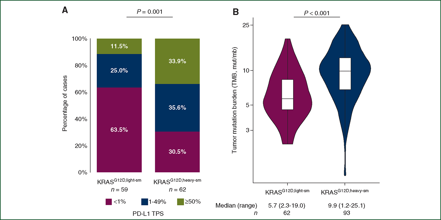
(A) Proportion of NSCLC samples with a PD-L1 TPS of <1%, 1%–49%, and ≥50%, in KRASG12D,light-sm (<10 pack-years smoking) and KRASG12D,heavy-sm (≥30 pack-years smoking) subsets. (B) TMB in KRASG12D,light-sm and KRASG12D,heavy-sm NSCLCs.
NSCLC, non-small-cell lung cancer; PD-L1, programmed death-ligand 1; TMB, tumor mutational burden; TPS, tumor proportion score.
Outcomes to PD-(L)1 blockade-based therapy in KRASG12D non-small-cell lung cancer
Finally, we explored clinical outcomes to PD-(L)1 blockade-based therapy in 850 patients with KRASG12D NSCLC at DFCI, MSKCC, MDACC, and Imperial College London. Clinicopathologic characteristics of patients who received immunotherapies are summarized in Supplementary Table S7, available at https://doi.org/10.1016/j.annonc.2022.07.005. Patients with KRASG12D and KRASnon-G12D were similar in terms of age, sex, histology, line of therapy for PD-(L)1 blockade-based therapy, and treatment received (92%–95% received carboplatin-pemetrexed backbone; Supplementary Table S7, available at https://doi.org/10.1016/j.annonc.2022.07.005). When we examined clinical outcomes to PD-(L)1 monotherapy, we found that patients with KRASG12D NSCLC had significantly lower ORR (15.8% versus 28.4%, P = 0.03), and shorter median PFS [2.1 versus 4.0 months, HR 1.51 (95% confidence interval [CI] 1.45–2.00), P = 0.003] and OS [7.4 versus 16.4 months, HR 1.45 (95%CI 1.05–1.99), P = 0.02] compared to KRASnon-G12D cases (Figure 5A-C). Among patients with KRASG12D NSCLC and a never/light pack-year smoking history (<10 pack-years) (n = 12), the ORR was 0% to PD-(L)1 blockade monotherapy. There was insufficient sample size to compare duration of response stratified by smoking history. By contrast, no significant differences in ORR, PFS, and OS were observed among patients who received chemotherapy combined with PD-(L)1 blockade therapy, according to KRAS mutation status (Figure 5D-F). However, given the small sample size of patients with KRASG12D who received chemotherapy combined with PD-(L)1 blockade therapy, these results should be interpreted with caution. When we comparedKRASG12D versus KRASG12C NSCLCs according to the regimen received, we found that KRASG12D was associated with a significantly lower ORR, mPFS, and mOS to PD-(L)1 monotherapy compared to KRASG12C (Supplementary Figure S9A-C, available at https://doi.org/10.1016/j.annonc.2022.07.005). Although no difference in ORR to chemo-immunotherapy was observed between patients with KRASG12D and KRASG12C NSCLC, mPFS and mOS were significantly shorter among KRASG12D cases (Supplementary Figure S9D-F, available at https://doi.org/10.1016/j.annonc.2022.07.005).
Figure 5. Clinical outcomes to PD-(L)1 ± chemotherapy in patients with KRAS G12D versus KRAS non-G12D NSCLC.
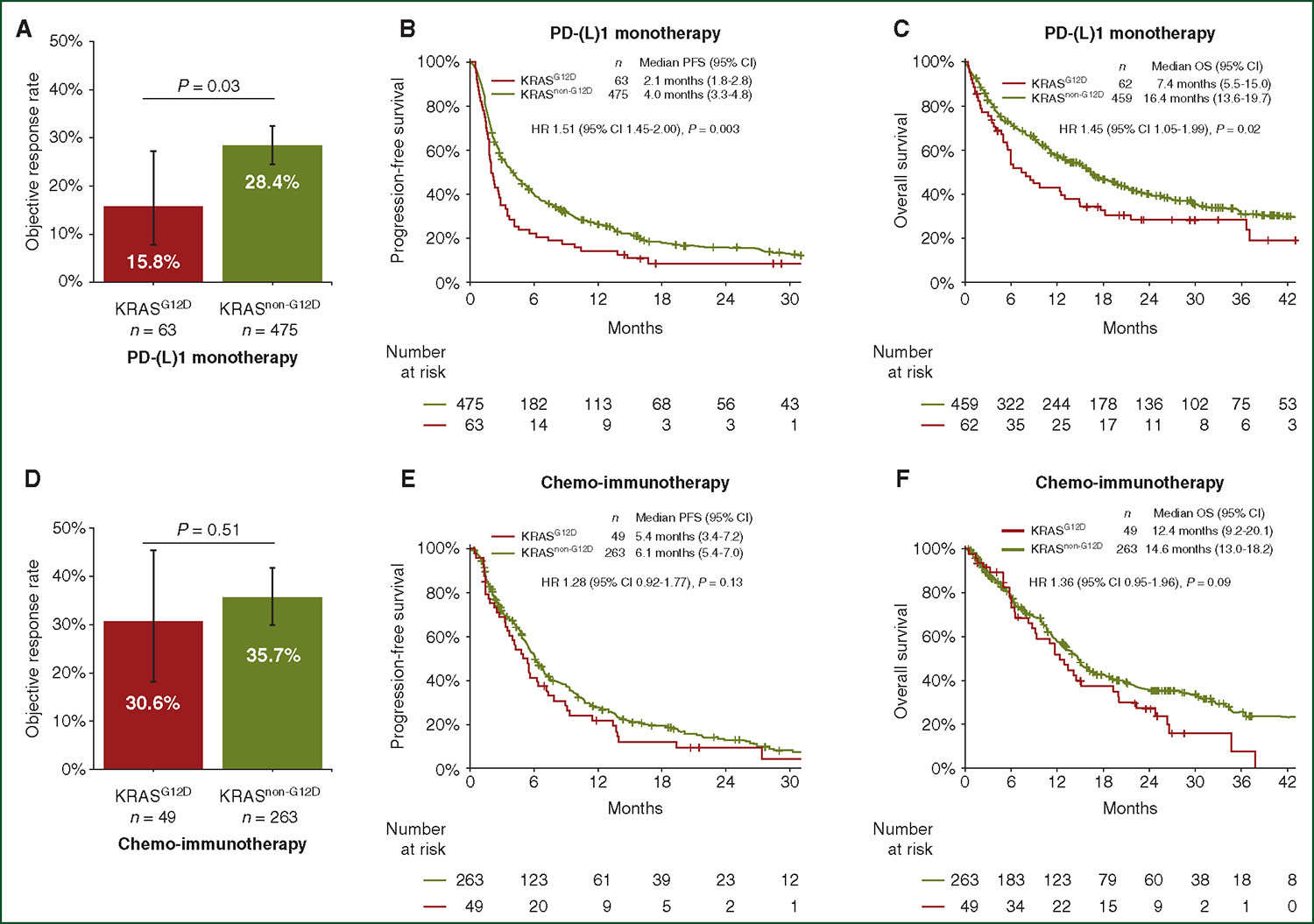
(A) Objective response rate, (B) progression-free survival, and (C) overall survival to PD-(L)1 blockade monotherapy in patients with advanced KRASG12D NSCLC versus KRASnon-G12D NSCLC. (D) Objective response rate, (E) progression-free survival, and (F) overall survival to PD-(L)1 blockade therapy in combination with chemotherapy in advanced KRASG12D NSCLC versus KRASnon-G12D NSCLC. HR, hazard ratio; NSCLC, non-small-cell lung cancer; PD-L1, programmed death-ligand 1; PFS, progression-free survival; OS, overall survival.
After adjusting for TMB, PD-L1 expression, line of therapy, performance status, age, sex, and smoking status, KRASG12D NSCLC retained association with worse PFS [aHR 1.49 (95%CI 1.10–2.02), P < 0.01], and OS [aHR 1.50 (95%CI 1.05–2.06), P = 0.03] in multivariable analysis among patients who received PD-(L)1 monotherapy, compared to KRASnon-G12D (Supplementary Table S8, available at https://doi.org/10.1016/j.annonc.2022.07.005).
We examined the impact of PD-L1 TPS <50% versus ≥50% and clinical outcomes to PD-(L)1 monotherapy in KRASG12D NSCLC. Importantly, we noted that among patients treated with PD-(L)1 monotherapy and a PD-L1 TPS <50%, those with KRASG12D had significantly lower ORR (P = 0.03) and PFS (HR 1.69, P = 0.02) compared to KRASnon-G12D (Supplementary Figure S10A-C, available at https://doi.org/10.1016/j.annonc.2022.07.005). By contrast, there were no differences in clinical outcomes between KRASG12D and KRASnon-G12D among NSCLCs with a PD-L1 TPS ≥50% (Supplementary Figure S10D-F, available at https://doi.org/10.1016/j.annonc.2022.07.005). This observation is limited by sample size and should be interpreted with caution.
DISCUSSION
In summary, KRAS NSCLC has heterogenous outcomes and should be considered in the context of the specific amino acid change and potential co-existing tumor suppressor mutations. Clinicopathologic analysis of KRASG12D NSCLC is timely given emerging allele-specific inhibitors targeting this subtype. Our analysis of KRASG12D NSCLC is the first and largest report characterizing this specific subset of lung cancers. We confirmed KRASG12D NSCLC is enriched for individuals with a never/light pack-year smoking history (<10 pack-years) compared to other KRAS subtypes13; unsurprisingly, KRASG12D NSCLC has lower TMB given this biomarker is largely a reflection of the amount of tobacco carcinogenesis in NSCLC. Biologically, this potentially leads to fewer tumor neoantigens that are being presented to tumor antigen-specific T cells in KRASG12D NSCLC compared to KRASnon-G12D NSCLC. The tumor microenvironment of KRASG12D NSCLC reflected these findings, with significantly lower CD8+ PD1+ T cells, and lower PD-L1 expression on both tumor and immune cells, compared to KRASnon-G12D.These data are consistent with previous reports demonstrating KRASG12D correlates with lower TMB and immune suppression in lung adenocarcinomas.19
Together, these findings suggest that within KRAS NSCLC, KRASG12D NSCLCs are immunologically colder tumors and may not benefit as much from PD-1 blockade compared to other KRAS subtypes. In agreement with our hypothesis, we found that patients with KRASG12D NSCLC had significantly worse outcomes to immunotherapy, compared to KRASnon-G12D NSCLC. This was the case even after controlling for baseline features, including PD-L1 TPS and TMB, suggesting that KRASG12D NSCLC is a distinct entity and the outcomes cannot be solely explained by existing biomarkers.
We further noticed the bimodal smoking history within KRASG12D NSCLC, with one-third of individuals with a never/light pack-year smoking history (<10 pack-years) and a third with heavy pack-year smoking history (≥30 pack-year). Our analysis suggests that pack-year smoking history correlates with PD-L1 expression and TMB within KRASG12D NSCLC, and has implications for response to PD-1 blockade in KRASG12D NSCLC. Despite some bias associated with self-reported history,20 assessment of smoking history should continue be a routine part of the social history even in KRASG12D NSCLC, especially when TMB is not routinely assessed.
Interestingly, NSCLCs harboring KRASG12D were significantly enriched in mutations in NKX2–1/TTF-1, compared to KRASnon-G12D tumors. This observation is consistent with previous reports showing that NKX2–1 haploinsufficiency induces mucinous adenocarcinoma of the lung to enhance KRASG12D tumor development and warrants further study.21 Similar to the observation in smaller cohorts,22 we also found that compared to KRASnon-G12D NSCLCs, KRASG12D NSCLC was significantly enriched for loss-of-function mutations in CDKN2A.
Our results have important implications both for thinking about existing treatment for KRASG12D NSCLC and for future drug development. Among KRAS NSCLC, KRASG12D NSCLC was associated with features of a colder immune microenvironment and worse outcomes to PD-(L)1 blockade monotherapy. This suggests that individuals with KRASG12D NSCLC might benefit from the addition of chemotherapy, particularly if they have a light/never-smoking history or if TMB is unknown. As new KRASmulti inhibitors are tested in the clinic, our findings suggest that these clinical trials should adopt enrollment measures to ensure that KRAS mutations are balanced. Otherwise, an imbalance in the number of KRASG12D NSCLC may lead to bias when extrapolating the results of small studies.
MRTX1133 is a selective inhibitor under preclinical development that binds to KRASG12D4. Should this or other KRASG12D targeting compounds lead to meaningful clinical benefit for patients with refractory KRASG12D NSCLC, sequencing for such a therapy might be prioritized before immunotherapy in subsets of KRASG12D NSCLC.
Limitations of our study include the retrospective design and the relatively small sample size of patients with KRASG12D NSCLC whose tumors were profiled by multiplex immunofluorescence and who received PD-(L)1 blockade. In addition, self-reported smoking history is subject to bias. Lastly, our gene expression analysis is limited by the small sample size and the lack of independent validation. Future studies with larger outcome datasets from other centers and functional studies of the clinical observations tested in murine modes of KRASG12D NSCLC23–25 will be needed to increase the robustness of our preliminary findings. Given the limited sample size of co-mutated KRASG12D with outcomes data, such as KRASG12D + STK11,26 we were not able to examine the impact of co-mutations on outcomes.
In summary, we found that KRASG12D NSCLC represents a subtype of KRAS-mutant lung cancers with distinct co-mutations, a colder immune microenvironment, and worse outcomes to PD-1 blockade monotherapy. Furthermore, an individual’s smoking history will need to be considered when thinking about this subtype. Future retrospective studies and prospective clinical trials of KRAS-mutant lung cancers should factor these unique features into analysis and trial design.
Supplementary Material
FUNDING
BR was supported by the Society for Immunotherapy of Cancer (SITC) AstraZeneca Clinical Lung Fellowship Award (no grant number).
DISCLOSURE
AC received grant consultancies from MSD, AstraZeneca, Roche, and Bristol-Myers Squibb. He also received speaker’s fees from Novartis, AstraZeneca, and EISAI. SJR reports receiving commercial research grants from Bristol-Myers Squibb, Merck, and KITE/Gilead. MN reports serving as consultant to Daiichi Sankyo and AstraZeneca; receiving research grant from Merck, Canon Medical Systems, AstraZeneca, and Daiichi Sankyo; receiving honorarium from Roche; and being supported by R01CA203636 and U01CA209414 (NCI). LMS reports consulting income from Genentech and Lilly (to my institution); research grant from Genentech (to my institution); and consulting income form GV20 therapeutics. DAB reports serving as consultant for N of One/Qiagen and Tango Therapeutics; receiving research grants from Bristol-Myers Squibb, Novartis, Eli Lilly, and Gilead Sciences; and being a cofounder and serving on the scientific advisory board of Xsphera Biosciences Inc. MVN received research funding to institution from Mirati, Novartis, Checkmate, Alaunos/Ziopharm, AstraZeneca, Pfizer, Genentech Consultant/Advisory Board: Mirati, Merck/MSD Meal expenses: Ziopharm. JZ reports receiving grants from Merck and Johnson and Johnson; and personal fees from Bristol-Myers Squibb, AZ, GenePlus, and Innovent. ADC received research support from Bayer. JVH reports serving as a consultant to AstraZeneca, Bristol-Myers Squibb, GlaxoSmithKline, Guardant Health, Kairos Venture Investments, BrightPath Biotherapeutics, Hengrui Therapeutics, Eli Lilly, Spectrum, EMD Serono, Roche, and Foundation One Medicine; receiving research grant from the National Institutes of Health/National Cancer Institute, American Cancer Society, Cancer Prevention & Research Institute of Texas, American Association for Cancer Research Johnson & Johnson Lung Cancer, AstraZeneca, Spectrum, and Checkmate Pharmaceuticals; and having royalties from Bio-Tree Systems, Inc. MM is the scientific advisory board chair of OrigiMed, is an inventor of a patent licensed to LabCorp for EGFR mutation diagnosis, and receives research funding from Bayer, Janssen, Novo, and Ono. MM receives research funds from IBM and Pharmacyclics, is an inventor on patent applications related to MuTect, ABSOLUTE, MutSig, MSMuTect, MSMutSig, MSIdetect, POLYSOLVER, and TensorQTL, and is a founder and consultant and has privately held equity in Scorpion Therapeutics. CA reports other support from Revolution Medicines, Verastem, and Boehringer Ingelheim. PAJ reports grants from The Mark Foundation for Cancer Research, grants from American Cancer Society, during the conduct of the study; grants and personal fees from AstraZeneca, grants and personal fees from Boehringer Ingelheim, personal fees from Pfizer, personal fees from Roche/Genentech, personal fees from Chugai Pharmaceuticals, personal fees from Ignyta, personal fees from LOXO Oncology, grants and personal fees from Eli Lilly, personal fees from SFJ Pharmaceuticals, personal fees from Voronoi, grants and personal fees from Daiichi Sankyo, personal fees from Biocartis, personal fees from Novartis, personal fees from Sanofi, grants and personal fees from Takeda Oncology, personal fees from Transcenta, personal fees from Silicon Therapeutics, personal fees from Syndax, personal fees from Nuvalent, personal fees from Bayer, personal fees from Esai, grants from PUMA, grants from Astellas Pharmaceuticals, grants from Revolution Medicines, personal fees from Mirati Therapeutics, outside the submitted work. KCA reports serving as a consultant for AstraZeneca and Iovance Biotherapeutics and receiving research support through her institution on her behalf from Novartis, Takeda, and Nektar. DJP has received lecture fees from ViiV Healthcare and Bayer Healthcare and travel expenses from BMS and Bayer Healthcare; consulting fees for Mina Therapeutics, EISAI, Roche, AstraZeneca, DaVolterra, and BMS; and received research funding (to institution) from MSD and BMS. FS reports receiving personal fees from Tango Therapeutics and grants from Amgen. AJS has been a consultant/advisor for Johnson & Johnson, KSQ Therapeutics, Heat Biologics, and Perceptive Advisors. AJS has received research funding from BMS, Merck, GlaxoSmithKline, Iovance Biotherapeutics, Achilles, and PACT pharma. MMA received consultant/advisory board fees for BMS, AstraZeneca, Achilles, AbbVie, Neon, Maverick, Nektar, Hegrui, Syndax, Gritstone, and research funding from BMS, AstraZeneca, Lilly, Genentech. JL has received honoraria from Physicians’ Education Resource and Targeted Therapies. All other authors have declared no conflicts of interest.
REFERENCES
- 1.Salgia R, Pharaon R, Mambetsariev I, Nam A, Sattler M. The improbable targeted therapy: KRAS as an emerging target in non-small cell lung cancer (NSCLC). Cell Rep Med. 2021;2:100186. [DOI] [PMC free article] [PubMed] [Google Scholar]
- 2.Skoulidis F, Li BT, Dy GK, et al. Sotorasib for lung cancers with KRAS p. G12C mutation. N Engl J Med. 2021;384:2371–2381. [DOI] [PMC free article] [PubMed] [Google Scholar]
- 3.Riely GJ, Ou SHI, Rybin I, et al. 99O_PR KRYSTAL-1: activity and preliminary pharmacodynamic (PD) analysis of adagrasib (MRTX849) in patients (Pts) with advanced non-small cell lung cancer (NSCLC) harboring KRASG12C mutation. J Thorac Oncol. 2021;16:S751–S752. [Google Scholar]
- 4.Wang X, Allen S, Blake JF, et al. Identification of MRTX1133, a non-covalent, potent, and selective KRAS G12D inhibitor. J Med Chem. 2022;65:3123–3133. [DOI] [PubMed] [Google Scholar]
- 5.Hood FE, Klinger B, Newlaczyl AU, et al. Isoform-specific Ras signaling is growth factor dependent. Mol Biol Cell. 2019;30:1108–1117. [DOI] [PMC free article] [PubMed] [Google Scholar]
- 6.Ihle NT, Byers LA, Kim ES, et al. Effect of KRAS oncogene substitutions on protein behavior: implications for signaling and clinical outcome. J Natl Cancer Inst. 2012;104:228–239. [DOI] [PMC free article] [PubMed] [Google Scholar]
- 7.Cook JH, Melloni GEM, Gulhan DC, Park PJ, Haigis KM. The origins and genetic interactions of KRAS mutations are allele- and tissue-specific. Nat Commun. 2021;12:1808. [DOI] [PMC free article] [PubMed] [Google Scholar]
- 8.Ricciuti B, Son J, Okoro JJ, et al. Comparative analysis and isoform-specific therapeutic vulnerabilities of KRAS mutations in non-small cell lung cancer. Clin Cancer Res. 2022;28:1640–1650. [DOI] [PMC free article] [PubMed] [Google Scholar]
- 9.Arbour KC, Jordan E, Kim HR, et al. Effects of co-occurring genomic alterations on outcomes in patients with KRAS-mutant non-small cell lung cancer. Clin Cancer Res. 2018;24:334–340. [DOI] [PMC free article] [PubMed] [Google Scholar]
- 10.Collisson EA, Campbell JD, Brooks AN, et al. Comprehensive molecular profiling of lung adenocarcinoma: the cancer genome atlas research network. Nature. 2014;511:543–550. [DOI] [PMC free article] [PubMed] [Google Scholar]
- 11.Dogan S, Shen R, Ang DC, et al. Molecular epidemiology of EGFR and KRAS mutations in 3,026 lung adenocarcinomas: higher susceptibility of women to smoking-related KRAS-mutant cancers. Clin Cancer Res. 2012;18:6169–6177. [DOI] [PMC free article] [PubMed] [Google Scholar]
- 12.El Osta B, Behera M, Kim S, et al. Characteristics and outcomes of patients with metastatic KRAS-mutant lung adenocarcinomas: the lung cancer mutation consortium experience. J Thorac Oncol. 2019;14:876–889. [DOI] [PMC free article] [PubMed] [Google Scholar]
- 13.Riely GJ, Kris MG, Rosenbaum D, et al. Frequency and distinctive spectrum of KRAS mutations in never smokers with lung adenocarcinoma. Clin Cancer Res. 2008;14:5731–5734. [DOI] [PMC free article] [PubMed] [Google Scholar]
- 14.Alexandrov LB, Ju YS, Haase K, et al. Mutational signatures associated with tobacco smoking in human cancer. Science. 2016;354:618–622. [DOI] [PMC free article] [PubMed] [Google Scholar]
- 15.Garcia EP, Minkovsky A, Jia Y, et al. Validation of oncopanel a targeted next-generation sequencing assay for the detection of somatic variants in cancer. Arch Pathol Lab Med. 2017;141:751–758. [DOI] [PubMed] [Google Scholar]
- 16.Cheng DT, Mitchell TN, Zehir A, et al. Memorial sloan kettering-integrated mutation profiling of actionable cancer targets (MSK-IMPACT): a hybridization capture-based next-generation sequencing clinical assay for solid tumor molecular oncology. J Mol Diagn. 2015;17:251–264. [DOI] [PMC free article] [PubMed] [Google Scholar]
- 17.Bogusz AM, Baxter RHG, Currie T, et al. Quantitative immunofluorescence reveals the signature of active B-cell receptor signaling in diffuse large B-cell lymphoma. Clin Cancer Res. 2012;18:6122–6135. [DOI] [PMC free article] [PubMed] [Google Scholar]
- 18.Eisenhauer EA, Therasse P, Bogaerts J, et al. New response evaluation criteria in solid tumours: revised RECIST guideline (version 1.1). Eur J Cancer. 2009;45:228–247. [DOI] [PubMed] [Google Scholar]
- 19.Gao G, Liao W, Ma Q, et al. KRAS G12D mutation predicts lower TMB and drives immune suppression in lung adenocarcinoma. Lung Cancer. 2020;149:41–45. [DOI] [PubMed] [Google Scholar]
- 20.Kukhareva PV, Caverly TJ, Li H, et al. Inaccuracies in electronic health records smoking data and a potential approach to address resulting underestimation in determining lung cancer screening eligibility. J Am Med Inform Assoc. 2022;29:779–788. [DOI] [PMC free article] [PubMed] [Google Scholar]
- 21.Maeda Y, Tsuchiya T, Hao H, et al. Kras(G12D) and Nkx2–1 haploinsufficiency induce mucinous adenocarcinoma of the lung. J Clin Invest. 2012;122:4388–4400. [DOI] [PMC free article] [PubMed] [Google Scholar]
- 22.Skoulidis F, Byers LA, Diao L, et al. Co-occurring genomic alterations define major subsets of KRAS-mutant lung adenocarcinoma with distinct biology, immune profiles, and therapeutic vulnerabilities. Cancer Discov. 2015;5:860–877. [DOI] [PMC free article] [PubMed] [Google Scholar]
- 23.Haigis KM, Kendall KR, Wang Y, et al. Differential effects of oncogenic K-Ras and N-Ras on proliferation, differentiation and tumor progression in the colon. Nat Genet. 2008;40:600–608. [DOI] [PMC free article] [PubMed] [Google Scholar]
- 24.Shaw AT, Winslow MM, Magendantz M, et al. Selective killing of K-ras mutant cancer cells by small molecule inducers of oxidative stress. Proc Natl Acad Sci U S A. 2011;108:8773–8778. [DOI] [PMC free article] [PubMed] [Google Scholar]
- 25.Winslow MM, Dayton TL, Verhaak RGW, et al. Suppression of lung adenocarcinoma progression by Nkx2–1. Nature. 2011;473:101–104. [DOI] [PMC free article] [PubMed] [Google Scholar]
- 26.Skoulidis F, Goldberg ME, Greenawalt DM, et al. STK11/LKB1 mutations and PD-1 inhibitor resistance in KRAS-mutant lung adenocarcinoma. Cancer Discov. 2018;8:822–835. [DOI] [PMC free article] [PubMed] [Google Scholar]
Associated Data
This section collects any data citations, data availability statements, or supplementary materials included in this article.