

Abstract
Filoviruses cause severe hemorrhagic fevers with case fatality rates of up to 90%, for which no antivirals are currently available. Their categorization as biosafety level 4 agents restricts work with infectious viruses to a few maximum containment laboratories worldwide, which constitutes a significant obstacle for the development of countermeasures. Reverse genetics facilitates the generation of recombinant filoviruses, including reporter-expressing viruses, which have been increasingly used for drug screening and development in recent years. Further, reverse-genetics based lifecycle modeling systems allow modeling of the filovirus lifecycle without the need for a maximum containment laboratory and have recently been optimized for use in high-throughput assays. The availability of these reverse genetics-based tools will significantly improve our ability to find novel antivirals against filoviruses.
Keywords: antiviral screening, Ebola virus, filoviruses, marburg virus, minigenome system, recombinant virus, reporter-expressing virus, reverse genetics, transcription and replication-competent particle system
Filoviruses are nonsegmented negative sense RNA viruses and are further divided into Marburg viruses and Ebola viruses [1]. They are the causative agents of severe hemorrhagic fevers in humans with case fatality rates of up to 90% depending on the virus species. While outbreaks of filoviruses have thus far been restricted to Central Africa, the recent discovery of the most virulent virus species, Zaire ebolavirus, in West Africa, where it is responsible for an ongoing outbreak that has so far resulted in 1440 clinical cases and 826 deaths (as of 30 July 2014), indicates that the endemic area of filoviruses is far larger than previously thought [2]. Filoviruses are classified as biosafety level (BSL) 4 agents, restricting work with infectious virus to a few maximum containment laboratories worldwide, which places a significant burden on research into their biology as well as the development of countermeasures.
Clinically, infections with filoviruses are initially characterized by relatively unspecific symptoms, including high fever, malaise, fatigue, headaches, myalgia, nausea, vomiting and diarrhea, which occur after an incubation time that ranges from 2 to 21 days [3]. The hemorrhagic symptoms associated with filovirus infections, including hematemesis, hematochezia, melena and bleeding from injection sites, as well as the often described maculopapular rash, manifest in only about half of patients and occur later in the disease course. Pathogenesis involves a combination of innate and adaptive immune response suppression, vascular dysfunction, coagulopathy (including disseminated intravascular coagulation) and dysregulation of cytokine responses, ultimately resulting in a syndrome resembling septic shock [4]. Currently there are no approved vaccines or specific therapies available for filoviruses, and as such treatment is restricted to supportive care [5].
Filovirus particles have a characteristic, threadlike appearance, giving rise to their name (Latin filum = thread), and contain a 19 kb non-segmented, single-stranded, negative-sense RNA genome that encodes seven structural proteins and, in the case of Ebola viruses, two additional nonstructural proteins [1]. These proteins are the nucleoprotein NP, which encapsidates the genome; the polymerase co-factor VP35, which also serves as an interferon antagonist; the matrix protein VP40, which is responsible for the budding of progeny virus particles; the structural glycoprotein GP1,2, which mediates virus entry, as well as the Ebola virus nonstructural proteins sGP and ssGP, all three of which are expressed from the GP gene via RNA editing; the transcriptional activator VP30, which is also involved in mRNA editing of the GP gene; the viral protein VP24, which serves as an additional interferon antagonist and is important in nucleocapsid assembly; and the viral polymerase L. Together, NP, VP35, VP30 and L are necessary and sufficient for genome replication and transcription [6,7] and, in conjunction with the viral genome, form ribonucleoprotein complexes (RNPs). In virus particles, condensed RNPs are found in association with VP24 as nucleocapsids in the center of the particles [8,9]. These nucleocapsids are surrounded by the matrix space, in which the matrix protein VP40 is located as well as a host-cell-derived lipid envelope, which is coated with the surface glycoprotein GP1,2.
The filovirus lifecycle begins with attachment of virus particles to a target cell using a variety of cellular attachment molecules and their subsequent internalization by macropinocytosis [10,11]. This is followed by proteolytic activation of GP1,2 and fusion through interaction with the cellular receptor NPC1 [12], which leads to the release of RNPs into the cytoplasm. The viral proteins that were brought into the cell within virus particles then transcribe the viral genomes into several mRNAs in a process called primary transcription, resulting in the expression of all viral proteins. RNP proteins that have been newly synthesized in the host cell then further transcribe the viral genomes, again into mRNAs, in a process called secondary transcription. In addition, these RNP proteins mediate replication of full-length copies of the viral genome through an antigenomic intermediate. This viral genome replication takes place in inclusion bodies, which begin to appear in the cytoplasm about 12 h postinfection [13,14]. Replicated genomes are then transported to the cell surface in the form of nucleocapsids and packaged into nascent progeny virus particles, which bud from the cell surface in a process mediated by VP40 [15–17].
While there are still no approved vaccines or therapeutics available to counteract infection with filoviruses, in recent years, there has been significant progress in the development of such countermeasures, with promising vaccines and therapeutics that are effective in nonhuman primates, the most stringent and authentic disease model, having been reported (reviewed in [18,19]). Importantly, reverse genetics has played a significant role in this progress, particularly for the development of antivirals. This review summarizes the current state of the art for filovirus reverse genetics systems and their use in the development of antivirals, and provides an outlook regarding future directions for the application of this technology.
Reverse genetics systems
While a wide range of definitions for reverse genetics are sometimes used in the literature, for this review, reverse genetics is defined as the generation, and subsequent replication and transcription, of viral genomes or genome analogues from cDNA plasmids [20]. Reverse genetics systems encompass, on the one hand, full-length clone systems [21,22], which allow the generation of recombinant, infectious filoviruses from cDNA, and on the other hand, lifecycle modeling systems [6,7,23–26], which use miniature versions of the viral genome (so-called minigenomes) with deletions of essential virus genes (Figure 1A), and allow the modeling of the virus lifecycle under BSL2 conditions. These lifecycle modeling systems can be further divided into minigenome systems, which model genome replication and transcription, and transcription- and replication-competent virus-like particle (trVLP) systems, which are an extension of minigenome systems that model the entire virus lifecycle.
Figure 1. Full-length clone and minigenome systems.
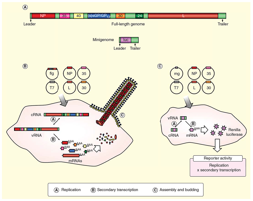
(A) Structure of a full-length ebolavirus genome and a minigenome. A full-length genome encoding all viral proteins (NP, VP35, VP40, sGP/GP1,2/ssGP, VP30, VP24 and L) as well as a miniature version of this genome (minigenome), in which the viral genes are removed and replaced with a reporter, for example luciferase, but in which the non-transcribed leader and trailer regions as well as the noncoding regions upstream of the first gene and downstream of the last gene are retained, are depicted. (B) Full-length clone system. A full-length cRNA antigenome (flg) is expressed from a cDNA-plasmid following initial transcription by T7 RNA polymerase (T7). Coexpressed RNP proteins (NP, L, VP35, VP30) recognize the cRNA, resulting in replication into vRNAs and subsequent transcription into mRNAs, which leads to expression of all viral proteins. Replicated vRNAs are then transported to the cell surface in the form of nucleocapsids, where they are incorporated into budding recombinant virus particles. (C) Minigenome systems. A vRNA minigenome is expressed from a cDNA-plasmid following initial transcription by a T7 RNA polymerase.
Coexpressed RNP proteins recognize the minigenome, resulting in replication through a cRNA intermediate and transcription into reporter mRNAs, leading to reporter activity that reflects genome replication and transcription.
cRNA: Complementary RNA; mg: Minigenome; RNP: Ribonucleoprotein complexes; vRNA: Viral RNA.
Full-length clone systems
Full-length clone systems (Figure 1B) are based on the generation of full-length complementary RNA (cRNA) antigenomes from a cDNA plasmid [21,22]. For filoviruses, this is usually achieved by initial transcription of the cRNA from the cDNA plasmid by T7 RNA polymerase. To this end, cells (the most commonly used being VeroE6 cells, BHK cells or a mixture of VeroE6 and 293 cells [22,27,28]) are transfected with the full-length genome cDNA plasmid and expression plasmids encoding the T7 polymerase (alternatively, in some cases, T7 is stably expressed by the cells used for the generation of recombinant viruses, rather than being expressed from a plasmid) as well as the viral RNP proteins NP, VP35, VP30 and L. Since T7 tends to incorporate nontemplated nucleotides at the transcript end, a hepatitis delta virus ribozyme is encoded downstream of the viral RNA sequence to ensure an authentic 3′ transcript end. After initial T7-driven transcription of a full-length cRNA, the RNP proteins replicate this cRNA into viral RNA genomes, which are then further transcribed into mRNAs, leading to the expression of all viral proteins, and thus jumpstarting the virus lifecycle and resulting in the rescue of recombinant viruses. Importantly, it is comparatively easy to introduce mutations into the cDNA plasmid, allowing for the generation of recombinant filoviruses tailored to answer specific scientific questions or to serve specific purposes, such as antiviral screening, for instance by expressing reporter proteins from additional genes inserted into the viral genome.
Minigenome systems
Minigenome systems (Figure 1C) resemble full-length clone systems; however, rather than using a cDNA copy of the full-length genome, these systems use a minigenome, in which the viral genes have been removed and replaced with a reporter gene such as luciferase, green fluorescent protein (GFP) or, in the case of the first filovirus minigenome systems, chloramphenicol acteyltransferase [6,7]. Importantly, in these minigenomes the 3’ and 5’ terminal noncoding regions are retained, and these regions, which encompass the leader and trailer regions of the genome, as well as the upstream noncoding region of the NP gene (the most 3’ gene in the virus genome), and the downstream noncoding region of the L gene (the most 5’ gene in the virus genome), harbor all of the signals necessary for recognition of the minigenome by the viral polymerase complex as an authentic template for genome replication and transcription. Minigenomes are expressed in mammalian cells either by T7-driven initial transcription [6,7] or by initial transcription using cellular RNA polymerases such as Pol I or Pol II [29]. Importantly, while Pol I generates authentic genome ends, Pol II-driven transcription leads to a 5’-capped and 3’-polyadenylated RNA transcript, so that in this case two ribozymes (usually a hammerhead ribozyme at the 5’ end and an hepatitis delta virus ribozyme at the 3’ end) have to be used to ensure authentic genome ends. Co-expressed RNP proteins then recognize the minigenome and replicate and transcribe it into reporter mRNAs, resulting in reporter activity reflecting both genome replication and transcription. In order to distinguish these two aspects of the viral lifecycle, replication-deficient minigenomes have also been developed. These carry mutations in the antigenomic replication promoter, resulting in a minigenome system in which reporter activity reflects only genome transcription, but not genome replication [30].
Transcription & replication competent virus-like particle systems
To model aspects of the virus lifecycle other than genome replication and transcription, trVLP systems (Figure 2) have been developed [24,25]. These systems resemble minigenome systems; however, in addition to the expression of a minigenome and the viral RNP proteins, the other viral structural proteins VP40 and GP1,2, and in some cases also VP24, are also expressed in cells (called producer or p0 cells). In these cells, the expression of VP40 leads to the formation of virus-like particles, which are coated with GP1,2 and harbor minigenome-containing nucleocapsids. These trVLPs are, therefore, able to infect target cells (also called p1 cells) and deliver their minigenome into these cells. In the case of untransfected, naive target cells, the minigenome then undergoes primary transcription to generate mRNAs, and ultimately resulting in reporter activity that reflects genome replication in p0 cells, budding of trVLPs from these p0 cells, the entry of trVLPs into p1 target cells and primary transcription in p1 cells [24]. As an alternative to the trVLP system with naive target cells, target cells can also be pretransfected with expression plasmids encoding the RNP proteins [25]. This leads to replication and secondary transcription of the trVLP-derived minigenome in target cells, so that reporter activity in this system reflects genome replication in p0 and p1 cells, trVLP budding in p0 cells, trVLP entry into p1 cells and secondary transcription in p1 cells.
Figure 2. Transcription and replication-competent virus-like particles systems.

A vRNA minigenome is expressed in p0 producer cells from a cDNA-plasmid following initial transcription by T7 RNA polymerase. Coexpressed RNP proteins recognize the minigenome, resulting in its replication and transcription. Additionally, coexpressed VP40 and GP1,2 lead to the formation of trVLPs that are coated in GP1,2 and harbor minigenome-containing nucleocapsids. These trVLPs can infect naive p1 target cells, where their minigenome undergoes primary transcription. Alternatively, p1 target cells can be pretransfected with the expression plasmids for the RNP proteins, which results in replication and secondary transcription of the trVLP-derived minigenome in p1 target cells.
cRNA: Complementary RNA; mg: Minigenome; RNP: Ribonucleoprotein complexes; trVLPs: Transcription and replication-competent virus-like particles; vRNA: Viral RNA.
Recently, trVLP systems have been further refined by utilizing multicistronic minigenomes as the source of the viral proteins needed for trVLP formation [23]. These systems now allow modeling of the complete virus lifecycle over multiple infectious cycles in the absence of foreign viral components under BSL2 conditions. Further, they alleviate a major limitation of classical trVLP systems, which is that these trVLP preparations contain large numbers of noninfectious particles, which can confound biochemical and structural analyses of trVLPs [31].
VP30-deficient Ebola virus
In order to study Ebola virus biology while circumventing the need for BSL4 containment, one additional reverse genetics-based approach that has been developed is that based on a recombinant Ebola virus with a genome in which the VP30 gene has been replaced by a reporter gene [32]. Since VP30 is essential for Ebola virus gene transcription, this virus requires VP30 to be provided in trans, which can be achieved by using a cell line stably expressing this protein. However, this virus is not able to productively infect cells (or organisms) that do not express VP30, resulting in biological containment of this virus to VP30-expressing cell lines. This virus is currently classified as a BSL3 organism, has been successfully used to study Ebola virus biology [14,33] and was evaluated as a potential vaccine candidate against ebolaviruses [34].
Application of reverse genetics systems for antiviral development
Reverse genetics systems can be used in many stages in the drug development process. Both full-length clone systems and lifecycle modeling systems allow for the identification of potential antiviral targets, and both systems can be used for antiviral screening approaches. Further, they can be used to confirm antiviral activity and, finally, to help elucidate the mechanism of action of novel antivirals. However, while reverse genetics systems have been available for filoviruses for more than 15 years [6], it is only in the last few years that there have been increased efforts to exploit them for antiviral drug development.
Antiviral screening
In order to facilitate antiviral screening against filoviruses, a number of recombinant viruses have been developed that carry reporter genes whose expression allows for easy detection and the quantification of infection in cell-based assays [35]. Most of these viruses express GFP from an additional transcriptional unit between the NP and VP35 gene, with such viruses having been developed for both Ebola virus [36,37] and Marburg virus [38]. In addition, recombinant Ebola and Marburg viruses expressing luciferase have also been recently reported, and these viruses allow for the extremely rapid detection of infection, with reporter activity first being detected as early as 2 h postinfection in the case of a luciferase-expressing Ebola virus [39,40]. However, as with reverse genetics systems in general, while such reporter-expressing viruses have been available for almost a decade [36], it is only in the recent years they have started to be used for drug screening, and so far, only GFP-expressing Ebola viruses have be utilized for this purpose.
Most of the antiviral screening studies with GFP-expressing Ebola viruses have been performed using high-content imaging (reviewed in [41]). This approach is based on infection of cells with the recombinant virus (usually in a 96-well format) under BSL4 conditions and fixation of these cells several days after infection, followed by removal from the BSL4 laboratory. Subsequently, the cells are subjected to high-content imaging, which allows not only the assessment of the extent of infection, but also of cell number and potential cytotoxic effects of the used drugs. Important parameters that have to be optimized for this technology are cell density and multiplicity of infection [42]. To date, five antiviral screening studies with different scopes have been performed using this technology and show the principal feasibility of this approach (Table 1). One additional study used a somewhat different approach and directly measured green fluorescence with a spectrofluorometer inside the BSL4 laboratory [43], while the cytotoxicity of the drugs was assessed in a parallel experiment using a commercially available luminescence-based cell viability assay.
Table 1.
Antiviral screens performed using a GFP-expressing Ebola virus (in each case the size and type of the library screened is indicated, as well as the detection method, the identified compounds and their protective efficacies in mice).
| Library composition | Method | Identified compounds | Survival of mice after treatment (prechallenge/postchallenge) | Ref. |
|---|---|---|---|---|
| 1990 compounds (National Cancer Institute small molecule diversity set) | HCI | NSC 62914 | 80/50% | [57] |
| 560 compounds (National Cancer Institute small molecule diversity set) | HCI | 16 primary hits, but with significant cytotoxicity | n.d. | [42] |
| 92 compounds (selected after a primary screen of 1012 US FDA-approved drugs using vesicular stomatitis virus pseudotyped with EBOV GP1,2) | HCI | Chloroquine | 80–90%/n.d. | [58] |
| 8 compounds (FDA-approved or former US-approved compounds) | SF | Clomiphene Toremifene |
n.d./90% n.d./50% |
[43] |
| Undisclosed number of compounds (National Cancer Institute) | HCI | FGI-103 | 100/100% | [59] |
| Undisclosed number of compounds (USAMRIID focused small molecule library) | HCI | FGI-106 | 100/80–90% | [60] |
HCI: High content imaging; n.d.: Not determined; SF: Spectrofluorometer.
A second strategy to screen for antivirals against filoviruses is the use of lifecycle modeling systems, which can be utilized without the need for a BSL4 laboratory. However, until now they only very rarely have been used for this purpose. The first study to show the principal feasibility of these systems for screening of antivirals was published in 2007 by Groseth et al., who tested a number of shRNAs for their efficacy in inhibiting Ebola virus genome replication and transcription [44]. In 2010, this strategy was revisited, and a luciferase-based Ebola virus minigenome assay suitable for high-throughput screening was developed [45]. To this end, it was necessary to exchange the promoter used for initial transcription of the minigenome from a T7-promoter to a PolI-promoter, as well as to change the cells used for screening from 293T cells to Cos7 cells, to optimize the Z’-factor (dynamic range divided by the separation band between positive and negative controls [46]; generally a Z’ factor >0.5 is considered indicative of a reliable screening assay) of the assay and to minimize background signals. Further, the authors reduced the number of plasmids required for this assay by expressing NP and VP35 from one plasmid. Following these changes, it was possible to miniaturize the minigenome into a 96-well format, and 960 compounds from the ‘Prestwick Chemical Known Bioactives’ library were screened, identifying 6 compounds that inhibited genome replication and transcription by 86–99% [45]. Recently, this approach was further refined by Uebelhoer et al. with the development of minigenome assays for both Ebola and Marburg viruses featuring the secreted Gaussia luciferase in a 96-well format [40]. These assays were established in BSR T7/5 cells, and interestingly it was necessary to use species codon-optimized expression plasmids for the RNP proteins in order to achieve a Z’ factor above 0.5. As a proof of concept, these assays were used to investigate the effect of a number of nucleoside analogues as well as siRNAs on genome replication and transcription, identifying 6-azauridine as a novel inhibitor of filovirus replication and/or transcription [46]. Other lifecycle modeling systems such as trVLP systems, which are available for Ebola and Marburg viruses [25,26], have so far not been utilized for antiviral drug screening.
An indication for the feasibility of using reverse genetics systems for antiviral screening is the fact that many of the compounds identified by these screens were protective in the mouse model of Ebola virus infection (Table 1). However, possibly due to the fact that antiviral screening is a rather new application of filovirus reverse genetics systems, so far none of the drugs have progressed beyond testing in mice, which, while a common and valuable first step in drug evaluation in vivo, is not necessarily predictive of protective effects in other more stringent models such as nonhuman primates [47].
Confirmation of antiviral activity
In addition to antiviral screening approaches, reporter-expressing filoviruses can also be used to confirm the activity of compounds identified as potential antivirals by different means. Perhaps the most comprehensive example for such an approach was published in 2013 by Filone et al., who screened a small molecule library containing about 2000 compounds for antiviral activity against vesicular stomatitis virus and then tested the activity of a number of identified compounds for their antiviral activity against Ebola viruses using a GFP-expressing Ebola virus. This approach yielded three compounds that show an antiviral activity against both vesicular stomatitis virus and Ebola virus [48]. Similarly, the kinase inhibitors genistein and tyrophostin, which had previously shown activity against the arenaviruses Pirital virus and Pichnide virus, and FGI-104, a potent inhibitor of hepatitis C virus release, were also shown to be active against Ebola viruses, again using a recombinant Ebola virus expressing GFP [49,50]. Further, the antiviral activity of BCX4430, a small molecule that has recently been shown to be protective against Marburg virus infection in nonhuman primates with treatment starting as late as 48 h postinfection was initially assessed using GFP-expressing filoviruses [51].
An important step in validating the antiviral activity of compounds identified using lifecycle modeling system-based screens involves confirming their antiviral effect in the context of a virus infection, and this is something that can also be accomplished using reporter-expressing viruses. To this end, in the aforementioned study by Uebelhoer et al., compounds showing a strong inhibition of minigenome replication and/or transcription were further tested for their antiviral activity using luciferase-expressing filoviruses [40]. Interestingly, while for most of the tested compounds antiviral activity matched the results from the minigenome-based screen, this was not the case for ribavirin, which had shown inhibition of genome replication and transcription in the minigenome assay but, at least under the experimental conditions chosen, did not exhibit antiviral activity against the recombinant Ebola virus, highlighting the importance of confirming results obtained with lifecycle modeling systems using live virus [40].
Elucidation of the mechanism of action of antivirals
Lifecycle modeling systems are particularly well suited for elucidating the mechanism of action of newly identified antivirals since they allow the dissection of the virus lifecycle in order to assess its individual aspects. One example of this approach is the aforementioned study by Filone et al., in which minigenome assays were used to show that one of the compounds active against both vesicular stomatitis virus and Ebola viruses strongly inhibits Ebola virus minigenome replication and transcription [48]. Similarly, the mechanism of action of BCX4430 was analyzed using minigenome systems and it was shown that this compound is indeed an inhibitor of virus genome replication and/or transcription, in line with it being designed as a nucleoside analogue [51].
However, reporter-expressing filoviruses can also be used to investigate the mechanism of action of antivirals. Brown et al. used a GFP-expressing Ebola virus as well as minigenome assays to identify the mechanisms of action of several in silico designed molecules predicted and experimentally confirmed to interact with VP35 [52]. GFP expression in infected cells, which is reflective of viral gene expression, was used as a means to assess genome replication and transcription and complemented by minigenome data, while GFP-based titration of supernatants was used as a read-out for inhibition of infectious particle production.
Identification of drug targets
One final and very broad application of reverse genetics for drug development is the identification of novel drug targets, an approach that bridges basic and applied research. Until now, this has mostly been done using minigenome systems. A straightforward example of this approach is a recent study in which peptides corresponding to fragments of the Ebola virus polymerase L were used to inhibit virus genome replication and/or transcription, as measured in minigenome assays, by blocking the formation of the active polymerase complex, which constitutes a potential novel target for antivirals [53]. Similarly, RNA aptamers binding to VP35 were developed and shown to inhibit genome replication and/or transcription using minigenome assays, again most likely by targeting the formation of an active polymerase complex [54]. Finally, the cellular factor heme oxygenase was identified as a potential drug target using a VP30-deficient GFP-expressing Ebola virus, with overexpression of this factor inhibiting Ebola virus replication [55]. Using minigenomes it was subsequently shown that this effect is due to a strong inhibition of genome replication and/or transcription.
All of these potential drug targets have yet to be exploited, and it remains to be shown if they are efficacious against actual infections in vitro and in vivo; nevertheless, these studies highlight the potential that exists in applying reverse genetics, and particularly lifecycle modeling systems, to identify novel drug targets.
Potential pitfalls when employing reverse genetics systems for drug development
While reverse genetics-based systems are very powerful tools for drug development, there are a number of points that have to be kept in mind when employing these systems. The first point that has to be taken into consideration is the extent to which these systems authentically reflect an infection with wild-type virus. Recombinant viruses expressing reporter genes are generally attenuated in vitro and in vivo [35]. In vitro the extent of attenuation is generally limited, with viruses expressing GFP or luciferase from an additional open reading frame located between the NP and VP35 genes, which is the standard strategy used for the design of such viruses, reaching identical end titers compared with wild-type virus and showing a delay in growth of only about 1 day [39]. As such, the use of these viruses for screening purposes is generally unproblematic. In contrast, in vivo these viruses show significant attenuation, and thus their utility for confirming antiviral activity in animal models is limited [37].
In the case of lifecycle modeling systems, understanding the extent to which these systems reflect infection with wild-type viruses, and particularly the inherent differences between these systems and actual infection, becomes even more important. Obviously, not all lifecycle modeling systems reflect the complete lifecycle, for example, minigenome systems only model genome replication and transcription, but not virus entry or budding, and trVLP systems with pretransfected target cells model most aspects of the virus lifecycle, but not primary transcription. This means that, when using these systems for screening purposes, potential antivirals that target steps not assessed by these assays will be missed. More importantly, these assays rely on a number of processes that do not have equivalents during virus infection, which can lead to false-positive findings. For instance, all lifecycle modeling systems rely on plasmid-based expression of viral proteins, which involves transcription by the cellular polymerase Pol II; in contrast, during virus infection this enzyme is not required for expression of viral proteins. Also, initial transcription of minigenomes in these assays, a step that has no equivalent during virus infection, uses either T7 polymerase, or the cellular polymerases Pol I or Pol II. Therefore, compounds targeting these enzymes will appear as false-positives during antiviral screens. This can be, to some extent, controlled for by including additional reporters expressed by these polymerases, and indeed for minigenome assays in most cases a second Pol II-expressed reporter is used for this purpose. One example of such discordant findings between lifecycle modeling systems and virus infection is the result for ribavirin, which in one study inhibited minigenome replication and transcription, but did not exhibit antiviral activity when assessed with infectious virus [40]. As a consequence, any compounds identified in a lifecycle modeling system-based screen clearly have to be validated using live virus; nevertheless, these systems remain an excellent option for a first step in the screening process.
A second point that needs to be considered is the use of proper readouts. There are a number of different reporters available, with the most commonly used ones being fluorescent reporters, such as GFP, and luminescent reporters, such as the various luciferases, each of which have their respective limitations. With the luciferases additional reagents are required for detection, while for the fluorescent reporters sophisticated imaging equipment is often required. Furthermore, while luciferase reporters are most often assessed by measuring overall reporter activity in a sample, in the case of fluorescent reporters, either the average reporter activity or the number of cells exceeding a certain threshold (i.e., the number of ‘positive’ cells) can be measured (Figure 3A). The latter strategy is well suited to assessing the rate of infection of cells, either by reporter-expressing viruses or trVLPs, in order to determine infectious titers or to screen for entry inhibitors. However, one has to make sure that the threshold for positive cells is low enough that low levels of replication and transcription, for example due to off-target effects of drugs on these processes, do not skew the results (Figure 3A, lower-right panels). Potentially very problematic is the use of this method to assess effects on replication and transcription because choosing an appropriate threshold becomes absolutely essential in order to avoid skewing results (Figure 3A, lower-left panels); rather, average reporter activity should be used for this purpose. However, measuring average reporter activity can be problematic if only a small percentage of cells are infected since then the dynamic range of the assay becomes very small due to the large number of uninfected cells and real differences can become obscured by experimental variations or background noise (figure 3B, upper half). In this case, a feasible alternative is the measurement of the average reporter activity in positive cells, with a low threshold defining these positive cells (figure 3B, lower half).
Figure 3. Effects of reporter readout on the interpretation of experimental results.

(A) Different readout methods. Cells that are infected with either a fluorescent reporter-expressing virus or with transcription and replication-competent virus-like particles encoding such a reporter (shown in green) can be assessed either by directly measuring average reporter activity in those cells throughout the whole sample, or by assessing the ratio of positive cells as defined by a threshold value. Importantly, in the presence of decreased genome replication and transcription or impaired entry these different methods will give different experimental readings, as indicated below each example. Values exhibiting overestimation of antiviral activity (i.e., failure to detect infected cells) are shown in red, whereas values exhibiting underestimation of antiviral activity (i.e., failure to detect a reduction in reporter activity in infected cells) are shown in blue. (B) Problem of low dynamic range due to low infection rates. Under conditions where there are low infection rates, differences in replication and transcription in infected cells result in only small differences in average reporter activity. This results in a very low dynamic range of the assay and exacerbates the risk that differences become obscured by experimental variation or background noise. By first identifying positive cells using a low threshold and then measuring reporter activity only in these cells, this problem can be mitigated.
Overall, it is clear that care must be taken when choosing experimental systems for drug screening, confirmation of antiviral activity or elucidation of mechanisms of action and that appropriate readouts must be selected to avoid false-positive or false-negative results. Nevertheless, reverse genetics-based systems remain powerful tools for antiviral drug development.
Expert commentary
The development of antivirals for filoviruses has been hindered by the fact that live viruses can only be handled under BSL4 condition, making classical drug development strategies such as high-throughput screening extremely problematic. Consequently, until now, no high-throughput screening assay conducted under BSL4 conditions has been reported for filoviruses, although recently a single laboratory reported such a screen for Nipah virus under BSL4 conditions [56]. Reverse genetics provides two main strategies that can be used to alleviate this problem.
First, reporter-expressing viruses facilitate a much easier and more rapid detection and quantification of infection, allowing antiviral screens to be performed manually in a 96-well format with thousands of compounds under BSL4 conditions, with either direct evaluation of reporter activity inside a BSL4 laboratory or removal of plates from the BSL4 laboratory and subsequent analysis by more sophisticated methods such as high-content imaging [57]. These viruses, which were introduced for filoviruses almost 10 years ago [36], are now widely used as tools for antiviral development and particularly for this type of screen.
Second, the availability of lifecycle modeling systems that have been successfully adapted to high-throughput assay formats in the last few years allow initial screening to be performed under BSL2 conditions, and thus taking advantage of already existing highly sophisticated facilities dedicated to this purpose. This technology, while in principle available for more than 15 years [6], has only recently been adopted for drug development purposes and has still not reached its full potential with regards to this application. This might be due to the fact that lifecycle modeling systems can be experimentally demanding, and for example requires the transfection of numerous plasmids (in general 5–7 for minigenome systems, and 8–10 for trVLP systems) at optimized ratios into cells. However, recent studies have significantly improved the robustness and ease of use of minigenome assays, and similar improvements also are feasible for the more comprehensive trVLP assays [23]. Thus, it is only a matter of time until these systems, which provide a viable alternative to experiments with infectious viruses, particularly for groups without access to BSL4 facilities, will be more widely used for the development of antivirals.
Five-year view
There are three main developments that we expect to see over the next 5 years regarding the use of reverse genetics for antiviral development.
First, reporter-expressing viruses will be more extensively used for screening purposes. Robust high-content imaging protocols have been established using GFP-expressing Ebola viruses, but have so far only been exploited to a limited extent, with the largest screen involving about 2000 compounds [57]. In the near future, we expect to see an increase in the number and scope of these screens. In addition, given that a GFP-expressing Marburg virus also exists [38], one can also expect to see similar studies performed with this virus.
Second, lifecycle modeling systems have just started to be used for antiviral development, and so far only classical minigenome systems have been used for this purpose. However, we expect the more sophisticated lifecycle modeling systems, which allow modeling of the complete virus lifecycle under BSL2 conditions, to also be optimized for high-throughput applications and become more widely used in this field.
Finally, the recent publication of the first high-throughput antiviral screen for Nipah virus under BSL4 conditions at the Galveston National Laboratory [56] has now opened up the possibility of performing this type of screen for filoviruses as well. Reporter-expressing viruses are most amenable to this purpose, particularly since this facility is set up to use luminescence as an assay readout. Due to the availability of recombinant Ebola viruses expressing luciferases [39,40], everything is in place for these studies to be performed over the next years.
Key issues.
Reverse genetics systems encompass full-length clone systems, which allow the generation of recombinant viruses, and lifecycle modeling systems.
Recombinant filoviruses expressing reporter genes can be used under biosafety level 4 conditions for rapid screening of antivirals, as well as for the confirmation of antiviral activity, and in some cases also the elucidation of the mechanism of action of these antivirals.
Over the last years reporter-expressing filoviruses have been increasingly employed for antiviral screening using high-content imaging.
Lifecycle modeling systems allow modeling of the lifecycle of filoviruses under biosafety level 2 conditions and can be used for antiviral screening purposes, as well as for the elucidation of mechanisms of action of antiviral drugs.
These lifecycle modeling systems have only recently been optimized for high-throughput applications.
The near future will see an increase in studies using reverse genetics-based tools for the development of antivirals.
Acknowledgements
The authors are very grateful to Allison Groseth (NIH/NIAID) for critical reading of the manuscript.
Financial & competing interests disclosure
This research was supported by the Intramural Research Program of the NIH, NIAID. The authors have no other relevant affiliations or financial involvement with any organization or entity with a financial interest in or financial conflict with the subject matter or materials discussed in the manuscript apart from those disclosed.
No writing assistance was utilized in the production of this manuscript.
References
Papers of special note have been highlighted as:
• of interest
•• of considerable interest
- 1.Sanchez A, Geisbert TW, Feldmann H. Filoviridae: Marburg and Ebola Viruses. In: Fields BN, Knipe DM, Howley PM, editors. Fields virology. 6th edition. Lippincott Williams & Wilkins; Philadelphia, USA: 2013 [Google Scholar]
- 2.Baize S, Pannetier D, Oestereich L, et al. Emergence of Zaire Ebola virus disease in Guinea - preliminary report. N Engl J Med 2014. [Epub ahead of print] [DOI] [PubMed] [Google Scholar]
- 3.Kortepeter MG, Bausch DG, Bray M. Basic clinical and laboratory features of filoviral hemorrhagic fever. J Infect Dis 2011;204(Suppl 3):S810–16 [DOI] [PubMed] [Google Scholar]
- 4.Hoenen T, Groseth A, Falzarano D, Feldmann H. Ebola virus: unravelling pathogenesis to combat a deadly disease. Trends Mol Med 2006;12(5):206–15 [DOI] [PubMed] [Google Scholar]
- 5.Roddy P, Colebunders R, Jeffs B, et al. Filovirus hemorrhagic fever outbreak case management: a review of current and future treatment options. J Infect Dis 2011;204(Suppl 3):S791–5 [DOI] [PubMed] [Google Scholar]
- 6.•.Muhlberger E, Lötfering B, Klenk HD, Becker S. Three of the four nucleocapsid proteins of Marburg virus, NP, VP35, and L, are sufficient to mediate replication and transcription of Marburg virus-specific monocistronic minigenomes. J Virol 1998;72(11):8756–64. [DOI] [PMC free article] [PubMed] [Google Scholar]; Describes the establishment of the first minigenome system for filoviruses.
- 7.Muhlberger E, Weik M, Volchkov VE, et al. Comparison of the transcription and replication strategies of Marburg virus and Ebola virus by using artificial replication systems. J Virol 1999;73(3):2333–42 [DOI] [PMC free article] [PubMed] [Google Scholar]
- 8.Bharat TA, Riches JD, Kolesnikova L, et al. Cryo-electron tomography of Marburg virus particles and their morphogenesis within infected cells. PLoS Biol 2011;9(11): e1001196. [DOI] [PMC free article] [PubMed] [Google Scholar]
- 9.Beniac DR, Melito PL, Devarennes SL, et al. The organisation of Ebola virus reveals a capacity for extensive, modular polyploidy. PLoS One 2012;7(1):e29608. [DOI] [PMC free article] [PubMed] [Google Scholar]
- 10.Aleksandrowicz P, Marzi A, Biedenkopf N, et al. Ebola virus enters host cells by macropinocytosis and clathrin-mediated endocytosis. J Infect Dis 2011;204(Suppl 3):S957–67 [DOI] [PMC free article] [PubMed] [Google Scholar]
- 11.Nanbo A, Imai M, Watanabe S, et al. Ebolavirus is internalized into host cells via macropinocytosis in a viral glycoprotein-dependent manner. PLoS Pathog 2010;6(9):e1001121. [DOI] [PMC free article] [PubMed] [Google Scholar]
- 12.Miller EH, Chandran K. Filovirus entry into cells - new insights. Curr Opin Virol 2012;2(2):206–14 [DOI] [PMC free article] [PubMed] [Google Scholar]
- 13.Hoenen T, Shabman RS, Groseth A, et al. Inclusion bodies are a site of ebolavirus replication. J Virol 2012;86(21):11779–88 [DOI] [PMC free article] [PubMed] [Google Scholar]
- 14.Nanbo A, Watanabe S, Halfmann P, Kawaoka Y. The spatio-temporal distribution dynamics of Ebola virus proteins and RNA in infected cells. Sci Rep 2013;3:1206. [DOI] [PMC free article] [PubMed] [Google Scholar]
- 15.Harty RN, Brown ME, Wang G, et al. A PPxY motif within the VP40 protein of Ebola virus interacts physically and functionally with a ubiquitin ligase: implications for filovirus budding. Proc Natl Acad Sci USA 2000;97(25):13871–6 [DOI] [PMC free article] [PubMed] [Google Scholar]
- 16.Timmins J, Scianimanico S, Schoehn G, Weissenhorn W. Vesicular release of ebola virus matrix protein VP40. Virology 2001;283(1):1–6 [DOI] [PubMed] [Google Scholar]
- 17.Jasenosky LD, Neumann G, Lukashevich I, Kawaoka Y. Ebola virus VP40-induced particle formation and association with the lipid bilayer. J Virol 2001;75(11):5205–14 [DOI] [PMC free article] [PubMed] [Google Scholar]
- 18.Falzarano D, Feldmann H. Possible leap ahead in filovirus therapeutics. Cell Res 2014;24(6):647–8 [DOI] [PMC free article] [PubMed] [Google Scholar]
- 19.Hoenen T, Groseth A, Feldmann H. Current ebola vaccines. Expert Opin Biol Ther 2012;12(7):859–72 [DOI] [PMC free article] [PubMed] [Google Scholar]
- 20.•.Hoenen T, Groseth A, de Kok-Mercado F, et al. Minigenomes, transcription and replication competent virus-like particles and beyond: reverse genetics systems for filoviruses and other negative stranded hemorrhagic fever viruses. Antiviral Res 2011;91(2):195–208. [DOI] [PMC free article] [PubMed] [Google Scholar]; Reviews the principles of reverse genetics systems, including the different lifecycle modeling systems, for negative strand RNA viruses.
- 21.Neumann G, Feldmann H, Watanabe S, et al. Reverse genetics demonstrates that proteolytic processing of the Ebola virus glycoprotein is not essential for replication in cell culture. J Virol 2002;76(1):406–10 [DOI] [PMC free article] [PubMed] [Google Scholar]
- 22.Volchkov VE, Volchkova VA, Muhlberger E, et al. Recovery of infectious Ebola virus from complementary DNA: RNA editing of the GP gene and viral cytotoxicity. Science 2001;291(5510):1965–9 [DOI] [PubMed] [Google Scholar]
- 23.Watt A, Moukambi F, Banadyga L, et al. A novel lifecycle modeling system for Ebola virus shows a genome length-dependent role of VP24 on virus infectivity. J Virol 2014. [Epub ahead of print] [DOI] [PMC free article] [PubMed] [Google Scholar]
- 24.Hoenen T, Groseth A, Kolesnikova L, et al. Infection of naive target cells with virus-like particles: implications for the function of ebola virus VP24. J Virol 2006;80(14):7260–4 [DOI] [PMC free article] [PubMed] [Google Scholar]
- 25.•.Watanabe S, Watanabe T, Noda T, et al. Production of novel ebola virus-like particles from cDNAs: an alternative to ebola virus generation by reverse genetics. J Virol 2004;78(2):999–1005. [DOI] [PMC free article] [PubMed] [Google Scholar]; First description of a transcription-and replication-competent virus-like particle system for filoviruses.
- 26.Wenigenrath J, Kolesnikova L, Hoenen T, et al. Establishment and application of an infectious virus-like particle system for Marburg virus. J Gen Virol 2010;91(Pt 5):1325–34 [DOI] [PubMed] [Google Scholar]
- 27.Shabman RS, Hoenen T, Groseth A, et al. An upstream open reading frame modulates ebola virus polymerase translation and virus replication. PLoS Pathog 2013;9(1):e1003147. [DOI] [PMC free article] [PubMed] [Google Scholar]
- 28.Neumann G, Ebihara H, Takada A, et al. Ebola virus VP40 late domains are not essential for viral replication in cell culture. J Virol 2005;79(16):10300–7 [DOI] [PMC free article] [PubMed] [Google Scholar]
- 29.Groseth A, Feldmann H, Theriault S, et al. RNA polymerase I-driven minigenome system for Ebola viruses. J Virol 2005;79(7):4425–33 [DOI] [PMC free article] [PubMed] [Google Scholar]
- 30.Hoenen T, Jung S, Herwig A, et al. Both matrix proteins of Ebola virus contribute to the regulation of viral genome replication and transcription. Virology 2010;403(1):56–66 [DOI] [PubMed] [Google Scholar]
- 31.Spiegelberg L, Wahl-Jensen V, Kolesnikova L, et al. Genus-specific recruitment of filovirus ribonucleoprotein complexes into budding particles. J Gen Virol 2011;92(Pt 12):2900–5 [DOI] [PMC free article] [PubMed] [Google Scholar]
- 32.Halfmann P, Kim JH, Ebihara H, et al. Generation of biologically contained Ebola viruses. Proc Natl Acad Sci USA 2008;105(4):1129–33 [DOI] [PMC free article] [PubMed] [Google Scholar]
- 33.Takahashi K, Halfmann P, Oyama M, et al. DNA topoisomerase 1 facilitates the transcription and replication of the Ebola virus genome. J Virol 2013;87(16):8862–9 [DOI] [PMC free article] [PubMed] [Google Scholar]
- 34.Halfmann P, Ebihara H, Marzi A, et al. Replication-deficient ebolavirus as a vaccine candidate. J Virol 2009;83(8):3810–15 [DOI] [PMC free article] [PubMed] [Google Scholar]
- 35.•.Falzarano D, Groseth A, Hoenen T. Development and application of reporter-expressing mononegaviruses: Current challenges and perspectives. Antiviral Res 2014;103:78–87. [DOI] [PMC free article] [PubMed] [Google Scholar]; Reviews the generation and use of reporter-expressing viruses and provides guidelines for their design.
- 36.•.Towner JS, Paragas J, Dover JE, et al. Generation of eGFP expressing recombinant Zaire ebolavirus for analysis of early pathogenesis events and high-throughput antiviral drug screening. Virology 2005;332(1):20–7. [DOI] [PubMed] [Google Scholar]; First description of a green fluorescent protein-expressing Ebola virus, which is currently the most widely used recombinant filovirus for antiviral screening.
- 37.Ebihara H, Theriault S, Neumann G, et al. In vitro and in vivo characterization of recombinant Ebola viruses expressing enhanced green fluorescent protein. J Infect Dis 2007;196(Suppl 2):S313–22 [DOI] [PubMed] [Google Scholar]
- 38.Schmidt KM, Schümann M, Olejnik J, et al. Recombinant Marburg virus expressing EGFP allows rapid screening of virus growth and real-time visualization of virus spread. J Infect Dis 2011;204(Suppl 3)):S861–70 [DOI] [PMC free article] [PubMed] [Google Scholar]
- 39.Hoenen T, Groseth A, Callison J, et al. A novel Ebola virus expressing luciferase allows for rapid and quantitative testing of antivirals. Antiviral Res 2013;99(3):207–13 [DOI] [PMC free article] [PubMed] [Google Scholar]
- 40.Uebelhoer LS, Albariño CG, McMullan LK, et al. High-throughput, luciferase-based reverse genetics systems for identifying inhibitors of Marburg and Ebola viruses. Antiviral Res 2014;106:86–94 [DOI] [PubMed] [Google Scholar]
- 41.••.Pegoraro G, Bavari S, Panchal RG. Shedding light on filovirus infection with high-content imaging. Viruses 2012;4(8):1354–71. [DOI] [PMC free article] [PubMed] [Google Scholar]; Reviews the development of high-content imaging systems for filoviruses.
- 42.Panchal RG, Kota KP, Spurgers KB, et al. Development of high-content imaging assays for lethal viral pathogens. J Biomol Screen 2010;15(7):755–65 [DOI] [PubMed] [Google Scholar]
- 43.Johansen LM, Brannan JM, Delos SE, et al. FDA-approved selective estrogen receptor modulators inhibit Ebola virus infection. Sci Transl Med 2013;5(190):190ra79. [DOI] [PMC free article] [PubMed] [Google Scholar]
- 44.Groseth A, Hoenen T, Alimonti JB, et al. In vitro evaluation of antisense RNA efficacy against filovirus infection, by use of reverse genetics. J Infect Dis 2007;196(Suppl 2):S382–9 [DOI] [PubMed] [Google Scholar]
- 45.••.Jasenosky LD, Neumann G, Kawaoka Y. Minigenome-based reporter system suitable for high-throughput screening of compounds able to inhibit Ebolavirus replication and/or transcription. Antimicrob Agents Chemother 2010;54(7):3007–10. [DOI] [PMC free article] [PubMed] [Google Scholar]; Detailed description of the development and optimization of a minigenome assay suitable for high-throughput screening.
- 46.Zhang JH, Chung TD, Oldenburg KR. A simple statistical parameter for use in evaluation and validation of high throughput screening assays. J Biomol Screen 1999;4(2):67–73 [DOI] [PubMed] [Google Scholar]
- 47.Geisbert TW, Bausch DG, Feldmann H. Prospects for immunisation against Marburg and Ebola viruses. Rev Med Virol 2010;20(6):344–57 [DOI] [PMC free article] [PubMed] [Google Scholar]
- 48.Filone CM, Hodges EN, Honeyman B, et al. Identification of a broad-spectrum inhibitor of viral RNA synthesis: validation of a prototype virus-based approach. Chem Biol 2013;20(3):424–33 [DOI] [PMC free article] [PubMed] [Google Scholar]
- 49.Kolokoltsov AA, Adhikary S, Garver J, et al. Inhibition of Lassa virus and Ebola virus infection in host cells treated with the kinase inhibitors genistein and tyrphostin. Arch Virol 2012;157(1):121–7 [DOI] [PubMed] [Google Scholar]
- 50.Kinch MS, Yunus AS, Lear C, et al. FGI-104: a broad-spectrum small molecule inhibitor of viral infection. Am J Transl Res 2009;1(1):87–98 [PMC free article] [PubMed] [Google Scholar]
- 51.Warren TK, Wells J, Panchal RG, et al. Protection against filovirus diseases by a novel broad-spectrum nucleoside analogue BCX4430. Nature 2014;508(7496):402–5 [DOI] [PMC free article] [PubMed] [Google Scholar]
- 52.Brown CS, Lee MS, Leung DW, et al. In silico derived small molecules bind the filovirus VP35 protein and inhibit its polymerase cofactor activity. J Mol Biol 2014;426(10):2045–58 [DOI] [PMC free article] [PubMed] [Google Scholar]
- 53.Trunschke M, Conrad D, Enterlein S, et al. The L-VP35 and L-L interaction domains reside in the amino terminus of the Ebola virus L protein and are potential targets for antivirals. Virology 2013;441(2):135–45 [DOI] [PMC free article] [PubMed] [Google Scholar]
- 54.Binning JM, Wang T, Luthra P, et al. Development of RNA aptamers targeting Ebola virus VP35. Biochemistry 2013;52(47):8406–19 [DOI] [PMC free article] [PubMed] [Google Scholar]
- 55.Hill-Batorski L, Halfmann P, Neumann G, Kawaoka Y. The cytoprotective enzyme heme oxygenase-1 suppresses Ebola virus replication. J Virol 2013;87(24):13795–802 [DOI] [PMC free article] [PubMed] [Google Scholar]
- 56.•.Tigabu B, Rasmussen L, White EL, et al. A BSL-4 high-throughput screen identifies sulfonamide inhibitors of Nipah virus. Assay Drug Dev Technol 2014;12(3):155–61. [DOI] [PMC free article] [PubMed] [Google Scholar]; Reports the first successful high-throughput screening assay under BSL4 conditions.
- 57.•.Panchal RG, Reid SP, Tran JP, et al. Identification of an antioxidant small-molecule with broad-spectrum antiviral activity. Antiviral Res 2012;93(1):23–9. [DOI] [PubMed] [Google Scholar]; Most comprehensive antiviral screening against filoviruses performed so far using reporter-expressing viruses.
- 58.Madrid PB, Chopra S, Manger ID, et al. A systematic screen of FDA-approved drugs for inhibitors of biological threat agents. PLoS One 2013;8(4):e60579. [DOI] [PMC free article] [PubMed] [Google Scholar]
- 59.Warren TK, Warfield KL, Wells J, et al. Antiviral activity of a small-molecule inhibitor of filovirus infection. Antimicrob Agents Chemother 2010;54(5):2152–9 [DOI] [PMC free article] [PubMed] [Google Scholar]
- 60.Aman MJ, Kinch MS, Warfield K, et al. Development of a broad-spectrum antiviral with activity against Ebola virus. Antiviral Res 2009;83(3):245–51 [DOI] [PubMed] [Google Scholar]