Abstract
Background
Rotational deformities in children are currently treated with an osteotomy, acute de-rotation, and surgical fixation. Meanwhile, guided growth is now the gold standard in pediatric coronal deformity correction. This study aimed to evaluate the feasibility of a novel implant intended for rotational guided growth (RotOs Plate) in a large porcine animal model.
Methodology
A submuscular plate was inserted on the medial and lateral aspect of the distal femoral physis of the left femur in 6 pigs. Each plate was anchored with a screw in the metaphysis and epiphysis respectively. The plates were expected to rotate the femur externally. The right femur acted as a control in a paired design. The animals were housed for 12 weeks after surgery. MRI scanning of both femora was performed before euthanasia after 12 weeks. Rotation was determined as the difference in the femoral version on MRI between the operated and non-operated femur after 12 weeks.
Results
External rotation in all operated femurs was observed. The mean difference in the femoral version on MRI between operated and non-operated femurs was 12.5° (range 9°-16°). No significant changes in axial growth were detected.
Conclusions
This study shows encouraging results regarding rotational guided growth, which may replace current invasive surgical treatment options for malrotation in children. However, further studies addressing potential secondary deformities are paramount and should be carried out.
Keywords: innovative implant, 3d print, guided growth, rotational deformity, maltorsion
Introduction
Rotational deformities of long bones are currently corrected by osteotomies with internal or external fixation [1-6]. The concept of guided growth is state of the art for correction of valgus and varus deformities in the growing skeleton. Recently, the guided growth concept has also been advocated to correct rotational deformities because of the lesser invasive nature compared with corrective osteotomies [7-10]. A few studies have investigated oblique tension band plating with eight plates or similar implants in rabbits, i.e., small animal studies [11-14]. Recently, Metaizeau et al. published a study applying guided growth to correct torsion in humans using cable-connected cannulated screws, yielding promising results [15]. The concept of rotational guided growth in humans has furthermore been investigated by Paley and Shannon using oblique peripheral tethers [16]. All of these have proven to rotate long bones by guided growth. However, the nature of the plates applied may lead to growth retardation and changes in joint morphology, and they may not predictably rotate the bone [17,18]. However, these findings underline the possibility of applying guided growth to address torsional deformities in children.
An implant capable of rotating long bones in a controlled and predictable manner by guided growth is lacking. We designed a novel plate concept (RotOs Plate) for rotational guided growth, which was capable of rotating cadaverous femora in a controlled and predictable manner [19]. The plate is based on two curved oblique sliding holes interconnected at an angle of 105°. It is dependent on axial growth to obtain rotation and is thus unlikely to impair axial growth. This study aims to investigate the efficacy of the novel plate (RotOs Plate) in rotating porcine femora by guided growth.
Materials and methods
Design and surgical procedure
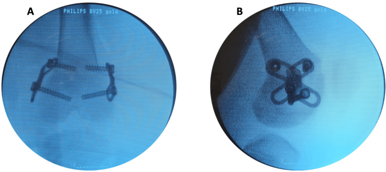
The study was carried out in a matched paired design. Six 12-week-old female pigs were included. The mean body weight was 43 kg (range 38-47 kg). The left femur was selected for surgical intervention. The right femur was left untreated in all animals. A submuscular plate (RotOs Plate) was inserted on the medial and lateral aspect of the distal femoral physis. The body of the plates was placed perpendicular to the physis in the sagittal plane with a sliding slot proximal and distal to the physis (Figure 1). Each plate was anchored proximal and distal to the physis using a 4.5 mm cannulated screw (titanium, DePuy Synthes). All surgeries were performed under general anesthesia with propofol (10 mg/kg/hour) and fentanyl (60 mg/kg/hour) as analgesics. All operated sites received a local anesthetic of 50 mg of Bupivacaine. The animals were housed and observed for 12 weeks after the operation, whereafter the plates were removed. MRI was subsequently performed to determine the left and right femoral versions. The difference in femoral version between the treated and nontreated femur was chosen as the primary outcome in this paired study.
Figure 1. Intraoperative images.
Intraoperative fluoroscopic images showing the insertion of the RotOs Plates in the porcine femur. Each plate is anchored with two 4.5 mm cannulated screws (DePuy Synthes): (A) coronal plane and (B) sagittal plane.
RotOs Plate
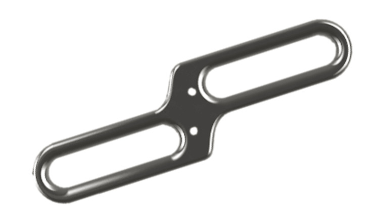
Two titanium (Ti6Al4V) plates per porcine were produced by additional manufactured, i.e., 3D printed by Danish Technological Institute, Aarhus, Denmark. RotOs Plate consists of two oblique and curved sliding slots interconnected at an angle of 105° through a body with two slots for temporary Kirschner-wire fixation (Figure 2). A screw was inserted proximally in each sliding slot. The length of the sliding holes was 13 mm. The distance between the two screws in the transverse plane, with the body aligned to the femoral axis, was 17 mm, as designed by the plate. Similar plates were applied in cadavers achieving rotation during axial growth [19].
Figure 2. RotOs Plate.
RotOs Plate concept plate with two sliding holes for screw fixation and two Kirschner-wire (K-wire) slots for temporary intraoperative fixation.
MRI and measurement of the femoral version
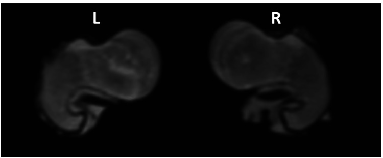
MRI with a 3.0 Tesla scanner (Siemens Skyra) was performed 12 weeks after surgery. T1 and T2 sequences were obtained. The femoral version was determined as the angle between an axis through the femoral condyles and the axis of the femoral neck in the axial plane. The difference in the femoral version between the right and left femur was assigned as obtained rotation (Figure 3).
Figure 3. MRI image of the femoral neck in the operated and non-operated femur with both femoral condyles aligned parallel to the scanning table showing the difference in femoral anteversion: L, left operated femur; R, right non-operated femur.
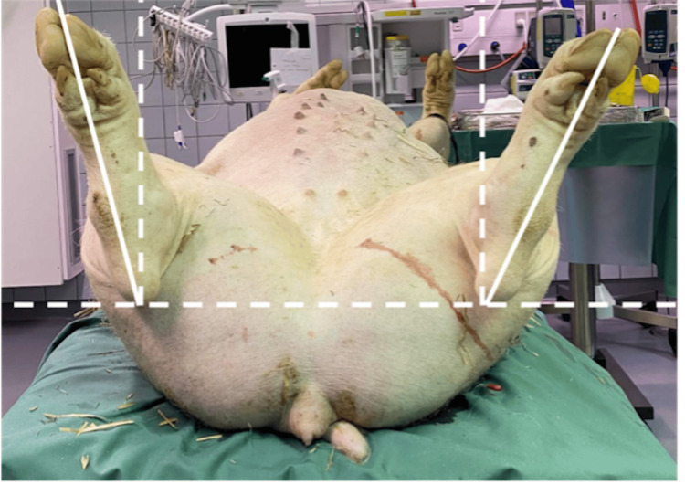
Clinical evaluation of the difference in torsion was done by measuring the angle between the foot and a perpendicular line to a line connecting the calcaneal tubercules. Measurements were done with all animals lying straight and supine on a leveled table (Figure 4).
Figure 4. Differences in clinical rotation of the hind limbs are shown in a clinical image obtained on the leveled table.
The femoral length was measured in the coronal plane from the top of the femoral head to the bottom of the medial femoral condyle.
Statistics
All calculations were conducted using Stata 16 software (Stata Statistical Software: Release 16, StataCorp LLC, College Station, TX). Paired t-tests were applied to determine the P-values. P-values ≤ 0.05 were considered statistically significant. Data are presented as mean (95% confidence interval) or median (range).
Results
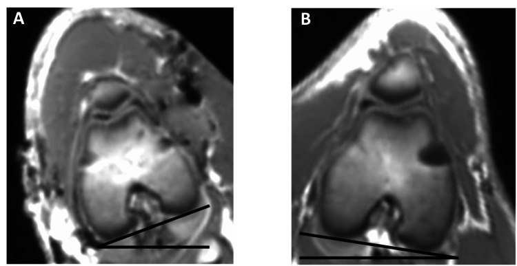
All animals tolerated the surgeries well and were ambulatory on the first postoperative day. One postoperative wound deficiency was observed, which required re-suturing. No infections or other complications were identified, and no animals were sacrificed before the end of the study. In two operated femora, only one of the two inserted plates was anchored proximal and distal to the physis as intended, resulting in only one plate guiding the growth. All operated left femurs were externally rotated and compared with the contralateral control on MRI (Figure 5). Clinical examination confirmed the rotation (Figure 4). The mean difference in the femoral version was 12.5° (range 9°-16°). Clinical examination indicated a torsional difference of 10.3° (range 7°-14°). The operated femur was 2.7 mm (-0.4 to 5.7) shorter than the non-operated femur (Table 1).
Table 1. Measured rotational differences.
Results showing measurements of torsional and length differences by MRI and clinical evaluation. The two animals marked with * only had one RotOs Plate spanning the physis.
| Difference in the femoral version MRI (°) | Difference in the femoral version clinical examination (°) | Difference in length MRI (mm) | |
| Animal 1 | 12 | 9 | - 1 |
| Animal 2* | 9 | 7 | 0 |
| Animal 3 | 15 | 14 | 3 |
| Animal 4* | 10 | 12 | 4 |
| Animal 5 | 16 | 9 | 3 |
| Animal 6 | 13 | 11 | 7 |
Figure 5. Rotational difference of the distal femur on MRI: (A) left operated femur and (B) right non-operated femur.
MRI images of the distal femora in the operated and non-operated femur showing the difference in the femoral version. For illustrational purposes, both femoral necks are aligned parallel to the axis of the table revealing the difference in the distal femur.
Discussion
This is the first study investigating the in vivo efficacy of the RotOs Plate to perform rotational guided growth in a large animal model. The efficacy of the plate to create the intended external rotation was confirmed in all six animals. This encourages the possibility of applying such a plate concept for rotational guided growth in case of a torsional malalignment in children. Although the analyses performed suggest an obtained rotation of 12.5°, the actual obtained rotation may differ as we lack baseline imaging. Hence, we assumed a symmetrical femoral growth and version in all animals, which may not be the case. We recently published a cadaverous study investigating the mechanical properties of the plate concept, which demonstrated a possible rotation of approximately 20° [19]. The difference in obtained rotation may be explained by several factors. Most importantly, measurement of the porcine femoral version is difficult due to the short femoral neck. Hence, these results should be understood as binary results, illustrating the efficacy of the plates in performing rotational guided growth and not quantifying the actual rotation caused by the plate insertion. Moreover, the possibility that soft tissue and/or the periosteum might play a role, reducing the actual rotational ability of the plates should not be ignored. Another likely explanation could be insufficient axial growth to fulfill the rotational potential of the plates. A previous study performed in similar animals indicates an axial growth of approximately 2 cm in the whole femur in a similar observation period [20]. This is comparable to the axial distraction in the cadaverous study, why axial growth of this magnitude was expected to be sufficient [19]. However, the plates were only spanning the distal femoral physis, meaning that growth in the proximal physis did not affect the plates. The difference in version would likely have been larger if observation time had been increased as rotation occurs during axial growth in the distal femoral physis. However, the sole purpose of this study was to determine whether the plates were able to create a torsional difference, rather than the size of it.
Despite these encouraging results, it is important to further investigate the possible creation of a secondary deformity in all planes due to the guided growth. Especially taking into consideration, that one functioning plate in two animals was sufficient to create a torsional difference. This raises the question if one plate with long screws might be adequate to perform rotational guided growth or if this will additionally create a secondary deformity or a change in joint morphology. Unfortunately, it was not possible to determine whether a significant secondary sagittal or coronal deformity was created in all animals due to the lack of baseline scans. We did not observe a significant difference in femoral length comparing the operated to the non-operated side. However, this is something to take into serious consideration in prolonged plate insertion, as the plates are likely to resemble eight plates, intended for leg length discrepancy (LLD) when the rotational potential of the plates has been achieved. In this case one should be aware of both decreased axial growth and central overgrowth [17]. Another critical issue to be aware of is the possibility of the rebound phenomenon [21-23]. This was not investigated in this study as all animals were euthanized after the removal of the plates. This phenomenon is more likely to be associated with the concept of rotational guided growth rather than the choice of implant or technique. Other studies have investigated the concept of rotational-guided growth. The study by Metaizeau et al. is arguably the most noticeable as it was performed in humans using an interesting technique [15]. The presented results are truly impressive regarding rotational guided growth, but the applied technique represents a tension-band-like implant immediately upon insertion. A concern however remains that this may decrease axial growth causing LLD. We have intended to deal with this issue by introducing the sliding slots in the plate, which allows the plate to travel in the axial plane during rotational guided growth and thus not impair axial growth.
The main limitations of this study are the lack of a baseline scan and the low number of animals. This limited our analyses to solely assessing the torsional difference between the operated and non-operated femur, excluding proper analyses regarding the size of the torsional difference, rebound phenomenon, secondary deformities, and growth inhibition. The study was carried out during the COVID-19 pandemic, which unfortunately limited access to the experimental facilities at our institute. We have already initiated other and more comprehensive studies to consider our known limitations including an optimized design of the plate to avoid having animals with only one plate spanning the physis.
Conclusions
The efficacy of RotOs Plate to achieve rotation in the intended direction has been proven. The need for further investigation looking into the accuracy of the predicted to the achieved rotation, as well as its efficacy to maintain the longitudinal growth, rebound phenomenon, and especially secondary deformities, is paramount.
The use of rotational guided growth is likely to increase in the future being a more tolerant technique when compared to the current alternatives, which likely may increase the spectrum for indications.
Acknowledgments
The authors would like to express their gratitude to the Danish Technological Institute for printing the implants and Idé Klinikken, Region Nord, for providing the STL files required for printing.
RotOs Plate TM is patented by the Danish Region of Northern Jutland/Region Nord Denmark (public Danish region) and the inventor of the plate design and concept is Ahmed A. Abood who is an author of this paper.
Ahmed A. Abood declare(s) a patent from Region Nord Denmark. Ahmed A. Abood is the inventor behind the investigated novel plate. The plate is patented by Region Nord Denmark.
Funding Statement
Funding for this study was provided by Sundhedsinnovationspuljen Region Nord. The funding was solely used to provide for the study animals and facilities.
Author Contributions
Concept and design: Ahmed A. Abood, Jan D. Rölfing, Ahmed Halloum, Steffen Ringgaard, Jeppe S. Byskov, Søren Kold, Ole Rahbek
Acquisition, analysis, or interpretation of data: Ahmed A. Abood
Drafting of the manuscript: Ahmed A. Abood, Ole Rahbek
Critical review of the manuscript for important intellectual content: Jan D. Rölfing, Ahmed Halloum, Steffen Ringgaard, Jeppe S. Byskov, Søren Kold
Human Ethics
Consent was obtained or waived by all participants in this study
Animal Ethics
The study was approved by the Danish Animal Experiments Inspectorate. Issued protocol number 2020-15-0201-00690
References
- 1.Derotational femoral osteotomy technique with locking nail fixation for adolescent femoral antetorsion: surgical technique and preliminary study. Pailhé R, Bedes L, Sales de Gauzy J, Tran R, Cavaignac E, Accadbled F. J Pediatr Orthop B. 2014;23:523–528. doi: 10.1097/BPB.0000000000000087. [DOI] [PubMed] [Google Scholar]
- 2.Femoral derotation for increased hip anteversion. A new surgical technique with a modified Ilizarov frame. Moens Moens, P.; Lammens, J.; Molenaers, G.; Fabry, G G. https://doi.org/10.1302/0301-620X.77B1.7822363. J Bone Joint Surg Br. 1995;77:107–109. [PubMed] [Google Scholar]
- 3.A review for pediatricians on limb lengthening and the Ilizarov method. Herbert AJ, Herzenberg JE, Paley D. Curr Opin Pediatr. 1995;7:98–105. doi: 10.1097/00008480-199502000-00019. [DOI] [PubMed] [Google Scholar]
- 4.Common rotational variations in children. Lincoln TL, Suen PW. J Am Acad Orthop Surg. 2003;11:312–320. doi: 10.5435/00124635-200309000-00004. [DOI] [PubMed] [Google Scholar]
- 5.Advances in management of limb length discrepancy and lower limb deformity. Friend L, Widmann RF. Curr Opin Pediatr. 2008;20:46–51. doi: 10.1097/MOP.0b013e3282f35eeb. [DOI] [PubMed] [Google Scholar]
- 6.Advances in modern osteotomies around the knee: Report on the Association of Sports Traumatology, Arthroscopy, Orthopaedic surgery, Rehabilitation (ASTAOR) Moscow International Osteotomy Congress 2017. Gao L, Madry H, Chugaev DV, et al. J Exp Orthop. 2019;6:9. doi: 10.1186/s40634-019-0177-5. [DOI] [PMC free article] [PubMed] [Google Scholar]
- 7.Complications of osteotomies about the knee in children. Mycoskie PJ. Orthopedics. 1981;4:1005–1015. doi: 10.3928/0147-7447-19810901-04. [DOI] [PubMed] [Google Scholar]
- 8.Recurrence after femoral derotational osteotomy in cerebral palsy. Kim H, Aiona M, Sussman M. J Pediatr Orthop. 2005;25:739–743. doi: 10.1097/01.bpo.0000173304.34172.06. [DOI] [PubMed] [Google Scholar]
- 9.Distal femoral torsional osteotomy increases the contact pressure of the medial patellofemoral joint in biomechanical analysis. Liska F, von Deimling C, Otto A, et al. Knee Surg Sports Traumatol Arthrosc. 2019;27:2328–2333. doi: 10.1007/s00167-018-5165-2. [DOI] [PubMed] [Google Scholar]
- 10.Corrective osteotomies of the lower limb show a low intra- and perioperative complication rate-an analysis of 1003 patients. Schenke M, Dickschas J, Simon M, Strecker W. Knee Surg Sports Traumatol Arthrosc. 2018;26:1867–1872. doi: 10.1007/s00167-017-4566-y. [DOI] [PubMed] [Google Scholar]
- 11.Torsional growth modulation of long bones by oblique plating in a rabbit model. Lazarus DE, Farnsworth CL, Jeffords ME, Marino N, Hallare J, Edmonds EW. J Pediatr Orthop. 2018;38:0–103. doi: 10.1097/BPO.0000000000001106. [DOI] [PubMed] [Google Scholar]
- 12.Guiding femoral rotational growth in an animal model. Arami A, Bar-On E, Herman A, Velkes S, Heller S. J Bone Joint Surg Am. 2013;95:2022–2027. doi: 10.2106/JBJS.L.00819. [DOI] [PubMed] [Google Scholar]
- 13.Rotational deformities of the long bones can be corrected with rotationally guided growth during the growth phase. Cobanoglu M, Cullu E, Kilimci FS, Ocal MK, Yaygingul R. Acta Orthop. 2016;87:301–305. doi: 10.3109/17453674.2016.1152450. [DOI] [PMC free article] [PubMed] [Google Scholar]
- 14.Effects of tibial rotational-guided growth on the geometries of tibial plateaus and menisci in rabbits. Sevil-Kilimci F, Cobanoglu M, Ocal MK, Korkmaz D, Cullu E. J Pediatr Orthop. 2019;39:289–294. doi: 10.1097/BPO.0000000000001004. [DOI] [PubMed] [Google Scholar]
- 15.New femoral derotation technique based on guided growth in children. Metaizeau JD, Denis D, Louis D. Orthop Traumatol Surg Res. 2019;105:1175–1179. doi: 10.1016/j.otsr.2019.06.005. [DOI] [PubMed] [Google Scholar]
- 16.Rotational guided growth: a preliminary study of its use in children. Paley D, Shannon C. Children (Basel) 2022;10 doi: 10.3390/children10010070. [DOI] [PMC free article] [PubMed] [Google Scholar]
- 17.Eight-plate epiphysiodesis: are we creating an intra-articular deformity? Sinha R, Weigl D, Mercado E, Becker T, Kedem P, Bar-On E. Bone Joint J. 2018;100-B:1112–1116. doi: 10.1302/0301-620X.100B8.BJJ-2017-1206.R3. [DOI] [PubMed] [Google Scholar]
- 18.Guided growth: Current perspectives and future challenges. Yang I, Gottliebsen M, Martinkevich P, Schindeler A, Little DG. JBJS Rev. 2017;5:0. doi: 10.2106/JBJS.RVW.16.00115. [DOI] [PubMed] [Google Scholar]
- 19.Controlled rotation of long bones by guided growth: a proof of concept study of a novel plate in cadavers. Abood AA, Hellfritzsch MB, Møller-Madsen B, et al. J Orthop Res. 2022;40:1075–1082. doi: 10.1002/jor.25148. [DOI] [PubMed] [Google Scholar]
- 20.Does retrograde femoral nailing through a normal physis impair growth? An experimental porcine model. Abood AA, Rahbek O, Olesen ML, Christensen BB, Møller-Madsen B, Kold S. Strategies Trauma Limb Reconstr. 2021;16:8–13. doi: 10.5005/jp-journals-10080-1515. [DOI] [PMC free article] [PubMed] [Google Scholar]
- 21.Temporary hemiepiphysiodesis using an eight-plate implant for coronal angular deformity around the knee in children aged less than 10 years: efficacy, complications, occurrence of rebound and risk factors. Dai ZZ, Liang ZP, Li H, Ding J, Wu ZK, Zhang ZM, Li H. BMC Musculoskelet Disord. 2021;22:53. doi: 10.1186/s12891-020-03915-w. [DOI] [PMC free article] [PubMed] [Google Scholar]
- 22.Rebound phenomenon and its risk factors after hemiepiphysiodesis using tension band plate in children with coronal angular deformity. Choi KJ, Lee S, Park MS, Sung KH. BMC Musculoskelet Disord. 2022;23:339. doi: 10.1186/s12891-022-05310-z. [DOI] [PMC free article] [PubMed] [Google Scholar]
- 23.Temporary hemiepiphyseal arrest using a screw and plate device to treat knee and ankle deformities in children: a preliminary report. Burghardt RD, Herzenberg JE, Standard SC, Paley D. J Child Orthop. 2008;2:187–197. doi: 10.1007/s11832-008-0096-y. [DOI] [PMC free article] [PubMed] [Google Scholar]


