Abstract
Recently it was discovered that van der Waals-bonded magnetic materials retain long range magnetic ordering down to a single layer, opening many avenues in fundamental physics and potential applications of these fascinating materials. One such material is FePS3, a large spin (S=2) Mott insulator where the Fe atoms form a honeycomb lattice. In the bulk, FePS3 has been shown to be a quasi-two-dimensional-Ising antiferromagnet, with additional features in the Raman spectra emerging below the Néel temperature () of approximately 120 K. Using magneto-Raman spectroscopy as an optical probe of magnetic structure, we show that one of these Raman-active modes in the magnetically ordered state is actually a magnon with a frequency of ≈3.7 THz (122 cm−1). Contrary to previous work, which interpreted this feature as a phonon, our Raman data shows the expected frequency shifting and splitting of the magnon as a function of temperature and magnetic field, respectively, where we determine the g-factor to be ≈2. In addition, the symmetry behavior of the magnon is studied by polarization-dependent Raman spectroscopy and explained using the magnetic point group of FePS3.
Keywords: Magnon, spin-wave, Raman spectroscopy, magneto-Raman, phonon, 2D materials, FePS3, Mott insulator, Ising antiferromagnet
I. INTRODUCTION
Since the isolation of monolayer graphene in 2004,1 there has been a surge of research into van der Waals layered materials, where the strong intralayer and weak interlayer coupling allows for isolation of layers that are only a few atoms thick. These materials exhibit a wide range of electronic properties, including semiconducting, metallic, insulating, superconducting, and charge density waves,2–4 allowing for device architectures composed of solely two-dimensional (2D) materials. While significant research has been dedicated thus far to studying the optical, mechanical, and electrical properties of 2D materials,3,5–9 exploring magnetism is still in its infancy, even though 2D magnetic materials provide a solid-state platform to experimentally access fundamental, low-dimensional physics.10,11 Additionally, any 2D magnetic material would likely still possess the captivating properties of 2D materials, including extremely large mechanical flexibility,12,13 efficient tuning of transport properties with an electric field,14–17 relative ease of chemical modification,18,19 as well as the ability to create van der Waals stacked heterostructures.20 These myriad of tuning parameters could unlock opportunities for custom-engineered magnetoelectric and magneto-optical devices, where 2D magnets coupled with other technologically relevant materials could realize unprecedented capabilities in fields such as spintronics.11,21,22
In early 2017, intrinsic ferromagnetism was observed down to the few-layer and monolayer limit in two different, layered materials with magnetic anisotropy, including Cr2Ge2Te6 and CrI3.23,24 For CrI3, it was also shown that the interlayer magnetic ordering (i.e. ferromagnetically or antiferromagnetically coupled) was dependent on the number of layers,24 and could be controlled by an external electric field.14,25 Transition metal phosphorus trisulfides XPS3 (X = Fe, Mn, Ni, etc.) are another class of van der Waals antiferromagnets that are being studied in the 2D limit. Interestingly, although FePS3 (),26 MnPS3 (),26 and NiPS3 ()26 are isostructural, they have different spin structures below the Néel temperature. The choice of transition metal results in varied magnetic phenomena, since the spins align antiferromagnetically in different fashions, including Néel, zigzag, or stripe ordering.27,28 FePS3, which is a Mott insulator,29,30 is especially intriguing as a 2D Ising antiferromagnet on a honeycomb lattice.26,31 In addition, long-distance magnon transport (several micrometers) has been experimentally observed in MnPS3, demonstrating that these materials are viable candidates for future magnonic devices.22
Raman spectroscopy, being non-destructive and highly sensitive to minute perturbations, is a powerful technique to study various properties of quantum materials, including the effects of layer number,32,33 strain,34 defects/doping,35 electron-phonon coupling,36,37 phase transitions,38 spin-phonon coupling,39 and magnetic excitations.40,41 In addition, unlike other measurements that require bulk, large-area crystals, such as neutron diffraction, X-ray diffraction, or magnetic susceptibility, Raman spectroscopy can probe atomically thin flakes with diffraction-limited spatial resolution. The Raman spectra of bulk XPS3 materials has been studied since the 1980’s,27,42,43 and only recently extended to samples in the monolayer limit for NiPS344 and FePS3.45,46 In particular, FePS3 is an interesting candidate to study using Raman spectroscopy because, due to the antiferromagnetic alignment of the spins and the resulting increase in the unit cell, Brillouin zone folding leads to new modes appearing below .45,46
In this work, we show that one of the modes that appears below , which has been recently attributed to a phonon,45 is actually a quantized spin wave, i.e. a magnon. This antiferromagnetic magnon, with a frequency of 3.7 THz (≈122 cm−1), softens with temperature more rapidly than typical phonon modes and splits upon application of a magnetic field, as expected for antiferromagnetic magnons. The frequency of the non-degenerate magnons depends linearly on magnetic field, with an effective g-factor . However, its symmetry behavior with respect to the polarization vectors of the incoming and scattered light contradicts the long accepted interpretation of the work of Fleury and Loudon in that Raman scattering of first-order magnon excitations involves antisymmetric Raman tensors and can thus only be observed in cross-polarized light configurations.40 Instead, we observe the magnon in FePS3 in both parallel and cross configurations, thus showing that the established magnon symmetry rule lacks the generality that has been suggested by previous Raman literature. We explain the symmetry of the observed magnon using the magnetic point group with the inclusion of complex tensor elements. Because of the small interlayer exchange coupling, and thus quasi-2D magnetic nature of bulk FePS3, the magnon observed herein is also expected to be quasi-2D. To the best of our knowledge, this work represents the first verification of a magnon in a quasi-2D magnet using magneto-Raman spectroscopy. Furthermore, this work highlights temperature-dependent, magneto-Raman spectroscopy as an important technique to explore properties of magnetic excitations, such as frequencies and lifetimes, and to aid in investigating next-generation, magnonic devices. FePS3 may prove a better candidate for magnon transport than the previously studied MnPS3 as the magnon frequency is an order of magnitude higher, promising faster switching capabilities.22,47
II. RESULTS AND DISCUSSIONS
A. Initial Characterization: Sample Details and Magnetization
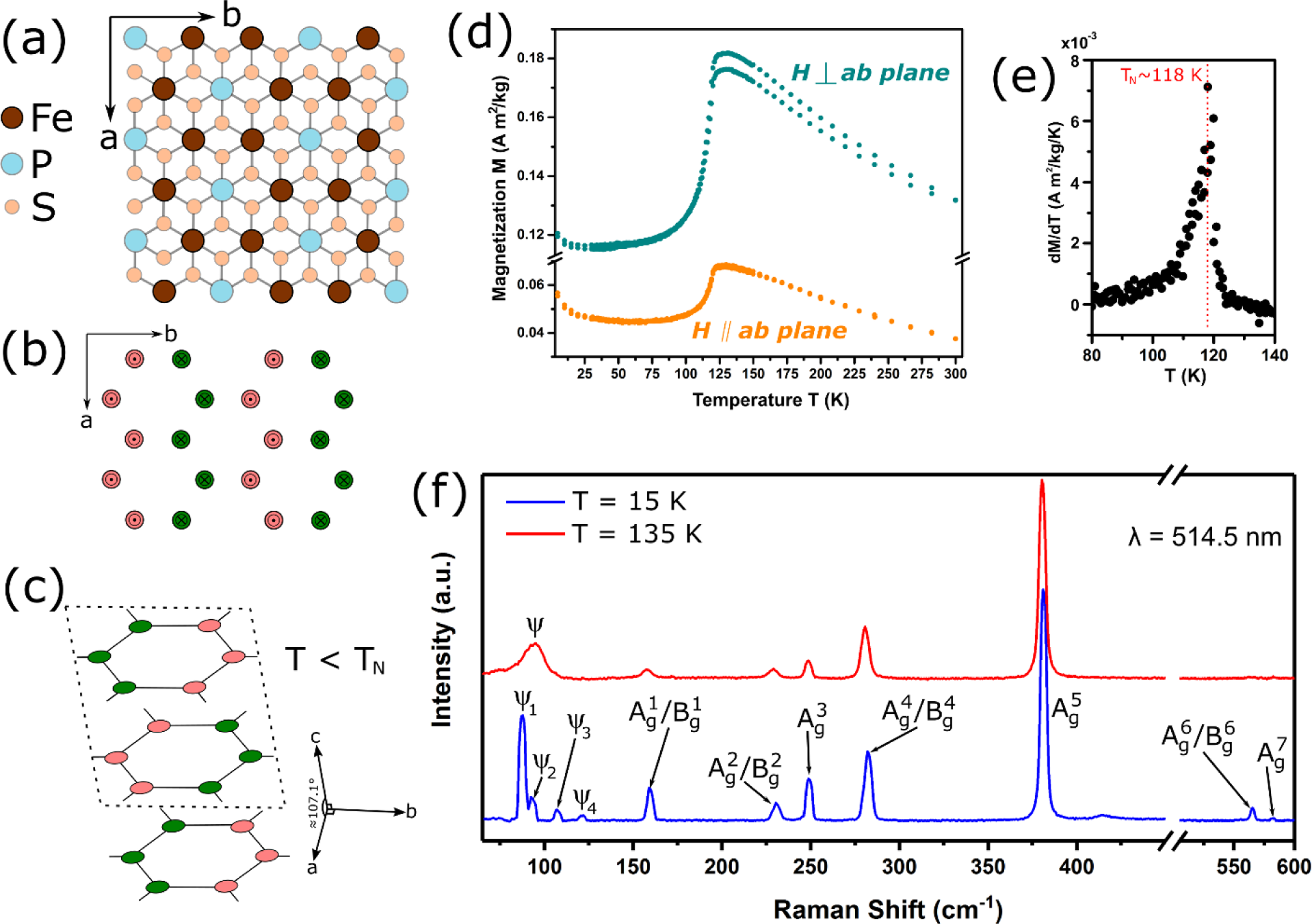
Large crystals of FePS3 were mechanically exfoliated using adhesive tape48 onto 300 nm SiO2 thermally grown on Si(100). Thicker flakes were selected for Raman spectroscopy measurements due to the more intense scattering signal. Bulk FePS3 crystallizes in the monoclinic structure with the space group C2/m. 49 In the plane, as is shown in Figure 1a, the Fe atoms form a honeycomb lattice. In the center of the Fe honeycomb lattice is a [P2S6]4− unit, where the P2 dimers orient normal to the surface and coordinate with six S atoms. The magnetic structure has been a source of controversy,31,49–52 where in both proposed structures, the moments of the large spin Fe2+ ions (S=2) are aligned normal to the plane and ordered in ferromagnetic chains that couple antiferromagnetically. The proposed structures differed from each other, however, by a 120° rotation of the ferromagnetic chains, as well as whether the layers themselves are ferromagnetically or antiferromagnetically coupled. Recent neutron scattering by D. Lançon et al.52 of large, single crystals of FePS3 confirmed the magnetic structure consists of ferromagnetic chains oriented along the -axis, with antiferromagnetically coupled layers along the -axis, as shown in Figures 1b and 1c.
Figure 1.

(a) Top view of the crystal structure of FePS3, where the Fe atoms form a honeycomb lattice. The a- and b-axes are labeled. (b) Magnetic structure of the Fe atoms in the plane below . The spins are collinear and normal to the surface, where pink indicates spin “up” (pointing out of the page) and green indicates spin “down” (pointing into the page). The ferromagnetic chains are aligned along the -axis.49 (c) Below , the layers are stacked antiferromagnetically, resulting in an increase in the unit cell along the c-axis. (d) Magnetization () vs. temperature () for a bulk crystal of FePS3, where the applied magnetic field () is parallel (orange) or perpendicular (cyan) to the plane. (e) of (d) with an inflection point at . (f) Raman spectra above () and below () .
Using a SQUID magnetometer, the magnetization of the FePS3 crystals (i.e. before exfoliation) was measured both parallel and perpendicular to the plane with an applied magnetic field of 0.1 T. The magnetization versus temperature from 5 K to 300 K is shown in Figure 1d. Similar to earlier reports,26,45,46 the strong anisotropy in FePS3 results in higher magnetization perpendicular to the plane, which is along the spin direction in the antiferromagnetic state. In a 2D antiferromagnet, is defined to be the temperature where there is a maximum in the slope of the magnetization vs. temperature curve.26 Thus, by taking the first derivative of the magnetization with respect to temperature (Figure 1e), is found to be ≈118 K. The broad maximum above in the magnetization vs. temperature for both parallel and perpendicular orientations is typical for low-dimensional magnetic systems and has been attributed to short-range spin-spin correlation.26
B. Raman Spectroscopy and Emergence of New Modes
The majority of the peaks in the Raman spectrum (Figure 1f) can be ascribed to phonons. From the symmetry of bulk FePS3, there are 30 zone-center phonons with irreducible representations: , of which only the and modes are Raman active. Figure 1f shows the Raman spectra of bulk FePS3 (laser excitation wavelength ) in the 180° back-scattering geometry at and , which corresponds to above and well below the Néel temperature, respectively. Phonons from the point () are labeled by whether they are an mode or a combination of / modes based on previous first principle calculations as well as their measured orientation-dependent frequency since a peak that is a combination of / modes will appear to shift in frequency as the light polarization is rotated with respect to the crystal orientation.46,53 The phonon modes , and are mostly ascribed to vibrations of the (P2S6)4− unit, and have similar frequencies for the different XPS3 materials due to the lack of contribution from the metal atom.53 While the and / modes in the spectra are similar in terms of frequency and intensity when comparing above and below the Néel temperature, significant changes are seen in the modes below 150 cm−1. Above the Néel temperature, there is a broad, asymmetric peak “” that exists even up to room temperature. At , however, there are four new modes that appear in this frequency range, labeled: , , , and .
The evolution of the Raman spectra of mode into four modes below has been observed since the 1980’s,27,42,43 yet there is still uncertainty regarding the nature of the transition and the origin of each component. It is well known that antiferromagnetic ordering in FePS3 leads to a doubling of the unit cell and subsequent zone folding, such that the M point in the paramagnetic Brillouin zone folds into the point of the antiferromagnetically ordered Brillouin zone. From the phonon dispersion calculated by X. Wang et al.,46 and appear to be M point phonons present below the Néel temperature due to magnetic-ordering induced zone folding. On the contrary, J.-U. Lee et al.45 attribute these modes to very different origins. From density functional theory calculations, they suggest is a point phonon without magnetic ordering, while and have both and point components, and would thus appear above . Other older publications also observed , but only comment on possible origins of and .43,54 Thus, although the emergence of these modes can provide insight on the transition to a magnetically ordered state in FePS3, there is still substantial debate as to the source of each mode. In this work, we shed light on the origin of , where we unequivocally show that it is a magnon.
C. Temperature Dependence of Raman Modes:
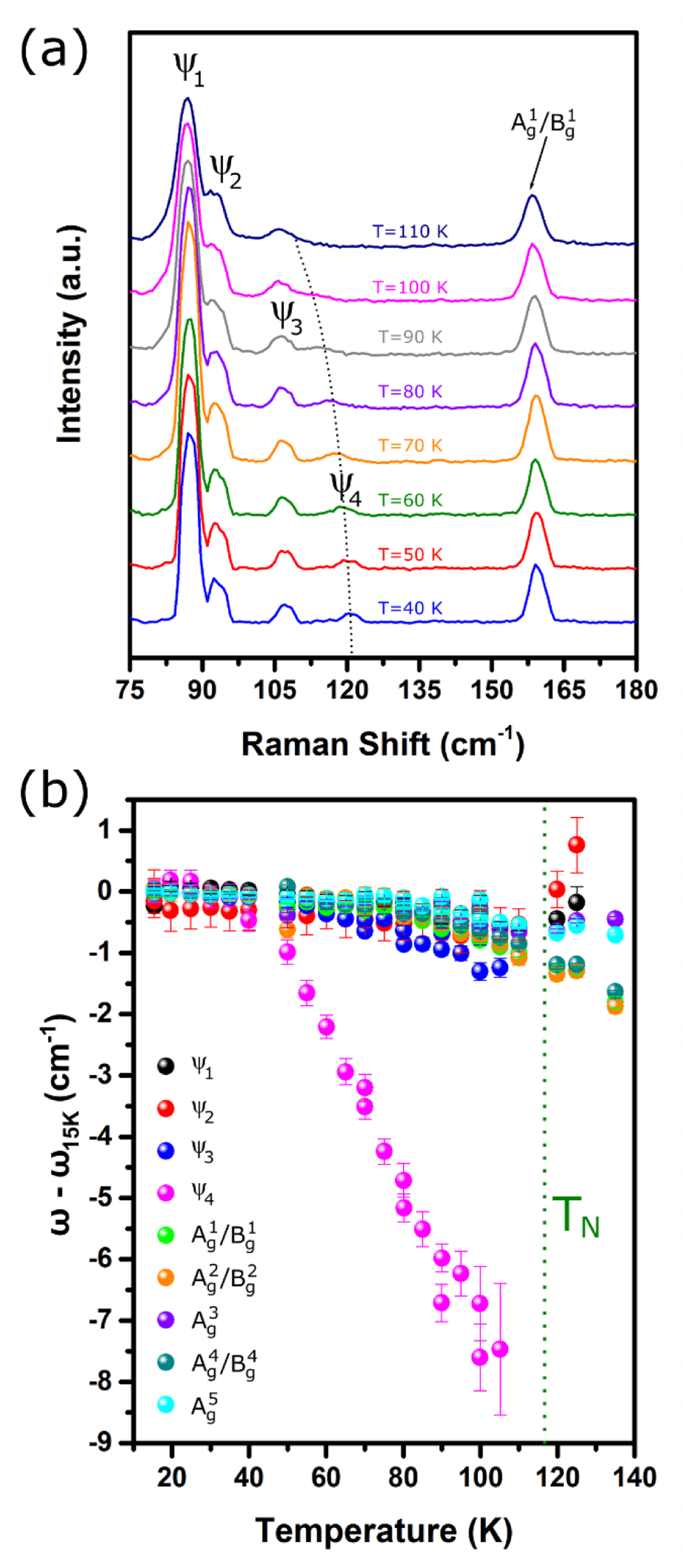
Figure 2a shows the temperature dependence of the Raman spectra from 110 K down to 40 K in steps of 10 K. is not observed until temperatures below 100 K, well below of ≈118 K. As the temperature is further decreased, displays a dramatic blueshift (higher in frequency) with temperature, especially compared with the other modes, including , , and the -point phonons. This effect is illustrated in Figure 2b, which shows the frequencies of the different modes relative to their frequency at 15 K (), including standard errors from fitting each peak with a Voigt function. While the frequency changes of the other modes only range from 0.01% to 0.57% (1 cm−1 to 2 cm−1 shifts), as is typical for anharmonic lattice effects, the frequency of changes by as much as 6.2% (8 cm−1 shift) as the temperature is increased to 100 K. The strong shift in frequency with respect to temperature offers the first indication that this mode is attributed to a magnon, as suggested by T. Sekine et al. in 1990 for FePS3.55 Similar magnon temperature dependence has been reported for three-dimensional (3D) antiferromagnets, such as FeF2,56,57 NiF2,58 and MnF2.59 The assignment of this mode to a magnon is further supported by recent inelastic neutron scattering measurements that observed a point magnon at ≈15.1 meV (122 cm−1),52,60 which coincides in frequency to our observed mode .
Figure 2.

(a) Temperature-dependent Raman spectra of bulk FePS3 below . The mode appears below 100 K and its frequency dramatically increases with decreasing temperature. (b) Frequencies of the various modes (relative to frequency at 15 K). While the other modes and phonon modes only shift by 1 cm−1 to 2 cm−1, as is expected by lattice anharmonic effects, the frequency of shifts by as much as 8 cm−1 before it is no longer measurable above 100 K.
D. Magnons and Magneto-Raman Spectroscopy of FePS3
Due to the contrasting behavior of phonons and magnons in an applied magnetic field, we utilize our unique magneto-Raman capabilities to investigate the magnetic field dependence of the modes in FePS3 to identify if they have magnetic character. We detail our findings from the magnetic-field dependent Raman spectra of FePS3. Then, by discussing how magnons behave under applied magnetic fields, we assign as a Raman-active magnon.
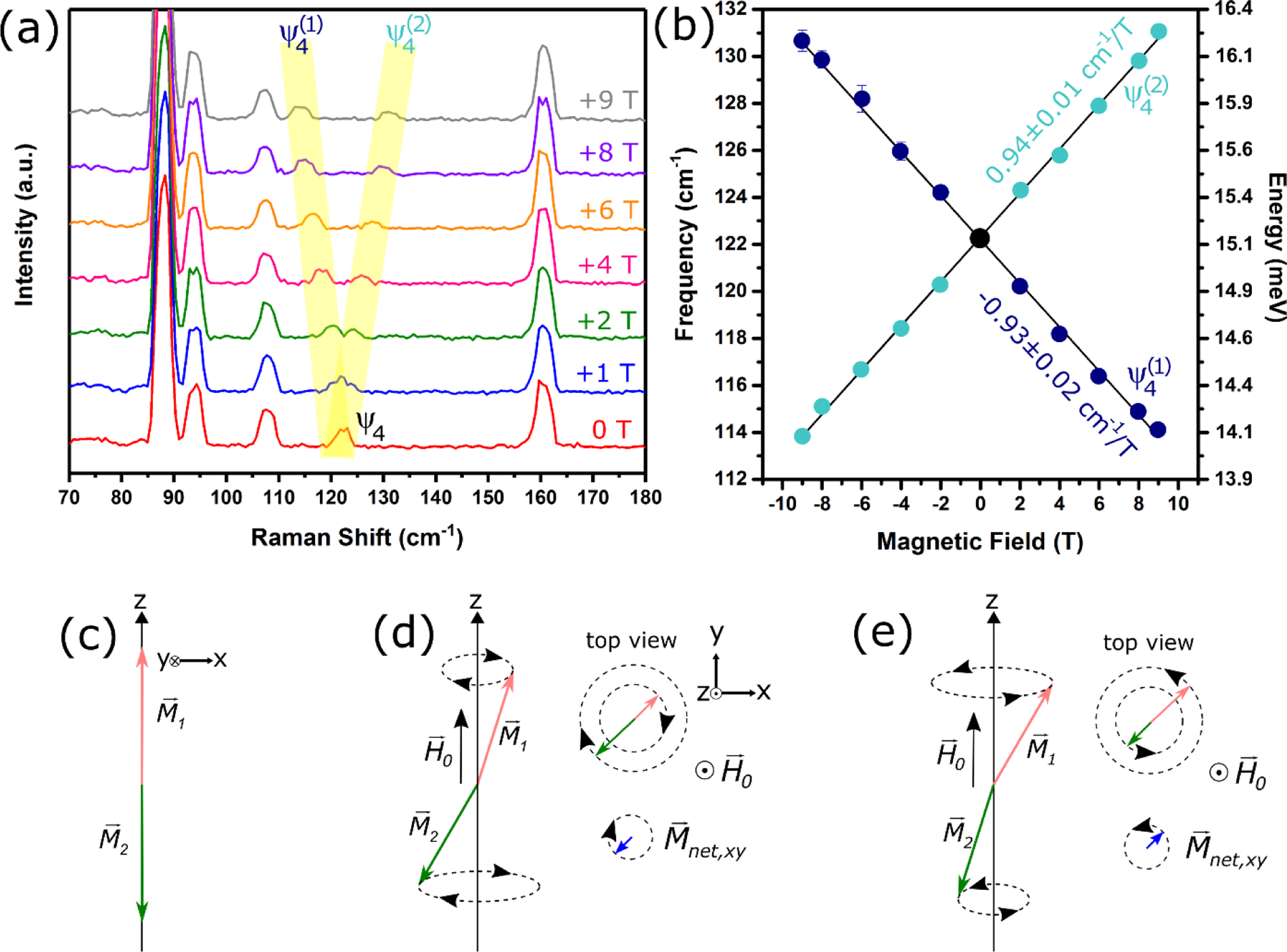
An exfoliated flake of bulk FePS3 was cooled to , after which a magnetic field was applied normal to the plane, i.e. parallel/antiparallel to the directions of the spins in the antiferromagnetically ordered state. As seen in Figure 3a, when the magnetic field is increased from 0 T to 9 T, splits into two modes, labeled as and , and the splitting increases with the applied magnetic field. Figure 3b shows the frequencies of and as a function of magnetic field, where the splitting is symmetric for positive and negative magnetic fields. From a linear fit, the extracted slopes for the and branches are 0.93 ± 0.02 cm−1/T and 0.94 ± 0.01 cm−1/T, respectively. This splitting only occurs for , where the other modes and phonon modes remain as single peaks. As discussed below, this behavior matches well with the expected magnetic field dependence of magnons.
Figure 3.

(a) Magnetic-field dependent Raman of FePS3 at , showing the splitting of into two components, and , where the frequency of () decreases (increases) with increasing magnetic field. (b) Frequency vs. magnetic field of and with the slopes of the linear fits for the two branches. (c) If we consider two magnetization sublattices (pink) and (green) in FePS3, in the ground state and point in the and directions. (d, e) The magnon spectrum includes two normal modes with net moments Mnet in the plane that precess in opposite directions. The degeneracy of these modes is broken in an applied field with the energies of the modes in (d) and (e) respectively decreasing and increasing with . In terms of this simple model, the modes in (d) and (e) would represent and in (b), respectively.
Spin waves, which as quasiparticles are called magnons, are collective excitations of the spins in magnetic materials. The classical dynamics of the magnetization at the -point in an antiferromagnet can be modeled following F. Keffer and C. Kittel61 as two interacting macrospins (Figure 3c) representing the spin-up sublattice magnetization (M1, pink) and spin-down sublattice magnetization (, green) that orient in the and directions in equilibrium, respectively. The excitation of a magnon causes the magnetization on both sublattices to precess about their equilibrium directions. Figures 3(d) and 3(e) show side and top view illustrations of the two normal modes for and precession. The frequencies of the normal modes are given by:
| (1) |
where is the exchange field, is the anisotropy field, is an applied field, and is the gyromagnetic ratio, which for free electrons ()62 is approximately 28.025 GHz/T, or 0.9348 cm−1/T. Without an applied magnetic field (), the frequencies of the magnon normal modes are degenerate (). However, an applied magnetic field along will cause a Zeeman splitting, leading to two separate modes with (Figure 3d) and (Figure 3e), both changing in magnetic field with a slope of . From Equation 1, a splitting of the mode with applied magnetic field offers a conclusive method to determine if a Raman mode is a magnon, as a magnetic field dependence is not expected for a typical phonon.
The behavior of the normal modes of magnons in a magnetic field described above matches with the observed magnetic-field dependence of , where the application of a magnetic field parallel to the spin direction leads to a lifting of the degeneracy of the magnon frequency into two components, and . The frequencies of the two non-degenerate magnon modes obey a linear response as a function of magnetic field, in agreement with Equation 1, with the two slopes of equal (within error) to the predicted slope for the free electron limit of . Using our experimental values of from the slopes of the linear fits in Figure 3b, we estimate that for bulk FePS3, the effective magnon g-factor is . From the full-width at half maximum (FWHM) value of approximately 3 cm−1, which includes instrument broadening, we estimate the lower bound of the magnon lifetime to be on the order of 10 picoseconds.
It should be noted that while it is possible that magneto-elastic effects could result in a phonon whose frequency strongly shifts with temperature, the splitting a phonon in a magnetic field due to symmetry lowering would require the phonon to be degenerate. For FePS3, only and modes are Raman active, and both are singlets. Based on the splitting of in a magnetic field, it cannot be a phonon. Thus, through magneto-Raman spectroscopy, we can unequivocally assign the mode at ≈122 cm−1 as a magnon. The extremely weak magnon dispersion measured by neutron diffraction experiments52 implies that this magnon we observed in bulk FePS3 herein is a quasi-2D magnon.
E. Magnon Symmetry Behavior and Magnetic Point Group
Raman scattering from magnons in both ferromagnets and antiferromagnets has been theoretically discussed by numerous authors, with one of the most cited works by P.A. Fleury and R. Loudon from 1968.40 In their work, the resulting polarization selection rules of Raman scattering from first order magnon excitations is purely antisymmetric with respect to the polarization of the incoming () and scattered () light. Thus, from their theory, a magnon mode can be observed only in the perpendicular (or crossed) polarization configurations (), regardless of the orientation with respect to the crystallographic axes. This theory has worked well to describe a variety of ferromagnetic and antiferromagnetic 3D materials with Raman-observed magnons, including FeF2,40 MnF2,40 YFeO3,63 and Cd1-xMnxTe.64
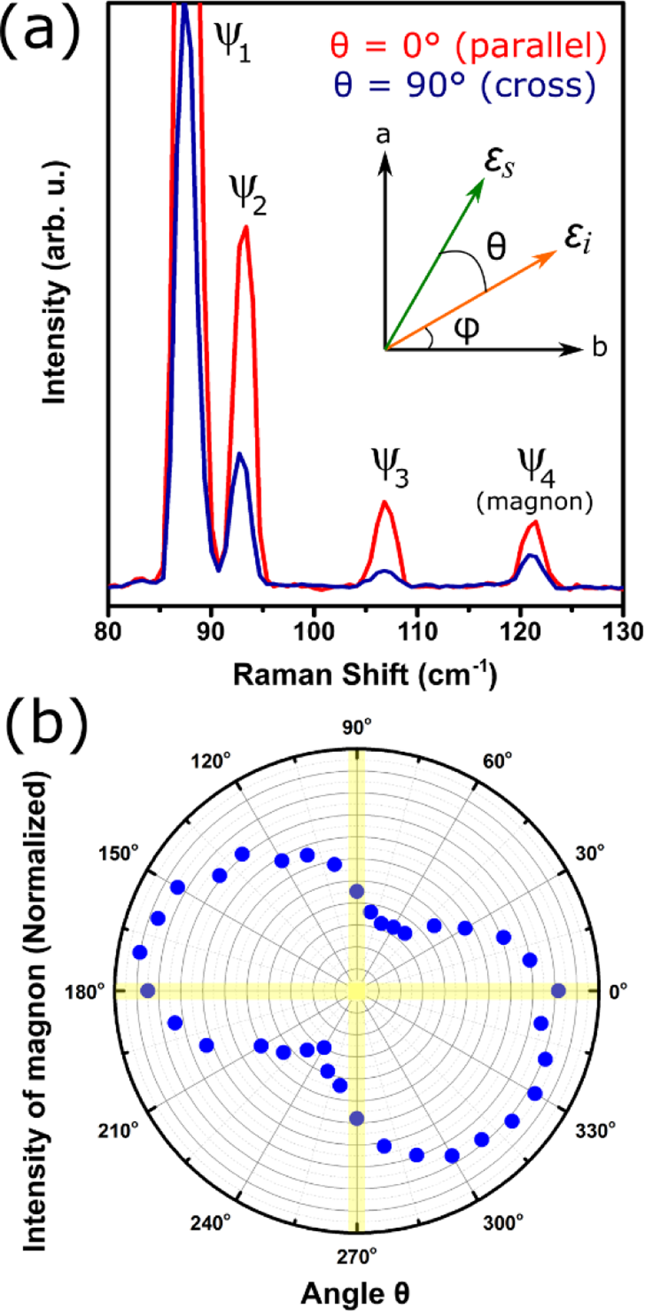
To probe the symmetry behavior of the magnon in FePS3, and were selected as shown in the inset of Figure 4a, where incoming light is at an arbitrary angle with respect to the crystallographic -axis and the scattered light is at a controlled angle with respect to . Figure 4a shows that the magnon in FePS3, can be observed in parallel polarization () in addition to perpendicular polarization (). When is varied between 0° and 360°, a two-fold symmetric pattern emerges where the magnon never disappears, but instead has a minimum at and maximum at , although these angle values are dependent on .
Figure 4.

(a) Raman spectra () showing intensity of magnon in parallel () and cross () polarization configurations at some arbitrary angle . Inset shows a schematic defining angles and with respect to the incoming () and scattered () light polarization and the - and - crystal axes. (b) Polar plot of intensity of the magnon as a function of at a constant angle . The intensity scale is represented by the radial lines that span from 0.1 to 1.
The theory presented in Fleury and Loudon applies rigorously to magnetic ions with orbital angular momentum ground states of L=0, or those with crystal-field quenching of the ground state to L=0,41 as is the case for the well-studied antiferromagnetic magnon example of FeF2.65,66 For materials that do not satisfy the L=0 ground state condition, such as FePS3,67 the magnetic excitations are more difficult to treat; the standard interpretation of the “antisymmetric” nature of the magnon Raman tensor does not apply. Instead, we use the symmetry properties of the magnetic point group of bulk FePS3 to understand the selection rules and polarization dependence of the magnon mode shown in Figure 4.
When a material orders antiferromagnetically, the symmetry generally lowers because the spin up and spin down sites are no longer equivalent. In this regard, for the magnetic point group, unitary operations are symmetry operations that preserve the spin direction. Other operations that do not preserve the spin direction may become allowed with the application of time reversal symmetry and are referred to as antiunitary operations. The magnetic point group for bulk FePS3, which considers both unitary and antiunitary operations is 2´/m, where the prime means time reversal. The Raman tensors for this magnetic group are given by the co-representations:68
The experimental 180° backscattering configuration incident on the honeycomb plane can access only the upper left block of these tensors, thus eliminating consideration of . The Raman intensity is proportional to , where and are the incident and scatter light polarization vectors, respectively.
It should be noted that if , , , and are purely real numbers, the intensity profile as a function of θ would necessarily have two nodes, i.e. two angles where the intensity goes to zero, which is contrary to the polar plot we observed in Figure 4b. These nodes, however, disappear from the symmetry response when the tensor elements are complex, which is the general case for absorbing materials:69
where , , and are the complex phase factors of the real tensor elements , , and with respect to . Considering the complex tensor elements, there are multiple combinations of the amplitude and phase factors that reproduce the polar plot in Figure 4b. However, there are two points that remain clear. First, at least one of the phase factors must be non-zero in FePS3 to agree with the non-nodal nature of the experimental polar plot. Second, the symmetry response of the magnon in FePS3 deviates strongly from the antisymmetric nature predicted by Fleury and Loudon40 for magnetic ions with L=0 ground state. Thus, there is no reason, a-priori, to expect that a magnon will appear only in cross-polarization configurations for these honeycomb-like structures, such as FePS3, -RuCl3, or CrI3,70 without considering the magnetic point group symmetry and associated Raman tensors.
In principle, one should be able to use the generators of rotations (i.e. angular momenta , , and ) generally given in the character table as guides for the magnon symmetry behavior. For example, in FePS3 where the spins are oriented along the -direction (normal to plane), the excitation of a magnon reduces the magnitude of the magnetization , which is then converted to the precession of the spins in the () plane, and , as depicted in Figures 3d and 3e. Thus, a magnon in FePS3, which is a magnetization rotation in the plane, can only have the same symmetry as and , which are generators of rotations. While the character tables that state these transformations are calculated for the non-magnetic space groups,71–73 we were unable to find a reference that tabulated these for the magnetic space groups. Deriving such transformations is out beyond the scope of this work.
III. CONCLUSIONS
In conclusion, we examined the temperature- and magnetic field-dependent Raman spectra of bulk FePS3, where new Raman-active modes appear in the antiferromagnetically ordered state. One of these modes (), with a frequency of ≈122 cm−1 (15.1 meV), was previously assigned as a phonon mode appearing due to zone folding. However, the strong temperature shift (compared to other phonon modes) and splitting (and linear shifting) of the mode with applied magnetic field observed herein allows this mode to be unequivocally assigned as a magnon. We also investigated the magnon symmetry behavior, which is not purely antisymmetric, and explained its behavior using the magnetic point group of FePS3 (2′/m). This work will aid in future studies of magnetic excitations in similar magnetic layered materials, such as -RuCl3 and CrI3, and demonstrates temperature- and magnetic field-dependent Raman spectroscopy as a technique to probe magnon phenomena in 2D materials, including for possible magnon transport applications. The quasi-2D magnetic nature of bulk FePS3, with weak interlayer exchange coupling, indicates that the magnon in bulk FePS3 is also quasi-2D. To the best of our knowledge, this is the first verification of a quasi-2D magnon in a layered material using magneto-Raman spectroscopy.
IV. EXPERIMENTAL DETAILS
Bulk FePS3 crystals were purchased from 2D Semiconductors† and then exfoliated via the adhesive tape onto 300 nm SiO2 thermally grown on Si(100).48 Based on optical contrast and the lack of silicon peak observed through the exfoliated flake studied herein, we estimate the thickness to be between 50 nm and 100 nm. Raman spectra were measured with the 514.5308 nm excitation wavelength of an Argon ion laser in the 180° backscattering configuration using a triple grating Raman spectrometer (Horiba JY T64000†, 1800 mm−1 grating) coupled to a liquid nitrogen cooled CCD detector. Polarization was selected and controlled using ultra broadband polarizers and achromatic half wave plates. To perform temperature- and magnetic-field dependent Raman, the sample was placed into an attoDRY1000 cryostat (Attocube Inc.†), where the sample holder is pumped to ≈1×10⁻³ Pa (≈7×10⁻⁶ Torr), backfilled with helium gas, and cooled. Micrometer-sized flakes were studied by focusing the laser with a white light camera onto the sample with a low-temperature, magnetic field compatible objective (50×, N.A. 0.82) and xyz nano-positioners. Integration times were approximately 12 minutes and the laser power was kept below 300 μW to reduce local heating of the sample. Spectra with applied magnetic field were corrected for Faraday rotation in the objective using half wave plates external to the cryostat. Magnetization vs. temperature measurements were performed on a Superconducting Quantum Interference Device-based Vibrating Sample Magnetometer (SQUID-VSM; Quantum Design, Inc.†). The piece of the unexfoliated FePS3 crystal was mounted on to a quartz holder using GE varnish (LakeShore Cryotronics, Inc.†), and then measured under vacuum (< 1 kPa). The magnetization was measured under an applied magnetic field μ0H = 0.1 T from 5 K to 300 K and back down to 5 K. The temperature was varied in steps of 5 K between 5 K – 300 K, 1 K between 30 K – 150 K, and then 5 K between 150 K – 300 K.
V. ACKNOWLEDGEMENTS
A.M., T.T.M., and A.R.H.W. would like to acknowledge the National Institute of Standards and Technology (NIST)/National Research Council Postdoctoral Research Associateship Program and NIST-STRS (Scientific and Technical Research and Services) for funding. Work at The Ohio State University was supported by the Center for Emergent Materials, an NSF MRSEC under grant DMR-1420451. We would also like to acknowledge Prof. Natalia Drichko for insightful discussions and Hans Nembach for a careful readthrough of the paper.
Footnotes
Certain commercial equipment, instruments, or materials are identified in this manuscript in order to specify the experimental procedure adequately. Such identification is not intended to imply recommendation or endorsement by the National Institute of Standards and Technology, nor is it intended to imply that the materials or equipment are necessarily the best available for the purpose.
REFERENCES:
- [1].Novoselov KS, Geim AK, Morozov SV, Jiang D, Zhang Y, Dubonos SV, Grigorieva IV, and Firsov AA Electric Field Effect in Atomically Thin Carbon Films. Science 306, 666–669 (2004). [DOI] [PubMed] [Google Scholar]
- [2].Wang QH, Kalantar-Zadeh K, Kis A, Coleman JN, and Strano MS Electronics and Optoelectronics of Two-Dimensional Transition Metal Dichalcogenides. Nature Nanotechnol. 7, 699–712 (2012). [DOI] [PubMed] [Google Scholar]
- [3].Novoselov KS, Mishchenko A, Carvalho A, and Neto AHC 2D Materials and Van der Waals Heterostructures. Science 353, aac9439 (2016). [DOI] [PubMed] [Google Scholar]
- [4].Schulman DS, Arnold AJ, Razavieh A, Nasr J, and Das S. The Prospect of Two-Dimensional Heterostructures: A Review of Recent Breakthroughs. IEEE Nanotechnol. Mag 11, 6–17 (2017). [Google Scholar]
- [5].Lin Z, McCreary A, Briggs N, Subramanian S, Zhang K, Sun Y, Li X, Borys NJ, Yuan H, Fullteron-Shirey SK, et al. 2D Materials Advances: From Large Scale Synthesis and Controlled Heterostructures to Improved Characterization Techniques, Defects and Applications. 2D Mater. 3, 042001 (2016). [Google Scholar]
- [6].Mak KF and Shan J. Photonics and Optoelectronics of 2D Semiconductor Transition Metal Dichalcogenides. Nat. Photonics 10, 216–226 (2016). [Google Scholar]
- [7].Schaibley JR, Yu H, Clark G, Rivera P, Ross JS, Seyler KL, Yao W, and Xu X. Valleytronics in 2D Materials. Nat. Rev. Mater 1, 16055 (2016). [Google Scholar]
- [8].Manzeli S, Ovchinnikov D, Pasquier D, Yazyev OV, and Kis A. 2D Transition Metal Dichalcogenides. Nat. Rev. Mater 2, 17033 (2017). [Google Scholar]
- [9].Akinwande D, Brennan CJ, Bunch JS, Egberts P, Felts JR, Gao H, Huang R, Kim J-S, Li T, Li Y, et al. A Review on Mechanics and Mechanical Properties of 2D Materials-Graphene and Beyond. Extreme Mech. Lett 13, 42–77 (2017). [Google Scholar]
- [10].Burch KS, Mandrus D, and Park JG Magnetism in Two-Dimensional van der Waals Materials. Nature 563, 47–52 (2018). [DOI] [PubMed] [Google Scholar]
- [11].Gong C, and Zhang X. Two-Dimensional Magnetic Crystals and Emergent Heterostructure Devices. Science 363, 706–717 (2019). [DOI] [PubMed] [Google Scholar]
- [12].Lee C, Wei XD, Kysar JW, and Hone J. Measurement of the Elastic Properties and Antrinsic Strength of Monolayer Graphene. Science 321, 385–388 (2008). [DOI] [PubMed] [Google Scholar]
- [13].Castellanos-Gomez A, Poot M, Steele GA, van der Zant HSJ, Agrait N, and Rubio-Bollinger G. Elastic Properties of Freely Suspended MoS2 Nanosheets. Adv. Mater 24, 772–775 (2012). [DOI] [PubMed] [Google Scholar]
- [14].Huang B, Clark G, Klein DR, MacNeill D, Navarro-Moratalla E, Seyler KL, Wilson N, McGuire MA, Cobden DH, Xiao D, et al. Electrical Control of 2D Magnetism in Bilayer CrI3. Nat. Nanotechnol 13, 544–548 (2018). [DOI] [PubMed] [Google Scholar]
- [15].Jiang SW, Shan JJ and Mak KF Electric-Field Switching of Two-Dimensional van der Waals Magnets. Nat. Mater 17, 406–410 (2018). [DOI] [PubMed] [Google Scholar]
- [16].Ross JS, Klement P, Jones AM, Ghimire NJ, Yan J, Mandrus DG, Taniguchi T, Watanabe K, Kitamura K, Yao W, et al. Electrically Tunable Excitonic Light-Emitting Diodes Based on Monolayer WSe2 p-n Junctions. Nat. Nanotechnol 9, 268–272 (2014). [DOI] [PubMed] [Google Scholar]
- [17].Akinwande D, Petrone N, and Hone J. Two-Dimensional Flexible Nanoelectronics. Nat. Commun 5, 5678 (2014). [DOI] [PubMed] [Google Scholar]
- [18].Zhang S, Hill HM, Moudgil K, Richter CA, Hight Walker AR, Barlow S, Marder SR, Hacker CA, and Pookpanratana SJ Controllable, Wide-Ranging n-Doping and p-Doping of Monolayer Group 6 Transition-Metal Disulfides and Diselenides. Adv. Mater 30, 1802991 (2018). [DOI] [PubMed] [Google Scholar]
- [19].Guo YQ, Xu K, Wu CZ, Zhao JY, and Xie Y. Surface Chemical-Modification for Engineering the Intrinsic Physical Properties of Inorganic Two-Dimensional Nanomaterials. Chem. Soc. Rev 44, 637–646 (2015). [DOI] [PubMed] [Google Scholar]
- [20].Li MY, Chen CH, Shi YM, and Li LJ Heterostructures Based on Two-Dimensional Layered Materials and Their Potential Applications. Mater. Today 19, 322–335 (2016). [Google Scholar]
- [21].Chumak AV, Vasyuchka VI, Serga AA, and Hillebrands B, B. Magnon Spintronics. Nat. Phys 11, 453–461 (2015). [Google Scholar]
- [22].Xing W, Qiu L, Wang X, Yao Y, Ma Y, Cai R, Jia S, Xie XC, and Han W. Magnon Transport in Quasi-Two-Dimensional van der Waals Antiferromagnets. Phys. Rev. X 9, 011026 (2019). [Google Scholar]
- [23].Gong C, Li L, Li Z, Ji H, Stern A, Xia Y, Cao T, Bao W, Wang C, Wang Y, et al. Discovery of Intrinsic Ferromagnetism in Two-Dimensional van der Waals Crystals. Nature 546, 265–269 (2017). [DOI] [PubMed] [Google Scholar]
- [24].Huang B, Clark G, Navarro-Moratalla E, Klein D, Cheng R, Seyler K, Zhong D, Schmidgall E, McGuire MA, Cobden DH, et al. Layer-Dependent Ferromagnetism in a van der Waals Crystal Down to the Monolayer Limit. Nature 546, 270–273 (2017). [DOI] [PubMed] [Google Scholar]
- [25].Jiang SW, Li LZ, Wang ZF, Mak KF, and Shan J. Controlling Magnetism in 2D CrI3 by Electrostatic Doping. Nat. Nanotechnol 13, 549–553 (2018). [DOI] [PubMed] [Google Scholar]
- [26].Joy PA and Vasudevan S. Magnetism in the Layered Transition-Metal Thiophosphates MPS3 (M = Mn, Fe, and Ni). Phys. Rev. B 46, 5425–5433 (1992). [DOI] [PubMed] [Google Scholar]
- [27].Balkanski M, Jouanne M, Ouvrard G, and Scagliotti M. Effects due to Spin Ordering in Layered MPX3 Compounds Revealed by Inelastic Light-Scattering. J. Phys. C-Solid State 20, 4397–4413 (1987). [Google Scholar]
- [28].Chittari BL, Park Y, Lee D, Han M, MacDonald AH, Hwang E, and Jung J. Electronic and Magnetic Properties of Single-layer MPX3 Metal Phosphorous Trichalcogenides. Phys. Rev. B 94, 184428 (2016). [Google Scholar]
- [29].Haines CRS, Coak MJ, Wildes AR, Lampronti GI, Liu C, Nahai-Williamson P, Hamidov H, Daisenberger D, and Saxena SS Pressure-Induced Electronic and Structural Phase Evolution in the van der Waals Compound FePS3. Phys. Rev. Lett 121, 266801 (2018). [DOI] [PubMed] [Google Scholar]
- [30].Tsurubayashi M, Kodama K, Kano M, Ishigaki K, Uwatoko Y, Watanabe T, Takase K, and Takano T. Metal-Insulator Transition in Mott-Insulator FePS3. AIP Adv. 8, 101307 (2018). [Google Scholar]
- [31].Rule KC, McIntyre GJ, Kennedy SJ, and Hicks TJ Single-Crystal and Powder Neutron Diffraction Experiments on FePS3: Search for the Magnetic Structure. Phys. Rev. B 76, 134402 (2007). [Google Scholar]
- [32].Li X-L, Qiao X-F, Han W-P, Lu Y, Tan Q-H, Liu X-L, and Tan P-H Layer Number Identification of Intrinsic and Defective Multilayered Graphenes Up to 100 Layers by the Raman Mode Intensity from Substrates. Nanoscale 7, 8135–8141 (2015). [DOI] [PubMed] [Google Scholar]
- [33].Liang LB, Zhang J, Sumpter BG, Tan Q-H, Tan P-H, and Meunier V. Low-Frequency Shear and Layer-Breathing Modes in Raman Scattering of Two-Dimensional Materials. ACS Nano 11, 11777–11802 (2017). [DOI] [PubMed] [Google Scholar]
- [34].Mohiuddin TMG, Lombardo A, Nair RR, Bonetti A, Savini G, Jalil R, Bonini N, Basko DM, Galiotis C, Marzari N, et al. Uniaxial Strain in Graphene by Raman Spectroscopy: G Peak Splitting, Gruneisen Parameters, and Sample Orientation. Phys. Rev. B 79, 205433 (2009). [Google Scholar]
- [35].Beams R, Cancado LG, and Novotny L. Raman Characterization of Defects and Dopants in Graphene. J. Phys.-Condens. Mat 27, 083002 (2015). [DOI] [PubMed] [Google Scholar]
- [36].del Corro E, Botello-Mendez A, Gillet Y, lias AL, Terrones H, Feng S, Fantini C, RHodes D, Pradhan N, Balicas L. et al. Atypical Exciton-Phonon Interactions in WS2 and WSe2 Monolayers Revealed by Resonance Raman Spectroscopy. Nano Lett. 16, 2363–2368, (2016). [DOI] [PubMed] [Google Scholar]
- [37].McCreary A, Simpson JR, Wang Y, Rhodes D, Fujisawa K, Balicas L, Dubey M, Crespi VH, Terrones M, and Hight Walker AR Intricate Resonant Raman Response in Anisotropic ReS2. Nano Lett. 17, 5897–5907 (2017). [DOI] [PubMed] [Google Scholar]
- [38].Zhang KN, Bao C, Gu Q, Ren X, Zhang H, Deng K, Wu Y, Li Y, Feng J, and Zhou S. Raman Signatures of Inversion Symmetry Breaking and Structural Phase Transition in Type-II Weyl Semimetal MoTe2. Nat. Commun 7, 13552 (2016). [DOI] [PMC free article] [PubMed] [Google Scholar]
- [39].Tian Y, Gray MJ, Ji HW, Cava RJ, Burch KS Magneto-Elastic Coupling in a Potential Ferromagnetic 2D Atomic Crystal. 2D Mater. 3, 025035 (2016). [Google Scholar]
- [40].Fleury PA and Loudon R. Scattering of Light By One- and Two-Magnon Excitations. Phys. Rev 166, 514–530 (1968). [Google Scholar]
- [41].Sethi A, Byrum T, McAuliffe RD, Gleason SL, Slimak JE, Shoemaker DP, and Cooper SL Magnons and Magnetodielectric Effects in CoCr2O4: Raman Scattering Studies. Phy. Rev. B 95, 174413 (2017). [Google Scholar]
- [42].Scagliotti M, Jouanne M, Balkanski M, Ouvrard G, and Benedek G. Raman-Scattering in Antiferromagnetic FePS3 and FePSe3 Crystals. Phys. Rev. B 35, 7097–7104 (1987). [DOI] [PubMed] [Google Scholar]
- [43].Bernasconi M, Marra GL, Benedek G, Miglio L, Jouanne M, Julien C, Scagliotti M, and Balkanski M. Lattice-Dynamics of Layered MPX3 (M=Mn, Fe, Ni, Zn, X=S, Se) Compounds. Phys. Rev. B 38, 12089–12099 (1988). [DOI] [PubMed] [Google Scholar]
- [44].Kuo C-T, Neumann M, Balamurugan K, Park HJ, Kang S, Shiu HW, Kang JH, Hong BH, Han M, Noh TW, and Park J-G Exfoliation and Raman Spectroscopic Fingerprint of Few-Layer NiPS3 van der Waals Crystals. Sci. Rep 6, 20904 (2016). [DOI] [PMC free article] [PubMed] [Google Scholar]
- [45].Lee J-U, Lee S, Ryoo SH, Kang S, Kim TY, Kim P, Park C-H, Park J-G, and Cheong H. Ising-Type Magnetic Ordering in Atomically Thin FePS3. Nano Lett. 16, 7433–7438 (2016). [DOI] [PubMed] [Google Scholar]
- [46].Wang XZ, Du K, Liu YYF, Hu P, Zhang J, Zhang Q, Owen MHS, Lu X, Gan CK, and Sengupta P. Raman Spectroscopy of Atomically Thin Two-Dimensional Magnetic Iron Phosphorus Trisulfide (FePS3) Crystals. 2D Mater. 3, 031009 (2016). [Google Scholar]
- [47].Wildes AR, Roessli B, Lebech B, and Godfrey W. Spin Waves and the Critical Behaviour of the Magnetization in MnPS3. J. Phys.: Condens. Matter 10, 6417–6428 (1998). [Google Scholar]
- [48].Novoselov KS, Jiang D, Schedin F, Booth TJ, Khotkevich VV, Morozov SV, and Geim AK Two-Dimensional Atomic Crystals. P. Natl. Acad. Sci. USA 102, 10451–10453 (2005). [DOI] [PMC free article] [PubMed] [Google Scholar]
- [49].Leflem G, Brec R, Ouvard G, Louisy A, and Segransan P. Magnetic-Interactions in the Layer Compounds MPX3 (M=Mn, Fe, Ni; X=S, Se). J. Phys. Chem. Solids 43, 455–461 (1982). [Google Scholar]
- [50].Kurosawa K, Saito S, and Yamaguchi Y. Neutron-Diffraction Study on MnPS3 and FePS3. J. Phys. Soc. Jpn 52, 3919–3926 (1983). [Google Scholar]
- [51].Rule KC, Kennedy SJ, Goossens DJ, Mulders AM, and Hicks TJ Contrasting Antiferromagnetic Order Between FePS3 and MnPS3. Appl. Phys. A-Mater 74, S811–S813 (2002). [Google Scholar]
- [52].Lancon D, Walker HC, Ressouche E, Ouladdiaf B, Rule KC, McIntyre GJ, Hicks TJ, Ronnow HM, and Wildes AR Magnetic Structure and Magnon Dynamics of the Quasi-Two-Dimensional Antiferromagnet FePS3. Phys. Rev. B 94, 214407 (2016). [Google Scholar]
- [53].Hashemi A, Komsa HP, Puska M, and Krasheninnikov AV Vibrational Properties of Metal Phosphorus Trichalcogenides from First-Principles Calculations. J. Phys. Chem C 121, 27207–27217 (2017). [Google Scholar]
- [54].Scagliotti M, Jouanne M, Balkanski M, and Ouvrard G. Spin Dependent Phonon Raman-Scattering in Antiferromagnetic FePS3 Layer-Type Compound. Solid State Commun. 54, 291–294 (1985). [Google Scholar]
- [55].Sekine T, Jouanne M, Julien C, and Balkanski M. Light-Scattering Study of Dynamic Behavior of Antiferromagnetic Spins in the Layered Magnetic Semiconductor FePS3. Phys. Rev. B 42, 8382–8393 (1990). [DOI] [PubMed] [Google Scholar]
- [56].Fleury PA, Porto SPS, Cheesman LE, and Guggenheim HJ Light Scattering by Spin Waves in FeF2. Phys. Rev. Lett 17, 84–87 (1966). [Google Scholar]
- [57].Ohlmann RC and Tinkham M. Antiferromagnetic Resonance in FeF2 at Far-Infrared Frequencies. Phys. Rev 123, 425–434 (1961). [Google Scholar]
- [58].Fleury PA Paramagnetic Spin Waves and Correlation Functions in NiF2. Phys. Rev 180, 591–593 (1969). [Google Scholar]
- [59].Johnson FM and Nethercot AH Antiferromagnetic Resonance in MnF2. Phys. Rev 114, 705–716 (1959). [Google Scholar]
- [60].Wildes AR, Rule KC, Bewley RI, Enderle M. and Hicks TJ The Magnon Dynamics and Spin Exchange Parameters of FePS3. J. Phys.-Condens. Mat 24, 416004 (2012). [DOI] [PubMed] [Google Scholar]
- [61].Keffer F. and Kittel C. Theory of Antiferromagnetic Resonance. Phys. Rev 85, 329–337 (1952). [Google Scholar]
- [62].Mohr PJ, Newell DB, and Taylor BN CODATA Recommended Values of the Fundamental Physical Constants: 2014. J. Phys. Chem. Ref. Data 45, 043102 (2016). [DOI] [PMC free article] [PubMed] [Google Scholar]
- [63].White RM, Nemanich RJ, and Tsang C. Raman-Scattering from Magnons in Rare-Earth Ortho-Ferrites. J. Magn. Magn. Mater 15–18, 773–774 (1980). [Google Scholar]
- [64].Venugopalan S, Petrou A, Galazka RR, Ramdas AK, and Rodriguez S. Raman-Scattering by Phonons and Magnons in Semimagnetic Semiconductors - Cd1-xMnxTe. Phys. Rev. B 25, 2681–2696 (1982). [Google Scholar]
- [65].Khomskii DI Transition Metal Compounds. (Cambridge University Press, 2014). [Google Scholar]
- [66].Stout JW and Reed SA The Crystal Structure of MnF2, FeF2, CoF2, NiF2 and ZnF2. J. Am. Chem. Soc 76, 5279–5281 (1954). [Google Scholar]
- [67].Jernberg P, Bjarman S, and Wappling R. FePS3: A First-Order Phase Transition in a “2D” Ising Antiferromagnet. J. Magn. Magn. Mater 46, 178–190 (1984). [Google Scholar]
- [68].Cracknell AP Scattering Matrices for Raman Effect in Magnetic Crystals. J. Physics Part C Solid 2, 500–511 (1969). [Google Scholar]
- [69].Ribeiro HB, Pimenta MA, de Matos CJS, Moreira RL, Rodin AS, Zapata JD, de Souza EAT, and Neto AHC Unusual Angular Dependence of the Raman Response in Black Phosphorus. ACS Nano 9, 4270–4276 (2015). [DOI] [PubMed] [Google Scholar]
- [70].Jin W, kim HH, Ye Z, Li S, Rezaie P, Diaz F, Siddiq S, Wauer E, Yang B, Li C, Tian S, et al. Raman Fingerprint of Two Terahertz Spin Wave Branches in a Two-Dimensional Honeycomb Ising Ferromagnet. Nat. Commun 9, 5122 (2018). [DOI] [PMC free article] [PubMed] [Google Scholar]
- [71].Aroyo MI, Kirov A, Capillas C, Perez-Mato JM, and Wondratschek H. Bilbao Crystallographic Server: II. Representations of Crystallographic Point Groups and Space Groups. Acta Crystallogr. A 62, 115–128 (2006). [DOI] [PubMed] [Google Scholar]
- [72].Aroyo MI, Perez-Mato JM, Capillas C, Kroumova E, Ivantchev S, Madariaga G, Kirov A, Wondratschek H. Bilbao Crystallographic Server: I. Databases and Crystallographic Computing Programs. Z. Kristallogr 221, 15–27 (2006). [Google Scholar]
- [73].Aroyo MI, Kirov A, Capillas C, Perez-Mato JM, and Wondratschek H. Crystallography Online: Bilbao Crystallographic Server. Bulg. Chem. Commun 43, 183–197 (2011). [Google Scholar]


