Abstract
Long non-coding RNAs (lncRNAs) have been previously researched in ankylosing spondylitis (AS). Nevertheless, there are few studies of lncRNAs and mRNAs associated with the pathogenesis of AS. Differentially expressed lncRNAs (DElncRNAs) and mRNAs (DEmRNAs) between AS and normal samples were assessed using the R limma package. DOSE packages and ‘clusterProfiler’ were exploited for gene enrichment analysis. The functional association of proteins and protein interactions was assessed using the STRING database. To investigate the important genes and subnetworks in the protein-protein interaction network, the MCODE plug-in in the Cytoscape software was utilized. The gene mRNA was examined via reverse transcription-quantitative PCR. In total, 152 DEmRNAs and 204 DElncRNAs were observed between normal and AS samples. A total of 68 candidate genes related to DElncRNA were identified. These candidate genes were enriched in 30 cellular component terms, 22 molecular functions, 83 biological processes, 9 Kyoto Encyclopedia of Genes and Genomes, and 36 disease ontology pathways. NONHSAG037054.2 was the most related lncRNA to genes, and GABPA was the most connected gene to lncRNA in AS. The NCBI/GenBank accession number of the lncRNA NONHSAG037054.2 was not found because it is not included in NCBI. The information of lncRNA NONHSAG037054.2 can be found at the website (http://www.noncode.org/show_gene.php?id=NONHSAG037054 and https://www.genecards.org/cgi-bin/carddisp.pl?gene=ACAP2-IT1). In total, 13 microRNAs (miRNAs) and 46 miRNAs associated with NONHSAG037054.2 and GABPA, respectively, were found. A total of 173 RNA-binding protein genes were associated with both NONHSAG037054.2 and GABPA. In addition, GABPA was downregulated in AS samples, suggesting it may have diagnostic value in AS. In conclusion, NONHSAG037054.2 and GABPA are associated with AS. GABPA was downregulated in AS, and it could serve as a novel diagnostic factor for AS.
Keywords: ankylosing spondylitis, long non-coding RNAs, NONHSAG037054.2, GABPA
Introduction
Ankylosing spondylitis (AS) is a long-term inflammatory rheumatic disease resulting from an autoimmune imbalance (1), and it is a type of spondyloarthropathy (2). Spinal stiffness and persistent back pain are the most typical signs of AS (3). In addition, peripheral (spondylitis and arthritis) and extra musculoskeletal manifestations, such as monocular uveitis, inflammatory bowel disease, psoriasis and osteoporosis, are also common (4,5). A meta-analysis, which included 8 studies, among 2236 AS patients revealed that the prevalence rates for arthritis (29.7%), enthesitis (28.8%), psoriasis (10.2%), and inflammatory bowel disease (4.1%) were similar to non-radiographic axial spondyloarthritis (nr-axSpA), except for that uveitis was higher in AS (23.0%) than nr-axSpA (6). The mainstay of treatment for AS is medication, including interleukin-17 inhibitors, tumor necrosis factor inhibitors and non-steroidal anti-inflammatory medications. Janus kinase inhibitors have also demonstrated effectiveness in easing AS symptoms (5). However, there remain some AS patients who do not respond to any of these drugs. Thus, it is crucial to explore the underlying mechanisms of AS to elucidate the pathogenesis of AS and provide additional information for the development of diagnostic, therapeutic and prognostic monitoring tools for patients with AS.
Long non-coding RNAs (lncRNAs) are transcripts >200 nucleotides that do not code for proteins (7). LncRNAs are involved in controlling gene expression at a variety of levels to create epigenetic, transcriptional and post-transcriptional effects and exert their biological functions through different molecular mechanisms (8,9). LncRNAs are important for the control of biological processes, including immune response (10), cell proliferation, migration, invasion (11,12) and apoptosis (13). Recently, it has become increasingly evident how lncRNAs modulate the pathophysiology of autoimmune disorders (14). For instance, Li et al (14) examined the profiles of mRNA and lncRNA and found that mRNA and lncRNA expression patterns differed between patients with AS vs. healthy controls, although the regulatory mechanism of lncRNA in AS remains unclear. Furthermore, in T cells of AS, peripheral blood mononuclear cells, whole blood cells, and lncRNAs were involved in modulating critical pro-inflammatory cytokines such as IL-6, TNF-α and IL-1β (15). However, to the best of the authors' knowledge, combining lncRNA and mRNA data for deep mining of pathogenic targets in AS has rarely been reported. Thus, in the present study, the key pathogenic-related lncRNA and mRNA of AS were explored and the mechanism of lncRNA-mRNA in AS was investigated.
Materials and methods
Study subjects
Peripheral blood was collected from patients hospitalized in the Beijing Jishuitan Hospital (Beijing, China) between January 5, 2021, and December 30, 2021, including five healthy volunteers and five AS patients. All experiments were authorized by the hospital's ethics committee (approval no. 201901-05-02) and conformed to the Declaration of Helsinki 2013 guidelines. Written informed consent was provided by all patients. Details of the patients (including sex and age distribution) are presented in Table SI.
A total of three datasets, GSE25101 (ncbi.nlm.nih.gov/geo/query/acc.cgi?acc=GSE25101) (16), GSE73754 (https://www.ncbi.nlm.nih.gov/geo/query/acc.cgi?acc=GSE73754) (17) and GSE221786 (https://www.ncbi.nlm.nih.gov/geo/query/acc.cgi?acc=GSE221786), were downloaded from the Gene Expression Omnibus database (GEO; https://www.ncbi.nlm.nih.gov/geo/). In total, 16 AS and 16 normal samples, 52 AS and 20 normal samples, and 20 AS and 8 normal samples were included from the datasets GSE25101, GSE73754 and GSE221786, respectively.
RNA sequencing
The NEBNext® Ultra™ RNA Library Prep Kit for Illumina® (cat. no. E7530S; New England BioLabs, Inc.) was used to construct libraries to be sequenced. The NEBNext® Poly(A) mRNA Magnetic Isolation Module (cat. no. E7490S; New England BioLabs, Inc.) kit was used to enrich poly(A)-tailed mRNA molecules from 1 g of total RNA, later generating first-strand cDNA and second-strand cDNA and repair. Purification and enrichment of the products were carried out utilizing PCR to amplify the library DNA. The obtained libraries were quantified with the Agilent 2100 Bioanalyzer and KAPA Library Quantification Kit (Kapa Biosystems; Roche Diagnostics). Lastly, the libraries were paired-end sequenced on an Illumina HiSeq sequencer (Illumina, Inc.) with a paired-end read length of 150 base pairs.
Differential gene expression analysis
Differential gene analysis between groups was conducted using the limma package in R software (version 4.2.1; https://www.r-project.org/). Differentially expressed mRNAs (DEmRNAs) and lncRNAs (DElncRNAs) were established using P<0.05 and |logFC| >1 criteria.
Functional enrichment analysis
The candidate genes acquired were analyzed with the Gene Ontology (GO) Resource [including cellular component (CC), molecular function (MF), biological process (BP)] and the Kyoto Encyclopedia of Genes and Genomes (KEGG) pathway enrichment using the R language package ‘clusterProfiler’ (18). Enriched KEGG pathways and GO terms were screened for those with P<0.05. In addition, a disease ontology (DO) enrichment analysis was implemented via the DOSE package (19) in R.
Protein-protein interaction (PPI) and co-expression network analyses
The functional correlation of PPI was examined with STRING (20) (https://string-db.org/, version 11.0). PPI networks are physical contacts between two or more protein molecules to mediate the assembly of proteins into protein complexes (21) which participate in various aspects of life processes such as biological signal transmission, gene expression regulation, energy and substance metabolism, and cell cycle regulation. The PPI network was visualized through Cytoscape (22) (version 3.7.2). Interactions among candidate genes were assessed through GENEMANIA (http://genemania.org/search/) (23). The MCODE plug-in (24) in the Cytoscape software was employed to investigate the key genes and subnetworks in the PPI network (degree cutoff=2, max.Depth=100, k-core=2, and node score cutoff=0.2).
Prediction of targeting miRNAs
The targeting miRNAs of GABPA were predicted using TargetScan (release 7.2; https://www.targetscan.org/vert_72/). The targeting miRNAs of NONHSAG037054.2 were predicted using lncRNASNP2 (http://bioinfo.life.hust.edu.cn/lncRNASNP#!/).
Reverse transcription-quantitative (RT-q) PCR
Extraction of total RNA from peripheral blood was conducted with TRIzol (cat. no. 15596-028; Beijing Solarbio Science & Technology Co., Ltd.) and RNA was converted to cDNA using a reverse transcription kit (cat. no. R202; EnzyArtisan Biotech Co., Ltd.) according to the manufacturer's instructions. Subsequently, qPCR was conducted utilizing 2X S6 Universal SYBR qPCR Mix (cat. no. Q204; Xinbei) on an ABI 7900HT instrument (Thermo Fisher Scientific, Inc.). The thermocycling conditions were as follows: Initial denaturation at 95˚C for 30 sec, followed by 40 cycles of denaturation at 95˚C for 3 sec, and annealing and extension at 60˚C for 10 sec. β-actin acted as an internal control. The primer sequences are listed in Table I. The mRNA fold variation was calculated with the 2-ΔΔCq method (25).
Table I.
The primer sequences used in reverse transcription-quantitative PCR.
| Gene name | Primer sequence (5'→3') |
|---|---|
| β-actin | F: CCTGGCACCCAGCACAAT |
| R: GGGCCGGACTCGTCATAC | |
| NONHSAG037054.2 | F: TGTGTGTATGTGAAGGTGGCA |
| R: TCCTTGAATGAAAGTGTTGGTGC | |
| GABPA | F: AAGAACGCCTTGGGATACCCT |
| R: GTGAGGTCTATATCGGTCATGCT |
F, forward; R, reverse.
Statistical analysis
Continuous variables with normal distribution are represented as mean (standard deviation). The student´s-test was used for comparison of means of continuous variables. Continuous variables with skewed distribution are represented with median (interquartile range) and compared using Wilcoxon rank-sum tests. The correlation between quantitative variables was measured using Pearson or Spearman coefficient.. Correlation analysis was performed with the ‘cor’ function in R software (version 4.2.1; https://www.r-project.org/) was used for all statistical analyses, and P<0.05 was considered to indicate a statistically significant difference.
Results
Identification of DEmRNAs related to DElncRNAs between AS and normal samples
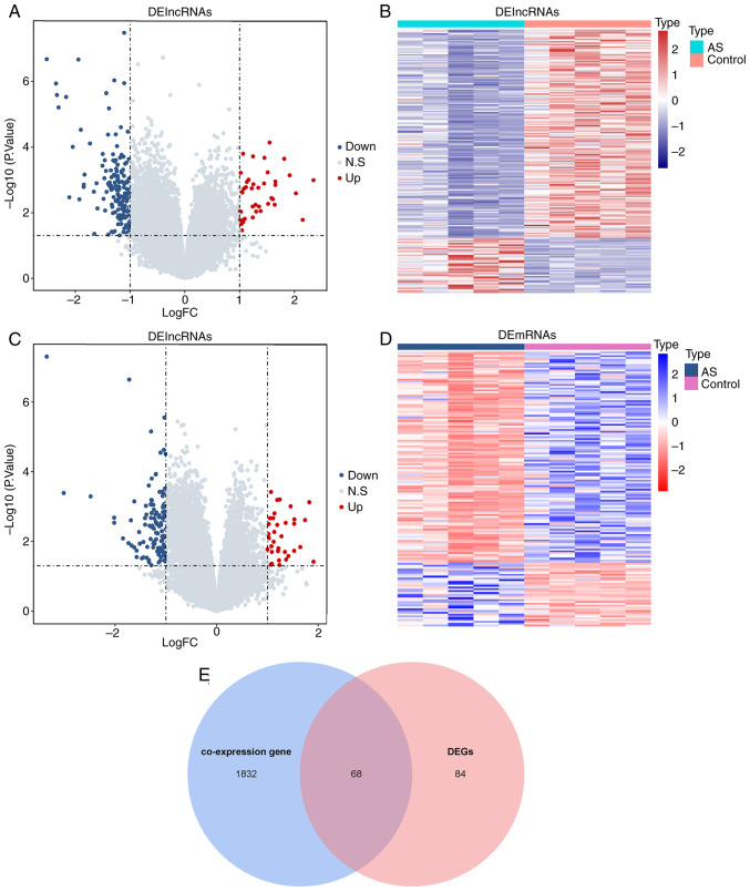
The DElncRNAs and DEmRNAs between AS and normal samples were first identified. As revealed in Fig. 1A and B, 204 DElncRNAs, 42 with increased expression and 162 with decreased expression, were found in AS samples compared with normal samples. Compared with normal samples, 152 DEmRNAs were detected in AS samples, including 35 upregulated mRNAs and 117 downregulated mRNAs (Fig. 1C and D). The Pearson correlation between DElncRNAs and all mRNAs was next analyzed. A total of 1900 mRNAs were significantly correlated with DElncRNAs and were defined as co-expressed genes. A cross-over analysis demonstrated that 68 overlapping genes (candidate genes) were present in co-expressed genes and DEmRNAs (Fig. 1E).
Figure 1.
Identification of DEGs related to DElncRNAs between AS and normal samples. (A) The volcano plot and (B) heatmap of DElncRNA between AS and normal samples. (C) The volcano plot and (D) heatmap of DEGs between AS and normal samples. (E) The overlapping genes between co-expressed genes and DEGs groups. DEGs, differentially expressed genes; DElncRNA, differentially expressed long non-coding RNA; AS, ankylosing spondylitis.
Functional enrichment of candidate genes
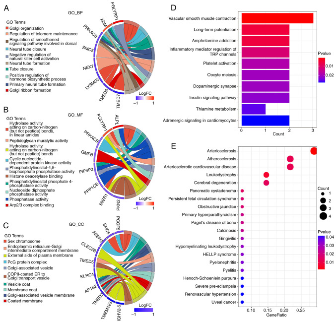
Next, an enrichment analysis was conducted on these 68 candidate genes. It was found that the candidate genes were enriched in 33 CC, 22 MF, 83 BP and nine KEGG pathways. The first 10 enrichment pathways for the BP, MF, CC and KEGG pathways are demonstrated in Fig. 2A-D, respectively. These candidate genes were also enriched in 36 DO pathways, and the first 20 pathways are shown in Fig. 2E. All findings of enrichment are shown in Table SII.
Figure 2.
Functional enrichment of candidate genes. The top significantly enriched 10 (A) BP, (B) MF, (C) CC and (D) Kyoto Encyclopedia of Genes and Genomes pathways. (E) The top 20 significantly enriched pathways. BP, biological process; MF, molecular function; CC, cellular component.
NONHSAG037054.2 and GABPA are tightly associated with AS
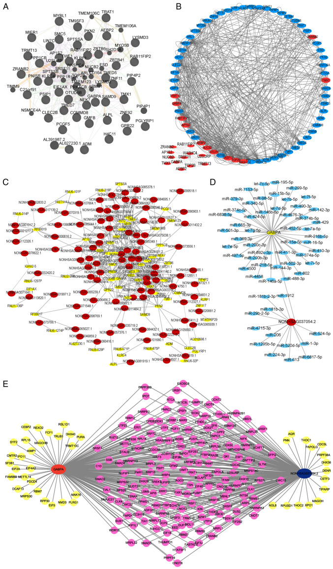
Based on these 68 candidate genes, an interaction network was constructed by GENEMANIA (http://genemania.org/search/). As revealed in Fig. 3A, a total of 491 interaction sites were identified among these candidate genes, including 360 co-expressions, 76 genetic interactions, 10 physical interactions, 35 shared protein domains and 10 predicted sites (Table SIII). Module analysis using the MCODE plug-in in Cytoscape software indicated five clusters were present. Among these, cluster 1 had the highest score (4.182) and included 12 genes (Fig. 3B). Next, a PPI network was constructed based on these 68 candidate genes and their related DElncRNAs to obtain the top 10 interacting pairs with the strongest interactions (Fig. 3C and Table II). NONHSAG037054.2 was the lncRNA most associated with genes and GABPA was the gene most connected to lncRNAs in AS patients (Fig. 3B and C; Table II). The term ‘most gene associated’ refers to ‘strongest association with’. The term ‘connected gene’ refers to highest level of connectivity. Therefore, NONHSAG037054.2 and GABPA were found to be tightly correlated with AS.
Figure 3.
GABPA and NONHSAG037054.2 are key genes and pathogenic targets of ankylosing spondylitis. (A) The PPI network in GENEMANIA website. (B) The clustered result of PPI network by MCODE plug-in. (C) The co-expression network of candidate genes and their associated differentially expressed long non-coding RNAs (red: lncRNA; yellow: candidate genes). (D) Competing endogenous RNA network of NONHSAG037054.2, gene GABPA and their predicted miRNAs (red: lncRNA; green: GABPA; blue: miRNA). (E) RBP network of NONHSAG037054.2, GABPA and their associated RBP genes (red: GABPA; blue: lncRNA; pink: the RBP genes associated with both NONHSAG037054.2 and GABPA; yellow: the RBP genes related to NONHSAG037054.2 or GABPA). PPI, protein-protein interaction; miRNAs, microRNAs; RBP, RNA-binding protein.
Table II.
The top 10 interacting pairs with the strongest interactions in the protein-protein interaction network.
| mRNA | Long non-coding RNA | rho | P-value |
|---|---|---|---|
| SMC5 | NONHSAG037054.2 | 0.993299 | 8.75x10⁻⁹ |
| TMX1 | NONHSAG070408.1 | 0.992897 | 1.10x10⁻⁸ |
| GABPA | NONHSAG037054.2 | 0.992772 | 1.18x10⁻⁸ |
| GABPA | NONHSAG111222.1 | 0.99078 | 3.13x10⁻⁸ |
| RAB11FIP2 | NONHSAG037054.2 | 0.989723 | 4.82x10⁻⁸ |
| LIN7C | NONHSAG006009.2 | 0.988781 | 6.84x10⁻⁸ |
| PKN2 | NONHSAG074750.1 | 0.988112 | 8.61x10⁻⁸ |
| LIN7C | NONHSAG065349.1 | 0.9872 | 1.16x10⁻⁷ |
| GPR22 | NONHSAG101839.1 | 0.986538 | 1.41x10⁻⁷ |
| AEBP2 | NONHSAG077454.1 | 0.986484 | 1.44x10⁻⁷ |
A total of 46 targeting miRNAs of GABPA were predicted by the TargetScan (https://www.targetscan.org/vert_72/) website, while 13 targeting miRNAs of NONHSAG037054.2 were predicted by lncRNASNP2 (http://bioinfo.life.hust.edu.cn/lncRNASNP#!/). ceRNA network refers to the interconnected regulatory network consisting of a class of RNAs with miRNA binding sites which can competitively bind miRNA, influencing gene expression and cellular processes (26). ceRNA network plays crucial roles in fine-tuning gene regulation, cell signaling and disease pathogenesis, providing a comprehensive understanding of RNA-mediated regulatory mechanisms. Next, a ceRNA network was constructed using NONHSAG037054.2, GABPA and their predicted miRNAs (Fig. 3D).
A total of 1542 RNA-binding protein (RBP) genes from a previous study (Table SIV) (27) were downloaded and the correlation of RBP genes with GABPA and NONHSAG037054.2 was calculated to select the RBP genes significantly associated with NONHSAG037054.2 and GABPA. An RBP interaction network was then constructed using NONHSAG037054.2, GABPA and their associated RBP genes. A total of 173 RBP genes were associated with both NONHSAG037054.2 and GABPA (Fig. 3E and Table SV).
NONHSAG037054.2 and GABPA are downregulated in AS samples
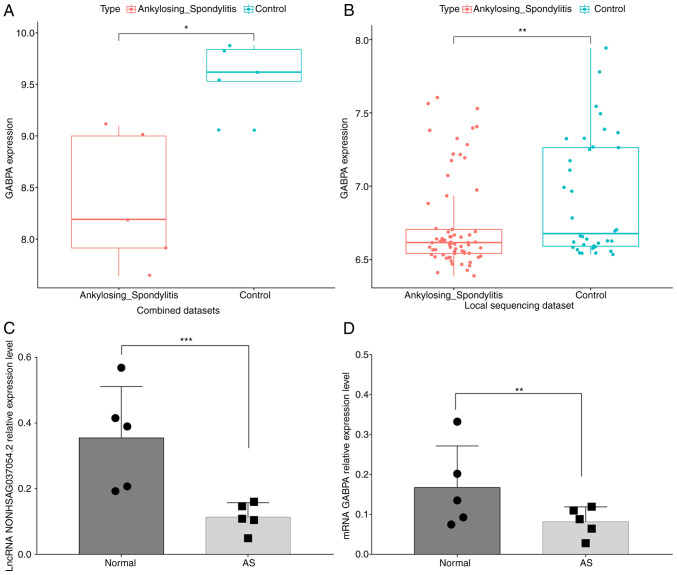
The GSE25101 and GSE73754 datasets were combined and the GABPA expression in normal and AS samples was analyzed. As demonstrated in Fig. 4A and B, GABPA was significantly downregulated in AS samples in both the combined dataset and the local sequencing dataset. Overall, the levels of both NONHSAG037054.2 and GABPA expression were significantly decreased in the peripheral blood of patients with AS (Fig. 4C and D).
Figure 4.
GABPA is downregulated in AS samples. (A and B) The expression of GABPA in AS and normal samples in (A) combined dataset and (B) sequenced dataset. (C and D) The levels of (C) NONHSAG037054.2 and (D) GABPA expression in the peripheral blood of AS patients. *P<0.05, **P≤0.01 and ***P<0.001. AS, ankylosing spondylitis.
GABPA has diagnostic value in AS
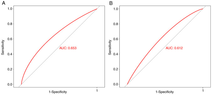
To determine whether GABPA has diagnostic value in AS, the receiver operating characteristic curve was plotted using a combined cohort (GSE25101 and GSE73754) and the GSE221786 cohort. GSE25101 and GSE73754 were merged as a combined cohort due to its low sequence quality and unbalanced ratio of female and male AS patients. There are differences in sequence quality between the combined cohort and GSE221786. The GSE25101 and GSE73754 datasets were released in 2010 and 2015, respectively, and were sequenced using the Illumina HumanHT-12 V3.0 and Illumina HumanHT-12 V4.0 platforms. GSE221786, was released in 2023 and sequenced using the Illumina NovaSeq 6000 platform. In total, GSE25101 and GSE73754 datasets belong to the same class from sequence quality. AS is more commonly diagnosed in men, with a ratio of 3:1 compared with women (28). In the AS patient group in GSE73754, there is an equal number of females and male patients (10 female and 11 male patients). However, both GSE25101 and GSE73754 exhibit an imbalance in sex distribution. To generate a more unbiased estimate of diagnostic value, GSE 25101 and GSE73754 were combined in order to achieve approximately an equal number of female and male samples. GABPA displayed diagnostic value for AS in both the combined [area under the curve (AUC)=0.653] and GSE221786 (AUC=0.612) cohorts (Fig. 5A and B).
Figure 5.
GABPA exhibits diagnostic value in ankylosing spondylitis. The receiver operating characteristic curves in (A) combined and (B) GSE221786 cohorts. AUC, area under the curve.
Discussion
LncRNAs affect the course of human diseases by regulating gene expression (29). In the present study, the mRNA and lncRNA associated with the pathogenesis of AS were investigated. It was found that NONHSAG037054.2 was a pathogenic target lncRNA of AS and GABPA was a key pathogenic gene of AS. NONHSAG037054.2 and GABPA shared 173 RBP genes. Moreover, GABPA was downregulated in AS samples and displayed diagnostic value in AS.
Li et al (14) conducted a thorough analysis of the mRNA and lncRNA profiles in AS peripheral blood mononuclear cells. They discovered that, compared with healthy controls, AS patients had 719 DEmRNAs (with 284 upregulated and 435 downregulated mRNAs) and 159 DElncRNAs (with 114 upregulated and 45 downregulated lncRNAs) (14). Nevertheless, no research has been conducted on the regulatory mechanism of these DEmRNAs and DElncRNAs in patients with AS. Between the normal and AS samples, 152 DEmRNAs and 204 DElncRNAs were found in the present investigation. Among these, 68 DEmRNAs were associated with DElncRNAs between AS and normal samples. Enrichment analysis indicated that these 68 genes are enriched in 30 CC, 22 MF and 83 BP terms, and in nine KEGG and 36 DO pathways. Of these, platelet activation, atherosclerosis and Henoch-Schoenlein purpura are notable due to their crucial association with AS.
Platelet counts and C-reactive protein levels are higher, and the erythrocyte sedimentation rate is slower in patients with AS (30). P-selectin (CD62P) is a marker of platelet activation, expressed in the α-granule membrane of standstill platelets (31). It has been identified that the CD62P level is greater in those with AS than in the control group (32). Fang et al (33) revealed that triptolide could affect the activation of platelets by regulating the levels of SDF-1, CXCR4, VEGFA and VEGFR mRNA to decrease TNF-α and IL-1β expression levels and increase IL-4 and IL-10 cytokine expression levels. It was hypothesized that drugs may influence platelet activation by regulating the 68 candidate genes identified in the present study. In addition, previous studies have shown that platelets participate in the process of atherosclerosis. CD62P targeting influences the formation of fatty streaks and the progression of mature atherosclerotic plaques (34). Huo et al (35) found that activated platelets increase atherosclerosis in apolipoprotein-E-deficient mice. Notably, the risk of atherosclerosis has been reported as 1.5-fold higher in patients with AS compared with the control group (36). The carotid intima-media thickness (an index for identifying early-stage atherosclerosis) is increased in AS patients compared with healthy individuals (37). These studies suggested that AS may accelerate the occurrence of atherosclerosis. In the present study, it was found that our 68 candidate genes were enriched in the atherosclerosis and platelet activation pathways. Thus, it was hypothesized that these 68 genes might be involved in the progress of AS through their regulation of the atherosclerosis and platelet activation pathways; this warrants further exploration in future studies.
The interaction and PPI networks revealed that NONHSAG037054.2 and GABPA interact with the most genes or lncRNAs in AS. LncRNA can bind to miRNA by complementary base pairing to modulate gene expression (38,39). Wang et al (40) suggested that lncRNA-UCAL can regulate expression of FGFR1 by binding to miR-216b, thereby increasing FGFR1 expression at the post-transcriptional regulation level in hepatocellular carcinoma. Bian et al (41) found that the lncRNA HOX transcript antisense RNA HOTAIR could serve as an endogenous ‘sponge’ for miR-148b to facilitate the expression of DNMT1, leading to the activation and proliferation of liver cancer cells. Thus, the miRNAs related to NONHSAG037054.2 and GABPA were analyzed. The findings suggested that 46 and 13 miRNAs are tightly correlated with NONHSAG037054.2 and GABPA, respectively. However, overlapping miRNAs associated with both NONHSAG037054.2 and GABPA were not found. Moreover, RBPs are important regulators of RNA metabolism, and RBPs participate in the transcription, translocation and translation of targeted mRNAs (42). In addition, lncRNAs include multiple RBP sites. LncRNAs may influence the interaction between mRNA and RBP by modulating the stability and bioactivity of RBP in ribonucleoprotein complexes (43). In addition, RBP can regulate the function, expression and stability of lncRNAs (44). In the present study, 173 RBP genes linked with both GABPA and NONHSAG037054.2 were identified. Thus, it was hypothesized that NONHSAG037054.2 might regulate the expression of GABPA by affecting the interaction between GABPA and RBP; this requires further study. There are certain limitations to the present study. Due to the small sample size, the reliability and accuracy of the study will be affected, thus affecting the generalization of the study conclusion. In future studies, more specimens will be collected for further verification.
It was also found that GABPA was markedly downregulated in the peripheral blood of AS patients. GABPA is a transcription factor in the ETS family (45) that is involved in the nuclear control of mitochondrial function and the expression of cytochrome oxidase c (46,47). Recent studies have indicated that GABPA is downregulated in clear cell renal cell carcinoma (48), endometrial carcinoma (49) and gastric cancer (50), and that high GABPA expression is associated with a lower survival rate of patients. In follicular thyroid carcinoma (FTC), GABPA directly regulates the expression of DICER1 and was also downregulated in clinical FTC samples (51). Therefore, GABPA might affect the occurrence and development of AS.
In summary, NONHSAG037054.2 and GABPA were closely correlated with AS, with GABPA downregulated in AS. Thus, GABPA could serve as a novel diagnostic factor for AS. The presents results provided more information for deeply understanding the mechanism of lncRNA-mRNA interactions in AS.
Supplementary Material
Acknowledgements
Not applicable.
Funding Statement
Funding: The present study was supported by the Beijing Municipal Health Commission (grant nos. BMHC-2021-6 and BJRITO-RDP-2023) and Beijing Postdoctoral Research Foundation (grant no. 2022-ZZ-020).
Availability of data and materials
The sequencing data generated in the present study may be found in the CNGB Sequence Archive (52) of the China National GeneBank DataBase (53) under accession number CNP0005132 or at the following URL: https://db.cngb.org/search/?q=CNP0005132. The other data generated in the present study may be requested from the corresponding author.
Authors' contributions
PC, CWu and WT designed the study. PC, YZ, CWa, BX, QW, LZ and HL performed the experiments. PC, YZ, CWa and QW acquired and analyzed the data. BX, LZ and HL obtained the clinical samples. PC, CWu and WT wrote and revised the manuscript. PC, CWu and WT confirm the authenticity of all the raw data. All authors read and approved the final version of the manuscript.
Ethics approval and consent to participate
All methods were conducted in compliance with all applicable rules and regulations. The peripheral blood samples were acquired from patients who were hospitalized in Beijing Jishuitan Hospital (Beijing, China) between January 05 and December 30, including five healthy volunteers and five AS patients. All procedures were authorized by the hospital's ethics committee (approval no. 201901-05-02) and conformed to the Declaration Helsinki guidelines. Written informed consent was obtained from all subjects.
Patient consent for publication
Not applicable.
Competing interests
The authors declare that they have no competing interests.
References
- 1.Gouveia EB, Elmann D, Morales MS. Ankylosing spondylitis and uveitis: overview. Rev Bras Reumatol. 2012;52:742–756. [PubMed] [Google Scholar]
- 2.Zhu W, He X, Cheng K, Zhang L, Chen D, Wang X, Qiu G, Cao X, Weng X. Ankylosing spondylitis: Etiology, pathogenesis, and treatments. Bone Res. 2019;7(22) doi: 10.1038/s41413-019-0057-8. [DOI] [PMC free article] [PubMed] [Google Scholar]
- 3.Sieper J, Poddubnyy D. Axial spondyloarthritis. Lancet. 2017;390:73–84. doi: 10.1016/S0140-6736(16)31591-4. [DOI] [PubMed] [Google Scholar]
- 4.Carron P, De Craemer AS, Van den Bosch F. Peripheral spondyloarthritis: A neglected entity-state of the art. RMD Open. 2020;6(e001136) doi: 10.1136/rmdopen-2019-001136. [DOI] [PMC free article] [PubMed] [Google Scholar]
- 5.Navarro-Compan V, Sepriano A, El-Zorkany B, van der Heijde D. Axial spondyloarthritis. Ann Rheum Dis. 2021;80:1511–1521. doi: 10.1136/annrheumdis-2021-221035. [DOI] [PubMed] [Google Scholar]
- 6.de Winter JJ, van Mens LJ, van der Heijde D, Landewe R, Baeten DL. Prevalence of peripheral and extra-articular disease in ankylosing spondylitis versus non-radiographic axial spondyloarthritis: A meta-analysis. Arthritis Res Ther. 2016;18(196) doi: 10.1186/s13075-016-1093-z. [DOI] [PMC free article] [PubMed] [Google Scholar]
- 7.Maruyama R, Suzuki H. Long noncoding RNA involvement in cancer. BMB Rep. 2012;45:604–611. doi: 10.5483/bmbrep.2012.45.11.227. [DOI] [PMC free article] [PubMed] [Google Scholar]
- 8.Mercer TR, Dinger ME, Mattick JS. Long non-coding RNAs: Insights into functions. Nat Rev Genet. 2009;10:155–159. doi: 10.1038/nrg2521. [DOI] [PubMed] [Google Scholar]
- 9.Engreitz JM, Ollikainen N, Guttman M. Long non-coding RNAs: Spatial amplifiers that control nuclear structure and gene expression. Nat Rev Mol Cell Biol. 2016;17:756–770. doi: 10.1038/nrm.2016.126. [DOI] [PubMed] [Google Scholar]
- 10.Kundu M, Basu J. The role of microRNAs and long non-coding RNAs in the regulation of the immune response to mycobacterium tuberculosis infection. Front Immunol. 2021;12(687962) doi: 10.3389/fimmu.2021.687962. [DOI] [PMC free article] [PubMed] [Google Scholar]
- 11.Shi G, Cheng Y, Zhang Y, Guo R, Li S, Hong X. Long non-coding RNA LINC00511/miR-150/MMP13 axis promotes breast cancer proliferation, migration and invasion. Biochim Biophys Acta Mol Basis Dis. 2021;1867(165957) doi: 10.1016/j.bbadis.2020.165957. [DOI] [PubMed] [Google Scholar]
- 12.Wang X, Li M, Wang Z, Han S, Tang X, Ge Y, Zhou L, Zhou C, Yuan Q, Yang M. Silencing of long noncoding RNA MALAT1 by miR-101 and miR-217 inhibits proliferation, migration, and invasion of esophageal squamous cell carcinoma cells. J Biol Chem. 2015;290:3925–3935. doi: 10.1074/jbc.M114.596866. [DOI] [PMC free article] [PubMed] [Google Scholar]
- 13.Sun J, Wang R, Chao T, Wang C. Long noncoding RNAs involved in cardiomyocyte apoptosis triggered by different stressors. J Cardiovasc Transl Res. 2022;15:588–603. doi: 10.1007/s12265-021-10186-w. [DOI] [PubMed] [Google Scholar]
- 14.Li C, Qu W, Yang X. Comprehensive lncRNA and mRNA profiles in peripheral blood mononuclear cells derived from ankylosing spondylitis patients by RNA-sequencing analysis. Medicine (Baltimore) 2022;101(e27477) doi: 10.1097/MD.0000000000027477. [DOI] [PMC free article] [PubMed] [Google Scholar]
- 15.Wang JX, Zhao X, Xu SQ. Screening key lncRNAs of ankylosing spondylitis using bioinformatics analysis. J Inflamm Res. 2022;15:6087–6096. doi: 10.2147/JIR.S387258. [DOI] [PMC free article] [PubMed] [Google Scholar]
- 16.Pimentel-Santos FM, Ligeiro D, Matos M, Mourão AF, Costa J, Santos H, Barcelos A, Godinho F, Pinto P, Cruz M, et al. Whole blood transcriptional profiling in ankylosing spondylitis identifies novel candidate genes that might contribute to the inflammatory and tissue-destructive disease aspects. Arthritis Res Ther. 2011;13(R57) doi: 10.1186/ar3309. [DOI] [PMC free article] [PubMed] [Google Scholar]
- 17.Gracey E, Yao Y, Green B, Qaiyum Z, Baglaenko Y, Lin A, Anton A, Ayearst R, Yip P, Inman RD. Sexual dimorphism in the Th17 signature of ankylosing spondylitis. Arthritis Rheumatol. 2016;68:679–689. doi: 10.1002/art.39464. [DOI] [PubMed] [Google Scholar]
- 18.Yu G, Wang LG, Han Y, He QY. clusterProfiler: An R package for comparing biological themes among gene clusters. OMICS. 2012;16:284–287. doi: 10.1089/omi.2011.0118. [DOI] [PMC free article] [PubMed] [Google Scholar]
- 19.Yu G, Wang LG, Yan GR, He QY. DOSE: An R/Bioconductor package for disease ontology semantic and enrichment analysis. Bioinformatics. 2015;31:608–609. doi: 10.1093/bioinformatics/btu684. [DOI] [PubMed] [Google Scholar]
- 20.Szklarczyk D, Gable AL, Lyon D, Junge A, Wyder S, Huerta-Cepas J, Simonovic M, Doncheva NT, Morris JH, Bork P, et al. STRING v11: Protein-protein association networks with increased coverage, supporting functional discovery in genome-wide experimental datasets. Nucleic Acids Res. 2019;47(D1):D607–D13. doi: 10.1093/nar/gky1131. [DOI] [PMC free article] [PubMed] [Google Scholar]
- 21.Lu H, Zhou Q, He J, Jiang Z, Peng C, Tong R, Shi J. Recent advances in the development of protein-protein interactions modulators: Mechanisms and clinical trials. Signal Transduct Target Ther. 2020;5(213) doi: 10.1038/s41392-020-00315-3. [DOI] [PMC free article] [PubMed] [Google Scholar]
- 22.Shannon P, Markiel A, Ozier O, Baliga NS, Wang JT, Ramage D, Amin N, Schwikowski B, Ideker T. Cytoscape: A software environment for integrated models of biomolecular interaction networks. Genome Res. 2003;13:2498–2504. doi: 10.1101/gr.1239303. [DOI] [PMC free article] [PubMed] [Google Scholar]
- 23.Warde-Farley D, Donaldson SL, Comes O, Zuberi K, Badrawi R, Chao P, Franz M, Grouios C, Kazi F, Lopes CT, et al. The GeneMANIA prediction server: Biological network integration for gene prioritization and predicting gene function. Nucleic Acids Res. 2010;38(Web Server issue):W214–W20. doi: 10.1093/nar/gkq537. [DOI] [PMC free article] [PubMed] [Google Scholar]
- 24.Bader GD, Hogue CW. An automated method for finding molecular complexes in large protein interaction networks. BMC Bioinformatics. 2003;4(2) doi: 10.1186/1471-2105-4-2. [DOI] [PMC free article] [PubMed] [Google Scholar]
- 25.Livak KJ, Schmittgen TD. Analysis of relative gene expression data using real-time quantitative PCR and the 2(-Delta Delta C(T)) method. Methods. 2001;25:402–408. doi: 10.1006/meth.2001.1262. [DOI] [PubMed] [Google Scholar]
- 26.Chen J, Song Y, Li M, Zhang Y, Lin T, Sun J, Wang D, Liu Y, Guo J, Yu W. Comprehensive analysis of ceRNA networks reveals prognostic lncRNAs related to immune infiltration in colorectal cancer. BMC Cancer. 2021;21(255) doi: 10.1186/s12885-021-07995-2. [DOI] [PMC free article] [PubMed] [Google Scholar]
- 27.Conga DF, Bowler M, Tantalean M, Montes D, Serra-Freire NM, Mayor P. Intestinal helminths in wild Peruvian red uakari monkeys (Cacajao calvus ucayalii) in the northeastern Peruvian Amazon. J Med Primatol. 2014;43:130–133. doi: 10.1111/jmp.12092. [DOI] [PubMed] [Google Scholar]
- 28. Abstract Supplement ACR Convergence 2022. Arthritis Rheumatology 74, 2022. [Google Scholar]
- 29.Schmitz SU, Grote P, Herrmann BG. Mechanisms of long noncoding RNA function in development and disease. Cell Mol Life Sci. 2016;73:2491–2509. doi: 10.1007/s00018-016-2174-5. [DOI] [PMC free article] [PubMed] [Google Scholar]
- 30.Orum H, Pamuk GE, Pamuk ON, Demir M, Turgut B. Does anti-TNF therapy cause any change in platelet activation in ankylosing spondylitis patients? A comparative study. J Thromb Thrombolysis. 2012;33:154–159. doi: 10.1007/s11239-011-0663-9. [DOI] [PubMed] [Google Scholar]
- 31.Pendl GG, Robert C, Steinert M, Thanos R, Eytner R, Borges E, Wild MK, Lowe JB, Fuhlbrigge RC, Kupper TS, et al. Immature mouse dendritic cells enter inflamed tissue, a process that requires E- and P-selectin, but not P-selectin glycoprotein ligand 1. Blood. 2002;99:946–956. doi: 10.1182/blood.v99.3.946. [DOI] [PubMed] [Google Scholar]
- 32.Wang F, Yan CG, Xiang HY, Xing T, Wang NS. The significance of platelet activation in ankylosing spondylitis. Clin Rheumatol. 2008;27:767–769. doi: 10.1007/s10067-008-0847-7. [DOI] [PubMed] [Google Scholar]
- 33.Fang YY, Wan L, Dong WZ, Wen JT, Liu J. Effect of triptolide in improving platelet activation in patients with ankylosing spondylitis by regulating VEGFA,SDF-1,CXCR4 pathway. Zhongguo Zhong Yao Za Zhi. 2019;44:3520–3525. doi: 10.19540/j.cnki.cjcmm.20181113.001. [DOI] [PubMed] [Google Scholar]
- 34.Dong ZM, Chapman SM, Brown AA, Frenette PS, Hynes RO, Wagner DD. The combined role of P- and E-selectins in atherosclerosis. J Clin Invest. 1998;102:145–152. doi: 10.1172/JCI3001. [DOI] [PMC free article] [PubMed] [Google Scholar]
- 35.Huo Y, Schober A, Forlow SB, Smith DF, Hyman MC, Jung S, Littman DR, Weber C, Ley K. Circulating activated platelets exacerbate atherosclerosis in mice deficient in apolipoprotein E. Nat Med. 2003;9:61–67. doi: 10.1038/nm810. [DOI] [PubMed] [Google Scholar]
- 36.Han C, Robinson DW Jr, Hackett MV, Paramore LC, Fraeman KH, Bala MV. Cardiovascular disease and risk factors in patients with rheumatoid arthritis, psoriatic arthritis, and ankylosing spondylitis. J Rheumatol. 2006;33:2167–2172. [PubMed] [Google Scholar]
- 37.Verma I, Krishan P, Syngle A. Predictors of atherosclerosis in ankylosing spondylitis. Rheumatol Ther. 2015;2:173–182. doi: 10.1007/s40744-015-0017-8. [DOI] [PMC free article] [PubMed] [Google Scholar]
- 38.Bloch S, Froc C, Pontiggia A, Yamamoto K. Existence of working memory in teleosts: Establishment of the delayed matching-to-sample task in adult zebrafish. Behav Brain Res. 2019;370(111924) doi: 10.1016/j.bbr.2019.111924. [DOI] [PubMed] [Google Scholar]
- 39.Gil N, Ulitsky I. Regulation of gene expression by cis-acting long non-coding RNAs. Nat Rev Genet. 2020;21:102–117. doi: 10.1038/s41576-019-0184-5. [DOI] [PubMed] [Google Scholar]
- 40.Wang F, Ying HQ, He BS, Pan YQ, Deng QW, Sun HL, Chen J, Liu X, Wang SK. Upregulated lncRNA-UCA1 contributes to progression of hepatocellular carcinoma through inhibition of miR-216b and activation of FGFR1/ERK signaling pathway. Oncotarget. 2015;6:7899–7917. doi: 10.18632/oncotarget.3219. [DOI] [PMC free article] [PubMed] [Google Scholar]
- 41.Bian EB, Wang YY, Yang Y, Wu BM, Xu T, Meng XM, Huang C, Zhang L, Lv XW, Xiong ZG, Li J. Hotair facilitates hepatic stellate cells activation and fibrogenesis in the liver. Biochim Biophys Acta Mol Basis Dis. 2017;1863:674–686. doi: 10.1016/j.bbadis.2016.12.009. [DOI] [PubMed] [Google Scholar]
- 42.Sternburg EL, Karginov FV. Global Approaches in studying RNA-binding protein interaction networks. Trends Biochem Sci. 2020;45:593–603. doi: 10.1016/j.tibs.2020.03.005. [DOI] [PubMed] [Google Scholar]
- 43.Tichon A, Gil N, Lubelsky Y, Havkin Solomon T, Lemze D, Itzkovitz S, Stern-Ginossar N, Ulitsky I. A conserved abundant cytoplasmic long noncoding RNA modulates repression by Pumilio proteins in human cells. Nat Commun. 2016;7(12209) doi: 10.1038/ncomms12209. [DOI] [PMC free article] [PubMed] [Google Scholar]
- 44.Lubelsky Y, Ulitsky I. Sequences enriched in Alu repeats drive nuclear localization of long RNAs in human cells. Nature. 2018;555:107–111. doi: 10.1038/nature25757. [DOI] [PMC free article] [PubMed] [Google Scholar]
- 45.Sizemore GM, Pitarresi JR, Balakrishnan S, Ostrowski MC. The ETS family of oncogenic transcription factors in solid tumours. Nat Rev Cancer. 2017;17:337–351. doi: 10.1038/nrc.2017.20. [DOI] [PubMed] [Google Scholar]
- 46.Wang KP, Zhu JQ, Zhang TT. Research progress in zygomatic implant technique. Zhonghua Kou Qiang Yi Xue Za Zhi. 2020;55:196–200. doi: 10.3760/cma.j.issn.1002-0098.2020.03.011. (In Chinese) [DOI] [PubMed] [Google Scholar]
- 47.Goto S, Takahashi M, Yasutsune N, Inayama S, Kato D, Fukuoka M, Kashiwaba SI, Murakami Y. Identification of GA-binding protein transcription factor alpha subunit (GABPA) as a novel bookmarking factor. Int J Mol Sci. 2019;20(1093) doi: 10.3390/ijms20051093. [DOI] [PMC free article] [PubMed] [Google Scholar]
- 48.Fang Z, Zhang N, Yuan X, Xing X, Li X, Qin X, Liu Z, Neo S, Liu C, Kong F, et al. GABPA-activated TGFBR2 transcription inhibits aggressiveness but is epigenetically erased by oncometabolites in renal cell carcinoma. J Exp Clin Cancer Res. 2022;41(173) doi: 10.1186/s13046-022-02382-6. [DOI] [PMC free article] [PubMed] [Google Scholar]
- 49.Ma X, Lin Q, Cui G, Zhao J, Wei X, Li R, Mao H, Ma Y, Liu P, Pang Y. GABPA expression in endometrial carcinoma: A prognostic marker. Dis Markers. 2021;2021(5552614) doi: 10.1155/2021/5552614. [DOI] [PMC free article] [PubMed] [Google Scholar]
- 50.Yin B, Dong B, Guo X, Wang C, Huo H. GABPA protects against gastric cancer deterioration via negatively regulating GPX1. J Med Biochem. 2022;41:355–362. doi: 10.5937/jomb0-35445. [DOI] [PMC free article] [PubMed] [Google Scholar]
- 51.Paulsson JO, Wang N, Gao J, Stenman A, Zedenius J, Mu N, Lui WO, Larsson C, Juhlin CC. GABPA-dependent down-regulation of DICER1 in follicular thyroid tumours. Endocr Relat Cancer. 2020;27:295–308. doi: 10.1530/ERC-19-0446. [DOI] [PMC free article] [PubMed] [Google Scholar]
- 52.Guo X, Chen F, Gao F, Li L, Liu K, You L, Hua C, Yang F, Liu W, Peng C, et al. CNSA: A data repository for archiving omics data. Database (Oxford) 2020;2020(baaa055) doi: 10.1093/database/baaa055. [DOI] [PMC free article] [PubMed] [Google Scholar]
- 53.Chen FZ, You LJ, Yang F, Wang LN, Guo XQ, Gao F, Hua C, Tan C, Fang L, Shan RQ, et al. CNGBdb: China national GeneBank database. Yi Chuan. 2020;42:799–809. doi: 10.16288/j.yczz.20-080. [DOI] [PubMed] [Google Scholar]
Associated Data
This section collects any data citations, data availability statements, or supplementary materials included in this article.
Supplementary Materials
Data Availability Statement
The sequencing data generated in the present study may be found in the CNGB Sequence Archive (52) of the China National GeneBank DataBase (53) under accession number CNP0005132 or at the following URL: https://db.cngb.org/search/?q=CNP0005132. The other data generated in the present study may be requested from the corresponding author.


