



ABSTRACT
Klebsiella pneumoniae, especially hypervirulent K. pneumoniae (hvKP), is a common opportunistic pathogen that often causes hospital- and community-acquired infections. Capsular polysaccharide (CPS) is an important virulence factor of K. pneumoniae. Some phages encode depolymerases that can recognize and degrade bacterial polysaccharides. In this study, the lytic bacteriophage vB_KpnP_ZK1 (abbreviated as ZK1) was isolated using serotype K1 hvKP as the host. Although amino acid sequence BLAST analysis indicated that the tail fiber protein Depo16 of phage ZK1 showed no significant similarity to any reported phage depolymerases, it displayed enzymatic activities that are characteristic of phage depolymerases. After expression and purification, Depo16 could efficiently remove the capsular polysaccharide layer that surrounds the surface of serotype K1 K. pneumoniae. Although no bactericidal activity was detected, Depo16 makes serotype K1 K. pneumoniae sensitive to peritoneal macrophages (PMs). In addition, in a mouse bacteremia model of serotype K1 K. pneumoniae, 25 µg of Depo16 was effective in significantly prolonging survival. Depo16 treatment can reduce the bacterial load in blood and major tissues and alleviate tissue damage in mice. This indicates that the putative depolymerase Depo16 is a potential antibacterial agent against serotype K1 K. pneumoniae infections.
IMPORTANCE
Klebsiella pneumoniae often causes hospital-acquired infections and community-acquired infections. Capsular polysaccharide (CPS) is one of the crucial virulence factors of K. pneumoniae. K1 and K2 capsular-type K. pneumoniae strains are the most prevalent serotypes of hypervirulent K. pneumoniae (hvKP). In this study, a novel K. pneumoniae phage named vB_KpnP_ZK1 was isolated, and its putative depolymerase Depo16 showed low homology with other reported phage depolymerases. Depo16 can specifically degrade the K. pneumoniae K1 capsule making this serotype sensitive to peritoneal macrophages. More importantly, Depo16 showed a significant therapeutic effect in a mouse bacteremia model caused by serotype K1 K. pneumoniae. Thus, Depo16 is a potential antibacterial agent to combat serotype K1 K. pneumoniae infections.
KEYWORDS: Klebsiella pneumoniae, serotype K1, phage depolymerases, bacteremia
INTRODUCTION
Klebsiella pneumoniae is one of the most common opportunistic pathogens and often causes hospital-acquired infections, as well as community-acquired infections (1–3). Most K. pneumoniae strains are enveloped by capsular polysaccharide (CPS), which is one of the crucial virulence factors. According to CPS diversity, the capsular serotypes of K. pneumoniae can be divided into at least 78 types (4). K1 and K2 are the most prevalent serotypes of hypervirulent K. pneumoniae (hvKP) strains, accounting for approximately 70% of these strains (5). Usually, K1 serotype K. pneumoniae shows hypermucoviscosity (HMV) and possesses rmpA/rmpA2 and magA genes are hvKP (5). In Korea, the most common capsular type of K. pneumoniae that causes liver abscess is K1 (59.4%) (6). Similarly, in Taiwan, K. pneumoniae capsular-type K1 has also been demonstrated to be the predominant cause of necrotizing fasciitis (4 of 15 strains; 26.7%) (7). The K1 capsular type, as the main serotype of hvKP, is associated with invasive infections caused by K. pneumoniae.
Some bacteriophages can produce enzymes called depolymerases. With the revival of phage therapy, depolymerases are attracting the attention of researchers (8–10). CPS is the first receptor of most phages. Correspondingly, depolymerases can recognize and degrade bacterial CPSs, thereby allowing the phage to recognize and bind to secondary receptors (usually outer membrane proteins) on the bacterial surface (11). Because CPS seriously affects the survival and virulence of bacterial cells in vivo (12), the destruction of CPS may render the bacteria more susceptible to being killed by complement-mediated immunity. The destruction of CPS also helps phagocytes effectively remove bacteria (13). Therefore, depolymerases hold great potential in the prevention and treatment of infections by capsular bacteria.
Phage depolymerases have been reported in many bacteria, such as Escherichia coli (14), Acinetobacter baumannii (15), Salmonella (16), and Pseudomonas aeruginosa (17). These depolymerases have functions such as inhibiting bacterial biofilm formation (14, 16–20) and sensitizing bacteria to serum or macrophage-mediated killing (13, 19, 21); these depolymerases increased the survival rate in a bacterial infection model (15). A small number of K. pneumoniae phage depolymerases have also been identified. However, although serotype K1 K. pneumoniae is one of the most common hvKPs, the phage depolymerases targeting this serotype of K. pneumoniae have not been adequately studied. To our knowledge, only depolymerases K1-ORF34 (22) of phage NTUHK2044-K1-1 and GBH001_056 (23) of phage GBH001 have been studied, and their biological properties and activity have been investigated in vitro. Thus, it is necessary to promote research on serotype K1 K. pneumoniae phage depolymerases.
In this study, a new phage, vB_KpnP_ZK1 (abbreviated as ZK1), was isolated using the K1 serotype hvKP. The depolymerase of phage ZK1, termed Depo16, was expressed and studied. The results showed that Depo16 can specifically degrade the K1 serotype CPS of K. pneumoniae and make serotype K1 K. pneumoniae sensitive to peritoneal macrophages. In addition, the depolymerase Depo16 had good therapeutic effects in a mouse model of bacteremia caused by K. pneumoniae infection.
MATERIALS AND METHODS
Animals and ethics statement
Six- to eight-week-old female C57BL/6 mice (weight 18–22 g) were purchased from the Liaoning Provincial Laboratory Animal Resource Center, Shenyang, China. For studies involving animals, all procedures were conducted according to regulations established by the Institutional Animal Care and Use Committee of Jilin University (no. SY202306021).
Bacterial strains and culture conditions
K. pneumoniae strains, including Wkp26, were isolated from clinical cases at the First Hospital of Jilin University. All K. pneumoniae strains were identified using the VITEK 2 Compact system (bioMérieux, Marcy-l’Étoile, France) and verified by specific identification primers. After 16S rRNA PCR amplification with universal primers (27F: AGAGTTTGATCCTGGCTCAG and 1429R: GGTTACCTTGTTACGACTT) (24), sequence analysis was used to confirm the identity of K. pneumoniae. As previously described (4, 25), serotypes of K. pneumoniae were identified by specific MagA gene primers (F: GGTGCTCTTTACATCATTGC and R: GCAATGGCCATTTGCGTTAG) and universal wzc gene primers (F: GGGTTTTTATCGGGTTGTAC and R: TTCAGCTGGATTTGGTGG). K. pneumoniae was incubated in Luria-Bertani (LB) broth at 37°C in a thermostatic oscillation incubator (180 rpm) (20).
String test for hypermucoviscosity and rmpA gene detection
The string test for hypermucoviscosity (HV) was conducted according to a previously described method (26). Briefly, Wkp26 was inoculated on a sheep blood agar plate at 37°C overnight, and a bacteriological loop was used to stretch the mucoviscous string from the colony. Hypermucoviscosity was defined as the formation of viscous strings >5 mm in length (positive line test). The presence of the rmpA gene was determined using specific primers (25) (F: ACTGGGCTACCTCTGCTTCA and R: CTTGCATGAGCCATCTTTCA).
Phage isolation, purification, and host spectrum determination
According to a previously described method (27), phage was isolated from sewage samples with K. pneumoniae Wkp26 as the host strain. The phage was purified three times by the double-layer agar plate method (27) and stored at either 4°C or 80°C in glycerol [3:1 (vol/vol)]. To determine the host spectrum of the phage, 95 clinical K. pneumoniae strains were analyzed using spot testing and the double-layer agar plate method, as previously described (27).
One-step growth curves
The one-step growth curve of the phage was generated as previously described (28) with some modifications. K. pneumoniae Wkp26 grew to the mid-exponential phase (OD600 ≈ 0.6) and was resuspended in fresh LB broth. Then, phage was added to a mid-exponential phase Wkp26 culture at a multiplicity of infection (MOI) of 0.1 and allowed to adsorb for 2 min at 37°C. The mixture was centrifuged at 4°C for 10 min at 12,000 × g, and the precipitate was resuspended in 10 mL of LB. Afterward, this suspension was incubated at 37°C with shaking at 180 rpm. The samples were collected at different time points to test the phage concentration. The first group of samples was immediately diluted and plated for phage titration. The second group of samples was processed using 1% (vol/vol) chloroform for 30 min before the release of intracellular phages and then quantified by a plaque assay. The burst size was calculated as the ratio of the average number of phages present at the start and end of the experiment (29, 30).
Phage stability
To measure the stability of the phage, the phage survival rate was measured after treatment at different pH values and temperatures as previously described (27). In short, the phage suspensions were incubated at 37°C (pH 1–14) for 1 h for pH stability tests. Phages were incubated at 4, 25, 37, 50, 60, 70, and 80°C for 1 h, and samples were collected every 20 min for a total of 80 min for the thermal stability test. Then, phage titers were determined by the double-layer agar plate method.
Transmission electron microscopy
The phage was concentrated and purified as previously described (31). Concentrated phage ZK1 was placed on copper grids and negatively stained with phosphotungstic acid (2%, wt/vol). Transmission electron microscopy (TEM) (JEOL JEM-1200EXII, Japan Electronics and Optics Laboratory, Tokyo, Japan) was used to observe ZK1 at an acceleration voltage of 80 kV.
Sequencing and analysis of the phage genome
The genome was extracted from concentrated phage samples using a viral genome extraction kit (Omega Bio-Tek Inc., Doraville, GA, United States). The NEBNext Ultra DNA Library Prep Kit for Illumina (NEB, USA) was used to complete the library preparation, and the genome was sequenced on an Illumina PE150. SPAdes v3.15.2 was used to assemble sequences (32). Potential open reading frames (ORFs) were predicted using GeneMarkS (Georgia Institute of Technology, Atlanta, GA, United States) (33). The functions of the encoded regions in ZK1 were annotated using PSI-BLAST (threshold = 0.0001) from the National Center for Biotechnology Information (NCBI) (https://blast.ncbi.nlm.nih.gov/Blast.cgi) (34). A schematic of the phage genome was generated using CLC Main Workbench, version 7.7.3 (CLC Bio-Qiagen, Aarhus, Denmark).
Analysis and expression of phage depolymerase
To determine the gene encoding the depolymerase, the protein structure was analyzed using Alfafold (https://colab.research.google.com/github/sokrypton/ColabFold/blob/main/AlphaFold2.ipynb) and Phyre2 (http://www.sbg.bio.ic.ac.uk/phyre2/html/page.cgi?id=index). The sequences of putative depolymerases were aligned using the ClustalW program. HHpred (https://toolkit.tuebingen.mpg.de/#/tools/hhpred) was used to analyze the protein domains online. The amino acid sequences of the depolymerase Depo16 and other depolymerases were compared using CLC Master Workbench, version 7.7.3 (CLC Bio-Qiagen, Aarhus, Denmark). The phylogenetic tree was constructed using BioEdit (7.0.4.1), MEGA 6, and PHYLIP software (version 3.697) (35).
PCR amplification of the depolymerase Depo16 was carried out with the specific primers 16 F (CGGGATCCATGGAAGTTCAAGGCTCGTTAGGT) and 16 R (CCCTCGAGTTATACCCTCTTTCTGGTTGGGTTATAGG). PCR products (containing XhoI and BamHI restriction sites) were cloned and inserted into the pET28a vector and transformed into Escherichia coli BL21 cells. An exponentially growing culture (OD600 = 0.6) was induced with 2.5 mM isopropyl-β-D-thiogalactopyranoside (IPTG). The cultures were collected by centrifugation at 8,000 × g and 4°C for 20 min. The cells were then collected and suspended in lysis buffer (50 mM Na2HPO4, 10 mM KH2PO4, 500 mM NaCl, and 2.7 mM KCl; pH 7.4) and disrupted with an ultrasonic disintegrator. The supernatant was obtained by centrifugation at 10,000 × g at 4°C for 15 min. For protein purification, elution was performed in a gravity column using nickel-nitrilotriacetic acid (Ni-NTA) resin (GE Healthcare, USA) according to the manufacturer’s instructions. The induced bacterial solution, the supernatant after ultrasonic fragmentation, and the purified recombinant protein Depo16 were analyzed using 12% sodium dodecyl sulfate-polyacrylamide gel electrophoresis (SDS-PAGE). The purified protein was quickly frozen in liquid nitrogen and stored at −80°C.
Depolymerase activity of Depo16
The activity of depolymerase Depo16 was determined as previously described with some modifications (36). The concentration of Depo16 was adjusted to 2 µg/mL, and 5 µL of continuously diluted Depo16 was dropped on a bacterial lawn of K. pneumoniae. Phage ZK1 was used as a positive control, and lysis buffer was used as a negative control. The plates were incubated at 37°C for 8 h before observation. The formation of a transparent zone halo on the bacterial lawn was a sign of the antibacterial activity of depolymerases.
Wkp26 cells treated with Depo16 or phosphate-buffered saline (PBS; negative control) were stained with CPS dye (37) and then observed by optical microscopy. In addition, the CPS of Wkp26 cells treated with Depo16 or PBS (negative control) was extracted as previously described (38). CPS samples were subjected to 12% SDS-PAGE. The obtained gels were stained with Alcian blue (39).
Wkp26 cells treated with Depo16 or PBS (negative control) were washed three times with sterile PBS and then fixed with 4% glutaraldehyde for 4 h. The bacterial cells were dehydrated with ethanol at different concentrations (10%, 30%, 50%, 70%, 90%, and 100%), dried overnight on coverslips, and observed by scanning electron microscopy (SEM) (Gemini Sigma 300, ZEISS, Germany).
The pH stability of Depo16 was evaluated at pH 3.0–11.0. The depolymerase was suspended in the different pH’s elution buffers (50 mM Na2HPO4, 10 mM KH2PO4, 137 mM NaCl, and 2.7 mM KCl) and incubated at 25°C for 1 h. The depolymerase used for thermal stability analysis was incubated in an elution buffer and maintained for 1 h in a temperature range of 4°C to 60°C. Afterward, the depolymerase activity was measured.
Effect of Depo16 on Wkp26 capture by peritoneal macrophages
Isolation and culture of mouse peritoneal macrophages (PMs) were conducted as previously described with some modifications (40). The PM cells were spread into 24-well cell culture plates (5 × 10⁴/well) and cultured with 10% fetal bovine serum and 1% penicillin-streptomycin RPMI-1640. Then, the mouse PM cells were washed twice with PBS, and a medium without bovine serum and antibiotics was used. Wkp26 (OD600 = 0.6–0.8) was washed three times. Furthermore, Wkp26 (OD600 = 0.6–0.8) was treated with 50 µg Depo16 for 10 min at 37°C and then washed twice to remove unbound Depo16. Afterward, Wkp26 was added to the wells and coincubated with PMs at an MOI = 50 (number of bacteria/number of PMs) at 37°C for 1 h. Afterward, the PMs were washed five times with PBS to remove nonadherent Wkp26 bacteria and then lysed for 60 s with trypsin. The bacteria were diluted, and colonies were counted.
Mouse infection model
A mouse bacteremia model of K. pneumoniae was established (41, 42). To determine the minimum lethal dose (MLD) that caused 100% mortality in mice, 40 C57BL/6 mice were randomly divided into four groups (10 mice in each group) and intraperitoneally injected with different concentrations of K. pneumoniae Wkp26 (1.5 × 10³, 1.5 × 10⁴, 1.5 × 10⁵ or 1.5 × 10⁶ CFU/mouse). The survival of mice in each group was recorded every 12 h for 5 days. MLD (2×) was used as the challenge dose in the mouse experiments.
Evaluation of the therapeutic effect of Depo16
To determine the therapeutic effect of different doses of Depo16 in a mouse bacteremia model (43), 50 mice were randomly divided into five groups (10 mice in each group). The control group was intraperitoneally injected with sterile PBS buffer, and the other groups were intraperitoneally injected with 2 × MLD Wkp26 (3 × 10⁵ CFU/mouse). A single dose of depolymerase (10 µg, 25 µg, or 50 µg/mouse) or PBS buffer was intraperitoneally injected 1 h after infection. The survival of mice was recorded every 24 h for 7 days.
To further evaluate the therapeutic effect of Depo16, mice were challenged with 2 × MLD Wkp26. The mice were randomly divided into the following two groups: the PBS treatment group and the Depo16 treatment group (24 mice in each group). One hour later, the mice were either intraperitoneally injected with 25 µg/mouse Depo16 or with an equal volume of PBS buffer (negative control). At 2, 3, 6, 12, 18, 24, 30, and 48 h, 3 mice in each group were randomly selected for euthanasia, and blood samples were collected to determine the bacterial loads. At the same time, the level of the cytokine IL-6 in the serum of blood (mice at 6, 12, 24, and 30 h after the challenge) was determined using an enzyme-linked immunosorbent assay (ELISA) kit (BioLegend, San Diego, CA, United States). The tissues, including the liver, spleen, kidney, and lung, were removed (mice at 3, 6, 12, 24, and 30 h), and part of the tissues were fixed with 4% formalin and then stained with hematoxylin-eosin (H&E). The homogenized tissue samples were collected to determine bacterial counts.
Statistical analysis
GraphPad Prism 5 (GraphPad Software, Inc., CA, USA) was used for all statistical analyses and chart generation. P values < 0.05 were considered statistically significant. Error bars represent the standard deviation of the mean.
RESULTS
Characteristics of K. pneumoniae Wkp26
The 16S rRNA sequencing results indicated that Wkp26 was a K. pneumoniae strain (Fig. S1a). The sequence of the wzc gene in Wkp26 was the same as the reference sequence of the K1 serotype (Fig. S1b), and the MagA gene was also detected in this bacterium (Fig. S1c). Therefore, K. pneumoniae Wkp26 was identified as the K1 serotype. In addition, results of the string test for hypermucoviscosity (Fig. S1d) and the rmpA gene test (Fig. S1e) of Wkp26 indicated that it was hvKP.
Characteristics of phage vB_KpnP_ZK1
Phage vB_KpnP_ZK1 (abbreviated as ZK1) was isolated using K. pneumoniae Wkp26. The phage formed a circular translucent plaque with a center diameter of 0.5–1 mm and a semitransparent halo 4–5 mm in diameter on a lawn of K. pneumoniae Wkp26 after 24 h. The halo expanded with time (Fig. 1a), suggesting that the phage most likely encoded a depolymerase.
Fig 1.

Characteristics of phage ZK1. (a) Plaques formed by phage ZK1. Plaques formed on double-layer agar LB plates of K. pneumoniae Wkp26 cells incubated for 24, 48, and 72 h. The scale bars represent 1 cm. (b) Morphology of the phage ZK1. ZK1 was observed by transmission electron microscopy (TEM). The scale bars represent 100 nm. (c) One-step growth curve of ZK1. The curve was generated at MOI = 0.1. Samples treated with (•) and without (▪) chloroform at different points in time are shown. Each data point is shown as the mean ± SD of three biological experiments. (d) Thermal stability of the phage ZK1. Phages were incubated at different temperatures for 1 h. Samples were collected after 20, 40, 60, and 80 min, and then the titers were determined. (e) pH stability of the phage ZK1. The phage was incubated under different pH conditions for 1 h, and then the titers were determined. (f) Graphical representation of the ZK1 genome. The directions of transcription are presented as arrows. The proposed modules are based on functions. The genome map was drawn using CLC Main Workbench, version 7.7.3 (CLC Bio-Qiagen, Aarhus, Denmark).
Morphological observations with TEM showed that ZK1 had an icosahedral head (approximately 50 ± 5 nm in diameter) and a short non-shrinkable tail (approximately 10 ± 5 nm in length). These characteristics indicate that phage ZK1 belongs to the family Przondovirus (Fig. 1b).
Among 95 clinical isolates of different serotypes of K. pneumoniae, ZK1 formed a clear spot on the lawn of all K1 capsular serotype strains except three strains (Wkp54, Wkp73, and Wkp93) (Table S1). The one-step growth curve of ZK1 showed that it had an eclipse period of 1 min and a latent phase of 2 min. The burst size was approximately 161 PFU per infected cell (Fig. 1c).
The stability of ZK1 at different temperatures and pH values is shown in Fig. 1d and e. The activity of ZK1 was stable, and the phage titer was basically unchanged at pH 3–11. In addition, phage ZK1 retained its infection activity at 4–50°C. At 60°C, the titer of phage significantly decreased.
Genome analyses of ZK1
The genome of phage ZK1 is a linear, double-stranded DNA that consists of 45,547 bp and has a GC content of 51.4%. Whole-genome sequence of ZK1 was deposited in the GenBank database under the accession number OK625527. The genome of ZK1 contained 57 ORFs. These ORFs include three different start codons (ATG, GTG, and TTG) and are encoded on a consistent strand. All ORFs were examined for similarity compared to known sequences deposited in the NCBI public databases, and the phage genome was annotated (Fig. 1f; Table S2). Among the identified ORFs, 23 ORFs (40%) had database matches to proteins with known functions. The other 32 ORFs were similar to other proteins with unknown functions. In addition, the remaining two gene products (ORF18 and ORF47) did not match any viral or prokaryotic sequences in any publicly available database. No drug-resistant, virulence, or lysogenic genes were found in the genome. All ORFs matching proteins in the databases with known functions were divided into nucleotide metabolism, replication, DNA packaging, morphogenesis, and host lysis coding modules.
The genome of phage ZK1 shows high coverage with seven other K. pneumoniae phages (KPN7, vB_KvaP_F4M1D, vB_KvaP_F4M1D, Kp7, VLCpiA2b, VLCpiA2a, and vB_KpP_FBKp) and one Proteus phage (PmP19), as shown in Table S3. Other phages showed low coverage (≤16%) with ZK1, including K. pneumoniae phage (K1-ULIP33, cp4), Pectobacterium phage (POP72), Escherichia phage (SP6, LL11, Mt1B1_P10), and Citrobacter phage (vB_CroP_CrRp3) (Table S3; Fig. S3). Phylogenetic analysis trees were generated based on DNA polymerase and the terminase large subunits, as shown in Fig. S4. K. pneumoniae phage vB_KvaP_F4M1D shows the closest relationship with ZK1.
Analysis of predicted genes encoding depolymerases
Among all 57 ORFs in ZK1, BLAST analysis indicated that ORF16 and ORF17 were related to the tail fiber protein or the tail spike protein. The protein size of ORF17 was only 27.2 kDa, which is too small to be a depolymerase. ORF16 showed similarity with a non-contractile tail tubular protein of K. pneumoniae phage KPN7 (99.62%, UVD41742.1), vB_KvaP_F4M1D (99.37%, UNA05359.1), and vB_KvaP_F5M1D (98.49%, UNA05475.1). In addition, ORF16 also had similarities with some other K. pneumoniae phage proteins, Proteus phage proteins, and Escherichia phage proteins (Fig. S5a). However, none of these homologous proteins have been studied or reported. Although no putative conserved domains were detected by NCBI BLAST, an HHpred search showed similarity to hydrolase in the region between amino acids 332–535 (Fig. S5b). Otherwise, structure prediction and analysis of ORF16 by Phyre2 indicated that part of this protein showed a structure similar to polysaccharide-associated hydrolases (Fig. S5c). Thus, ORF16 may encode a polysaccharide depolymerase, which we named Depo16. Depo16 has minimal amino acid similarity to other depolymerases (Fig. S6) and no structural similarity to any other proteins by Alfafold, yet exhibits activities characteristic of phage depolymerases. Thus, Depo16 may represent a new, independent branch of polysaccharide-associated hydrolases (Fig. 2).
Fig 2.
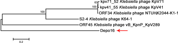
Phylogenetic analyses of Depo16. Sequences were compared using PHYLIP version 3.0, and phylogenetic trees were generated using the maximum likelihood method with 100 bootstrap replicates.
Expression and activity of Depo16
The putative depolymerase Depo16 of phage ZK1 was expressed and purified. SDS-PAGE analysis indicated that the size of Depo16 was approximately 88 kDa, which was the expected size (Fig. 3a). After continuous dilution, Depo16 was dropped on a lawn of K. pneumoniae Wkp26. Depo16 formed a halo zone on the lawn of K. pneumoniae Wkp26 at concentrations ≥ 0.5 µg/µL (Fig. 3b).
Fig 3.

Biological characteristics of Depo16. (a) SDS-PAGE analysis. Depo16 was separated by 10% SDS-PAGE along with Coomassie blue staining, and the molecular weight was approximately 88 kDa. Lane M, protein marker; Lane 1, purified Depo16. (b) Enzyme activity assay of Depo16. The activity of different concentrations of Depo16 (ranging from 5 ng/mL to 10 µg/mL) against K. pneumoniae strain Wkp26 on a plate. ZK1 and lysis buffer were used as controls. (c) Stability of Depo16 at different pH values. Different concentrations of Depo16 were spotted on lawns of K. pneumoniae Wkp26. (d) Stability of Depo16 at different temperatures. (e) Centrifugal precipitation test. Wkp26 (Depo16) represents K. pneumoniae Wkp26 grown to the mid-exponential phase (OD600 ≈ 0.6) and treated with Depo16 at a final concentration of 50 µg/mL at 37°C for 1 h. Wkp26 without any treatment was the control group. (f) CPS staining. CPS staining of the extracellular material of K. pneumoniae Wkp26 (left) and Wkp26 (Depo16) (right). (g) Alcian blue staining. CPS samples were extracted from equal amounts of the K. pneumoniae Wkp26 strains. After separation by 12% SDS-PAGE, the CPS phenotypes of Wkp26 (Lane 2) and Wkp26 (Depo16) (Lane 1) were visualized by alcian blue staining. (h) SEM assay. The surface morphology of K. pneumoniae Wkp26 and Wkp26 (Depo16) was observed. Scale bars represent 1 µm.
Characteristics of Depo16
In all, 95 strains of K. pneumoniae were utilized for activity testing, and Depo16 formed clear spots on 45 strains. All 45 strains belonged to the K1 capsular type (Table S1). Depo16 activity was maintained at pH 4–11 (Fig. 3c). At pH = 7, Depo16 had stable enzyme activity in a temperature range of 4 to 50°C. At 60°C, Depo16 was almost inactive (Fig. 3d).
Degradation of extracellular polysaccharides by Depo16
For the centrifugal precipitation test, the precipitate of the group without Depo16 treatment was loose, and the Depo16 treatment group exhibited a compact precipitate (Fig. 3e). Maneval staining and microscopic analyses showed that the Wkp26 strains were surrounded by blueish mucoid capsules. After treatment with Depo16, the blueish capsules disappeared (Fig. 3f). By alcian blue staining, the CPS in Wkp26 cells treated with Depo16 was much less than the CPS in Wkp26 cells not treated with Depo16 (Fig. 3g). In addition, SEM results showed that there was a thick capsule on the surface of Wkp26, while the capsule size decreased drastically when treated with Depo16 (Fig. 3h).
Depolymerase treatment makes K1 K. pneumoniae sensitive to macrophage capture
Depo16 treatment made serotype K1 K. pneumoniae Wkp26 sensitive to macrophage capture. The number of bacteria captured by PMs in the PBS-treated control group was 2.7 × 10⁵ CFU/mL, while the number of bacteria captured by PMs in the Depo16-treated group was 1.4 × 10⁶ CFU/mL (Fig. 4). The capture efficiency of the PMs on Wkp26 was 11.1% when Wkp26 was not treated with Depo16, and the capture efficiency increased to 56.0% when treated with Depo16.
Fig 4.
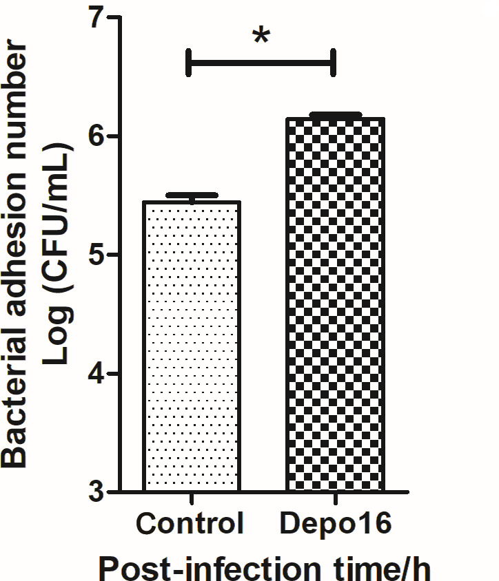
Effect of Depo16 on Wkp26 capture by peritoneal macrophages. Depolymerase treatment made K1 K. pneumoniae sensitive to capture by macrophages. Wkp26 (control group) and depolymerase Depo16-treated Wkp26 (Depo16 group) were added to 24-well cell culture plates with mouse peritoneal macrophage cells, and the plates were incubated for 1 h at 37°C. The values represent the mean and SD (n = 3). *P < 0.01, **P < 0.001, ***P < 0.0001.
Depo16 exhibits significant therapeutic effects in infected mice
The MLD of Wkp26 was 1.5 × 10⁵ CFU/mouse. After the challenge at this dose by intraperitoneal injection, all mice died within 36 h (Fig. 5a). At the same time, the infected mice showed severe clinical symptoms, such as a refusal to eat and drink, stiff limbs, and movement difficulties. A large amount of viscous yellow-white secretion appeared around the eyes of mice. After the challenge with 2 × MLD (3 × 10⁵ CFU/mouse), all untreated mice died within 48 h (Fig. 5b). By contrast, the Depo16 group had a significantly prolonged survival rate. A 10 µg/mouse dose of Depo16 increased the survival rate of mice to 80% in 7 days. The mice treated with Depo16 at a dose of 25 µg/mouse survived for 7 days. Moreover, no mice in either group died within 72 h after Depo16 treatment. By contrast, Depo16 did not show any therapeutic effects in mice infected with Depo16-insensitive serotype K2 K. pneumoniae bacteria (Fig. S8).
Fig 5.

Therapeutic effect of Depo16. (a) Survival curves of the infected mice. To determine the MLD, mice were intraperitoneally injected with different concentrations (1.5 × 10³, 1.5 × 10⁴, 1.5 × 10⁵, or 1.5 × 10⁶ CFU/mouse) of K. pneumoniae Wkp26 (10 mice in each group). The survival of mice in each group was recorded every 12 h for 5 days. (b) Survival curves of the infected mice treated with either PBS or Depo16. Each group contained 10 mice, which were intraperitoneally challenged with Wkp26 (3 × 10⁵ CFU/mouse). Various doses of Depo16 were administered intraperitoneally 1 h after infection, and the control group was treated with the same amount of sterile PBS. The survival of mice in each group was recorded every 24 h for 7 days. (c) Bacterial load in blood samples from mice. Blood samples were collected from each group at 2, 3, 6, 12, 18, 24, 30, and 48 h after the challenge. Bacterial load was determined based on the colony counts. (d) Levels of IL-6 in the blood of mice. Three mice from each group were euthanized at each time point. Blood samples were collected from each group of mice at 6, 12, 24, and 30 h after the challenge. The level of IL-6 cytokine in the blood was measured. Blood samples from healthy mice were used as controls (normal group). These values are expressed as the mean and SD (n = 3). *P < 0.01, **P < 0.001, ***P < 0.0001.
The therapeutic effect of Depo16 was further evaluated. At 12 h post-infection, the load of Wkp26 in the blood of the nontreatment control group reached approximately 1.5 × 10⁵ CFU/mL and continued to increase until 30 h, when it reached 1.29 × 10⁸ CFU/mL (Fig. 5c). However, the bacterial load in the Depo16-treated group was only 87 CFU/mL at 12 h and it was undetectable at 30 h. Comparatively, the mice in the control group all died within 30 h. In addition, the level of the inflammatory cytokine IL-6 in the blood was measured (Fig. 5d). Compared to healthy mice, the level of IL-6 in the blood of infected mice was elevated and reached >3.2 × 10³ pg/mL. However, the level of IL-6 was significantly lower in the Depo16-treated group, where it was continuously maintained at 673 pg/mL, similar to the level of healthy mice.
At 30 h, the bacterial load in the main tissues of mice in the infection group reached approximately 9.35 × 10⁸ CFU/g (lung) (Fig. 6a), 8.18 × 10⁸ CFU/g (liver) (Fig. 6b), 1.02 × 10⁹ CFU/g (spleen) (Fig. 6c), and 5.5 × 10⁸ CFU/g (kidney) (Fig. 6d). By contrast, the bacterial load in tissues of the mice in the Depo16 treatment group was significantly reduced (P < 0.001) to approximately 5.09 × 10⁵ CFU/g (lung) (Fig. 6a), 1.84 × 10⁵ CFU/g (liver) (Fig. 6b), 5.32 × 10⁵ CFU/g (spleen) (Fig. 6c), and 3.07 × 10³ CFU/g (kidney) (Fig. 6d). After 48 h of treatment with Depo16, the bacterial load was further reduced in all tissues in the treated group compared to that in the control group. Although the bacteria were still not clear from all the tissues of infected mice, H&E staining of the tissue showed hemorrhage, diffuse red blood cells, and the infiltration of neutrophils in the infection group (Fig. 6e). Some cells were even fragmented and necrotic. Comparatively, Depo16 treatment significantly alleviated pathological damage in liver and kidney tissue, and no obvious pathological changes were found in the lung and spleen.
Fig 6.

Bacterial load and histopathological examination of the major tissues of mice. The lung, liver, spleen, and kidney of mice from each group were removed at 3, 6, 12, 24, and 30 h after the challenge, and part of the tissue was homogenized to calculate the bacterial load. Bacterial loads in the lung (a), liver (b), spleen (c), and kidney (d) were determined. The values represent the mean and SD (n = 3). (e) H&E analysis. The mice were euthanized 30 h after infection, and lung, liver, spleen, and kidney tissues were removed and stained with H&E.
DISCUSSION
Compared with other K. pneumoniae, hvKP (especially serotypes K1 and K2) is more virulent and has a higher fatality rate (5, 6, 44). Phage derivative proteins, such as depolymerases, can destroy the virulence factor CPS and show good therapeutic effects for K. pneumoniae infections (45, 46). A putative depolymerase of Depo16 was expressed and studied. Depo16 can specifically degrade the K1 serotype CPS of K. pneumoniae and make bacteria sensitive to peritoneal macrophages. In addition, Depo16 had therapeutic effects in a mouse model of bacteremia.
Usually, a given depolymerase only shows depolymerization activity for a single serotyped capsule. For example, K1-ORF34 exhibited specificity for 21 strains with capsular-type K1 (22). Similarly, Depo16 also showed activity only against the K1 CPS of K. pneumoniae. However, our results showed that not all the K1 serotype strains were sensitive to Depo16. Three K1 serotype strains (Wkp54, Wkp73, and Wkp93) were insensitive to Depo16 and phage ZK1 (Table S1). We measured the adsorption of ZK1 on Wkp54, Wkp73, and Wkp93 and found that the adsorption of phages was significantly reduced (these data are not presented). Wkp54, Wkp73, and Wkp93 belong to the K1 serotype according to the wzc gene, but their CPS may differ in sugar chains or spatial structures compared to that of other K1 CPS (sensitive to Depo16). Although the wzc gene has allowed for the identification of more than 79 serotypes (4), the diversity of CPS sugar chains still makes genetic testing methods questionable. The diversity of CPS sugar chains may be the main reason for the insensitivity of Wkp54, Wkp73, and Wkp93.
More than 23 depolymerases derived from K. pneumoniae phages have been previously reported (47). In addition to Depo16, only six K. pneumoniae phage depolymerases targeting serotype K1 CPS have been studied (21, 23, 48, 49). Among these serotype K1 K. pneumoniae phage depolymerases, only K1-ORF34 has been expressed, and its functional characteristics have been investigated (22). Notably, Depo16 had almost no homology with K1-ORF34 and other K1 K. pneumoniae phage depolymerases. Depo16 can degrade the CPS (serotype K1), but exactly how it does is not clear because of its lack of similarity to other fully researched depolymerases. Also, the function of Depo16 may be only similar to depolymerase. It may be a different enzyme with an overlapping function. After the structure and molecular mechanism of this protein are revealed in subsequent research, the mystery can be revealed.
Some depolymerases are also capable of removing the biofilm or enhancing antibiotic susceptibility (45, 50). We found that Depo16 also enhances the sensitivity of K. pneumoniae to polymyxin B (Fig. S9). In terms of biofilm, the effect of Depo16 was not able to be observed in conventional crystal violet solution staining experiments (45).
Depolymerases have shown good antibacterial and therapeutic effects in vitro and in animal experiments (13–15). In our study, although Depo16 did not have bactericidal effects (Fig. S7), Depo16 sensitized the bacteria to peritoneal macrophages by degrading the capsular polysaccharide, just like other reported depolymerases (46). K. pneumoniae Wkp26 is highly virulent, and a challenge of only 1.5 × 10⁵ CFU/mouse was sufficient to cause 100% mortality in mice within 36 h. However, Depo16 still provides a good therapeutic effect, and small single doses of Depo16 led to survival for at least 7 days. Thus, Depo16 shows promising application prospects. Meanwhile, treatment with Depo16 did not result in the complete removal of bacteria from the major tissues after 48 h. This may be because depolymerase itself has no bactericidal ability, so it does not achieve as good of an effect as phages or antibiotics (13, 15, 19). Nonetheless, the remaining bacteria were not enough to cause serious or life-threatening infections in the mice.
The high specificity of depolymerases limits their application. Only a few phage depolymerases show depolymerization activity against strains of different serotypes (21). Their spectrum of activity may be expanded through some improvements. The artificial modification of depolymerases by genetic engineering is also feasible (51). Depolymerases against strains of different serotypes can be applied together, just like phage cocktails (52, 53). Furthermore, depolymerases can also be used in combination with antibiotics (54), antimicrobial peptides (55), or Chinese herbal medicines [such as Carthamus tinctorius oil (56), allicin (57), or Plantago major (58)].
Conclusions
In this study, the putative depolymerase Depo16 of the newly isolated phage ZK1 was identified. Depo16 showed no significant similarity to any reported phage depolymerases in terms of amino acid sequence and existed in an independent branch of evolution. The depolymerization activity of Depo16 was stable at different temperatures and pH values and could sensitize bacteria to peritoneal macrophages by degrading CPS. More importantly, Depo16 was able to protect mice against bacteremia. Thus, Depo16 is a potential antibacterial agent to combat serotype K1 K. pneumoniae infections.
ACKNOWLEDGMENTS
This work was financially supported through grants from the National Natural Science Foundation of China (Nos. 32072824 and 32222083), the Achievement Transformation Project of the First Hospital of Jilin University (Nos. JJKH20231216KJ and JDYYZH-2022006), and the Fundamental Research Funds for the Central Universities.
Contributor Information
Zhimin Guo, Email: amily@jlu.edu.cn.
Jingmin Gu, Email: jingmin0629@163.com.
Martha Vives, Unversidad de los Andes, Bogotá, Colombia.
DATA AVAILABILITY
The completed genome sequence of Klebsiella phage vB_KpnP_ZK1 has been deposited in the GenBank database with accession number OK625527.
SUPPLEMENTAL MATERIAL
The following material is available online at https://doi.org/10.1128/aem.01197-23.
Figures S1 to S9; Tables S1 to S3.
ASM does not own the copyrights to Supplemental Material that may be linked to, or accessed through, an article. The authors have granted ASM a non-exclusive, world-wide license to publish the Supplemental Material files. Please contact the corresponding author directly for reuse.
REFERENCES
- 1. Kuehn BM. 2013. "Nightmare" bacteria on the rise in US hospitals, long-term care facilities. JAMA 309:1573–1574. doi: 10.1001/jama.2013.2922 [DOI] [PubMed] [Google Scholar]
- 2. Paczosa MK, Mecsas J. 2016. Klebsiella pneumoniae: going on the offense with a strong defense. Microbiol Mol Biol Rev 80:629–661. doi: 10.1128/MMBR.00078-15 [DOI] [PMC free article] [PubMed] [Google Scholar]
- 3. Harada S, Aoki K, Yamamoto S, Ishii Y, Sekiya N, Kurai H, Furukawa K, Doi A, Tochitani K, Kubo K, et al. 2019. Clinical and molecular characteristics of Klebsiella pneumoniae isolates causing bloodstream infections in Japan: occurrence of hypervirulent infections in health care. J Clin Microbiol 57:e01206-19. doi: 10.1128/JCM.01206-19 [DOI] [PMC free article] [PubMed] [Google Scholar]
- 4. Pan YJ, Lin TL, Chen YH, Hsu CR, Hsieh PF, Wu MC, Wang JT. 2013. Capsular types of Klebsiella pneumoniae revisited by wzc sequencing. PLoS One 8:e80670. doi: 10.1371/journal.pone.0080670 [DOI] [PMC free article] [PubMed] [Google Scholar]
- 5. Choby JE, Howard-Anderson J, Weiss DS. 2020. Hypervirulent Klebsiella pneumoniae - clinical and molecular perspectives. J Intern Med 287:283–300. doi: 10.1111/joim.13007 [DOI] [PMC free article] [PubMed] [Google Scholar]
- 6. Chung DR, Lee SS, Lee HR, Kim HB, Choi HJ, Eom JS, Kim JS, Choi YH, Lee JS, Chung MH, Kim YS, Lee H, Lee MS, Park CK, Korean Study Group for Liver Abscess . 2007. Emerging invasive liver abscess caused by K1 serotype Klebsiella pneumoniae in Korea. J Infect 54:578–583. doi: 10.1016/j.jinf.2006.11.008 [DOI] [PubMed] [Google Scholar]
- 7. Cheng NC, Yu YC, Tai HC, Hsueh PR, Chang SC, Lai SY, Yi WC, Fang CT. 2012. Recent trend of necrotizing fasciitis in Taiwan: focus on monomicrobial Klebsiella pneumoniae necrotizing fasciitis. Clin Infect Dis 55:930–939. doi: 10.1093/cid/cis565 [DOI] [PubMed] [Google Scholar]
- 8. Nazir A, Song J, Chen Y, Liu Y. 2023. Phage-derived depolymerase: its possible role for secondary bacterial infections in COVID-19 patients. Microorganisms 11:424. doi: 10.3390/microorganisms11020424 [DOI] [PMC free article] [PubMed] [Google Scholar]
- 9. Yang Y, Dufault-Thompson K, Yan W, Cai T, Xie L, Jiang X. 2023. Deciphering phage-host specificity based on the association of phage depolymerases and bacterial surface glycan with deep learning. bioRxiv:2023.06.16.545366. doi: 10.1101/2023.06.16.545366 [DOI]
- 10. Magill DJ, Skvortsov TA. 2023. DePolymerase predictor (DePP): a machine learning tool for the targeted identification of phage depolymerases. BMC Bioinformatics 24:208. doi: 10.1186/s12859-023-05341-w [DOI] [PMC free article] [PubMed] [Google Scholar]
- 11. Pires DP, Oliveira H, Melo LDR, Sillankorva S, Azeredo J. 2016. Bacteriophage-encoded depolymerases: their diversity and biotechnological applications. Appl Microbiol Biotechnol 100:2141–2151. doi: 10.1007/s00253-015-7247-0 [DOI] [PubMed] [Google Scholar]
- 12. Rakhuba DV, Kolomiets EI, Dey ES, Novik GI. 2010. Bacteriophage receptors, mechanisms of phage adsorption and penetration into host cell. Pol J Microbiol 59:145–155. [PubMed] [Google Scholar]
- 13. Majkowska-Skrobek G, Latka A, Berisio R, Squeglia F, Maciejewska B, Briers Y, Drulis-Kawa Z. 2018. Phage-borne depolymerases decrease Klebsiella pneumoniae resistance to innate defense mechanisms. Front Microbiol 9:2517. doi: 10.3389/fmicb.2018.02517 [DOI] [PMC free article] [PubMed] [Google Scholar]
- 14. Guo Z, Huang J, Yan G, Lei L, Wang S, Yu L, Zhou L, Gao A, Feng X, Han W, Gu J, Yang J. 2017. Identification and characterization of Dpo42, a novel depolymerase derived from the Escherichia coli phage vB_EcoM_ECOO78. Front Microbiol 8:1460. doi: 10.3389/fmicb.2017.01460 [DOI] [PMC free article] [PubMed] [Google Scholar]
- 15. Wang C, Li P, Zhu Y, Huang Y, Gao M, Yuan X, Niu W, Liu H, Fan H, Qin Y, Tong Y, Mi Z, Bai C. 2020. Identification of a novel Acinetobacter baumannii phage-derived depolymerase and its therapeutic application in mice. Front Microbiol 11:1407. doi: 10.3389/fmicb.2020.01407 [DOI] [PMC free article] [PubMed] [Google Scholar]
- 16. Kim SH, Lee H, Park MK. 2023. Isolation, characterization, and application of a novel, lytic phage vB_SalA_KFSST3 with depolymerase for the control of Salmonella and its biofilm on cantaloupe under cold temperature. Food Res Int 172:113062. doi: 10.1016/j.foodres.2023.113062 [DOI] [PubMed] [Google Scholar]
- 17. Santiago AJ, Burgos-Garay ML, Kartforosh L, Mazher M, Donlan RM. 2020. Bacteriophage treatment of carbapenemase-producing Klebsiella pneumoniae in a multispecies biofilm: a potential biocontrol strategy for healthcare facilities. AIMS Microbiol 6:43–63. doi: 10.3934/microbiol.2020003 [DOI] [PMC free article] [PubMed] [Google Scholar]
- 18. Li M, Li P, Chen L, Guo G, Xiao Y, Chen L, Du H, Zhang W. 2021. Identification of a phage-derived depolymerase specific for KL64 capsule of Klebsiella pneumoniae and its anti-biofilm effect. Virus Genes 57:434–442. doi: 10.1007/s11262-021-01847-8 [DOI] [PubMed] [Google Scholar]
- 19. Oliveira H, Pinto G, Mendes B, Dias O, Hendrix H, Akturk E, Noben JP, Gawor J, Łobocka M, Lavigne R, Azeredo J. 2020. A tailspike with exopolysaccharide depolymerase activity from a new Providencia stuartii phage makes multidrug-resistant bacteria susceptible to serum-mediated killing. Appl Environ Microbiol 86:e00073-20. doi: 10.1128/AEM.00073-20 [DOI] [PMC free article] [PubMed] [Google Scholar]
- 20. Sun X, Pu B, Qin J, Xiang J. 2023. Effect of a depolymerase encoded by phage168 on a carbapenem-resistant Klebsiella pneumoniae and its biofilm. Pathogens 12:1396. doi: 10.3390/pathogens12121396 [DOI] [PMC free article] [PubMed] [Google Scholar]
- 21. Pan YJ, Lin TL, Chen CC, Tsai YT, Cheng YH, Chen YY, Hsieh PF, Lin YT, Wang JT. 2017. Klebsiella phage ΦK64-1 encodes multiple depolymerases for multiple host capsular types. J Virol 91:e02457-16. doi: 10.1128/JVI.02457-16 [DOI] [PMC free article] [PubMed] [Google Scholar]
- 22. Lin TL, Hsieh PF, Huang YT, Lee WC, Tsai YT, Su PA, Pan YJ, Hsu CR, Wu MC, Wang JT. 2014. Isolation of a bacteriophage and its depolymerase specific for K1 capsule of Klebsiella pneumoniae: implication in typing and treatment. J Infect Dis 210:1734–1744. doi: 10.1093/infdis/jiu332 [DOI] [PubMed] [Google Scholar]
- 23. Blundell-Hunter G, Enright MC, Negus D, Dorman MJ, Beecham GE, Pickard DJ, Wintachai P, Voravuthikunchai SP, Thomson NR, Taylor PW. 2021. Characterisation of bacteriophage-encoded depolymerases selective for key Klebsiella pneumoniae capsular exopolysaccharides. Front Cell Infect Microbiol 11:686090. doi: 10.3389/fcimb.2021.686090 [DOI] [PMC free article] [PubMed] [Google Scholar]
- 24. Sunagawa S, DeSantis TZ, Piceno YM, Brodie EL, DeSalvo MK, Voolstra CR, Weil E, Andersen GL, Medina M. 2009. Bacterial diversity and white plague disease-associated community changes in the Caribbean coral Montastraea faveolata. ISME J 3:512–521. doi: 10.1038/ismej.2008.131 [DOI] [PubMed] [Google Scholar]
- 25. Yu WL, Ko WC, Cheng KC, Lee HC, Ke DS, Lee CC, Fung CP, Chuang YC. 2006. Association between rmpA and magA genes and clinical syndromes caused by Klebsiella pneumoniae in Taiwan. Clin Infect Dis 42:1351–1358. doi: 10.1086/503420 [DOI] [PubMed] [Google Scholar]
- 26. Fang CT, Chuang YP, Shun CT, Chang SC, Wang JT. 2004. A novel virulence gene in Klebsiella pneumoniae strains causing primary liver abscess and septic metastatic complications. J Exp Med 199:697–705. doi: 10.1084/jem.20030857 [DOI] [PMC free article] [PubMed] [Google Scholar]
- 27. Cai R, Wang Z, Wang G, Zhang H, Cheng M, Guo Z, Ji Y, Xi H, Wang X, Xue Y, Ur Rahman S, Sun C, Feng X, Lei L, Tong Y, Han W, Gu J. 2019. Biological properties and genomics analysis of vB_KpnS_GH-K3, a Klebsiella phage with a putative depolymerase-like protein. Virus Genes 55:696–706. doi: 10.1007/s11262-019-01681-z [DOI] [PubMed] [Google Scholar]
- 28. Xi H, Dai J, Tong Y, Cheng M, Zhao F, Fan H, Li X, Cai R, Ji Y, Sun C, Feng X, Lei L, Rahman SU, Han W, Gu J. 2019. The characteristics and genome analysis of vB_AviM_AVP, the first phage infecting Aerococcus viridans. Viruses 11:104. doi: 10.3390/v11020104 [DOI] [PMC free article] [PubMed] [Google Scholar]
- 29. Jhandai P, Mittal D, Gupta R, Kumar M, Khurana R. 2023. Therapeutics and prophylactic efficacy of novel lytic Escherichia phage vB_EcoS_PJ16 against multidrug-resistant avian pathogenic E. coli using in vivo study. Int Microbiol. doi: 10.1007/s10123-023-00420-7 [DOI] [PubMed] [Google Scholar]
- 30. Tang F, Zhang P, Zhang Q, Xue F, Ren J, Sun J, Qu Z, Zhuge X, Li D, Wang J, Jiang M, Dai J. 2019. Isolation and characterization of a broad-spectrum phage of multiple drug resistant Salmonella and its therapeutic utility in mice. Microb Pathog 126:193–198. doi: 10.1016/j.micpath.2018.10.042 [DOI] [PubMed] [Google Scholar]
- 31. Sambrook J, Russell DW. 2006. Precipitation of bacteriophage λ particles from large-scale lysates. CSH Protoc 2006:pdb.prot3966. doi: 10.1101/pdb.prot3966 [DOI] [PubMed] [Google Scholar]
- 32. Margulies M, Egholm M, Altman WE, Attiya S, Bader JS, Bemben LA, Berka J, Braverman MS, Chen Y-J, Chen Z, et al. 2005. Genome sequencing in microfabricated high-density picolitre reactors. Nature 437:376–380. doi: 10.1038/nature03959 [DOI] [PMC free article] [PubMed] [Google Scholar]
- 33. Besemer J, Lomsadze A, Borodovsky M. 2001. GeneMarkS: a self-training method for prediction of gene starts in microbial genomes. Implications for finding sequence motifs in regulatory regions. Nucleic Acids Res 29:2607–2618. doi: 10.1093/nar/29.12.2607 [DOI] [PMC free article] [PubMed] [Google Scholar]
- 34. Altschul SF, Madden TL, Schäffer AA, Zhang J, Zhang Z, Miller W, Lipman DJ. 1997. Gapped BLAST and PSI-BLAST: a new generation of protein database search programs. Nucleic Acids Res 25:3389–3402. doi: 10.1093/nar/25.17.3389 [DOI] [PMC free article] [PubMed] [Google Scholar]
- 35. Shimada MK, Nishida T. 2017. A modification of the PHYLIP program: a solution for the redundant cluster problem, and an implementation of an automatic bootstrapping on trees inferred from original data. Mol Phylogenet Evol 109:409–414. doi: 10.1016/j.ympev.2017.02.012 [DOI] [PubMed] [Google Scholar]
- 36. Wang C, Li P, Niu W, Yuan X, Liu H, Huang Y, An X, Fan H, Zhangxiang L, Mi L, Zheng J, Liu Y, Tong Y, Mi Z, Bai C. 2019. Protective and therapeutic application of the depolymerase derived from a novel KN1 genotype of Klebsiella pneumoniae bacteriophage in mice. Res Microbiol 170:156–164. doi: 10.1016/j.resmic.2019.01.003 [DOI] [PubMed] [Google Scholar]
- 37. Ren G, Wang Z, Li Y, Hu X, Wang X. 2016. Effects of lipopolysaccharide core sugar deficiency on colanic acid biosynthesis in Escherichia coli. J Bacteriol 198:1576–1584. doi: 10.1128/JB.00094-16 [DOI] [PMC free article] [PubMed] [Google Scholar]
- 38. Lee KJ, Kim JA, Hwang W, Park SJ, Lee KH. 2013. Role of capsular polysaccharide (CPS) in biofilm formation and regulation of CPS production by quorum-sensing in Vibrio vulnificus. Mol Microbiol 90:841–857. doi: 10.1111/mmi.12401 [DOI] [PubMed] [Google Scholar]
- 39. Hsieh PF, Lin HH, Lin TL, Chen YY, Wang JT. 2017. Two T7-like bacteriophages, K5-2 and K5-4, each encodes two capsule depolymerases: isolation and functional characterization. Sci Rep 7:4624. doi: 10.1038/s41598-017-04644-2 [DOI] [PMC free article] [PubMed] [Google Scholar]
- 40. Zhang X, Goncalves R, Mosser DM. 2008. The isolation and characterization of murine macrophages. Curr Protoc Immunol Chapter 14:14. doi: 10.1002/0471142735.im1401s83 [DOI] [PMC free article] [PubMed] [Google Scholar]
- 41. Wang Z, Cai R, Wang G, Guo Z, Liu X, Guan Y, Ji Y, Zhang H, Xi H, Zhao R, Bi L, Liu S, Yang L, Feng X, Sun C, Lei L, Han W, Gu J. 2021. Combination therapy of phage vB_KpnM_P-KP2 and gentamicin combats acute pneumonia caused by K47 serotype Klebsiella pneumoniae. Front Microbiol 12:674068. doi: 10.3389/fmicb.2021.674068 [DOI] [PMC free article] [PubMed] [Google Scholar]
- 42. Hung CH, Kuo CF, Wang CH, Wu CM, Tsao N. 2011. Experimental phage therapy in treating Klebsiella pneumoniae-mediated liver abscesses and bacteremia in mice. Antimicrob Agents Chemother 55:1358–1365. doi: 10.1128/AAC.01123-10 [DOI] [PMC free article] [PubMed] [Google Scholar]
- 43. Ji Y, Song L, Zhou Z, Liu X, Li F, Guo Z, Guan Y, Yang L, Feng X, Sun C, Lei L, Han W, Gu J. 2021. vB-ApyS-JF1, the first Trueperella pyogenes phage, shows potential as an alternative treatment strategy for Trueperella pyogenes infections. Front Microbiol 12:736304. doi: 10.3389/fmicb.2021.736304 [DOI] [PMC free article] [PubMed] [Google Scholar] [Retracted]
- 44. Liu C, Guo J. 2019. Hypervirulent Klebsiella pneumoniae (hypermucoviscous and aerobactin positive) infection over 6 years in the elderly in China: antimicrobial resistance patterns, molecular epidemiology and risk factor. Ann Clin Microbiol Antimicrob 18:4. doi: 10.1186/s12941-018-0302-9 [DOI] [PMC free article] [PubMed] [Google Scholar]
- 45. Wu Y, Wang R, Xu M, Liu Y, Zhu X, Qiu J, Liu Q, He P, Li Q. 2019. A novel polysaccharide depolymerase encoded by the phage SH-KP152226 confers specific activity against multidrug-resistant Klebsiella pneumoniae via biofilm degradation. Front Microbiol 10:2768. doi: 10.3389/fmicb.2019.02768 [DOI] [PMC free article] [PubMed] [Google Scholar]
- 46. Cai R, Ren Z, Zhao R, Lu Y, Wang X, Guo Z, Song J, Xiang W, Du R, Zhang X, Han W, Ru H, Gu J. 2023. Structural biology and functional features of phage-derived depolymerase Depo32 on Klebsiella pneumoniae with K2 serotype capsular polysaccharides. Microbiol Spectr 11:e0530422. doi: 10.1128/spectrum.05304-22 [DOI] [PMC free article] [PubMed] [Google Scholar]
- 47. Pan YJ, Lin TL, Chen YY, Lai PH, Tsai YT, Hsu CR, Hsieh PF, Lin YT, Wang JT. 2019. Identification of three podoviruses infecting Klebsiella encoding capsule depolymerases that digest specific capsular types. Microb Biotechnol 12:472–486. doi: 10.1111/1751-7915.13370 [DOI] [PMC free article] [PubMed] [Google Scholar]
- 48. Volozhantsev NV, Myakinina VP, Popova AV, Kislichkina AA, Komisarova EV, Knyazeva AI, Krasilnikova VM, Fursova NK, Svetoch EA. 2016. Complete genome sequence of novel T7-like virus vB_KpnP_KpV289 with lytic activity against Klebsiella pneumoniae. Arch Virol 161:499–501. doi: 10.1007/s00705-015-2680-z [DOI] [PubMed] [Google Scholar]
- 49. Solovieva EV, Myakinina VP, Kislichkina AA, Krasilnikova VM, Verevkin VV, Mochalov VV, Lev AI, Fursova NK, Volozhantsev NV. 2018. Comparative genome analysis of novel podoviruses lytic for hypermucoviscous Klebsiella pneumoniae of K1, K2, and K57 capsular types. Virus Res 243:10–18. doi: 10.1016/j.virusres.2017.09.026 [DOI] [PubMed] [Google Scholar]
- 50. Majkowska-Skrobek G, Łątka A, Berisio R, Maciejewska B, Squeglia F, Romano M, Lavigne R, Struve C, Drulis-Kawa Z. 2016. Capsule-targeting depolymerase, derived from Klebsiella KP36 phage, as a tool for the development of anti-virulent strategy. Viruses 8:324. doi: 10.3390/v8120324 [DOI] [PMC free article] [PubMed] [Google Scholar]
- 51. Lin TL, Yang FL, Ren CT, Pan YJ, Liao KS, Tu IF, Chang YP, Cheng YY, Wu CY, Wu SH, Wang JT. 2022. Development of Klebsiella pneumoniae capsule polysaccharide-conjugated vaccine candidates using phage depolymerases. Front Immunol 13:843183. doi: 10.3389/fimmu.2022.843183 [DOI] [PMC free article] [PubMed] [Google Scholar]
- 52. Oliveira H, Drulis-Kawa Z, Azeredo J. 2022. Exploiting phage-derived carbohydrate depolymerases for combating infectious diseases. Trends Microbiol 30:707–709. doi: 10.1016/j.tim.2022.05.002 [DOI] [PubMed] [Google Scholar]
- 53. Drobiazko AY, Kasimova AA, Evseev PV, Shneider MM, Klimuk EI, Shashkov AS, Dmitrenok AS, Chizhov AO, Slukin PV, Skryabin YP, Volozhantsev NV, Miroshnikov KA, Knirel YA, Popova AV. 2022. Capsule-targeting depolymerases derived from Acinetobacter baumannii prophage regions. Int J Mol Sci 23:4971. doi: 10.3390/ijms23094971 [DOI] [PMC free article] [PubMed] [Google Scholar]
- 54. Chen X, Liu M, Zhang P, Xu M, Yuan W, Bian L, Liu Y, Xia J, Leung SSY. 2022. Phage-derived depolymerase as an antibiotic adjuvant against multidrug-resistant Acinetobacter baumannii. Front Microbiol 13:845500. doi: 10.3389/fmicb.2022.845500 [DOI] [PMC free article] [PubMed] [Google Scholar]
- 55. da Silva Duarte V, Dias RS, Kropinski AM, da Silva Xavier A, Ferro CG, Vidigal PMP, da Silva CC, de Paula SO. 2018. A T4virus prevents biofilm formation by Trueperella pyogenes. Vet Microbiol 218:45–51. doi: 10.1016/j.vetmic.2018.03.025 [DOI] [PubMed] [Google Scholar]
- 56. Saeed J, Hussain Shah SN, Javed H, Aslam A, Ali A, Siddique F, Zahra T, Bin Jardan YA, Wondmie GF, Nafidi HA, Bourhia M. 2023. Experimental and in silico evaluation of Carthamus tinctorius L. oil emulgel: a promising treatment for bacterial skin infections. Front Cell Infect Microbiol 13:1253095. doi: 10.3389/fcimb.2023.1253095 [DOI] [PMC free article] [PubMed] [Google Scholar]
- 57. Tao Z, Geng D, Tao J, Wang J, Liu S, Wang Q, Xu F, Xiao S, Wang R. 2023. Synergistic antibacterial effect and mechanism of allicin and an Enterobacter cloacae bacteriophage. Microbiol Spectr 11:e0315522. doi: 10.1128/spectrum.03155-22 [DOI] [PMC free article] [PubMed] [Google Scholar]
- 58. Farid A, Sheibani M, Shojaii A, Noori M, Motevalian M. 2022. Evaluation of anti-inflammatory effects of leaf and seed extracts of Plantago major on acetic acid-induced ulcerative colitis in rats. J Ethnopharmacol 298:115595. doi: 10.1016/j.jep.2022.115595 [DOI] [PubMed] [Google Scholar]
Associated Data
This section collects any data citations, data availability statements, or supplementary materials included in this article.
Supplementary Materials
Figures S1 to S9; Tables S1 to S3.
Data Availability Statement
The completed genome sequence of Klebsiella phage vB_KpnP_ZK1 has been deposited in the GenBank database with accession number OK625527.