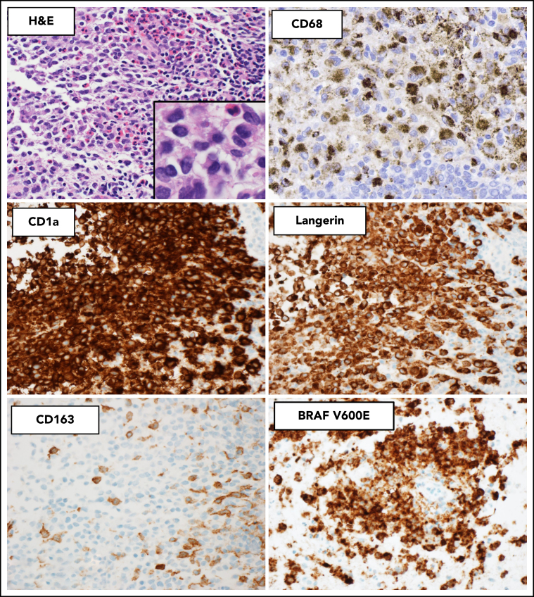
Figure 2.
Histopathologic and immunophenotypic features of LCH. The characteristic infiltrate of LCH on hematoxylin and eosin shows mononuclear cells with grooved nuclei and abundant eosinophilic cytoplasm, with a mixture of eosinophils and small lymphocytes. The LCH cells show expression of CD68 (cytoplasmic, Golgi dot-like staining with background macrophages darkly stained), CD1a (surface), and langerin (cytoplasmic) and are negative for CD163. The mutant-specific antibody clone VE1 detects the BRAF-V600E mutation by immunohistochemistry with 2-3+ strong cytoplasmic staining. Images magnification ×400; inset magnification ×600.

