



Abstract
Elevated blood pressure is a well-established risk factor for age-related cognitive decline. Long linked to cognitive impairment on vascular bases, increasing evidence suggests a potential association of hypertension with the neurodegenerative pathology underlying Alzheimer’s disease. Hypertension is well known to disrupt the structural and functional integrity of the cerebral vasculature. However, the mechanisms by which these alterations lead to brain damage, enhance Alzheimer pathology, and promote cognitive impairment remain to be established. Furthermore, critical questions concerning whether lowering blood pressure by antihypertensive medications prevents cognitive impairment have not been answered. Recent developments in neurovascular biology, brain imaging, and epidemiology, as well as new clinical trials, have provided insights into these critical issues. In particular, clinical and basic findings on the link between neurovascular dysfunction and the pathobiology of neurodegeneration have shed new light on the overlap between vascular and Alzheimer pathology. In this review, we will examine the progress made in the relationship between hypertension and cognitive impairment and, after a critical evaluation of the evidence, attempt to identify remaining knowledge gaps and future research directions that may advance our understanding of one of the leading health challenges of our time.
Keywords: Cerebral blood flow, neurovascular dysfunction, Alzheimer disease, dementia
Introduction
The impacts of elevated blood pressure (BP) on brain health significantly contribute to cognitive decline over the life course1–3. Although associations between chronically elevated BP and cognitive impairment were noted by the end of the 19th century, systematic investigations into the relationship did not begin in earnest until the 1940s-1960s. The introduction of the Kety and Schmidt method to measure cerebral blood flow (CBF) led to the discovery that cerebrovascular resistance is increased in hypertensives4 and correlates with hypertensive retinopathy5. Shortly thereafter, pioneering observations of diminished psychomotor speed in hypertensive air traffic controllers by Spieth6 and intellectual decline in patients with hypertension by Wilkie and Eisdorfer7 provided the initial evidence associating chronically elevated BP with worsened cognition.
More recently, hypertension has emerged as a pathogenic factor both in cognitive impairment on vascular bases and in Alzheimer’s disease (AD)8. This hypertension-dementia relationship has led to hypertension being targeted as a treatable condition that could delay the onset of cognitive deterioration. Longitudinal studies indicate preservation of cognition following adequate BP control9, particularly in intensive lowering regimens10, although such outcomes are not universally observed11. Results from investigations into the differential efficacy of antihypertensive agents in combating cognitive decline are similarly conflicting12–14. Consequently, emphasis has been placed on the identification of early biomarkers of hypertension-induced brain damage and novel therapeutic targets. To this end, recent studies have focussed on identifying the white matter tracts and brain regions affected in hypertension, the mechanisms by which hypertension impairs cognition, and the nature of the association of hypertension with AD pathology.
The present review will explore the consequences of hypertensive sequelae on cerebrovascular health, cognition, and dementia risk; the associations between hypertension and AD pathology; and potential therapeutic targets and strategies to mitigate the impacts of hypertension on cognitive decline. Finally, we will attempt to highlight outstanding questions that remain unaddressed.
Hypertension and cognitive decline: epidemiological evidence
BP trends over the life course, as observed in the Framingham Heart Study15, suggest an age-related pattern of systolic (SBP) and diastolic (DBP) BP changes in hypertension. Characterized initially by increasing DBP and SBP in early adulthood, DBP falls in late-life concurrent with continual elevation of SBP. Accordingly, mean arterial pressure progressively increases until reaching an asymptote around the seventh decade, whereas pule pressure, beginning in middle-age, steadily rises throughout the remaining lifespan15.
The hypertension-cognition relationship changes over the life course (for in-depth reviews, see references16,17). Thus, elevated BP in the 4th to 5th decade of life is associated with late-life cognitive deterioration1–3, while a U-shaped relationship is noted in the elderly where both high and low BP portend cognitive impairment18. Of interest, elevated SBP through mid-life followed by a rapid drop in late-life is associated with severe cognitive decline19 and brain atrophy20, suggesting that late-life reductions in BP may be driven by pathological brain changes impacting autonomic control, as has been advocated for AD21. Whether preventing such late-life BP decline would be beneficial remains to be established.
BP variability (BPV), defined as variations in BP stratified by very short term (beat-to-beat), short term (24-hours), mid-term (day-to-day), or long term (visit-to-visit)22 variability, has emerged as an additional contributor to cognitive impairment, particularly in the elderly. In recent meta-analyses combining 53 (9 very short-, 12 short-, 9 mid-, 23 long-term) and 19 (4 short-, 4 mid-, 11 long-term) studies, worse cognitive performance was associated with short, medium, and long term BPV23,24. In contrast, low BPV over the very-short term elevates the risk of poor cognitive outcomes, perhaps due to impairment of the ability to adjust perfusion commensurate with demand (e.g., postural changes)23.
In summary, hypertension, particularly in mid-life, is associated with worse long-term cognitive outcomes, and increased short term, mid-term, and long term BPV are emergent risk factors for cognitive decline and dementia, especially in the elderly.
Hypertension and the pathobiology of cerebrovascular impairment
In this section, we will first review basic concepts on the blood supply of the brain, then examine the consequences of hypertension on the structure and function of cerebral vessels.
The cerebral blood supply and the neurovasculome
The carotid arteries enter the skull and merge to form the circle of Willis, a collateral flow loop from which the anterior, middle, and posterior cerebral arteries originate25. Branches of these vessels travel along the surface of the brain within the subarachnoid space, forming an anastomotic network (pial arteries)25, before diving into the substance of the brain (penetrating arteries and arterioles) while ensheathed within an extension of the subarachnoid space delimited by the vascular basement membrane and glia limitans (perivascular space; PVS). As these penetrating arterioles advance into the brain parenchyma, the PVS disappears, and the vascular basal lamina and glia limitans merge25. At this level, arterioles are surrounded by a single layer of smooth muscle cells which, as the vessels get smaller, becomes discontinuous until replaced by pericytes at the capillary level25.
Interactions between cerebral vessels and neural elements are crucial determinants of CBF. Structural and functional associations between vascular cells, glia, and neurons, dubbed the neurovascular unit (NVU), regulate blood-brain barrier (BBB) maintenance and the adjustment of cerebral perfusion commensurate with local metabolic demands25. Owing to the segmental specificity of these neurovascular interactions, the concept of the NVU has been recently expanded to include large extra- and intra-cerebral vessels, as well as meningeal vessels and lymphatics (the neurovasculome)26.
Hypertension alters cerebrovascular structure
Hypertension is often preceded by arterial stiffening27 which, as a result of diminished vascular compliance, drives an elevation in pulse pressure27 that exposes the downstream circulation to increased tensile strain28, triggering adaptive remodeling responses throughout the cerebrovascular tree in an attempt to preserve vessel integrity (Figure 1)16,29. Remodeling is defined as inward or outward depending on whether luminal diameters increase or decrease, and eutrophic or hypertrophic based on the nature of changes to the vascular wall. Eutrophic remodeling is characterized by an altered luminal diameter sans modification of wall thickness29, whereas increased wall thickness consequent to cellular hyperplasia/hypertrophy and deposition of extracellular matrix signifies hypertrophic remodeling29. Although indeterminate, stiffening and remodeling involve the convergence of hemodynamic stress, endothelial dysfunction, immune infiltration, and inflammation30,31, as well as the actions of overlapping mediators, such as cytokines, angiotensin-II (AngII), endothelin, and oxidative stress32.
Figure 1: Structural alterations and segmental pathology induced by hypertension.
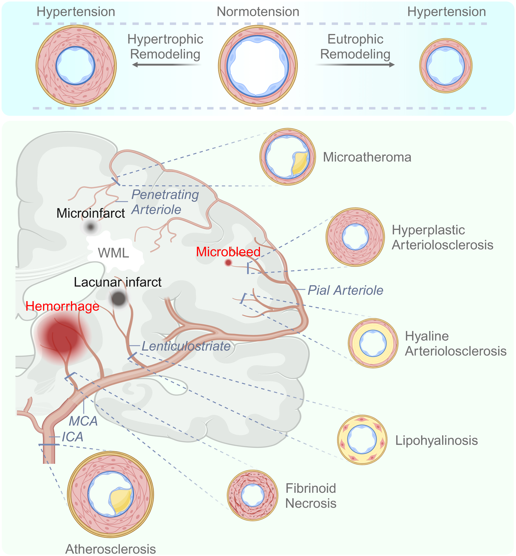
Top: Vascular remodeling proceeds in either an outward or inward manner in response to mechanical, cellular, inflammatory, and oxidative factors. Bottom: The predominant vascular structural alterations associated with hypertension are indicated according to the affected segment of the neurovasculature. Key pathological outcomes are also depicted. Details are provided in the text. Abbreviations: WML, white matter lesion; ICA – internal carotid artery; MCA – middle cerebral artery.
Large extracranial and intracranial cerebral arteries
Hypertension contributes to formation of atherosclerotic plaques in both extracranial and intracranial cerebral arteries16,33, increasing the risk of ischemic stroke34,35. Extracranial lesions are characterized by elevated accumulation of lipids in vertebral and carotid arteries36, increasing the risk for artery-to-artery embolism36, while intracranial lesions affecting the circle of Willis and its branches display a fibrous expression more likely to result in local vascular occlusion34. How hypertension promotes atherosclerosis remains incompletely understood, but AngII is thought to contribute by generating reactive oxygen species (ROS) and upregulating cytokines, chemokines, and growth factors37. The resultant cellular and molecular milieu triggers an inflammatory response in the vascular wall that precipitates endothelial dysfunction, neointima formation, and atherosclerotic lesions37.
In addition to atherosclerosis, concurrent vascular stiffening and stenosis resulting from sclerosis of the vascular wall (arteriosclerosis) negatively affects the cerebral blood supply and downstream microvasculature. These changes are implicated in CBF reduction38; diminution of cerebrovascular reserves39, which can elevate stroke risk40; and increased hydrodynamic impacts on the microvasculature41. Modification of the extracellular matrix is a major contributor to arterial stiffening, and is characterized, in part, by collagen and fibronectin accumulation, metalloprotease-mediated elastin fragmentation, and pro-fibrotic cascades related to transforming growth factor-β30.
Pial vessels and penetrating arterioles
Pial and penetrating arteries and arterioles are uniquely sensitive to chronic elevations in BP16. In humans, hypertension-induced microatheroma in pial arteries and small perforating arteries (300 to 800 μm external diameter42–44) can occlude vessels and instigate lacunar infarction or microinfarction44. Moving further into the brain, small penetrating arteries and arterioles, from 300 μm to 20 μm42–44, arising from either the first segment of the middle cerebral artery or terminal branches of the pial arteries, converge on deep white matter territories16. As these vessels display vulnerability to shifts in upstream pressure, scant collateralization, and limited anastomosis16, they are strongly implicated in formation of the white matter damages attendant to vascular dysfunction45,46.
Three vascular lesions predominate within these smaller vessels: lipohyalinosis, fibrinoid necrosis, and arteriolosclerosis. Lipohyalinosis involves asymmetric deposition of an amorphous, glass-like material composed of collagen and degenerated smooth muscle tissue into the vascular wall, while fibrinoid necrosis, observed in more advanced lesions, is signified by infiltration of fibrin and its degradation by-products42–44. More common is arteriolosclerosis43, characterized by myocyte degradation and loss of elastin alongside concentric accumulation of fibro-hyaline materials and collagens in the vascular wall, leading to stenosis and reduced elasticity (hyaline arteriolosclerosis)42–44, or the extensive intimal fibromuscular proliferation, luminal narrowing, and necrotic degeneration often associated with malignant hypertension (hyperplastic arteriolosclerosis)42–44. Capillaries and pre-capillary arterioles are also affected, marked by loss of endothelial cells and pericytes (string vessels), increased thickness of the basal lamina and accompanying tortuosity, fibrin deposition, and overall rarefaction47.
Collectively, these changes form the basis of hypertension-associated small vessel disease (SVD), a disorder affecting arterioles, venules, and capillaries of the subcortical and periventricular white matter in a segment specific manner42–44; atheromatous lesions are typically observed in larger arteries/arterioles situated upstream of distal perforating vessels supplying the deep parenchymal tissues, which are in turn affected by arteriolosclerosis, lipohyalinosis, and fibrinoid necrosis (Figure 1)42–44. The sequelae of SVD, detectable via magnetic resonance imaging (MRI), include WML, small ischemic foci (lacunes and microinfarcts) and microhemorrhages48, venous collagenosis49, and enlarged PVS50. SVD is also influenced by short term and long term BPV, evidenced by brain imaging and autopsy data (arteriolosclerosis, microinfarcts, WMLs, enlarged perivascular spaces, atherosclerosis of the circle of Willis,)51,52.
Hypertension impairs cerebrovascular function
The brain is dependent on a continuous and highly regulated supply of oxygen- and glucose-rich blood. Accordingly, neurovascular mechanisms assure that perfusion is commensurate with the brain’s regionally and temporally diverse energetic and waste clearance requirements25. In this section, the mechanisms regulating CBF are reviewed, focusing on factors disrupted in hypertension which may lead to cognitive impairment. A more comprehensive description of CBF regulation is found in recent reviews25,26,53,54.
Autoregulation
The cerebral vasculature maintains CBF within approximately ±20 mmHg of baseline53, dampening the BP changes attendant to daily life. Traditionally, autoregulatory responses have been studied using stepwise changes in BP coupled with measurement of CBF; the stabilization of perfusion during these manipulations is known as static autoregulation55. In the 1990s, the introduction of transcranial Doppler flowmetry enabled assessment of flow velocity in response to rapid changes in BP56, leading to the characterization of dynamic autoregulation57, which refers to the latency between BP changes and corresponding adjustments to vascular resistance53. These studies revealed that while rapid fluctuations in BP escape autoregulation and result in flow changes, they are not as large as if the autoregulatory process was absent.
On a cellular level, autoregulation depends on the ability of smooth muscle cells to constrict or dilate in accordance with intravascular pressure (myogenic response). Multiple mechanisms have been implicated, including (a) ion channels and mechanosensors, which adjust intracellular calcium levels commensurate with fluctuating transmural pressures58, (b) alteration of contractile apparatus calcium sensitivity59, and (c) depolarization-mediated amplification of intracellular calcium signaling60.
Animal studies have provided evidence that hypertension shifts the static autoregulatory curve to the right, rendering the brain more susceptible to reductions in BP16. In contrast, most human studies suggest that both static and dynamic autoregulation are preserved among hypertensives53, except for malignant hypertension (SBP: 180–260 mmHg), where failure of the autoregulation mechanism causes flow to passively follow BP61.
Overall, autoregulation appears to be a resilient mechanism that is largely maintained in human hypertension, but the available data have limitations that need to be considered. Since in most studies flow was assessed in a single vessel by transcranial Doppler, regional changes in dynamic autoregulation could have been missed. Furthermore, the inherent risk associated with lowering BP has been an obstacle to assessing the lower limit of static autoregulation in patients with hypertension.
Neurovascular coupling
Neurovascular coupling, which pertains to the distribution of blood to active brain regions commensurate with their metabolic demands25, requires participation of all constituents of the NVU at each level of the cerebrovasculaure, and is driven by diffusible mediators (nitric oxide (NO), prostanoids, adenosine, ions, etc.) and segment-specific intrinsic vascular mechanisms25. In brief, activation of neurons deep within the brain parenchyma initiates a series of vascular alterations which begin at the level of the capillary endothelium and are transmitted upstream in a retrograde fashion through intramural signaling, resulting in propagation of smooth muscle cell relaxation and attendant vasodilation62–64. This retrograde propagation ultimately reaches larger pial vessels, which must relax to facilitate adequate blood flow to the activated region and prevent flow steal from neighboring vascular territories25.
At present, a paucity of human studies have examined neurovascular coupling in hypertension. In untreated hypertensives, increased baseline BP is associated with diminished regional alterations to CBF during cognitive tasks65. Similar impairments are reported for blood flow responses to visual stimuli in the retina66 or posterior cerebral artery67. These observations, albeit limited, support the presence of impaired functional hyperemia in hypertensives and, in turn, increased susceptibility to vascular insufficiency.
Endothelial cells: vasoregulation and the BBB
Cerebrovascular endothelial cells regulate vasomotor tone by releasing vasoactive messengers in response to select chemical and mechanical stimuli54. Perhaps the most well-studied endothelium-dependent vasoactive mechanism is NO-mediated vasodilation in response to cholinergic agonists activating endothelial nitric oxide synthase (eNOS)68. Signaling in a paracrine manner, NO reaches the vascular smooth muscle cells where it initiates cGMP-mediated relaxation of the smooth muscle69. As mentioned above, the cerebral capillary endothelium contributes to functional hyperemia. Release of K+ by neuronal depolarization activates endothelial inward rectified K+ channels, leading to a hyperpolarization cascade transmitted upstream through inter-endothelial junctions which, upon reaching smooth muscle cells, culminates in their relaxation62.
Endothelial cells are also the site of the BBB70. The bidirectional exchange of molecules between blood and brain is regulated by (a) molecular transporters on their surface, which gate substrate entry, (b) tight junctions between adjacent endothelial cells, which largely prevents paracellular solute flux, and (c) paucity of vesicular transport, which limits transcytosis70.
In humans, endothelial dysfunction in peripheral arteries precedes BP elevation and correlates with the severity of hypertension post-development71. While direct in vivo evaluation of cerebral endothelial function in humans is not feasible, inhibition of NO synthesis does not attenuate arteriolar blood flow in the retina of hypertensive patients, suggesting NO deficiency concomitant with endothelial dysfunction66, and endothelium-dependent vasodilatory responses to acetylcholine are diminished in arteriolar samples collected post-mortem from SVD/AD mixed pathology patients72.
These endothelial aberrations may affect cognition: impaired endothelium-dependent vasodilation in peripheral arteries is associated with brain microhemorrhages73 and WMLs74, both of which are implicated in cognitive decline75,76; and some hypertensive patients present with BBB impairment, which can contribute to brain damages and SVD (e.g., WMLs)77.
Perivascular spaces, waste clearance and neuroimmune regulation
PVS and the vessels within have emerged as major routes for the elimination of potentially-deleterious by-products of brain activity, such as amyloid-β (Aβ) and tau78. Several clearance systems have been proposed78. A transvascular pathway facilitates the passage of select molecules through the vascular wall by way of abluminal transporters70. Since the brain parenchyma lacks conventional lymphatic vessels, other pathways are thought to use the PVS as a conduit to carry solutes out of the brain. In the perivascular pathway, solutes are thought to exit the parenchyma through retrograde transport along the vascular basal lamina or PVS, eventually mixing with the CSF in the subarachnoid space and draining into the cervical lymph nodes79. Running opposed to this route is the glymphatic pathway. Aided by aquaporin-4 (AQP4) water channels in astrocytic endfeet, convective and diffusive forces are hypothesized to drive CSF from the periarterial space into the parenchyma where it blends with the interstitial fluid and picks up waste products. This waste rich fluid is thought to exit into the perivenous space, travel to the subarachnoid compartment, and drain into the meningeal lymphatics80.
Cerebral vessels and PVS are also involved in immune regulation, and the constituent vessels of the cerebrovasculature are surrounded by an assortment of immune cells. Yolk sack derived myeloid cells, collectively termed border associated macrophages, populate the meninges, PVS, and choroid plexus81, and have emerged as key correlates of the neurovascular and cognitive dysfunction associated with hypertension82,83.
In humans, hypertension is linked to enlarged PVS84,85. Although incompletely understood, a proposed mechanism invokes hypertension-induced stiffening of large arteries exposing the microvasculature to enhanced pulsatile pressures86, which increases the mechanical stress under which the PVS is placed and drives their enlargement85. In support, a recent neuroimaging study found that the glymphatic removal of an intravenously administered tracer was reduced in SVD-positive participants, most of whom were hypertensive87. This hypertension-clearance dynamic is discussed further in Impaired clearance of amyloid and tau peptides.
Hypertension and neurovascular dysfunction: mechanistic studies
Angiotensin II
Arguably the most well-established models of hypertension involve use of pressor doses of AngII that elicit a sharp increase in BP, or sub-pressor doses (slow-pressor model) which drive a gradual elevation in BP over several days. The slow-pressor model has gained in popularity for its potential to capture the progressive increase in BP of primary hypertension88. Neurovascular dysfunction, i.e., impaired functional hyperemia and endothelial vasodilation, is provoked by both acute and chronic AngII treatment82,89,90, and is driven by the interaction of AngII with one of its receptors, AngII receptor type 1 (AT1R), on vascular and perivascular cells (Figure 2)82,89, as well as brain structures involved in autonomic regulation, such as the subfornical organ and periventricular hypothalamus90.
Figure 2: Potential mechanisms of neurovascular dysfunction in AngII hypertension.
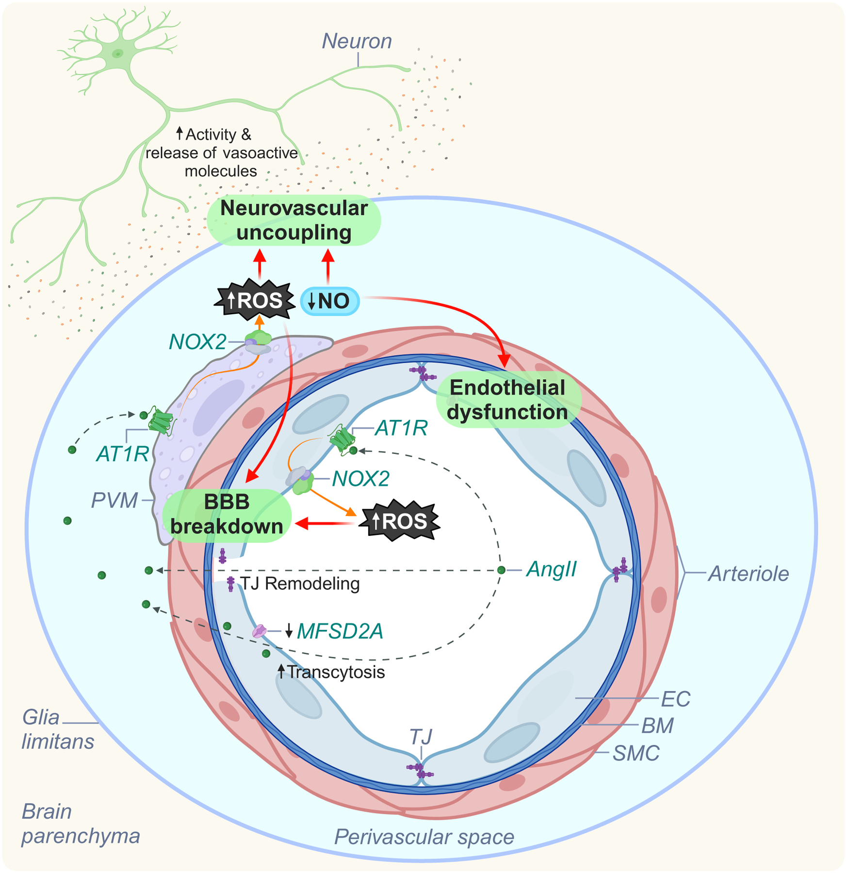
Circulating AngII interacts with AT1R on endothelial cells to initiate BBB disruption (tight junction remodeling and suppression of MFSD2A, leading to increased transcytosis) and enable its entry into the PVS. Next, circulating and brain-derived AngII engage with AT1R on PVM, leading to NOX2 activation, vascular oxidative (superoxide) and nitrosative (peroxynitrite) stress, reduced NO, further BBB disruption, and neurovascular dysfunction. Abbreviations: NO, nitric oxide; SMC, smooth muscle cell; EC, endothelial cell; PVS, perivascular space; PVM, perivascular macrophage; ROS, reactive oxygen species; BBB, blood-brain-barrier; AngII, angiotensin II; NOX2, NADPH oxidase 2; AT1R, angiotensin II receptor type I; TJ, tight junction.
Intriguingly, the effects of AngII on neurovascular function may be independent of BP elevation. Direct neocortical application of AngII, which does not increase BP, elicits neurovascular uncoupling89; non-pressor doses of AngII induce neurovascular dysfunction91; and topical application of the AngII receptor blocker (ARB) losartan to the neocortex rescues functional hyperemia without lowering BP89. Additional evidence for BP independent effects is provided by the BP high mouse (BPH)82 and SHR92, genetic models of lifelong hypertension, as well as the deoxycorticosterone acetate+salt (DOCA-salt) model of salt-sensitive hypertension93, in which neurovascular dysfunction can be rescued despite increased BP82,91,93. Furthermore, elevations in BP provoked by an α-adrenergic agonist fail to induce neurovascular dyfunction82, suggesting that not all pressor agents will affect these regulatory mechanisms. While these experimental observations highlight the BP-independence of neurovascular dysfunction, the pathogenic impacts of elevated BP in human hypertension cannot be underestimated. After all, antihypertensive intervention dramatically lowers stroke risk and94, in some studies, improves cognitive function10,95; however, as the data on the efficacy of BP lowering against cognitive decline is conflicting (see Therapeutic Interventions), BP-independent effects warrant further consideration.
Cytokines
In recent years, interleukin-17 (IL-17), a proinflammatory molecule, has received increased attention for its contributions to neurovascular dysfunction. In the AngII96 and DOCA-salt93 models, IL-17 is required for the development of neurovascular dysfunction and cognitive impairment, whereas neutralization of IL-1796, inhibition of its receptor96, or elimination of its cellular sources93 prevents such outcomes. Intriguingly, IL-17 knock-out mice treated with DOCA-salt develop increased BP without neurovascular dysfunction93, attesting to the BP-independent effects of hypertension-associated mediators.
Oxidative stress
Oxidative stress is a principal driver of the cerebrovascular sequelae of hypertension16. While several enzymatic sources have been identified97, a NOX2 containing NADPH oxidase appears to be the prime source of the ROS involved in neurovascular dysfunction, particularly in AngII-induced hypertension82,89, but also in other models, such as salt-sensitive93 and in BPH mice82. Accordingly, both genetic deletion and pharmacological inhibition of NOX2 prevents neurovascular dysfunction and/or cognitive impairment in models of hypertension82,89,93. However, how ROS precipitate these alterations requires elucidation. In AngII hypertension, peroxynitrite, a diffusible reaction product of NOX-2-derived superoxide with NO98, is the ultimate mediator of the neurovascular dysfunction99. Alternative mechanisms may include NO scavenging, and redox modifications of proteins, lipids and DNA, among others98.
Innate immunity: Perivascular macrophages and microglia
Perivascular macrophages (PVM) are found closely apposed to the outer vascular wall of intracerebral arterioles and venules within the PVS81. PVM express both AT1R and IL-17RA, are richly endowed with the ROS producing enzyme NOX2100,101, and have emerged as key players in the neurovascular and cognitive dysfunction observed in animal models of hypertension82,93. In the slow-pressor model, AngII disrupts the BBB and subsequently enters the PVS where it interacts with AT1R expressed on PVMs to elicit vascular impairments, as evidenced by the restoration of neurovascular function which follows PVM depletion or deletion of At1r or Nox2 from PVM82. The disruption of the BBB is mediated by remodeling of tight-junctions and an increase in vesicular transport consequent to suppression of Mfs2da102, a negative regulator of transcytosis103. Although activation of endothelial AT1R initiates the BBB dysfunction, PVMs are required for its full expression (Figure 2)102. In addition, depletion of PVMs abrogates the neurovascular dysfunction and cognitive impairments attendant to life-long hypertension in BPH mice82, as well as to salt-sensitive hypertension in the DOCA-salt model93, supporting the inter-model generalizability of their contributions.
Less is known on the role of microglia. In the slow-pressor model, microglia acquire a proinflammatory phenotype at sites of BBB leakage, and their depletion (along with PVM) by the colony-stimulating factor 1 receptor inhibitor PLX5622 partially curtails cognitive impairments without rescuing BBB integrity104. However, how microglia promote cognitive impairment independently of BBB disruption and whether PVMs are also involved remains to be determined.
Adaptive immunity: T-cells
T cells, namely of the T helper 17 (Th17) and γδ17 subtypes, and IL-17, their principal effector cytokine105, have emerged as crucial players in the induction of hypertension106 and its sequelae, both centrally93 and peripherally106. Concerning central immunity, a recent study using the DOCA-salt paradigm revealed T cell-dependent mechanisms of neurovascular dysfunction (Figure 3): in the circulation, IL-17 derived from gut localized and circulating Th17 and γδ17 cells acts on cerebral endothelial IL-17RA to reduce NO production and disrupt endothelial vasoactivity; in the brain, IL-17 secreted from γδ17 cells situated within the dura acts on IL-17RA in PVMs to induce vascular oxidative stress and concomitant neurovascular uncoupling. While endothelial IL-17RA deletion partially restores cognition, antagonizing the central sources (T-cells) or targets of IL-17 (PVMs) rescues cognition in full93, revealing a previously unappreciated involvement of meningeal immunity in the cognitive effects of salt-sensitive hypertension.
Figure 3: Putative contributions of meningeal immunity to neurovascular dysfunction in salt-sensitive hypertension.
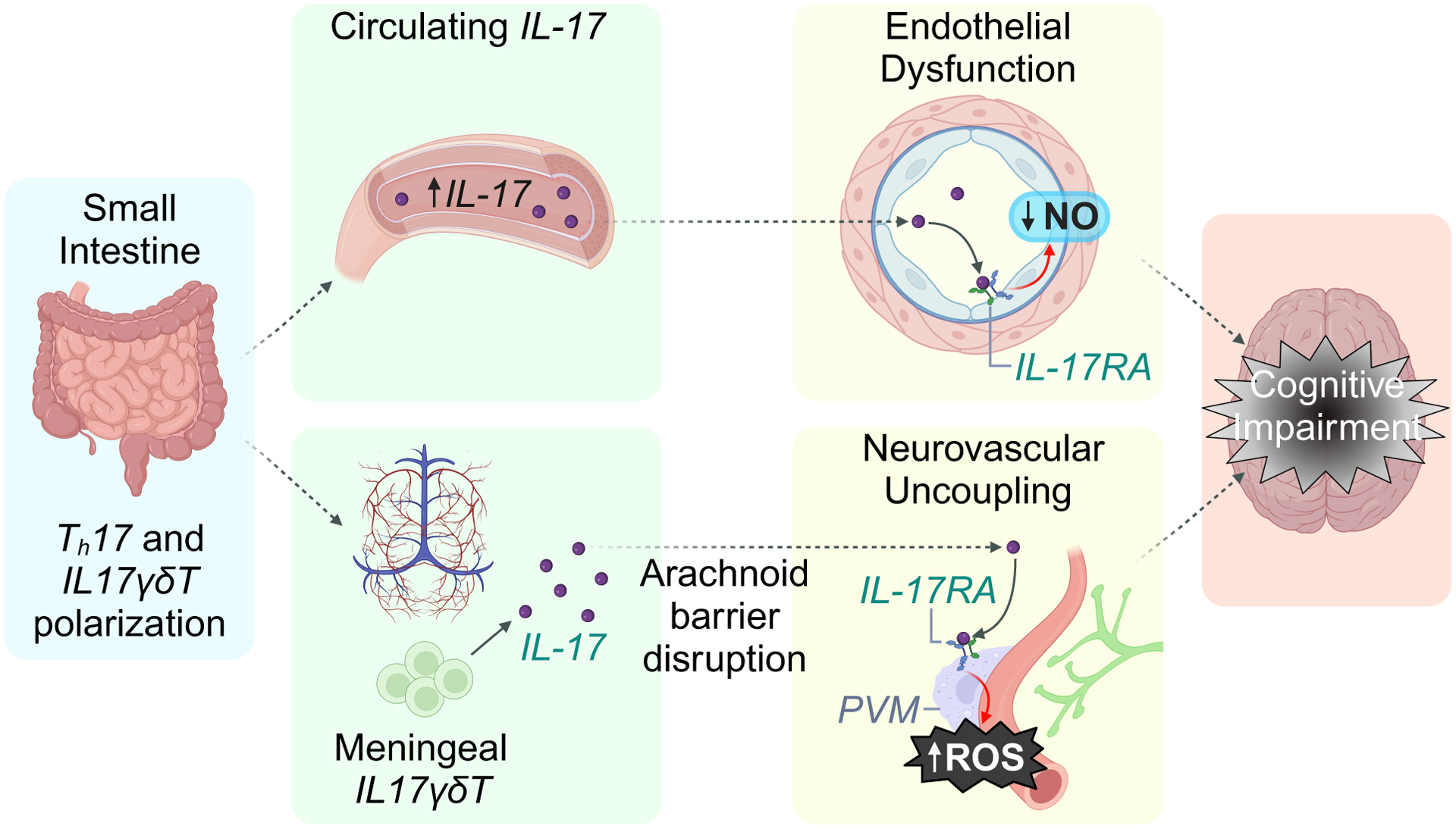
DOCA-salt treatment leads to production of IL-17 from T cells in the small intestine and dural immune compartment. Gut-derived circulating IL-17 interacts with IL-17RA on endothelial cells to disrupt endothelial function. IL17 produced in the meninges enters the subarachnoid space, travels to the PVS, and engages with IL-17RA on PVMs to induce vascular oxidative stress (via NOX2) and neurovascular dysfunction. Abbreviations: NO, nitric oxide; SMC, smooth muscle cell; EC, endothelial cell; PVS, perivascular space; PVM, perivascular macrophage; ROS, reactive oxygen species; IL-17, interleukin 17; Nox2, NADPH oxidase 2; IL-17RA, interleukin 17 receptor A; Th17, T helper 17 cell; IL17γδT – IL-17-producing gamma delta T cell.
How does hypertension promote cognitive decline?
The most profound impacts of hypertension on cognition appear to center on executive function2, motor speed, and attention107, domains classically associated with vascular cognitive impairment16. Typically, memory is involved more so in AD-related cognitive decline than in vascular cognitive impairment, although this is more of a trend than a rule (see ref.108).
In this section, the potential mechanisms underlying hypertension-induced cognitive impairment are discussed, with a particular focus on two hallmarks of vascular cognitive impairment: ischemic and hemorrhagic brain lesions and white matter disease16.
Ischemia, hemorrhage, white matter lesions and atrophy
Hypertension is major risk factor for both hemorrhagic and ischemic stroke which, in turn, is associated with a 3–6 fold increased risk of cognitive impairment16. Lacunar infarcts, microinfarcts, and microbleeds similarly portend cognitive deterioration109–111.
WML are among the most common lesions associated with hypertension112. SVD, often a consequence of hypertension113, is a major contributor to WML burden114, and associations between SVD, WML, and cognitive decline have been reported in the epidemiological literature. In the Radboud University Nijmegen Diffusion Tensor and Magnetic Resonance Cohort (RUN DMC), baseline SVD severity and progression were independently correlated with dementia risk over 14 years115; SVD progression over time was accelerated by baseline vascular risk factors, a phenomenon primarily driven by hypertension116; and reduced processing speed was associated with WML load75. Supporting these observations, WML risk loci overlapped with BP traits in a meta-analysis of genome-wide association studies, with subsequent Mendelian randomization suggesting a causal association of WML volume with genetically predicted SBP117.
Mechanistically, hypoxia-ischemia following occlusion or rarefaction of the microvessels supplying the subcortical white matter likely contributes to white matter disease consequent to SVD16,118. Indeed, reduced cerebrovascular reactivity to CO2 was observed to precede the progression of normal-appearing white matter to WML in middle aged subjects with moderate-to-severe WML burden at baseline119; arteriolosclerosis and a hypoxic milieu have been found in WMLs post-mortem120; and increased severity of arteriolosclerosis in watershed regions located between arterial territories has been shown to associate with greater WML and neurofibrillary tangles at autopsy121. However, reverse causation has been suggested122 wherein reductions in blood supply could be consequent to BBB dysfunction-induced damage of the microvasculature via extravasation of neurotoxic molecules, edema, and microvascular compression123, or by WMLs lowering local CBF requirements secondary to deafferentation, tissue atrophy, and reduced energy demands124.
Disrupted functional connectivity
Hypertension-induced loss of white matter integrity and consequent derangement of functional connectivity may affect cognition. Continued progression of WMLs125, microstructural alterations126, and network functional deficits127,128 correlate with cognitive decline in hypertensive patients126–128. In recent years, location-based image analyses have revealed that strategically placed WMLs are particularly relevant to cognitive decline110. In a study employing white matter tractography and resting state fMRI126, hypertension was associated with early alterations in the superior longitudinal fasciculus, the forceps minor, and the anterior thalamic radiation. Hypertensive patients performed worse in the domains ascribable to these affected tracts in subsequent cognitive testing (executive functions, processing speed, and memory)126. These observations are corroborated by an analysis combining imaging in middle aged adults from the UK Biobank cohort with genetic causal inference approaches, which found elevated SBP to associate with alterations to the external capsule, anterior corona radiata, and anterior thalamic radiation, the latter two of which are linked to cognitive decline129.
Potential contributions of hypertension to AD
Once considered to be purely a neurodegenerative disease, an increasing body of evidence indicates the involvement of vascular brain lesions in AD, such that most cases diagnosed clinically present mixed vascular and AD pathologies8,130. Relative to age-matched controls, AD brains display elevated intracranial atherosclerosis and numerous microvascular alterations131. Furthermore, BBB permeability is increased132 and hemodynamic responses to neuronal activity are suppressed133 in the prodromal period, implicating vascular factors early in disease progression.
Epidemiological associations of hypertension with AD biomarkers
Amyloid positron emission tomography (PET) data concerning the link between amyloid pathology and hypertension are conflicting. In the ARIC cohort, heightened vascular risk factor burden in mid-life was associated with elevated PET Aβ in old age; however, hypertension alone did not significantly correlate with amyloid burden134. In the British 1946 birth cohort, increased BP in middle age associated with late-life WML burden and brain atrophy, but not PET Aβ135. While these findings suggest that hypertension fails to exacerbate Aβ burden, this does not preclude the possibility that amyloid pathology in some of the hypertensive patients was driven, in part, by vascular contributions. Of note, the impact of hypertension on Aβ may be modulated by genetic risk factors. In carriers of the ε4 allele of the apolipoprotein E gene, mid-life vascular burden correlated with increased brain amyloid in ARIC participants at 20-year follow-up134, while in the Rotterdam study, hypertension was associated with Aβ burden 7 years after initial assessment136.
As for tau, increased pulsatility secondary to aortic stiffness correlated with tau burden in the rhinal and entorhinal cortices of dementia-free subjects from the Framingham Heart Study137, and a clear age-related pattern of BP was delineated in the Chinese Alzheimer’s Biomarker and LifestylE (CABLE) cohort, where higher mid-life SBP, late-life lower DBP, and increased pulse pressure in both mid- and late-life was associated with tau-related biomarkers and cognitive dysfunction, but not amyloid burden138.
Intriguingly, inconsistencies in BP might be important for both amyloid and tau pathologies. An association of BPV with phosphorylated tau (p-tau) and decreased Aβ was observed in the CSF of older participants from the Alzheimer’s Disease Neuroimaging Initiative139.
Overall, these observations present a plausible association of hypertension with AD pathology, particularly at the level of tau, although it remains unclear whether this relation is causative or incidental, or if the effects of hypertension on cognition involve or simply co-occur with AD pathology.
Hypertension and AD pathology: mechanistic considerations
Amyloidogenesis and tau hyperphosphorylation
Hypertension promotes tau hyperphosphorylation and Aβ accumulation in animal models (Figure 4). In transgenic models of brain amyloid build up, AngII-induced hypertension increases Aβ deposition140 and p-tau immunoreactivity141. Similar findings are reported in other models, such as SHR142. While the underlying mechanisms have yet to be elucidated, AngII-induced elevation of β-140 and γ-secretase143 activity, which shifts APP processing toward β-amyloidogenesis, could be involved. Experimentally, genetic ablation of AT1R curtails γ-secretase activity and subsequent Aβ generation143, and AT1R blockers abrogate amyloid pathology: telmisartan reduces Aβ formation in stroke-resistant SHR144 and valsartan diminishes Aβ burden in Tg2576 mice145. Regarding tauopathies, AngII146 and IL-1793,147 disrupt eNOS-mediated NO production and reduce NO bioavailability. Since suppression of endothelial NO has been linked to tau phosphorylation via activation of cyclin-dependent kinase 5141, AngII and IL-17 could promote p-tau through this mechanism, although supporting evidence is missing.
Figure 4: Potential mechanisms associating hypertension and AD.
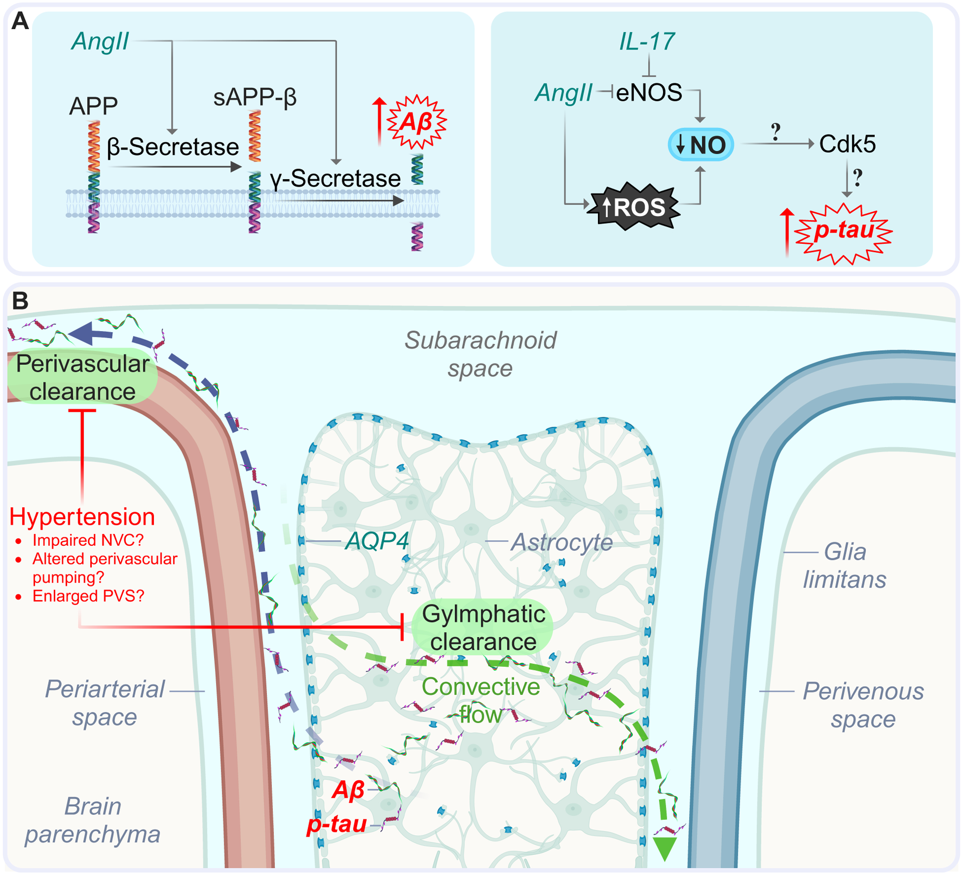
The oxidative and inflammatory sequela of hypertension could promote AD pathology by increasing Aβ and p-tau (A), and disrupting perivascular and glymphatic clearance (B). Hypertension may enhance Aβ accumulation through increased processing of APP by secretase enzymes. Tau phosphorylation may be elevated under hypertensive conditions consequent to reductions in endothelial NO bioavailability and attendant activation of cyclin-dependent kinase 5. Abbreviations: SMC, smooth muscle cell; EC, endothelial cell; PVS, perivascular space; AngII, angiotensin II; IL-17, interleukin 17; AQP4, aquaporin 4; Aβ, amyloid-β; APP, amyloid precursor protein; sAPPβ, soluble peptide APPβ; BM, basement membrane; p-tau, phosphorylated tau.
Impaired clearance of amyloid and tau peptides
Aβ clearance is facilitated by the glymphatic and perivascular pathways148,149, as well as utilization of transporters situated within the BBB , namely the low-density lipoprotein receptor-related protein 1 (LRP1) for egress and the receptor for advanced glycation end products (RAGE) for ingress70; thus, disruption of these systems would be expected to worsen amyloid accumulation.
Hypertension-induced alterations in perivascular pumping, a fluid transport mechanism wherein vascular wall kinetics drive CSF flow in the PVS150, could aggravate proteinopathy by disrupting Aβ and tau clearance. In a study using particle tracking velocimetry concurrent with measurements of arterial diameter, acute elevations in BP induced by intravenous infusion of AngII in mice altered arterial wall motion during the cardiac cycle, leading to reduced CSF flux in the PVS consequent to increased backflow150. This diminished CSF flow within the PVS could precipitate a similar decrease in glymphatic transport, a phenomenon recently demonstrated in the SHR model using dynamic contrast-enhanced MRI-aided quantification of the glymphatic transport of a contrast agent151. These perturbations are likely to diminish Aβ and tau removal. Indeed, TGN-020-mediated inhibition of AQP4 in astrocytic end-feet149, which are crucial for glymphatic flow80, suppressed tau protein clearance in mice149. Considering recent observations that changes in vasomotion caused by neurovascular coupling or optogenetically-induced vascular constriction-dilation facilitate CSF flow152,148, the neurovascular uncoupling observed in experimental and human hypertension67,82,89,93 could compromise metabolite clearance. Lastly, RAGE, which is activated in hypertensive models153, modulates the entry of Aβ into the brain and influences β- and γ-secretase activity154, suggesting that hypertension could promote Aβ accumulation by enhancing Aβ influx153 and/or β-amyloidogenesis through RAGE induction154.
Evidence for disruption of these pathways in human hypertension is limited. Elevated peripheral expression of extracellular RAGE binding protein was associated with increased risk of cognitive decline in the Rotterdam Study155, suggesting a role for RAGE in AD pathogenesis; however, this cross-sectional association was attenuated in a longitudinal setting155, indicating potential reverse causation. Human studies of SVD, in which the majority of participants were hypertensive, showed evidence of reduced glymphatic clearance156, but a specific role of elevated BP has not been established.
Collectively, these clinical and basic observations suggest that hypertension could contribute to AD pathology through induction of amyloidogenesis and, based mainly on animal studies, diminution of amyloid/tau clearance (Figure 4), although the degree to which hypertension affects these pathways in humans requires elucidation.
Summary: how does hypertension promote cognitive deterioration?
Hypertension likely contributes to cognitive decline through a confluence of pathogenic mechanisms (Figure 5). First, vascular damages, inflammation, neurovascular dysfunction, and BBB disruption threaten the health of the cortical tissue and subcortical white matter. The resulting WMLs, microinfarcts, microbleeds, and disruption of metabolite clearance may contribute to network dysfunction, neuronal loss, and brain atrophy. Lastly, by fostering Aβ and p-tau accumulation, hypertension could conceivably contribute to the overlap between vascular and AD pathology to accelerate the development of cognitive impairment.
Figure 5: Hypertensive sequalae which underlie cognitive impairment.
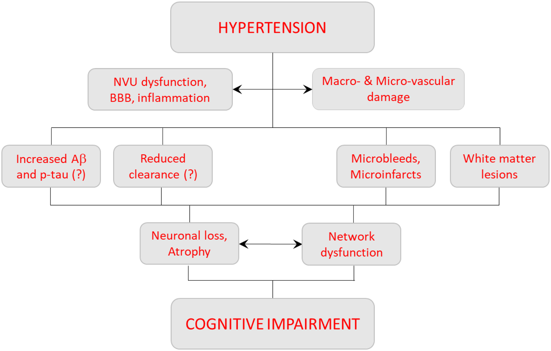
Hypertension promotes structural alterations to the cerebrovasculature concurrent with NVU functional deficits. The resultant microbleeds, microinfarcts, and local hypoxia-ischemia drive neuronal loss and degradation of white matter tracts (especially in thalamo-cortico circuits), leading to brain atrophy and network disruption. Additionally, hypertension may provoke β-amyloidogenesis (upregulated secretase activity) and disrupt clearance of toxic metabolites, giving rise to Alzheimer’s-associated proteinopathies. Collectively, these events likely contribute to cognitive impairment. Abbreviations: WML, white matter lesion; CBF, cerebral blood flow; NVU, neurovascular unit.
Therapeutic interventions
A recent post-mortem investigation of samples taken from 4 separate birth cohorts (1905–1914, 19-15–1919, 1920–1924, and 1925–1930) found that although neurodegenerative pathologies did not differ by birth year, dementia incidence decreased over time concurrently with atherosclerosis and arteriosclerosis157. The dramatic reduction in vascular pathology, possibly due to better control of vascular risk factors, including hypertension, could have contributed to the decrease in dementia incidence. Considering epidemiological associations of hypertension with dementia, elevated BP presents a particularly inviting modifiable target in the effort to diminish global dementia burden.
A role for antihypertensive therapy?
As previously discussed (see Hypertension and cognitive decline: epidemiological evidence), the relationship between hypertension and cognitive decline is most pronounced when considered in mid-life1–3. Thus, while randomized clinical trials would best evaluate the efficacy of antihypertensive therapy in dementia prophylaxis, the temporal characteristics of the hypertension-dementia relationship necessitate reliance on long-term observational studies.
Several large cohort longitudinal analyses indicate an association of antihypertensive treatment with lesser cognitive decline2,158,159. Consistent with the relationship of hypertension chronicity to dementia risk160, therapy duration appears important in prophylaxis: in the Rotterdam cohort, each additional year of antihypertensive treatment, prior to age 75, reduced dementia risk by 8%159.
Clinical trial data are comparatively conflicting, likely due to issues surrounding the age of treatment onset and duration of medication use. Indeed, the most recent update of the Cochrane Review found that pharmacological treatment of hypertension in patients without prior cerebrovascular disease provides only low certainty evidence for the prevention of dementia onset and cognitive deterioration11, with investigators citing insufficient study duration as a probable confound. In contrast, a recent meta-analysis of 12 major clinical trials, including the recently completed SBP Intervention Trial – Memory and Cognition in Decreased Hypertension (SPRINT-MIND) study, reported a significant association of BP control with decreased risk of dementia or cognitive impairment161.
Intriguingly, intensive BP-lowering regimens may offer increased benefit relative to standard therapy. Recent findings from secondary analyses of the SPRINT-MIND data set indicate diminished risk of cognitive decline10,95, white matter lesion development162, and probable dementia9 in patients undergoing intensive blood-pressure-lowering (SBP < 120 mmHg). Intensive regimens, but not standard, were also found to ameliorate cognitive decline in patients with high BPV163. Overall, these findings are corroborated by a recent meta-analysis where late-mid-to-late-life BP correction dramatically mitigated dementia risk, with intensive lowering showing the most benefit164. Taking into account the demonstrated safety of BP lowering interventions in the elderly165, aggressive BP control warrants further consideration as a means to delay dementia onset.
Comparative assessments of differing antihypertensive agents have yielded inconsistent results. While some meta-analyses show no heterogeneity of antihypertensive class on risk of incident dementia12, an emerging body of evidence seemingly supports improved outcomes for drugs that preserve AngII synthesis (such as ARBs) over those that inhibit it (ACE inhibitors)13. The BBB permeability of drugs may also be consequential, with recent reports suggesting BBB-permeable ARBs14 and β-blockers166 are superior to their impermeable counterparts. Lastly, the recent development of a selective aldosterone synthase inhibitor167 and powerful angiotensinogen silencing antisense oligos168 could allow for blood pressure control in treatment resistant hypertensive patients.
Conclusions
The data presented herein show that hypertension remains among the most insidious factors influencing cognition over the lifespan, largely due to impacts on cerebrovascular structure and function which threaten the health of the brain16. Despite advances in the field, numerous questions remain regarding the temporality of hypertension-induced cognitive decline, the underlying mechanisms, and potential interventions.
Because the best clinical evidence for preserving cognition in hypertension currently supports BP control, it is important to consider whether certain drug classes are more efficacious than others. While many studies fail to show heterogeneity among the antihypertensive classes12, AngII-preserving drugs13 and BBB-crossing antihypertensives14 and β-blockers166, as discussed in the previous section, warrant further exploration.
Another important question is whether intensive BP control provides greater cognitive benefit than standard treatment. Results from SPRINT-MIND secondary analyses suggest this to be the case10, particularly in those with increased BPV163. While this more aggressive approach does not compromise cerebral perfusion165, the risk/reward ratio of intensive treatment requires further validation (will adverse effects emerge as treatment length increases, or in the elderly?), and it remains to be determined whether aggressive control in mid-life, when the hypertension-cognitive decline relationship is strongest1–3, will still provide greater benefit.
Does combating cognitive deterioration by controlling BP also reduce AD pathology? Since the neurohumoral dysfunction underlying hypertension may promote Aβ and p-tau accumulation, it would be of interest to assess if antihypertensive medications reduce AD pathology independently of effects on BP, as suggested by animal studies145. If so, antihypertensives could be combined with Aβ immunotherapy, which has recently been approved by the FDA.
Considering preclinical evidence that the cerebrovascular sequelae of hypertension are not entirely ascribable to elevated BP82,89,93,102, further elucidation of the mechanistic underpinnings of neurovascular and cognitive impairment in hypertension are needed. Are immune cells a major driving force? In addition to the growing body of experimental evidence connecting PVMs to hypertensive pathology82,93,102, the recently unveiled contributions of meningeal T-cells to neurovascular dysfunction in a model of salt-sensitive hypertension93 links meningeal immunity to hypertension-induced cognitive impairment.
What about pericytes, which may be involved in BBB regulation and cerebral perfusion169? Reduced capillary pericyte coverage is observed alongside BBB disruption in slow-pressor AngII hypertension170, and mouse models of pericyte deficiency exhibit impairment of the BBB171 and CBF regulation172. However, the precise contributions pericyte alteration to the neurovascular and cognitive dysfunction in hypertension remain to be determined and would be an area of interest for future studies.
The events linking vascular dysfunction with disruption of neuronal function and cognitive impairment have yet to be defined. How does derangement of specific vascular cells by hypertension contribute to neuronal dysfunction? scRNA-seq-based studies could help unveil the hypertension-induced molecular changes which occur within these cells, potentially leading to new diagnostic insights. Is a mismatch between energy demand and blood supply consequent to hemodynamic insufficiency enough to drive cognitive decline? Or are events such as loss of endothelial trophic support and diminished clearance of metabolites of equal (or greater) importance? Answering these questions would provide new leads and, potentially, new therapeutic targets.
How do aging and sex modulate these relationships? At present, most animal studies have been performed in young males88. Given that age and sex modulate the development and expression of hypertension in the AngII, SHR, Dahl salt-sensitive, and DOCA-salt models173, future research into the influence of these factors on neurovascular and cognitive dysfunction in hypertension would be illuminating and relevant to the human disease, which affects mainly aged individuals of both sexes.
These are a few of the outstanding questions that remain to be addressed. Rapid advances in neurovascular biology, an increased appreciation for the role of the neurovasculome in health and disease, and powerful new methodological approaches will expand our knowledge of the impacts of vascular sequela on cognitive function in hypertension. In closing, synchrony of biomarker and clinical-pathological studies with basic science mechanistic investigations will be essential for the advancement of new diagnostic tools and treatment interventions in the effort to preserve cognitive health over the lifespan.
Acknowledgements
Support from the Feil Family Foundation is gratefully acknowledged. Figures were prepared using BioRender.com
Sources of Funding
Supported by NIH grants NS126467, NS095441, NS/HL37853, and NS128947.
Non-standard Abbreviations and Acronyms
- Aβ
Amyloid-β
- AD
Alzheimer’s disease
- AngII
Angiotensin II
- APOE4
Apolipoprotein E ε4
- AQP4
Aquaporin 4
- ARB
Angiotensin receptor blocker
- AT1R
AngII type 1 receptor
- BBB
Blood-brain barrier
- BP
Blood pressure
- BPV
BP variability
- CBF
Cerebral blood flow
- CSF
Cerebrospinal fluid
- DBP
Diastolic blood pressure
- IL-17
Interleukin 17
- IL-17RA
IL-17 receptor A
- MAP
Mean arterial pressure
- MRI
Magnetic resonance imaging
- PET
Positron emission tomography
- PP
Pulse pressure
- p-tau
Phosphorylated tau
- PVM
Perivascular macrophage
- PVS
Perivascular space
- RAGE
Receptor for advanced glycation end products
- SBP
Systolic blood pressure
- SHR
Spontaneously hypertensive rat
- SVD
Small vessel disease
- WML
White matter lesion
Footnotes
Disclosure
CI serves on the scientific advisory board of Broadview Ventures.
References
- 1.Swan GE, DeCarli C, Miller BL, et al. Association of midlife blood pressure to late-life cognitive decline and brain morphology. Neurology. 1998;51(4):986–983. [DOI] [PubMed] [Google Scholar]
- 2.Gottesman RF, Schneider AL, Albert M, et al. Midlife hypertension and 20-year cognitive change: the atherosclerosis risk in communities neurocognitive study. JAMA neurology. 2014;71(10):1218–1227. [DOI] [PMC free article] [PubMed] [Google Scholar]
- 3.Gottesman RF, Albert MS, Alonso A, et al. Associations Between Midlife Vascular Risk Factors and 25-Year Incident Dementia in the Atherosclerosis Risk in Communities (ARIC) Cohort. JAMA neurology. 2017;74(10):1246–1254. [DOI] [PMC free article] [PubMed] [Google Scholar]
- 4.Kety SS, Hafkenschiel JH. The blood flow, vascular resistance, and oxygen consumption of the brain in essential hypertension. The Journal of clinical investigation. 1948;27(4):511–514 [PubMed] [Google Scholar]
- 5.Leopold IH, Kety SS. Correlation of the cerebrovascular resistance and the grade of hypertensive retinal findings. American journal of ophthalmology. 1949;32(3):365–368. [DOI] [PubMed] [Google Scholar]
- 6.Spieth W CARDIOVASCULAR HEALTH STATUS, AGE, AND PSYCHOLOGICAL PERFORMANCE. Journal of gerontology. 1964;19:277–284. [DOI] [PubMed] [Google Scholar]
- 7.Wilkie F, Eisdorfer C. Intelligence and blood pressure in the aged. Science (New York, NY). 1971;172(3986):959–962. [DOI] [PubMed] [Google Scholar]
- 8.Iadecola C, Duering M, Hachinski V, et al. Vascular Cognitive Impairment and Dementia: JACC Scientific Expert Panel. J Am Coll Cardiol. 2019;73(25):3326–3344. [DOI] [PMC free article] [PubMed] [Google Scholar]
- 9.Williamson JD, Pajewski NM, Auchus AP, et al. Effect of Intensive vs Standard Blood Pressure Control on Probable Dementia: A Randomized Clinical Trial. JAMA. 2019;321(6):553–561. [DOI] [PMC free article] [PubMed] [Google Scholar]
- 10.Elahi FM, Alladi S, Black SE, et al. Clinical trials in vascular cognitive impairment following SPRINT-MIND: An international perspective. Cell reports Medicine. 2023;4(6):101089. [DOI] [PMC free article] [PubMed] [Google Scholar]
- 11.Cunningham EL, Todd SA, Passmore P, Bullock R, McGuinness B. Pharmacological treatment of hypertension in people without prior cerebrovascular disease for the prevention of cognitive impairment and dementia. The Cochrane database of systematic reviews. 2021;5(5):CD004034. [DOI] [PMC free article] [PubMed] [Google Scholar]
- 12.Ding J, Davis-Plourde KL, Sedaghat S, et al. Antihypertensive medications and risk for incident dementia and Alzheimer’s disease: a meta-analysis of individual participant data from prospective cohort studies. The Lancet Neurology. 2020;19(1):61–70. [DOI] [PMC free article] [PubMed] [Google Scholar]
- 13.Marcum ZA, Cohen JB, Zhang C, et al. Association of Antihypertensives That Stimulate vs Inhibit Types 2 and 4 Angiotensin II Receptors With Cognitive Impairment. JAMA network open. 2022;5(1):e2145319. [DOI] [PMC free article] [PubMed] [Google Scholar]
- 14.Ho JK, Moriarty F, Manly JJ, et al. Blood-Brain Barrier Crossing Renin-Angiotensin Drugs and Cognition in the Elderly: A Meta-Analysis. Hypertension (Dallas, Tex : 1979). 2021;78(3):629–643. [DOI] [PMC free article] [PubMed] [Google Scholar]
- 15.Franklin SS, Gustin Wt, Wong ND, et al. Hemodynamic patterns of age-related changes in blood pressure. The Framingham Heart Study. Circulation. 1997;96(1):308–315. [DOI] [PubMed] [Google Scholar]
- 16.Iadecola C, Gottesman RF. Neurovascular and Cognitive Dysfunction in Hypertension. Circulation research. 2019;124(7):1025–1044. [DOI] [PMC free article] [PubMed] [Google Scholar]
- 17.Ungvari Z, Toth P, Tarantini S, et al. Hypertension-induced cognitive impairment: from pathophysiology to public health. Nat Rev Nephrol. 2021;17(10):639–654. [DOI] [PMC free article] [PubMed] [Google Scholar]
- 18.Glynn RJ, Beckett LA, Hebert LE, Morris MC, Scherr PA, Evans DA. Current and remote blood pressure and cognitive decline. JAMA. 1999;281(5):438–445. [DOI] [PubMed] [Google Scholar]
- 19.Stewart R, Xue QL, Masaki K, et al. Change in blood pressure and incident dementia: a 32-year prospective study. Hypertension. 2009;54(2):233–240. [DOI] [PMC free article] [PubMed] [Google Scholar]
- 20.Power MC, Schneider AL, Wruck L, et al. Life-course blood pressure in relation to brain volumes. Alzheimers Dement. 2016;12(8):890–899. [DOI] [PMC free article] [PubMed] [Google Scholar]
- 21.Burke WJ, Coronado PG, Schmitt CA, Gillespie KM, Chung HD. Blood pressure regulation in Alzheimer’s disease. J Auton Nerv Syst. 1994;48(1):65–71. [DOI] [PubMed] [Google Scholar]
- 22.Rosei EA, Chiarini G, Rizzoni D. How important is blood pressure variability? Eur Heart J Suppl. 2020;22:E1–e6. [DOI] [PMC free article] [PubMed] [Google Scholar]
- 23.Asmuje NF, Mat S, Myint PK, Tan MP. Blood Pressure Variability and Cognitive Function: a Scoping Review. Curr Hypertens Rep. 2022;24(10):375–383. [DOI] [PubMed] [Google Scholar]
- 24.de Heus RAA, Tzourio C, Lee EJL, et al. Association Between Blood Pressure Variability With Dementia and Cognitive Impairment: A Systematic Review and Meta-Analysis. Hypertension (Dallas, Tex : 1979). 2021;78(5):1478–1489. [DOI] [PMC free article] [PubMed] [Google Scholar]
- 25.Iadecola C The Neurovascular Unit Coming of Age: A Journey through Neurovascular Coupling in Health and Disease. Neuron. 2017;96(1):17–42. [DOI] [PMC free article] [PubMed] [Google Scholar]
- 26.Iadecola C, Smith EE, Anrather J, et al. The Neurovasculome: Key Roles in Brain Health and Cognitive Impairment: A Scientific Statement From the American Heart Association/American Stroke Association. Stroke. 2023;54(6):e251–e271. [DOI] [PMC free article] [PubMed] [Google Scholar]
- 27.Kaess BM, Rong J, Larson MG, et al. Aortic stiffness, blood pressure progression, and incident hypertension. JAMA. 2012;308(9):875–881. [DOI] [PMC free article] [PubMed] [Google Scholar]
- 28.O’Rourke MF, Safar ME. Relationship between aortic stiffening and microvascular disease in brain and kidney: cause and logic of therapy. Hypertension. 2005;46(1):200–204. [DOI] [PubMed] [Google Scholar]
- 29.Pires PW, Dams Ramos CM, Matin N, Dorrance AM. The effects of hypertension on the cerebral circulation. American journal of physiology Heart and circulatory physiology. 2013;304(12):H1598–1614. [DOI] [PMC free article] [PubMed] [Google Scholar]
- 30.Iulita MF, Noriega de la Colina A, Girouard H. Arterial stiffness, cognitive impairment and dementia: confounding factor or real risk? Journal of neurochemistry. 2018;144(5):527–548. [DOI] [PubMed] [Google Scholar]
- 31.Norlander AE, Madhur MS, Harrison DG. The immunology of hypertension. The Journal of experimental medicine. 2018;215(1):21–33. [DOI] [PMC free article] [PubMed] [Google Scholar]
- 32.Ogola BO, Zimmerman MA, Clark GL, et al. New insights into arterial stiffening: does sex matter? Am J Physiol Heart Circ Physiol. 2018;315(5):H1073–h1087. [DOI] [PMC free article] [PubMed] [Google Scholar]
- 33.Hollander W, Prusty S, Kemper T, Rosene DL, Moss MB. The effects of hypertension on cerebral atherosclerosis in the cynomolgus monkey. Stroke. 1993;24(8):1218–1226. [DOI] [PubMed] [Google Scholar]
- 34.Qureshi AI, Caplan LR. Intracranial atherosclerosis. Lancet (London, England). 2014;383(9921). [DOI] [PubMed] [Google Scholar]
- 35.Li Hui C, Antonio S-A, Dixon Y, Ye Q, Jose G. Epidemiology, Pathophysiology, and Imaging of Atherosclerotic Intracranial Disease. review-article. Stroke. 2024;55:311–323. [DOI] [PMC free article] [PubMed] [Google Scholar]
- 36.Agmon Y, Khandheria BK, Meissner I, et al. Independent association of high blood pressure and aortic atherosclerosis: A population-based study. Circulation. 2000;102(17):2087–2093. [DOI] [PubMed] [Google Scholar]
- 37.Weiss D, Sorescu D, Taylor WR. Angiotensin II and atherosclerosis. The American journal of cardiology. 2001;87(8A):25C–32C. [DOI] [PubMed] [Google Scholar]
- 38.Jefferson AL, Cambronero FE, Liu D, et al. Higher Aortic Stiffness Is Related to Lower Cerebral Blood Flow and Preserved Cerebrovascular Reactivity in Older Adults. Circulation. 2018;138(18):1951–1962. [DOI] [PMC free article] [PubMed] [Google Scholar]
- 39.DuBose LE, Boles Ponto LL, Moser DJ, Harlynn E, Reierson L, Pierce GL. Higher Aortic Stiffness Is Associated With Lower Global Cerebrovascular Reserve Among Older Humans. Hypertension. 2018;72(2):476–482. [DOI] [PMC free article] [PubMed] [Google Scholar]
- 40.Gupta A, Chazen JL, Hartman M, et al. Cerebrovascular reserve and stroke risk in patients with carotid stenosis or occlusion: a systematic review and meta-analysis. Stroke. 2012;43(11):2884–2891. [DOI] [PMC free article] [PubMed] [Google Scholar]
- 41.van Sloten TT, Protogerou AD, Henry RM, Schram MT, Launer LJ, Stehouwer CD. Association between arterial stiffness, cerebral small vessel disease and cognitive impairment: A systematic review and meta-analysis. Neurosci Biobehav Rev. 2015;53:121–130. [DOI] [PMC free article] [PubMed] [Google Scholar]
- 42.Blevins BL, Vinters HV, Love S, et al. Brain arteriolosclerosis. Acta Neuropathol. 2021;141(1):1–24. [DOI] [PMC free article] [PubMed] [Google Scholar]
- 43.Hainsworth AH, Markus HS, Schneider JA. Cerebral Small Vessel Disease, Hypertension, and Vascular Contributions to Cognitive Impairment and Dementia. Hypertension. 2024;81(1):75–86. [DOI] [PMC free article] [PubMed] [Google Scholar]
- 44.Lammie GA. Hypertensive cerebral small vessel disease and stroke. Brain pathology (Zurich, Switzerland). 2002;12(3):358–370. [DOI] [PMC free article] [PubMed] [Google Scholar]
- 45.Blanco PJ, Müller LO, Spence JD. Blood pressure gradients in cerebral arteries: a clue to pathogenesis of cerebral small vessel disease. Stroke and vascular neurology. 2017;2(3):108–117. [DOI] [PMC free article] [PubMed] [Google Scholar]
- 46.Shih AY, Blinder P, Tsai PS, et al. The smallest stroke: occlusion of one penetrating vessel leads to infarction and a cognitive deficit. Nat Neurosci. 2013;16(1):55–63. [DOI] [PMC free article] [PubMed] [Google Scholar]
- 47.Brown WR, Moody DM, Thore CR, Challa VR, Anstrom JA. Vascular dementia in leukoaraiosis may be a consequence of capillary loss not only in the lesions, but in normal-appearing white matter and cortex as well. Journal of the neurological sciences. 2007;257(1–2):62–66. [DOI] [PMC free article] [PubMed] [Google Scholar]
- 48.Ter Telgte A, van Leijsen EMC, Wiegertjes K, Klijn CJM, Tuladhar AM, de Leeuw FE. Cerebral small vessel disease: from a focal to a global perspective. Nature reviews Neurology. 2018;14(7):387–398. [DOI] [PubMed] [Google Scholar]
- 49.Keith J, Gao FQ, Noor R, et al. Collagenosis of the Deep Medullary Veins: An Underrecognized Pathologic Correlate of White Matter Hyperintensities and Periventricular Infarction? Journal of neuropathology and experimental neurology. 2017;76(4):299–312. [DOI] [PubMed] [Google Scholar]
- 50.Wardlaw JM, Benveniste H, Nedergaard M, et al. Perivascular spaces in the brain: anatomy, physiology and pathology. Nature reviews Neurology. 2020;16(3):137–153. [DOI] [PubMed] [Google Scholar]
- 51.Tully PJ, Yano Y, Launer LJ, et al. Association Between Blood Pressure Variability and Cerebral Small-Vessel Disease: A Systematic Review and Meta-Analysis. J Am Heart Assoc. 2020;9(1):e013841. [DOI] [PMC free article] [PubMed] [Google Scholar]
- 52.Ma Y, Blacker D, Viswanathan A, et al. Visit-to-Visit Blood Pressure Variability, Neuropathology, and Cognitive Decline. Neurology. 2021;96(23):e2812–e2823. [DOI] [PMC free article] [PubMed] [Google Scholar]
- 53.Claassen JAHR, Thijssen DHJ, Panerai RB, Faraci FM. Regulation of cerebral blood flow in humans: physiology and clinical implications of autoregulation. Physiological reviews. 2021;101(4):1487–1559. [DOI] [PMC free article] [PubMed] [Google Scholar]
- 54.Ashby JW, Mack JJ. Endothelial Control of Cerebral Blood Flow. The American journal of pathology. 2021;191(11):1906–1916. [DOI] [PubMed] [Google Scholar]
- 55.LASSEN NA. Cerebral blood flow and oxygen consumption in man. Physiological reviews. 1959;39(2):183–238. [DOI] [PubMed] [Google Scholar]
- 56.Aaslid R, Lindegaard KF, Sorteberg W, Nornes H. Cerebral autoregulation dynamics in humans. Stroke. 1989;20(1):45–52. [DOI] [PubMed] [Google Scholar]
- 57.Newell DW, Aaslid R, Lam A, Mayberg TS, Winn HR. Comparison of flow and velocity during dynamic autoregulation testing in humans. Stroke. 1994;25(4):793–797. [DOI] [PubMed] [Google Scholar]
- 58.Mederos y Schnitzler M, Storch U, Meibers S, et al. Gq-coupled receptors as mechanosensors mediating myogenic vasoconstriction. Embo j. 2008;27(23):3092–3103. [DOI] [PMC free article] [PubMed] [Google Scholar]
- 59.Jarajapu YP, Knot HJ. Relative contribution of Rho kinase and protein kinase C to myogenic tone in rat cerebral arteries in hypertension. Am J Physiol Heart Circ Physiol. 2005;289(5):1917–1922. [DOI] [PubMed] [Google Scholar]
- 60.Knot HJ, Nelson MT. Regulation of arterial diameter and wall [Ca2+] in cerebral arteries of rat by membrane potential and intravascular pressure. J Physiol. 1998;508:199–209. [DOI] [PMC free article] [PubMed] [Google Scholar]
- 61.Immink RV, van den Born BJ, van Montfrans GA, Koopmans RP, Karemaker JM, van Lieshout JJ. Impaired cerebral autoregulation in patients with malignant hypertension. Circulation. 2004;110(15):2241–2245. [DOI] [PubMed] [Google Scholar]
- 62.Longden TA, Dabertrand F, Koide M, et al. Capillary K(+)-sensing initiates retrograde hyperpolarization to increase local cerebral blood flow. Nat Neurosci. 2017;20(5):717–726. [DOI] [PMC free article] [PubMed] [Google Scholar]
- 63.Iadecola C, Yang G, Ebner TJ, Chen G. Local and propagated vascular responses evoked by focal synaptic activity in cerebellar cortex. J Neurophysiol. 1997;78(2):651–659. [DOI] [PubMed] [Google Scholar]
- 64.Chen BR, Kozberg MG, Bouchard MB, Shaik MA, Hillman EM. A critical role for the vascular endothelium in functional neurovascular coupling in the brain. J Am Heart Assoc. 2014;3(3):e000787. [DOI] [PMC free article] [PubMed] [Google Scholar]
- 65.Jennings JR, Muldoon MF, Ryan C, et al. Reduced cerebral blood flow response and compensation among patients with untreated hypertension. Neurology. 2005;64(8):1358–1365. [DOI] [PubMed] [Google Scholar]
- 66.Delles C, Michelson G, Harazny J, Oehmer S, Hilgers KF, Schmieder RE. Impaired endothelial function of the retinal vasculature in hypertensive patients. Stroke. 2004;35(6):1289–1293. [DOI] [PubMed] [Google Scholar]
- 67.Junejo RT, Braz ID, Lucas SJ, et al. Neurovascular coupling and cerebral autoregulation in atrial fibrillation. J Cereb Blood Flow Metab. 2020;40(8):1647–1657. [DOI] [PMC free article] [PubMed] [Google Scholar]
- 68.Toda N, Ayajiki K, Okamura T. Cerebral blood flow regulation by nitric oxide: recent advances. Pharmacol Rev. 2009;61(1):62–97. [DOI] [PubMed] [Google Scholar]
- 69.Kraehling JR, Sessa WC. Contemporary Approaches to Modulating the Nitric Oxide-cGMP Pathway in Cardiovascular Disease. Circulation research. 2017;120(7):1174–1182. [DOI] [PMC free article] [PubMed] [Google Scholar]
- 70.Sweeney MD, Zhao Z, Montagne A, Nelson AR, Zlokovic BV. Blood-Brain Barrier: From Physiology to Disease and Back. Physiological reviews. 2019;99(1):21–78. [DOI] [PMC free article] [PubMed] [Google Scholar]
- 71.Daiber A, Steven S, Weber A, et al. Targeting vascular (endothelial) dysfunction. Br J Pharmacol. 2017;174(12):1591–1619. [DOI] [PMC free article] [PubMed] [Google Scholar]
- 72.Bagi Z, Brandner DD, Le P, et al. Vasodilator dysfunction and oligodendrocyte dysmaturation in aging white matter. Annals of neurology. 2018;83(1):142–152. [DOI] [PMC free article] [PubMed] [Google Scholar]
- 73.Nezu T, Hosomi N, Aoki S, et al. Endothelial dysfunction is associated with the severity of cerebral small vessel disease. Hypertension research : official journal of the Japanese Society of Hypertension. 2015;38(4):291–297. [DOI] [PubMed] [Google Scholar]
- 74.Hoth KF, Tate DF, Poppas A, et al. Endothelial function and white matter hyperintensities in older adults with cardiovascular disease. Stroke. 2007;38(2):308–312. [DOI] [PMC free article] [PubMed] [Google Scholar]
- 75.da Silva PHR, de Leeuw FE, Zotin MCZ, Neto OMP, Leoni RF, Tuladhar AM. Cortical Thickness and Brain Connectivity Mediate the Relation Between White Matter Hyperintensity and Information Processing Speed in Cerebral Small Vessel Disease. Brain topography. 2023;36(4):613–630. [DOI] [PubMed] [Google Scholar]
- 76.Akoudad S, Wolters FJ, Viswanathan A, et al. Association of Cerebral Microbleeds With Cognitive Decline and Dementia. JAMA neurology. 2016;73(8):934–943. [DOI] [PMC free article] [PubMed] [Google Scholar]
- 77.Muñoz Maniega S, Chappell FM, Valdés Hernández MC, et al. Integrity of normal-appearing white matter: Influence of age, visible lesion burden and hypertension in patients with small-vessel disease. Journal of cerebral blood flow and metabolism : official journal of the International Society of Cerebral Blood Flow and Metabolism. 2017;37(2):644–656. [DOI] [PMC free article] [PubMed] [Google Scholar]
- 78.Tarasoff-Conway JM, Carare RO, Osorio RS, et al. Clearance systems in the brain-implications for Alzheimer disease. Nature reviews Neurology. 2015;11(8):457–470. [DOI] [PMC free article] [PubMed] [Google Scholar]
- 79.Morris AW, Sharp MM, Albargothy NJ, et al. Vascular basement membranes as pathways for the passage of fluid into and out of the brain. Acta neuropathologica. 2016;131(5):725–736. [DOI] [PMC free article] [PubMed] [Google Scholar]
- 80.Rasmussen MK, Mestre H, Nedergaard M. Fluid transport in the brain. Physiol Rev. 2022;102(2):1025–1151. [DOI] [PMC free article] [PubMed] [Google Scholar]
- 81.Amann L, Masuda T, Prinz M. Mechanisms of myeloid cell entry to the healthy and diseased central nervous system. Nat Immunol. 2023;24(3):393–407. [DOI] [PubMed] [Google Scholar]
- 82.Faraco G, Sugiyama Y, Lane D, et al. Perivascular macrophages mediate the neurovascular and cognitive dysfunction associated with hypertension. The Journal of clinical investigation. 2016;126(12):4674–4689. [DOI] [PMC free article] [PubMed] [Google Scholar]
- 83.Pires PW, Girgla SS, McClain JL, Kaminski NE, van Rooijen N, Dorrance AM. Improvement in middle cerebral artery structure and endothelial function in stroke-prone spontaneously hypertensive rats after macrophage depletion. Microcirculation (New York, NY : 1994). 2013;20(7):650–661. [DOI] [PMC free article] [PubMed] [Google Scholar]
- 84.Zhu YC, Tzourio C, Soumaré A, Mazoyer B, Dufouil C, Chabriat H. Severity of dilated Virchow-Robin spaces is associated with age, blood pressure, and MRI markers of small vessel disease: a population-based study. Stroke. 2010;41(11):2483–2490. [DOI] [PubMed] [Google Scholar]
- 85.van den Kerkhof M, van der Thiel MM, van Oostenbrugge RJ, et al. Impaired damping of cerebral blood flow velocity pulsatility is associated with the number of perivascular spaces as measured with 7T MRI. Journal of cerebral blood flow and metabolism : official journal of the International Society of Cerebral Blood Flow and Metabolism. 2023;43(6):937–946. [DOI] [PMC free article] [PubMed] [Google Scholar]
- 86.van den Kerkhof M, van der Thiel MM, Postma AA, et al. Hypertension Correlates With Stronger Blood Flow Pulsatility in Small Perforating Cerebral Arteries Assessed With 7 Tesla Magnetic Resonance Imaging. Hypertension (Dallas, Tex : 1979). 2023;80(4):802–810. [DOI] [PubMed] [Google Scholar]
- 87.Zhang M, Tang J, Xia D, et al. Evaluation of glymphatic-meningeal lymphatic system with intravenous gadolinium-based contrast-enhancement in cerebral small-vessel disease. European radiology. 2023:6096–6106. [DOI] [PubMed] [Google Scholar]
- 88.Lerman LO, Kurtz TW, Touyz RM, et al. Animal Models of Hypertension: A Scientific Statement From the American Heart Association. Hypertension. 2019;73(6):e87–e120. [DOI] [PMC free article] [PubMed] [Google Scholar]
- 89.Kazama K, Anrather J, Zhou P, et al. Angiotensin II impairs neurovascular coupling in neocortex through NADPH oxidase-derived radicals. Circulation research. 2004;95(10):1019–1026. [DOI] [PubMed] [Google Scholar]
- 90.Capone C, Faraco G, Peterson JR, et al. Central cardiovascular circuits contribute to the neurovascular dysfunction in angiotensin II hypertension. The Journal of neuroscience : the official journal of the Society for Neuroscience. 2012;32(14):4878–4886. [DOI] [PMC free article] [PubMed] [Google Scholar]
- 91.Capone C, Faraco G, Park L, Cao X, Davisson RL, Iadecola C. The cerebrovascular dysfunction induced by slow pressor doses of angiotensin II precedes the development of hypertension. American journal of physiology Heart and circulatory physiology. 2011;300(1):H397–H407. [DOI] [PMC free article] [PubMed] [Google Scholar]
- 92.Calcinaghi N, Wyss MT, Jolivet R, et al. Multimodal imaging in rats reveals impaired neurovascular coupling in sustained hypertension. Stroke. 2013;44(7):1957–1964. [DOI] [PubMed] [Google Scholar]
- 93.Santisteban MM, Schaeffer S, Anfray A, et al. Meningeal interleukin-17-producing T cells mediate cognitive impairment in a mouse model of salt-sensitive hypertension. Nature Neuroscience. 2024;27(1):63–77. [DOI] [PMC free article] [PubMed] [Google Scholar]
- 94.Gorelick PB, Whelton PK, Sorond F, Carey RM. Blood Pressure Management in Stroke. Hypertension. 2020;76(6):1688–1695. [DOI] [PMC free article] [PubMed] [Google Scholar]
- 95.Ghazi L, Shen J, Ying J, et al. Identifying Patients for Intensive Blood Pressure Treatment Based on Cognitive Benefit: A Secondary Analysis of the SPRINT Randomized Clinical Trial. JAMA network open. 2023;6(5):e2314443. [DOI] [PMC free article] [PubMed] [Google Scholar]
- 96.Youwakim J, Vallerand D, Girouard H. Neurovascular Coupling in Hypertension Is Impaired by IL-17A through Oxidative Stress. International journal of molecular sciences. 2023;24(4):3959. [DOI] [PMC free article] [PubMed] [Google Scholar]
- 97.Paravicini TM, Touyz RM. NADPH oxidases, reactive oxygen species, and hypertension: clinical implications and therapeutic possibilities. Diabetes care. 2008;31:S170–S180. [DOI] [PubMed] [Google Scholar]
- 98.Dröge W Free radicals in the physiological control of cell function. Physiol Rev. 2002;82(1):47–95. [DOI] [PubMed] [Google Scholar]
- 99.Girouard H, Park L, Anrather J, Zhou P, Iadecola C. Cerebrovascular nitrosative stress mediates neurovascular and endothelial dysfunction induced by angiotensin II. Arterioscler Thromb Vasc Biol. 2007;27(2):303–309. [DOI] [PubMed] [Google Scholar]
- 100.Faraco G, Park L, Anrather J, Iadecola C. Brain perivascular macrophages: characterization and functional roles in health and disease. Journal of molecular medicine (Berlin, Germany). 2017;95(11):1143–1152. [DOI] [PMC free article] [PubMed] [Google Scholar]
- 101.Van Hove H, Martens L, Scheyltjens I, et al. A single-cell atlas of mouse brain macrophages reveals unique transcriptional identities shaped by ontogeny and tissue environment. Nat Neurosci. 2019;22(6):1021–1035. [DOI] [PubMed] [Google Scholar]
- 102.Santisteban MM, Ahn SJ, Lane D, et al. Endothelium-Macrophage Crosstalk Mediates Blood-Brain Barrier Dysfunction in Hypertension. Hypertension (Dallas, Tex : 1979). 2020;76(3):795–807. [DOI] [PMC free article] [PubMed] [Google Scholar]
- 103.Ben-Zvi A, Lacoste B, Kur E, et al. Mfsd2a is critical for the formation and function of the blood-brain barrier. Nature. 2014;509(7501):507–511. [DOI] [PMC free article] [PubMed] [Google Scholar]
- 104.Kerkhofs D, van Hagen BT, Milanova IV, et al. Pharmacological depletion of microglia and perivascular macrophages prevents Vascular Cognitive Impairment in Ang II-induced hypertension. Theranostics. 2020;10(21):9512–9527. [DOI] [PMC free article] [PubMed] [Google Scholar]
- 105.Korn T, Bettelli E, Oukka M, Kuchroo VK. IL-17 and Th17 Cells. Annu Rev Immunol. 2009;27:485–517. [DOI] [PubMed] [Google Scholar]
- 106.Guzik TJ, Hoch NE, Brown KA, et al. Role of the T cell in the genesis of angiotensin II induced hypertension and vascular dysfunction. J Exp Med. 2007;204(10):2449–2460. [DOI] [PMC free article] [PubMed] [Google Scholar]
- 107.Goldstein FC, Hajjar IM, Dunn CB, Levey AI, Wharton W. The Relationship Between Cognitive Functioning and the JNC-8 Guidelines for Hypertension in Older Adults. The journals of gerontology Series A, Biological sciences and medical sciences. 2017;72(1):121–126. [DOI] [PMC free article] [PubMed] [Google Scholar]
- 108.Reed BR, Mungas DM, Kramer JH, et al. Profiles of neuropsychological impairment in autopsy-defined Alzheimer’s disease and cerebrovascular disease. Brain : a journal of neurology. 2007;130:731–739. [DOI] [PubMed] [Google Scholar]
- 109.van Veluw SJ, Shih AY, Smith EE, et al. Detection, risk factors, and functional consequences of cerebral microinfarcts. The Lancet Neurology. 2017;16(9):730–740. [DOI] [PMC free article] [PubMed] [Google Scholar]
- 110.Biesbroek JM, Weaver NA, Biessels GJ. Lesion location and cognitive impact of cerebral small vessel disease. Clinical science (London, England : 1979). 2017;131(8):715–728. [DOI] [PubMed] [Google Scholar]
- 111.Martinez-Ramirez S, Greenberg SM, Viswanathan A. Cerebral microbleeds: overview and implications in cognitive impairment. Alzheimer’s Research & Therapy. 2014;6(3):33. [DOI] [PMC free article] [PubMed] [Google Scholar]
- 112.Habes M, Erus G, Toledo JB, et al. White matter hyperintensities and imaging patterns of brain ageing in the general population. Brain : a journal of neurology. 2016;139:1164–1179. [DOI] [PMC free article] [PubMed] [Google Scholar]
- 113.Wardlaw JM, Smith C, Dichgans M. Small vessel disease: mechanisms and clinical implications. The Lancet Neurology. 2019;18(7):684–696. [DOI] [PubMed] [Google Scholar]
- 114.Joutel A, Chabriat H. Pathogenesis of white matter changes in cerebral small vessel diseases: beyond vessel-intrinsic mechanisms. Clinical science (London, England : 1979). 2017;131(8):635–651. [DOI] [PubMed] [Google Scholar]
- 115.Jacob MA, Cai M, van de Donk V, et al. Cerebral Small Vessel Disease Progression and the Risk of Dementia: A 14-Year Follow-Up Study. The American journal of psychiatry. 2023;180(7):508–518. [DOI] [PubMed] [Google Scholar]
- 116.Cai M, Jacob MA, van Loenen MR, et al. Determinants and Temporal Dynamics of Cerebral Small Vessel Disease: 14-Year Follow-Up. Stroke. 2022;53(9):2789–2798. [DOI] [PMC free article] [PubMed] [Google Scholar]
- 117.Sargurupremraj M, Suzuki H, Jian X, et al. Cerebral small vessel disease genomics and its implications across the lifespan. Nature communications. 2020;11(1):6285. [DOI] [PMC free article] [PubMed] [Google Scholar]
- 118.Arvanitakis Z, Capuano AW, Leurgans SE, Buchman AS, Bennett DA, Schneider JA. The Relationship of Cerebral Vessel Pathology to Brain Microinfarcts. Brain Pathol. 2017;27(1):77–85. [DOI] [PMC free article] [PubMed] [Google Scholar]
- 119.Sam K, Crawley AP, Conklin J, et al. Development of White Matter Hyperintensity Is Preceded by Reduced Cerebrovascular Reactivity. Annals of neurology. 2016;80(2):277–285. [DOI] [PubMed] [Google Scholar]
- 120.Fernando MS, Simpson JE, Matthews F, et al. White matter lesions in an unselected cohort of the elderly: molecular pathology suggests origin from chronic hypoperfusion injury. Stroke. 2006;37(6):1391–1398. [DOI] [PubMed] [Google Scholar]
- 121.Kapasi A, Yu L, Petyuk V, Arfanakis K, Bennett DA, Schneider JA. Association of small vessel disease with tau pathology. Acta neuropathologica. 2022;143(3):349–362. [DOI] [PMC free article] [PubMed] [Google Scholar]
- 122.Shi Y, Thrippleton MJ, Makin SD, et al. Cerebral blood flow in small vessel disease: A systematic review and meta-analysis. Journal of cerebral blood flow and metabolism : official journal of the International Society of Cerebral Blood Flow and Metabolism. 2016;36(10):1653–1667. [DOI] [PMC free article] [PubMed] [Google Scholar]
- 123.Wardlaw JM, Makin SJ, Valdes Hernandez MC, et al. Blood‐brain barrier failure as a core mechanism in cerebral small vessel disease and dementia: evidence from a cohort study - Wardlaw - 2017 - Alzheimer’s & Dementia - Wiley Online Library. Alzheimer’s & Dementia. 2017;13(6):634–643. [Google Scholar]
- 124.van der Veen PH, Muller M, Vincken KL, et al. Longitudinal relationship between cerebral small-vessel disease and cerebral blood flow: the second manifestations of arterial disease-magnetic resonance study. Stroke. 2015;46(5):1233–1238. [DOI] [PubMed] [Google Scholar]
- 125.Nam KW, Kwon HM, Jeong HY, Park JH, Kwon H, Jeong SM. Cerebral Small Vessel Disease and Stage 1 Hypertension Defined by the 2017 American College of Cardiology/American Heart Association Guidelines. Hypertension (Dallas, Tex : 1979). 2019;73(6):1210–1216. [DOI] [PubMed] [Google Scholar]
- 126.Carnevale L, D’Angelosante V, Landolfi A, et al. Brain MRI fiber-tracking reveals white matter alterations in hypertensive patients without damage at conventional neuroimaging. Cardiovascular research. 2018;114(11):1536–1546. [DOI] [PubMed] [Google Scholar]
- 127.Carnevale L, Maffei A, Landolfi A, Grillea G, Carnevale D, Lembo G. Brain Functional Magnetic Resonance Imaging Highlights Altered Connections and Functional Networks in Patients With Hypertension. Hypertension (Dallas, Tex : 1979). 2020;76(5):1480–1490. [DOI] [PubMed] [Google Scholar]
- 128.Shen J, Tozer DJ, Markus HS, Tay J. Network Efficiency Mediates the Relationship Between Vascular Burden and Cognitive Impairment: A Diffusion Tensor Imaging Study in UK Biobank. Stroke. 2020;51(6):1682–1689. [DOI] [PMC free article] [PubMed] [Google Scholar]
- 129.Siedlinski M, Carnevale L, Xu X, et al. Genetic analyses identify brain structures related to cognitive impairment associated with elevated blood pressure. European heart journal. 2023;44(23):2114–2125. [DOI] [PMC free article] [PubMed] [Google Scholar]
- 130.Azarpazhooh MR, Avan A, Cipriano LE, Munoz DG, Sposato LA, Hachinski V. Concomitant vascular and neurodegenerative pathologies double the risk of dementia. Alzheimer’s & dementia : the journal of the Alzheimer’s Association. 2018;14(2):406–408. [DOI] [PubMed] [Google Scholar]
- 131.Love S, Miners JS. Cerebrovascular disease in ageing and Alzheimer’s disease. Acta neuropathologica. 2016;131(5):645–658. [DOI] [PMC free article] [PubMed] [Google Scholar]
- 132.Sweeney MD, Sagare AP, Zlokovic BV. Blood-brain barrier breakdown in Alzheimer disease and other neurodegenerative disorders. Nat Rev Neurol. 2018;14(3):133–150. [DOI] [PMC free article] [PubMed] [Google Scholar]
- 133.van Dijk SE, Drenth N, Hafkemeijer A, et al. Neurovascular coupling in early stage dementia - A case-control study. J Cereb Blood Flow Metab. 2023;0(0). [DOI] [PMC free article] [PubMed] [Google Scholar]
- 134.Gottesman RF, Schneider AL, Zhou Y, et al. Association Between Midlife Vascular Risk Factors and Estimated Brain Amyloid Deposition. JAMA. 2017;317(14):1442–1450. [DOI] [PMC free article] [PubMed] [Google Scholar]
- 135.Lane CA, Barnes J, Nicholas JM, et al. Associations between blood pressure across adulthood and late-life brain structure and pathology in the neuroscience substudy of the 1946 British birth cohort (Insight 46): an epidemiological study. The Lancet Neurology. 2019;18(10):942–952. [DOI] [PMC free article] [PubMed] [Google Scholar]
- 136.van Arendonk J, Neitzel J, Steketee RME, et al. Diabetes and hypertension are related to amyloid-beta burden in the population-based Rotterdam Study. Brain : a journal of neurology. 2023;146(1):337–348. [DOI] [PMC free article] [PubMed] [Google Scholar]
- 137.Cooper LL, O’Donnell A, Beiser AS, et al. Association of Aortic Stiffness and Pressure Pulsatility With Global Amyloid-β and Regional Tau Burden Among Framingham Heart Study Participants Without Dementia. JAMA neurology. 2022;79(7):710–719. [DOI] [PMC free article] [PubMed] [Google Scholar]
- 138.Hu H, Meng L, Bi YL, et al. Tau pathologies mediate the association of blood pressure with cognitive impairment in adults without dementia: The CABLE study. Alzheimer’s & dementia : the journal of the Alzheimer’s Association. 2022;18(1):53–64. [DOI] [PubMed] [Google Scholar]
- 139.Sible IJ, Nation DA. Visit-to-Visit Blood Pressure Variability and CSF Alzheimer Disease Biomarkers in Cognitively Unimpaired and Mildly Impaired Older Adults. Neurology. 2022;98(24):e2446–e2453. [DOI] [PMC free article] [PubMed] [Google Scholar]
- 140.Faraco G, Park L, Zhou P, et al. Hypertension enhances Aβ-induced neurovascular dysfunction, promotes β-secretase activity, and leads to amyloidogenic processing of APP. Journal of cerebral blood flow and metabolism : official journal of the International Society of Cerebral Blood Flow and Metabolism. 2016;36(1):241–252. [DOI] [PMC free article] [PubMed] [Google Scholar]
- 141.Faraco G, Hochrainer K, Segarra SG, et al. Dietary salt promotes cognitive impairment through tau phosphorylation. 2018:686–690. [DOI] [PMC free article] [PubMed]
- 142.Schreiber S, Drukarch B, Garz C, et al. Interplay between age, cerebral small vessel disease, parenchymal amyloid-β, and tau pathology: longitudinal studies in hypertensive stroke-prone rats. Journal of Alzheimer’s disease : JAD. 2014;42:S205–S215. [DOI] [PubMed] [Google Scholar]
- 143.Liu J, Liu S, Matsumoto Y, et al. Angiotensin type 1a receptor deficiency decreases amyloid β-protein generation and ameliorates brain amyloid pathology. Scientific reports. 2015;5:12059. [DOI] [PMC free article] [PubMed] [Google Scholar]
- 144.Kurata T, Lukic V, Kozuki M, et al. Telmisartan reduces progressive accumulation of cellular amyloid beta and phosphorylated tau with inflammatory responses in aged spontaneously hypertensive stroke resistant rat. Journal of stroke and cerebrovascular diseases : the official journal of National Stroke Association. 2014;23(10):2580–2590. [DOI] [PubMed] [Google Scholar]
- 145.Wang J, Ho L, Chen L, et al. Valsartan lowers brain beta-amyloid protein levels and improves spatial learning in a mouse model of Alzheimer disease. The Journal of clinical investigation. 2007;117(11):3393–3402. [DOI] [PMC free article] [PubMed] [Google Scholar]
- 146.Loot AE, Schreiber JG, Fisslthaler B, Fleming I. Angiotensin II impairs endothelial function via tyrosine phosphorylation of the endothelial nitric oxide synthase. J Exp Med. 2009;206(13):2889–2896. [DOI] [PMC free article] [PubMed] [Google Scholar]
- 147.Nguyen H, Chiasson VL, Chatterjee P, Kopriva SE, Young KJ, Mitchell BM. Interleukin-17 causes Rho-kinase-mediated endothelial dysfunction and hypertension. Cardiovascular research. 2013;97(4):696–704. [DOI] [PMC free article] [PubMed] [Google Scholar]
- 148.van Veluw SJ, Hou SS, Calvo-Rodriguez M, et al. Vasomotion as a Driving Force for Paravascular Clearance in the Awake Mouse Brain. Neuron. 2020;105(3):549–561. [DOI] [PMC free article] [PubMed] [Google Scholar]
- 149.Harrison IF, Ismail O, Machhada A, et al. Impaired glymphatic function and clearance of tau in an Alzheimer’s disease model. Brain : a journal of neurology. 2020;143(8):2576–2593. [DOI] [PMC free article] [PubMed] [Google Scholar]
- 150.Mestre H, Tithof J, Du T, et al. Flow of cerebrospinal fluid is driven by arterial pulsations and is reduced in hypertension. Nature communications. 2018;9(1):4878. [DOI] [PMC free article] [PubMed] [Google Scholar]
- 151.Mortensen KN, Sanggaard S, Mestre H, et al. Impaired Glymphatic Transport in Spontaneously Hypertensive Rats. J Neurosci. 2019;39(32):6365–6377. [DOI] [PMC free article] [PubMed] [Google Scholar]
- 152.Holstein-Rønsbo S, Gan Y, Giannetto MJ, et al. Glymphatic influx and clearance are accelerated by neurovascular coupling. Nature neuroscience. 2023;26(6):1042–1053. [DOI] [PMC free article] [PubMed] [Google Scholar]
- 153.Carnevale D, Mascio G, D’Andrea I, et al. Hypertension induces brain β-amyloid accumulation, cognitive impairment, and memory deterioration through activation of receptor for advanced glycation end products in brain vasculature. Hypertension (Dallas, Tex : 1979). 2012;60(1):188–197. [DOI] [PMC free article] [PubMed] [Google Scholar]
- 154.Fang F, Yu Q, Arancio O, et al. RAGE mediates Aβ accumulation in a mouse model of Alzheimer’s disease via modulation of β- and γ-secretase activity. Human molecular genetics. 2018;27(6):1002–1014. [DOI] [PMC free article] [PubMed] [Google Scholar]
- 155.Chen J, Mooldijk SS, Licher S, et al. Assessment of Advanced Glycation End Products and Receptors and the Risk of Dementia. JAMA Netw Open. 2021;4(1):e2033012. [DOI] [PMC free article] [PubMed] [Google Scholar]
- 156.Zhang W, Zhou Y, Wang J, et al. Glymphatic clearance function in patients with cerebral small vessel disease. Neuroimage. 2021;238:118257. [DOI] [PubMed] [Google Scholar]
- 157.Grodstein F, Leurgans SE, Capuano AW, Schneider JA, Bennett DA. Trends in Postmortem Neurodegenerative and Cerebrovascular Neuropathologies Over 25 Years. JAMA neurology. 2023;80(4):370–376. [DOI] [PMC free article] [PubMed] [Google Scholar]
- 158.Tzourio C, Dufouil C, Ducimetière P, Alpérovitch A. Cognitive decline in individuals with high blood pressure: a longitudinal study in the elderly. EVA Study Group. Epidemiology of Vascular Aging. Neurology. 1999;53(9):1948–1952. [DOI] [PubMed] [Google Scholar]
- 159.Haag MD, Hofman A, Koudstaal PJ, Breteler MM, Stricker BH. Duration of antihypertensive drug use and risk of dementia: A prospective cohort study. Neurology. 2009;72(20):1727–1734. [DOI] [PubMed] [Google Scholar]
- 160.McGrath ER, Beiser AS, DeCarli C, et al. Blood pressure from mid- to late life and risk of incident dementia. Neurology. 2017;89(24):2447–2454. [DOI] [PMC free article] [PubMed] [Google Scholar]
- 161.Hughes D, Judge C, Murphy R, et al. Association of Blood Pressure Lowering With Incident Dementia or Cognitive Impairment: A Systematic Review and Meta-analysis. JAMA. 2020;323(19):1934–1944. [DOI] [PMC free article] [PubMed] [Google Scholar]
- 162.Rashid T, Li K, Toledo JB, et al. Association of Intensive vs Standard Blood Pressure Control With Regional Changes in Cerebral Small Vessel Disease Biomarkers: Post Hoc Secondary Analysis of the SPRINT MIND Randomized Clinical Trial. JAMA network open. 2023;6(3):e231055. [DOI] [PMC free article] [PubMed] [Google Scholar]
- 163.Sible IJ, Nation DA. Blood Pressure Variability and Cognitive Decline: A Post Hoc Analysis of the SPRINT MIND Trial. American journal of hypertension. 2023;36(3):168–175. [DOI] [PMC free article] [PubMed] [Google Scholar]
- 164.Peters R, Xu Y, Fitzgerald O, et al. Blood pressure lowering and prevention of dementia: an individual patient data meta-analysis. European heart journal. 2022;43(48):4980–4990. [DOI] [PubMed] [Google Scholar]
- 165.van Rijssel AE, Stins BC, Beishon LC, et al. Effect of Antihypertensive Treatment on Cerebral Blood Flow in Older Adults: a Systematic Review and Meta-Analysis. Hypertension (Dallas, Tex : 1979). 2022;79(5):1067–1078. [DOI] [PMC free article] [PubMed] [Google Scholar]
- 166.Beaman EE, Bonde AN, Larsen SMU, et al. Blood-brain barrier permeable β-blockers linked to lower risk of Alzheimer’s disease in hypertension. Brain : a journal of neurology. 2023;146(3):1141–1151. [DOI] [PMC free article] [PubMed] [Google Scholar]
- 167.Freeman MW, Halvorsen YD, Marshall W, et al. Phase 2 Trial of Baxdrostat for Treatment-Resistant Hypertension. The New England journal of medicine. 2023;388(5):395–405. [DOI] [PubMed] [Google Scholar]
- 168.Desai AS, Webb DJ, Taubel J, et al. Zilebesiran, an RNA Interference Therapeutic Agent for Hypertension. N Engl J Med. 2023;389(3):228–238. [DOI] [PubMed] [Google Scholar]
- 169.Kisler K, Nelson AR, Montagne A, Zlokovic BV. Cerebral blood flow regulation and neurovascular dysfunction in Alzheimer disease. Nature reviews Neuroscience. 2017;18(7):419–434. [DOI] [PMC free article] [PubMed] [Google Scholar]
- 170.Toth P, Tucsek Z, Sosnowska D, et al. Age-related autoregulatory dysfunction and cerebromicrovascular injury in mice with angiotensin II-induced hypertension. Journal of cerebral blood flow and metabolism : official journal of the International Society of Cerebral Blood Flow and Metabolism. 2013;33(11):1732–1742. [DOI] [PMC free article] [PubMed] [Google Scholar]
- 171.Bell RD, Winkler EA, Sagare AP, et al. Pericytes control key neurovascular functions and neuronal phenotype in the adult brain and during brain aging. Neuron. 2010;68(3):409–427. [DOI] [PMC free article] [PubMed] [Google Scholar]
- 172.Kisler K, Nelson AR, Rege SV, et al. Pericyte degeneration leads to neurovascular uncoupling and limits oxygen supply to brain. Nat Neurosci. 2017;20(3):406–416. [DOI] [PMC free article] [PubMed] [Google Scholar]
- 173.Drury ER, Wu J, Gigliotti JC, Le TH. Sex differences in blood pressure regulation and hypertension: renal, hemodynamic, and hormonal mechanisms. Physiol Rev. 2024;104(1):199–251. [DOI] [PMC free article] [PubMed] [Google Scholar]