Figure.
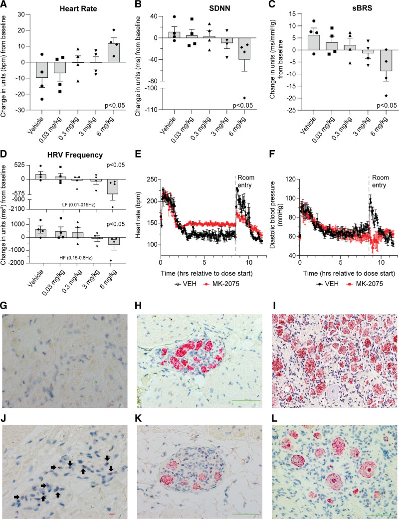
Effects of MK-2075 on cardiac autonomic nervous system balance and reflex control through the assessment of HRV and sBRS in telemetered rhesus monkey at 2 to 6 hours postdose (A–D). Adult male rhesus monkeys (n=4) were given increasing subcutaneous doses of MK-2075 and continuous ECG was evaluated for changes in heart rate (HR, A), the time domain HRV parameter, standard deviation of N-N intervals (SDNN, B), baroreceptor effectiveness index (sBRS, C), and both high-frequency (HF) and low-frequency (LF) domain heart rate variability (HRV, D). To further clarify persistent effects during the diurnal phase, continuous data extracted as 15-minute means were aggregated into a 4-hour time period (2–6 hours postdose). A dose-dependent trend in SDNN, sBRS, and HRV decreases can be observed with a statistically significant (P<0.05) difference in all parameters at the 6 mg/kg SC dose. Statistical significance from vehicle was determined for change from baseline data using a linear mixed-effects model (Y=Group×Time+ID+error) where Group×Time capture the fixed group and time effects and their interactions; ID characterizes the between-subject random effects. E and F, Effect of supratherapeutic doses of MK-2075 on heart rate and diastolic blood pressure. Adult male rhesus monkeys (n=5) were given a continuous 8-hour intravenous infusion through jacketed infusion pumps to determine the effect of MK-2075 on HR (E) and diastolic blood pressure (F) parameters at supratherapeutic exposures (50 mg/kg over 8 hours). Increased HR and no change in diastolic pressure were observed during the infusion. Note that postinfusion handling procedures (noted as “room entry” on the graphs) caused an expected increase in HR and diastolic blood pressure in vehicle-treated animals (VEH), but the HR increase was attenuated and the diastolic blood pressure paradoxically decreased after administration of MK-2075 suggestive of compromised cardiovascular autonomic regulation. G through L, In situ hybridization Nav1.7 mRNA expression in rhesus and human tissues. G, H, and I represent Nav1.7 staining in formalin-fixed paraffin-embedded rhesus sinoatrial node, cardiac ganglia, and dorsal root ganglia, and J, K, and L represent Nav1.7 staining in human sinoatrial node, cardiac ganglia, and dorsal root ganglia, respectively. Nav1.7 is not detected in rhesus sinoatrial node, but mildly expressed in human sinoatrial node (40×). It highly expressed in cardiac ganglia and dorsal root ganglia of both rhesus and human subjects (20×).

