Abstract
Increasing numbers of trials employing anti-PD-1 immunotherapy emphasize the requirement for predictive biomarkers of clinical response. Many studies examine the cell surface expression of PD-1 and other key regulators of T-cell activation and inhibition. Here, we compared common commercially available anti-PD-1 diagnostic antibodies and tested whether they can bind the PD-1 receptor in the presence of the therapeutic antagonists pembrolizumab and nivolumab. We observed that currently no antibodies are available that can reliably stain all PD-1 receptors on T-cells from patients treated with anti-PD-1 antibodies. Furthermore, none of the diagnostic antibodies detected the entire population of PD-1+ T-cells relative to indirect staining using the therapeutic antibodies themselves. To overcome this problem, here we present a reliable method for quantifying PD-1 expression on immune cells from treated patients which can be included in any conventional flow or mass cytometry antibody panel used for patient monitoring.
Electronic supplementary material
The online version of this article (10.1007/s00262-018-2244-7) contains supplementary material, which is available to authorized users.
Keywords: Checkpoint blockade, Immunomonitoring, Pembrolizumab, Nivolumab, Flow cytometry
Introduction
After the groundbreaking success of checkpoint therapy in late-stage melanoma [1, 2], increasing numbers of monotherapies targeting PD-1 are steadily being approved for other cancer entities by the FDA/EMA. However, although some patients experience clinical benefits, many fail to respond. Hence, predictive biomarkers for clinical response to checkpoint therapy are being urgently sought. Currently, the expression of PD-L1 on tumor cells and high tumor mutational burdens are the most sensitive biomarkers of clinical response to anti-PD-1 treatment [3, 4]. However, blood-based markers are mostly unexplored. So far, serum LDH is the only accepted biomarker in late-stage melanoma [5]. Recently, reinvigoration of circulating exhausted T-cells, in relation to pretreatment tumor burden was introduced as a potential on-treatment predictor of response to PD-1 blockade [6]. Other promising candidates are frequencies of myeloid-derived suppressor cells (MDSCs), T-cells and regulatory T-cells (Tregs) in peripheral blood, which have been shown to be associated with clinical outcome under ipilimumab treatment [7, 8]. In late-stage melanoma patients that did not receive checkpoint therapy, the presence of circulating melanoma-associated antigen-specific T-cells was associated with prolonged overall survival [9, 10]. Whether any or all these blood-based marker-candidates also possess similar predictive power for PD-1 blockade or combined CTLA-4/PD-1 blockade remains to be seen. Generally, the expression of PD-1 on T-cells is considered beneficial, as it might provide a window on tumor-resident cells with anti-cancer activity [11, 12]. Thus, many studies examining immune cells from cancer patients before and during treatment with the therapeutic PD-1 antagonists pembrolizumab (pembro) or nivolumab (nivo) seek to quantify cell surface expression of PD-1 and other key regulators of T-cell activation/inhibition. For that purpose, the targeted receptor should of course be detected as accurately as possible. Currently, most studies use commercially available fluorescence-labelled anti-PD-1 diagnostic antibodies. Here we have tested and compared different antibodies to detect PD-1 on the surface of CD4+ and CD8+ T-cells to establish the most feasible and reliable methodology to investigate PD-1-expressing cells.
Materials and methods
Patients
Fresh blood samples from n = 11 late-stage melanoma patients undergoing pembrolizumab (2 mg/kg body weight every 3 weeks) or nivolumab (3 mg/kg body weight every 2 weeks) therapy were obtained from the Department of Dermatology, University Medical Center, Tübingen. Samples were obtained before and at a median of 42 days (range 42–106 days) after the first injection. Fresh blood samples from n = 10 healthy donors were obtained from the local blood bank. Peripheral blood mononuclear cells (PBMCs) were isolated by Ficoll-Hypaque density gradient centrifugation and cryopreserved using freezing medium containing 10% DMSO until use.
Flow cytometry
After thawing, cells were pre-incubated for 20 min with 10 µg/ml nivolumab (dissolved in staining buffer) or 10 µg/ml pembrolizumab (dissolved in staining buffer) or pure staining buffer (PBS, 2% FCS, 2 mM EDTA, 0.01% sodium azide), followed by staining with anti-human IgG4-PE (Southern Biotech; 1 µg/ml). Afterwards, cells were stained for surface markers using CD4-BB515 (clone RPA-T4, BD Biosciences), CD8-APC-H7 (clone SK1, BD Biosciences), PD-1-BV421 (clone MIH4, BD Biosciences) or various PD-1-APC antibodies (clone EH12.2H7, BioLegend; clone MIH4, BD Biosciences; clone PD-1.3.1.3, Miltenyi Biotec; clone NAT105, BioLegend; clone J105, eBioscience) or corresponding isotype control antibodies from the same manufacturer. Selected samples were washed and fixed and permeabilized using the fixation/permeabilization solution (BD Biosciences) according to the manufacturer’s instructions. Dead cells were excluded by ethidium monoazide (Biotinum) staining. Cells were acquired on a LSR II cytometer (BD Biosciences) and analysed using FlowJo (Tree Star). PD-1-expression was determined on CD4+ and CD8+ T-cells within viable lymphocytes.
Statistics
Paired/unpaired t tests were performed using GraphPad Prism 5.
Results
Commercially available diagnostic antibodies identify significantly fewer amounts of PD-1-expressing cells
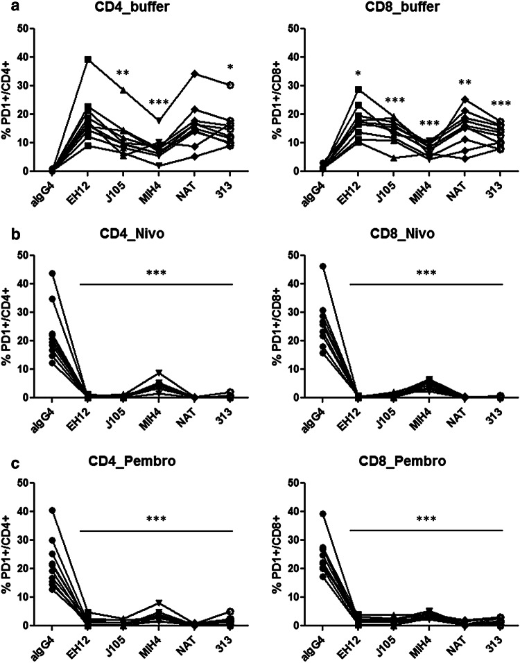
We first compared detection of PD-1 using different commercially available diagnostic antibodies versus PD-1 detection by indirect staining with the therapeutic antibodies themselves [pembrolizumab (pembro) or nivolumab (nivo)]. Cryopreserved PBMCs from healthy donors were incubated with or without the indicated anti-PD-1 therapeutic antibodies, followed by staining with a human anti-IgG4 antibody and/or commercially available anti-PD-1 diagnostic antibodies. We determined the percentage of PD-1+ cells (by either looking at the anti-IgG4 or anti-PD-1 signal) within the CD4+ and CD8+ T-cell populations. For both CD4+ and CD8+ T-cells, the percentage of PD-1+ cells assessed using direct staining was significantly lower compared to indirect approaches using nivo or pembro as the primary antibody. Within CD4+ T-cells, the average percentage of PD-1+ T-cells was 22.6 ± 9.6% using nivo or 21.8 ± 8.5% with pembro (p = 0.8431; unpaired t test compared to nivo), or the commercially available anti-PD-1 antibodies at 18.8 ± 8.3% for clone EH12.2H7 (later referred to as EH12; p = 0.3548), 16.5 ± 7.8% for NAT105 (NAT; p = 0.1354), 14.9 ± 6.3% for PD-1.3.1.3 (313; p = 0.0472), for 11.9 ± 6.5% J105 (p = 0.0091) and 8.0 ± 4.0% for MIH4 (p = 0.0003). Within CD8+ T-cells, the average percentage of PD-1+ T-cells was 26.8 ± 8.5% using nivo, 24.7 ± 6.1% with pembro (p = 0.5367), 17.7 ± 5.5% with EH12 (p = 0.0105), 15.4 ± 6.3% with NAT (p = 0.0031), 14.2 ± 4.4% with J105 (p = 0.0006), 12.7 ± 3.6% with 313 (p = 0.0001) and 7.6 ± 2.2% with MIH4 (p < 0.0001) (Fig. 1a).
Fig. 1.
Cryopreserved PBMCs from n = 10 healthy donors were pre-incubated with buffer (a), Nivo (b) or Pembro (c), followed by staining with a human anti-IgG4 antibody or diagnostic anti-PD-1 antibodies. Percentages of positive cells within all viable CD4+ (left) and CD8+ T-cells (right) are displayed. Each line connects samples from one healthy donor. Asterisks indicate results from t test (*p ≤ 0.05; **p ≤ 0.01; ***p ≤ 0.001)
Commercially available antibodies fail to bind all expressed PD-1 molecules in the presence of therapeutic PD-1 antagonists
We next tested whether the commercially available anti-PD-1 diagnostic antibodies can bind the PD-1 receptor in presence of the therapeutic antagonists pembro or nivo. Importantly, none of the diagnostic antibody clones we used for monitoring was able to bind to PD-1 at a significant level when the therapeutic antibodies were present, although staining using the MIH4 clone antibody led to the highest percentage of PD-1+ cells. After pre-incubation with nivo, within CD4+ T-cells, the average percentage of PD-1+ T-cells was 4.3 ± 1.8% for MIH4 (p < 0.0001; paired t test compared to MIH4 signal from pre-incubation with buffer), 0.5 ± 0.4% for J105 (p < 0.0001), 0.4 ± 0.6% for 313 (p < 0.0001), 0.3 ± 0.4% for EH12 (p < 0.0001) and 0.2 ± 0.1% for NAT (p < 0.0001) (Fig. 1b).
After pre-incubation with pembro, within CD4+ T-cells, the average percentage of PD-1+ T-cells was 3.4 ± 2.0% for MIH4 (p = 0.0001; paired t test compared to MIH4 signal from pre-incubation with buffer), 1.7 ± 1.5% for 313 (p < 0.0001), 1.6 ± 1.4% for EH12 (p < 0.0001), 1.1 ± 0.9% for J105 (p < 0.0001) and 0.5 ± 0.4% for NAT (p < 0.0001) (Fig. 1c).
Accurate detection of PD-1-expressing cells via PD-1 antagonists is possible in patients undergoing checkpoint therapy
The above results suggested that accurately assessing the percentage of PD-1+ T-cells from patients being treated with therapeutic anti-PD-1 is likely to be problematic. Thus, we aimed to determine whether accurate detection of PD-1 via the in vivo-bound therapeutic antibody itself is feasible. To this end, cryopreserved PBMCs from n = 11 melanoma patients undergoing pembrolizumab (n = 7) or nivolumab (n = 4) treatment obtained before and during therapy were stained with an anti-IgG4 antibody followed by staining of surface PD-1 using clone MIH4 (Fig. 2) or clone EH12 antibody (Supplementary Figure 1). MIH4 was chosen because of all the diagnostic antibodies, it was able to bind the highest frequency of PD-1+ cells in the presence of the therapeutic antibodies. Prior to this staining, samples were incubated either with buffer, pembro or nivo. We included this step, as we noticed an undefined loss of bound pembro on isolated PBMCs after freezing and thawing. This may have been due to a complete loss or degradation of the antagonistic antibody. We assume the latter, because neither anti-IgG4 nor other anti-PD-1 antibodies were able to bind. This phenomenon was absent or very minor in nivolumab-treated patients (see Supplementary Figure 2).
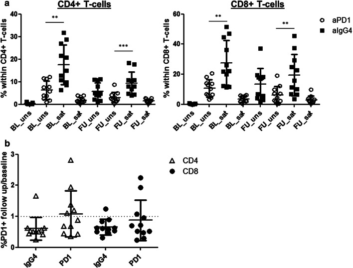
Fig. 2.
a Cryopreserved PBMCs from n = 11 melanoma patients undergoing pembro/nivo treatment were obtained before (baseline, BL) and 42 days (median) after the first anti-PD-1 injection (follow up, FU). PBMCs were stained with anti-IgG4 antibody followed by staining of surface PD-1 using clone MIH4. Prior to this staining, samples were incubated either with pembro/nivo (saturation step, sat) or buffer (unsaturated, uns). Mean percentages + SD of PD-1+ cells, detected directly (white circles) or indirectly (black squares) within CD4+ (left) and CD8+ T-cells (right) are displayed. Asterisks indicate results from t test (*p ≤ 0.05, **p ≤ 0.01, ***p ≤ 0.001). b Alterations to PD-1 expression during treatment were evaluated by calculation of fold changes at follow up/baseline. Mean fold changes + SD, detected directly (PD-1) or indirectly (IgG4) within CD4+ (white triangles) and CD8+ T-cells (black circles) are displayed
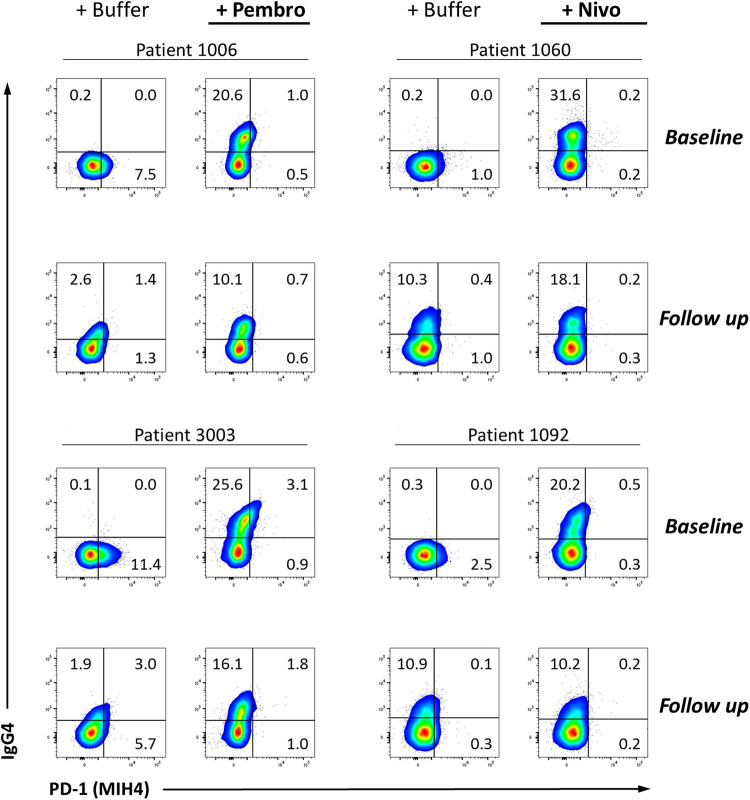
Again, MIH4 antibodies bound significantly lower amounts of PD-1-expressing cells compared to the therapeutic antibodies. At baseline, within CD4+ T-cells, the average percentage of PD-1+ T-cells was 17.5 ± 8.9% using pembro/nivo and 6.3 ± 4.0% using MIH4 clone (p = 0.0011; unpaired t test compared to pembro/nivo). Within CD8+ T-cells, the percentage of PD-1+ T-cells was 27.6 ± 14.8% using pembro/nivo and 10.6 ± 5.7% using MIH4 clone (p = 0.0019). Interestingly, therapeutic antibodies were still detectable on the cell surface in follow-up samples, while the MIH4 antibody failed to identify all PD-1 expressing cells in those samples. Within CD4+ T-cells, the average percentage of PD-1+ T-cells was 5.7 ± 3.8% using pembro/nivo and 3.0 ± 2.3% using MIH4 clone (p = 0.0521; unpaired t test compared to pembro/nivo). Within CD8+ T-cells, the percentage of PD-1+ T-cells was 13.4 ± 10.5% using pembro/nivo and 6.2 ± 5.8% using MIH4 clone (p = 0.0585) (Fig. 2a). For both pembro- and nivo-treated patients, an additional incubation of the follow-up samples with the corresponding therapeutic antagonist to saturate bound antibody, led to an increased frequency of PD-1+ T-cells and an improved staining pattern (CD4+ T-cells: 9.4 ± 4.8%; p = 0.0008, unpaired t test MIH4 versus pembro/nivo. CD8+ T-cells: 19.4 ± 13.8%; p = 0.0080) (see examples of staining in Fig. 3). Comparing baseline and follow-up samples, we saw a lower frequency of PD-1+ T-cells (both CD4+ and CD8+) using pembro/nivo for PD-1 detection in samples from 10 of 11 patients under therapy, while using the MIH4 clone, such a decrease was only observed in 6 of these patients (for CD4; CD8: 8 of 11 patients) (Fig. 2b). We have also tested whether this methodology can be combined with additional protocols and performed fixation and permeabilization for intracellular staining in 7 of the 11 patient samples. Interestingly, we did not observe alterations regarding PD-1 expression in fixed versus unfixed samples (Supplementary Figure 3).
Fig. 3.
Examples of stainings from four melanoma patients undergoing pembro (left) or nivo (right) treatment were obtained before (baseline) and 42 days (median) after the first anti-PD-1 injection (follow up). PBMCs were stained with anti-IgG4 antibody followed by staining of surface PD-1 using clone MIH4. Similar results were obtained using the EH12 clone antibody (Supplementary Figure 1). Prior to this staining, samples were incubated either with buffer, pembro or nivo (saturation step). Signals from the anti-PD-1 antibody (x-axis) and the anti-IgG4 antibody (y-axis) within all viable CD8+ T-cells are displayed. All gates were placed according to corresponding Isotype control-stained cells
Discussion
It remains the case that only a minority of patients responds to checkpoint blockade and many centers are seeking biomarkers predicting clinical response to anti-PD-1 therapy. In this respect, it is important to accurately assess PD-1 expression in patients’ T-cells. However, our study revealed that at the time of writing, no anti-PD-1-antibodies that can reliably stain all PD-1 receptors on ex vivo T-cells from patients being treated with therapeutic anti-PD-1 antibodies were available. Consistent with this, Ribas et al. reported that the therapeutic anti-PD-1 antibody pembrolizumab and both of the diagnostic anti-PD-1 antibody clones EH12 [13] and MIH4 [14] compete for the same epitope. Additionally, Tan et al. reported that although pembro and a second therapeutic anti-PD-1 antibody nivolumab showed very similar patterns of binding to PD-1, their targeted epitopes do not overlap. Nonetheless, due to the comparably small size of the PD-1 molecule and the close proximity of these two binding sites, there is steric hindrance between pembro- and nivo-binding sites. Furthermore, because structural analyses of the PD-1 receptor indicated different conformations taken up on binding to different ligands (pembro, nivo, PD-L1), it is unlikely that PD-1 can be reliably detected when ligands are present/bound [15]. Thus, using commercially available antibodies for the ex vivo detection of PD-1 in samples from patients undergoing anti-PD-1 therapy, a large fraction of PD-1-expressing cells probably remains unidentified (i.e., false-negative events). This technical failure would not be expected to be homogeneously distributed amongst all tested individuals, and therefore interpretation of such data will necessarily lead to misinterpretations leading to, for example, incorrect clustering of patients into clinically relevant groups.
Although Brahmer et al. already used this improved PD-1 detection method for patients undergoing anti-PD-1 therapy in 2010 [16], many studies published thereafter used suboptimal PD-1 detection. For example, Das et al. analysed PBMCs from patients undergoing nivo therapy and failed to detect PD-1+ cells during therapy using clone J105 antibody [17]. Huang et al. failed to confirm associations between PD-1+ Ki-67+ CD8+ T-cells with tumor burden and prolonged overall survival in the one of their two cohorts where PD-1 was detected using clone MIH4 antibody [6]. Ribas et al. abstained from analysing PD-1 expression in a cohort of pembro-treated patients for exactly the reason discussed above, i.e., competition for diagnostic and therapeutic antibody-binding epitopes [13, 14]. Most recently, Krieg et al. used EH12 clone to detect PD-1-expressing cells in patients undergoing pembro/nivo treatment [18].
To overcome this limitation, here we propose a method allowing the identification of PD-1-expressing cells by an indirect detection method using visualization of the therapeutic PD-1 antibody itself, which remains bound to cells ex vivo after their isolation and even cryopreservation. This can be accomplished using a fluorescent, or metal labelled secondary anti-IgG4 antibody. In agreement with Brahmer et al., we have observed that the therapeutic PD-1 antagonist shows highly saturated binding (98%) on CD4+ and CD8+ T-cells even weeks after the last intravenous injection [16, 17]. However, we recommend staining all patient samples, both before and during anti-PD-1 treatment, with the therapeutic anti-PD-1 antibody ex vivo followed by its detection via a secondary antibody. This protocol leads to a very high degree of saturated PD-1 receptors with anti-PD-1 antibodies, even in cryopreserved samples.
In addition to the ex vivo “patient setting” where therapeutic antibodies are present, a commonly employed PD-1 staining protocol for untreated samples from healthy blood bank donors consistently identified fewer PD-1+ cells relative to indirect targeting of the therapeutic antibodies, in an experimental setting using the same diagnostic antibodies, fluorochromes, compensation matrix and antibody concentrations. We therefore conclude that indirect PD-1 staining using the therapeutic antibody itself is a feasible and reliable methodology that can be combined with conventional flow and mass cytometry antibody panels and protocols, ensuring a consistent and comparable quantification of PD-1 expression patterns. This is of major importance as the T-cells, the main target of PD-1 blockade, must be identified and analysed as precisely as possible, especially because the PD-1+ T-cell compartment includes the tiny fraction of cells that recognize tumor-associated antigens and tumor neoantigens [11].
Electronic supplementary material
Below is the link to the electronic supplementary material.
Acknowledgements
We thank Janine Spreuer for technical help.
Abbreviations
- CTLA-4
Cytotoxic T-lymphocyte-associated protein 4
- DMSO
Dimethyl sulfoxide
- EDTA
Ethylenediaminetetraacetic acid
- EMA
European Medicines Agency
- FCS
Fetal calf serum
- FDA
U.S. Food and Drug Administration
- IgG4
Immunoglobulin G4
- LDH
Lactate dehydrogenase
- MDSCs
Myeloid-derived suppressor cells
- Nivo
Nivolumab
- PBMC
Peripheral blood mononuclear cell
- PBS
Phosphate-buffered saline
- PD-1
Programmed cell death protein 1
- PD-L1
Programmed cell death protein 1 ligand 1
- Pembro
Pembrolizumab
Author contributions
HZ conceived and designed the study. JB executed and analysed the experiments. All authors discussed and interpreted the data, wrote and edited the manuscript.
Funding
This work was partially funded by the Klaus Tschira Stiftung (Project no. 00.316.2017).
Compliance with ethical standards
Informed consent
All patients/donors gave written informed consent for biobanking, and use of biomaterials and clinical data for scientific purposes. This study was approved by the ethics committee in Tübingen (Ethik-Kommission an der Medizinischen Fakultät der Eberhard-Karls-Universität und am Universitätsklinikum Tübingen; approvals 490/2014BO1 and 792/2016BO2).
Conflict of interest
C. Garbe reports receiving commercial research grants from Bristol-Myers Squibb, Novartis, and Roche; and is a consultant/advisory board member for Amgen, Bristol-Myers Squibb, Merck Sharp & Dohme, Novartis, and Roche. B. Weide reports receiving commercial research grants from, is a consultant/advisory board member for, and reports receiving travel reimbursement from Bristol-Myers Squibb and Merck Sharp & Dohme. No potential conflicts of interest were disclosed by the other authors.
Footnotes
Kilian Wistuba-Hamprecht and Benjamin Weide contributed equally to this work.
References
- 1.Hodi FS, O’Day SJ, McDermott DF, Weber RW, Sosman JA, Haanen JB, Gonzalez R, Robert C, Schadendorf D, Hassel JC, Akerley W, van den Eertwegh AJ, Lutzky J, Lorigan P, Vaubel JM, Linette GP, Hogg D, Ottensmeier CH, Lebbe C, Peschel C, Quirt I, Clark JI, Wolchok JD, Weber JS, Tian J, Yellin MJ, Nichol GM, Hoos A, Urba WJ. Improved survival with ipilimumab in patients with metastatic melanoma. N Engl J Med. 2010;363(8):711–723. doi: 10.1056/NEJMoa1003466. [DOI] [PMC free article] [PubMed] [Google Scholar]
- 2.Hamid O, Robert C, Daud A, Hodi FS, Hwu WJ, Kefford R, Wolchok JD, Hersey P, Joseph RW, Weber JS, Dronca R, Gangadhar TC, Patnaik A, Zarour H, Joshua AM, Gergich K, Elassaiss-Schaap J, Algazi A, Mateus C, Boasberg P, Tumeh PC, Chmielowski B, Ebbinghaus SW, Li XN, Kang SP, Ribas A. Safety and tumor responses with lambrolizumab (anti-PD-1) in melanoma. N Engl J Med. 2013;369(2):134–144. doi: 10.1056/NEJMoa1305133. [DOI] [PMC free article] [PubMed] [Google Scholar]
- 3.Taube JM, Klein A, Brahmer JR, Xu H, Pan X, Kim JH, Chen L, Pardoll DM, Topalian SL, Anders RA. Association of PD-1, PD-1 ligands, and other features of the tumor immune microenvironment with response to anti-PD-1 therapy. Clin Cancer Res. 2014;20(19):5064–5074. doi: 10.1158/1078-0432.CCR-13-3271. [DOI] [PMC free article] [PubMed] [Google Scholar]
- 4.Rizvi NA, Hellmann MD, Snyder A, Kvistborg P, Makarov V, Havel JJ, Lee W, Yuan J, Wong P, Ho TS, Miller ML, Rekhtman N, Moreira AL, Ibrahim F, Bruggeman C, Gasmi B, Zappasodi R, Maeda Y, Sander C, Garon EB, Merghoub T, Wolchok JD, Schumacher TN, Chan TA. Cancer immunology. Mutational landscape determines sensitivity to PD-1 blockade in non-small cell lung cancer. Science. 2015;348(6230):124–128. doi: 10.1126/science.aaa1348. [DOI] [PMC free article] [PubMed] [Google Scholar]
- 5.Eton O, Legha SS, Moon TE, Buzaid AC, Papadopoulos NE, Plager C, Burgess AM, Bedikian AY, Ring S, Dong Q, Glassman AB, Balch CM, Benjamin RS. Prognostic factors for survival of patients treated systemically for disseminated melanoma. J Clin Oncol. 1998;16(3):1103–1111. doi: 10.1200/JCO.1998.16.3.1103. [DOI] [PubMed] [Google Scholar]
- 6.Huang AC, Postow MA, Orlowski RJ, Mick R, Bengsch B, Manne S, Xu W, Harmon S, Giles JR, Wenz B, Adamow M, Kuk D, Panageas KS, Carrera C, Wong P, Quagliarello F, Wubbenhorst B, D’Andrea K, Pauken KE, Herati RS, Staupe RP, Schenkel JM, McGettigan S, Kothari S, George SM, Vonderheide RH, Amaravadi RK, Karakousis GC, Schuchter LM, Xu X, Nathanson KL, Wolchok JD, Gangadhar TC, Wherry EJ. T-cell invigoration to tumour burden ratio associated with anti-PD-1 response. Nature. 2017;545(7652):60–65. doi: 10.1038/nature22079. [DOI] [PMC free article] [PubMed] [Google Scholar]
- 7.Martens A, Wistuba-Hamprecht K, Geukes Foppen M, Yuan J, Postow MA, Wong P, Romano E, Khammari A, Dreno B, Capone M, Ascierto PA, Di Giacomo AM, Maio M, Schilling B, Sucker A, Schadendorf D, Hassel JC, Eigentler TK, Martus P, Wolchok JD, Blank C, Pawelec G, Garbe C, Weide B. Baseline peripheral blood biomarkers associated with clinical outcome of advanced melanoma patients treated with ipilimumab. Clin Cancer Res. 2016;22(12):2908–2918. doi: 10.1158/1078-0432.CCR-15-2412. [DOI] [PMC free article] [PubMed] [Google Scholar]
- 8.Martens A, Wistuba-Hamprecht K, Yuan J, Postow MA, Wong P, Capone M, Madonna G, Khammari A, Schilling B, Sucker A, Schadendorf D, Martus P, Dreno B, Ascierto PA, Wolchok JD, Pawelec G, Garbe C, Weide B. Increases in absolute lymphocytes and circulating CD4+ and CD8+ T cells are associated with positive clinical outcome of melanoma patients treated with ipilimumab. Clin Cancer Res. 2016;22(19):4848–4858. doi: 10.1158/1078-0432.CCR-16-0249. [DOI] [PMC free article] [PubMed] [Google Scholar]
- 9.Weide B, Zelba H, Derhovanessian E, Pflugfelder A, Eigentler TK, Di Giacomo AM, Maio M, Aarntzen EH, de Vries IJ, Sucker A, Schadendorf D, Buttner P, Garbe C, Pawelec G. Functional T cells targeting NY-ESO-1 or Melan-A are predictive for survival of patients with distant melanoma metastasis. J Clin Oncol. 2012;30(15):1835–1841. doi: 10.1200/JCO.2011.40.2271. [DOI] [PubMed] [Google Scholar]
- 10.Zelba H, Weide B, Martens A, Derhovanessian E, Bailur JK, Kyzirakos C, Pflugfelder A, Eigentler TK, Di Giacomo AM, Maio M, Aarntzen EH, de Vries J, Sucker A, Schadendorf D, Buttner P, Garbe C, Pawelec G. Circulating CD4+ T cells that produce IL4 or IL17 when stimulated by melan-A but not by NY-ESO-1 have negative impacts on survival of patients with stage IV melanoma. Clin Cancer Res. 2014;20(16):4390–4399. doi: 10.1158/1078-0432.CCR-14-1015. [DOI] [PubMed] [Google Scholar]
- 11.Gros A, Parkhurst MR, Tran E, Pasetto A, Robbins PF, Ilyas S, Prickett TD, Gartner JJ, Crystal JS, Roberts IM, Trebska-McGowan K, Wunderlich JR, Yang JC, Rosenberg SA. Prospective identification of neoantigen-specific lymphocytes in the peripheral blood of melanoma patients. Nat Med. 2016;22(4):433–438. doi: 10.1038/nm.4051. [DOI] [PMC free article] [PubMed] [Google Scholar]
- 12.Wistuba-Hamprecht K, Martens A, Haehnel K, Geukes Foppen M, Yuan J, Postow MA, Wong P, Romano E, Khammari A, Dreno B, Capone M, Ascierto PA, Demuth I, Steinhagen-Thiessen E, Larbi A, Schilling B, Schadendorf D, Wolchok JD, Blank CU, Pawelec G, Garbe C, Weide B. Proportions of blood-borne Vdelta1+ and Vdelta2+ T-cells are associated with overall survival of melanoma patients treated with ipilimumab. Eur J Cancer. 2016;64:116–126. doi: 10.1016/j.ejca.2016.06.001. [DOI] [PMC free article] [PubMed] [Google Scholar]
- 13.Ribas A, Dummer R, Puzanov I, VanderWalde A, Andtbacka RHI, Michielin O, Olszanski AJ, Malvehy J, Cebon J, Fernandez E, Kirkwood JM, Gajewski TF, Chen L, Gorski KS, Anderson AA, Diede SJ, Lassman ME, Gansert J, Hodi FS, Long GV. Oncolytic virotherapy promotes intratumoral T cell infiltration and improves anti-PD-1 immunotherapy. Cell. 2017;170(6):1109–1119.e10. doi: 10.1016/j.cell.2017.08.027. [DOI] [PMC free article] [PubMed] [Google Scholar]
- 14.Ribas A, Shin DS, Zaretsky J, Frederiksen J, Cornish A, Avramis E, Seja E, Kivork C, Siebert J, Kaplan-Lefko P, Wang X, Chmielowski B, Glaspy JA, Tumeh PC, Chodon T, Pe’er D, Comin-Anduix B. PD-1 blockade expands intratumoral memory T cells. Cancer Immunol Res. 2016;4(3):194–203. doi: 10.1158/2326-6066.CIR-15-0210. [DOI] [PMC free article] [PubMed] [Google Scholar]
- 15.Tan S, Zhang H, Chai Y, Song H, Tong Z, Wang Q, Qi J, Wong G, Zhu X, Liu WJ, Gao S, Wang Z, Shi Y, Yang F, Gao GF, Yan J. An unexpected N-terminal loop in PD-1 dominates binding by nivolumab. Nat Commun. 2017;8:14369. doi: 10.1038/ncomms14369. [DOI] [PMC free article] [PubMed] [Google Scholar]
- 16.Brahmer JR, Drake CG, Wollner I, Powderly JD, Picus J, Sharfman WH, Stankevich E, Pons A, Salay TM, McMiller TL, Gilson MM, Wang C, Selby M, Taube JM, Anders R, Chen L, Korman AJ, Pardoll DM, Lowy I, Topalian SL. Phase I study of single-agent anti-programmed death-1 (MDX-1106) in refractory solid tumors: safety, clinical activity, pharmacodynamics, and immunologic correlates. J Clin Oncol. 2010;28(19):3167–3175. doi: 10.1200/JCO.2009.26.7609. [DOI] [PMC free article] [PubMed] [Google Scholar]
- 17.Das R, Verma R, Sznol M, Boddupalli CS, Gettinger SN, Kluger H, Callahan M, Wolchok JD, Halaban R, Dhodapkar MV, Dhodapkar KM. Combination therapy with anti-CTLA-4 and anti-PD-1 leads to distinct immunologic changes in vivo. J Immunol. 2015;194(3):950–959. doi: 10.4049/jimmunol.1401686. [DOI] [PMC free article] [PubMed] [Google Scholar]
- 18.Krieg C, Nowicka M, Guglietta S, Schindler S, Hartmann FJ, Weber LM, Dummer R, Robinson MD, Levesque MP, Becher B. High-dimensional single-cell analysis predicts response to anti-PD-1 immunotherapy. Nat Med. 2018;24(2):144–153. doi: 10.1038/nm.4466. [DOI] [PubMed] [Google Scholar]
Associated Data
This section collects any data citations, data availability statements, or supplementary materials included in this article.


