Abstract
The timely restoration of lost blood in hemorrhaging patients with trauma, especially those who are hemodynamically unstable, is of utmost importance. While intravenous access has traditionally been considered the primary method for vascular access, intraosseous (IO) access is gaining popularity as an alternative for patients with unsuccessful attempts. Previous studies have highlighted the higher success rate and easier training process associated with IO access compared with peripheral intravenous (PIV) and central intravenous access. However, the effectiveness of IO access in the early aggressive resuscitation of patients remains unclear. This review article aims to comprehensively discuss various aspects of IO access, including its advantages and disadvantages, and explore the existing literature on the clinical outcomes of patients with trauma undergoing resuscitation with IO versus intravenous access.
Keywords: Intraosseous, resuscitation, hypotension, blood transfusion
Introduction
Resuscitation of critically injured patients with trauma necessitates vascular access in order to restore lost blood volume. Ideally, this is done by establishing at least two large-bore intravenous catheters.1 These are typically placed in the antecubital veins. However, in the shocked patients with trauma, the veins may collapse. Additional factors such as obesity, destruction of veins due to repeated intravenous drug abuse or significant injury to the extremities may preclude the successful placement of an intravenous catheter.
Intraosseous (IO) access represents an alternative method of delivering fluids and medications directly into the bone marrow, typically the long bones such as the tibia or humerus. Moreover, the sternum is another common site of IO insertion, particularly in the military setting, as a convenient and efficient site for the infusion of large volumes.2 This technique is often used in emergency situations, particularly in trauma scenarios, where obtaining intravenous access may be challenging or time-consuming. The use of IOs for patients in clinical cardiac arrest during ongoing cardiopulmonary resuscitation has been endorsed by the American Heart Association and the International Liaison Committee on Resuscitation3
While IO access is a valuable tool in trauma and emergency medicine, it is important to note that it is typically considered a bridge to more definitive vascular access. Once the patient’s condition stabilizes, efforts should be made to establish traditional intravenous access for ongoing treatment. IO access should not be maintained beyond the acute resuscitation phase unless dire circumstances preclude obtaining intravenous access. Nevertheless, IO access is a key resuscitation procedure.
This review will provide an overview of IO access, review its benefits (namely, the ease of training, higher success rates and comparable flow rates when compared with intravenous access), its challenges (including the complications) and define an alternative to its use.
Types of IO access
There are different types of IO access, and the choice of method may depend on factors such as patient age, anatomical considerations and available equipment. IOs can be used in both adults and pediatric patients. The common types of IO access include:
Manual IO access: this involves manually inserting an IO needle into the bone marrow using the thumb or finger to apply pressure. This technique is generally used in emergency situations and is considered a basic method.
Mechanical IO access: these devices use a spring-loaded mechanism to insert the IO needle into the bone. The device is typically placed on the bone surface, and on activation, the needle is quickly and forcefully inserted into the marrow. This method is relatively quick and requires less manual force.
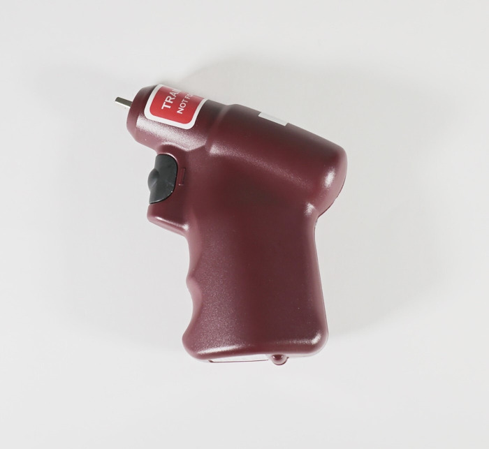
Power drill devices (figure 1): some IO devices are designed to be used with a power drill. These devices use a drill to insert the IO needle into the bone marrow, providing a rapid and controlled method of access. This type of IO access is commonly used in both prehospital and hospital settings.
Figure 1.
- Power Drill Intraosseous Device
Sites of IO access
The choice of IO access method and site may depend on the patient’s age, the clinical situation and the preferences or training of the healthcare clinician. In emergency situations, the goal is to establish vascular access quickly and efficiently to facilitate the delivery of life-saving interventions.
Tibial access: the tibia is a commonly used site for IO access, particularly in adults and older children. The correct site is 1–2 cm inferior and medial to the tibial tuberosity.
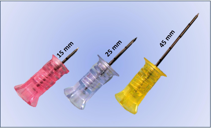
Humeral access: the humeral head is often used for IO access in pediatric patients as well as in adults when tibial access is not feasible. Care must be taken after needle insertion, as internal and external rotation of the humerus may dislodge or bend the needle. In addition, a longer (45 mm) needle is typically needed on this site. Figure 2 demonstrates the 15-gauge IO needles, which are available in 15 mm, 25 mm and 45 mm lengths.
Figure 2.
Different needle sizes for 15-gauge intraosseous access
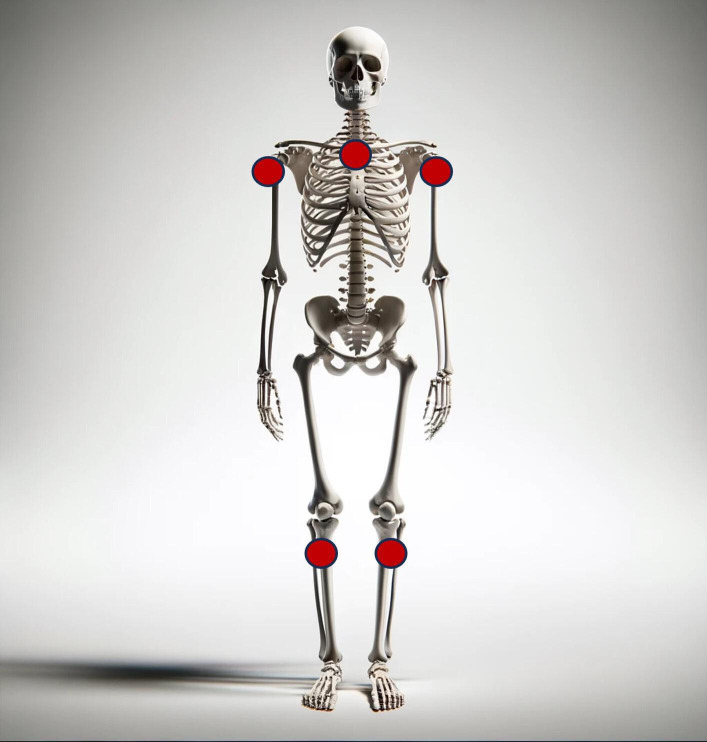
Sternum access: in certain situations, the sternum may be considered as an alternative site for IO access. However, this is less common and may be used when other sites are not accessible. Of note, this site requires a specific type of IO needle, which is typically then sited into the middle of the manubrium sternum. In addition, sternal IO route is contraindicated in children due to possibility of serious complications.4 Care must be taken due to the relatively shallow width of the sternum. Placement off-midline risks infusion of fluids directly into the mediastinum rather than the sternal marrow. Figure 3 demonstrates the common sites of IO access for adult patients.
Figure 3.
Common sites of intraosseous access for adult patients.
Notably, some studies have found that tibial IO access might be comparable to a large PIV, while humeral and sternal access are more similar to central venous access and result in rapid absorption into the heart, when IO is used for drug delivery.5
Training for IO placement
Clinical staff who are required to attend to critically ill or injured patients must have the skills to place an IO access device. Inadequate knowledge and experience risks an increased complication and failure rate. However, training is straightforward. Although use had initially been primarily aimed at physicians, non-physician staff including nurses and paramedics can be successfully trained to place IO needles. Training on IO technique can be incorporated into national life support courses or can be delivered as stand-alone training programs.
In a study of emergency department staff, 80.8% of whom have never placed an IO needle, Levitan et al demonstrated that 97.3% of insertions were successful after a short training program incorporating a 5 min video and cadaveric instruction.6 Similarly, in a prospective multicenter trial of prehospital clinicians (paramedics and nurses), Anderson et al showed that a 1-hour standardized training session followed by hands-on supervised simulation allowed for the successful implementation of IO access in the field, with an 87% success rate.7 This simplicity of training allows for versatile use of IO access in all acute-care areas prehospital and in-hospital.
Speed and success of IO placement
The key reported benefit of IO access over intravenous access in the shocked patient is the speed with which vascular access is obtained. Chreiman et al utilized trauma video recordings to compare times for completion of obtaining intravenous, IO or central venous access.8 In 145 attempts in 38 patients, although attempts at IO and intravenous were equally fast, IO access was more successful than both intravenous and central access (95% vs 42% vs 46%). Dumas et al conducted a multicenter trial using video review and collected numerous data points to quantify both the time and success of vascular access attempts.9 Of 1410 access attempts in 581 patients at 19 centers, IO was found to have higher success than intravenous and central access (93% vs 67% vs 59%) despite access times being similar for IO and intravenous attempts (both under 1 min). This group also showed the time to resuscitation initiation was faster in the IO group (5.8 min) vs intravenous (6.7 min).
Analgesia considerations for IO access
Although it can be assumed that placement of a large-bore needle into the bone is painful, few studies have clearly demonstrated this. What appears to be more likely to cause pain is the expansion of the marrow space as fluid or medications are pushed through the device.10 If time permits, local anesthetic can be administered prior to insertion, though this must infiltrate the periosteum to be successful. Notably, concentrations of lidocaine typical for managing arrhythmias (ie, 1%–2% at 1–1.5 mg/kg) are preferred. Time pressures may make this impossible during active trauma resuscitations. However, experts recommend administering 0.5 mg/kg of lidocaine through the IO needle prior to fluid or medication infusion.11
Flow rates through IO needles
The utilization of IO access exploits the vascularity of cancellous bone, which is the sponge-like bone found inside the hard external compact bone. Cancellous bone consists of a porous structure made up of spicules or trabeculae and hematopoietic red marrow. It is important to acknowledge that the properties of cancellous bone vary significantly based on factors such as age, gender and ethnicity.12–14
Any medication or fluid that can be administered intravenous can also be given IO, with the exception of hypertonic saline, as some animal studies suggest that there is a risk of soft tissue and bone necrosis with IO hypertonic saline administration.15 However, it is not reported in any currently available human trial studies. It is recommended that fluid be administered using a pressure bag to maximize flow rates. Ong et al performed a prospective observational study in 24 patients (with a total of 24 tibial and 11 humeral IO insertions) and found that when administered under pressure, crystalloid flow rates of 165 mL/min could be achieved through the tibial route.16 This dropped to 73 mL/min if pressure was not used. At the humeral site, rates of 153 mL/min with pressure and 84 mL/min without pressure were documented.
Pasley et al performed a cadaveric study to demonstrate crystalloid flow rates at tibial, humeral and sternal IO sites.17 Mean flow rates were fastest at the sternum (93.7 mL/min), followed by humeral (57.1 mL/min) and then tibial (18.7 mL/min) sites. Hence, the sternal site showed flow rates just over three times that of the tibial site. Nevertheless, the presence of an intact circulation likely increases the flow rates as is demonstrated when comparing the results of this study with those from Ong et al. Although human trials of blood product administration through IO devices are limited, those that have been done show both safety and reliability while achieving sufficient flow.18 Table 1 compares flow rates from various vascular access devices. When fluid is administered via pressure bag, IO access, especially at the humeral site, can allow for comparable flow rates to gravity flow through a 16-gauge PIV. Even at lower gravity flow rates, given the demonstrated higher success rates of IO access, this will allow at least initial fluid resuscitation to occur prior to further access being obtained.
Table 1.
Flow rates through various access devices
| Size of access device | Approximate flow rate to gravity (mL/min) | Time to infused 1 L (min) |
| 14G IV | 250 | 4 |
| 16G IV | 150 | 7 |
| 7.5 French introducer sheath | 130 | 8 |
| 18G IV | 100 | 10 |
| 15G humeral IO | 80 | 13 |
| 16G distal port triple-lumen central venous catheter | 70 | 15 |
| 15G tibial IO | 70 | 15 |
| 20G IV | 60 | 17 |
| 22G IV | 35 | 29 |
| 18G proximal port central venous catheter | 30 | 34 |
IO, intraosseous.
The comparison of flow rates also highlights the importance of at least two sites of vascular access being established during the acute resuscitation phase. A single access site may not allow for sufficient volume return in an actively bleeding patient. While ideally this is via two large-bore intravenous catheters, two IO needles or a combination of an intravenous and IO needle can allow for initial resuscitation until more definitive access is obtained.
Disadvantages of IO fluid administration
Access devices used in trauma cases must be able to administer large amounts of blood products promptly and provide flow rates that allow for frequent patient evaluation, enabling therapy adjustment based on specific physiological parameters. Although there has been a recent increase in the use of IO access for patients with trauma, current guidelines recommend IO catheters as a temporary solution until definitive intravenous access can be established, rather than as a replacement for it19 20
Despite the advantages and endorsement of IOs, the efficacy and evidence supporting their use in modern trauma resuscitation continue to be debated.21 Additionally, the lack of comprehensive studies comparing the outcomes of shocked patients with trauma resuscitated with IO devices versus those with intravenous catheters is another obstacle preventing the widespread adoption of IO access as a substitute for intravenous access. In the remainder of this review, we will discuss the major drawbacks of IO access devices that healthcare professionals should consider before considering them as a primary route of access for resuscitation of unstable trauma cases.
Inadequate IO access flow rate
Despite the reported higher rates of success in IO versus intravenous access among hemodynamically unstable patients with trauma8 22 and non-trauma population,23–25 one of the major concerns regarding the use of IO devices is the inadequate flow rates of blood products.26 In 2014, Burgert et al conducted an intervention study on swine models to compare the transfusion rates of IO and intravenous access.27 The authors found that it took approximately two times as long to transfuse 900 mL of blood using IO compared with intravenous access. In a separate prospective observational study on volunteer professional military personnel, 450 mL of autologous whole blood from each participant was collected and reinfused with IO vs intravenous routes, using gravity only.18 Notably, the IO groups had a median infusion rate of 32.4 mL/min, which was nearly half of the intravenous group’s rate of 74.1 mL/min. The study used the sternal site, which is known to have the fastest flow rate compared with the humeral and tibial insertion sites.17
One commonly overlooked factor is the impact of a patient’s initial blood pressure (BP) on the flow rate of different IO accesses. Studies have found that in animal models experiencing hemorrhagic shock, the flow rate is significantly lower compared with those with normal blood volume.28 Despite the limited studies comparing the flow rates between IO versus intravenous access in normotensive and hypotensive models, an animal study on a piglet model showed that hypovolemia results in average decreased infusion rates of 32% within various sites of IO access.29 It is worth noting that intravenous access was found to be the most effective method for immediate volume replacement, as even with IO access using 300 mmHg pressure, the flow rates were significantly lower compared with intravenous access.
In addition, it should be noted that the IO catheter itself does not restrict the flow rate of blood products, as it belongs to the same size category as the intravenous catheters commonly used in trauma patient resuscitation. Therefore, the parameters of the IO space, including bone density, play a crucial role in defining this area. Haris et al, utilizing Darcy’s law and accounting for the low flow rates of blood transfusion into the IO space, argued that transfusing blood via the IO route does not yield substantial results in the resuscitation of patients with trauma.21 They, in fact, recommended that medical personnel receive training in ultrasound technologies as an alternative to enhance successful PIV access and to completely avoid the IO route when blood transfusion is necessary.
A blood loss of 150 mL/min or more is considered a major hemorrhage, according to one definition.30 This implies that the current data indicate insufficient transfusion flow rates through a single IO catheter in trauma patients experiencing hemorrhagic shock. This acknowledgment is crucial and mentioned by those who support the use of IO devices in trauma cases. Consequently, some experts recommend the use of two catheters during the initial resuscitative phase to ensure adequate transfusion volumes and as a temporary solution until definitive access can be obtained.8 31–33 Furthermore, they recommend IO access be a temporary solution until definitive intravenous access is obtained. It is important to consider this recommendation when comparing IO devices with other access route alternatives.
Potential for red cell hemolysis
Another contentious issue regarding the utilization of IO devices is the possibility of red cell hemolysis.34 This is of particular significance in the context of trauma, where prompt replenishment of sufficient blood volume is crucial for patients. Based on theoretical models, the only adjustable factor for medical professionals to enhance flow rate in an IO system during a device closure reperfusion procedure is pressure.21 Heightened pressure not only places strain on the connections within the infusion system itself but also amplifies the shearing forces exerted on the fluid. These shearing forces have the potential to induce red blood cell destruction, resulting in the loss of oxygen-carrying capacity and the subsequent development of rhabdomyolysis.
Despite previous studies on animal models attempting to address the concern regarding the red cell hemolysis,27 33 35–37 one crucial aspect has been overlooked—the bone densitometry of these models does not accurately reflect that of young adult humans. A recent systematic review, consisting of nine papers on red cell hemolysis following IO blood transfusion, revealed a lack of high-quality evidence regarding the risks associated with red cell hemolysis in IO blood transfusion.34 However, findings from one study suggest that the use of a three-way tap to administer blood transfusion to young adult male patients with trauma may increase the likelihood of red cell hemolysis. Notably, among the nine papers included, seven were animal studies, while only one prospective human study was reported. This human study documented a significant increase in lactate dehydrogenase levels and a decrease in hemoglobin levels following the infusion within the IO groups.18
Complications
Despite a dearth of data comparing the rates of complications between IO and intravenous access, various studies have examined the complications of IO access within specific sites that should be taken into consideration before utilizing them.38 In an online questionnaire-based study of 386 Scandinavian physicians, 1802 clinical cases of IO use were reported, of which nearly one-fourth (23.4%) was indicated following a hemorrhage.39 The authors concluded that the overall complication rate exceeded what is typically reported from model and cadaver studies, with responders reporting that 68.6% experienced some form of complication during the procedure, infusion or late after the infusion. While this study included factors like severe patient pain as a complication, it also shed light on the challenges faced by clinicians and patients when employing IO devices. Consequently, future research on IO devices should encompass all stages of IO use.
Although previous research has demonstrated the safety and feasibility of IO access in pediatric patients,40 it can be challenging to successfully cannulate the hardened bones of adult patients using IO devices.41 Hence, variations in osseous anatomy between pediatric and adult patients are expected to affect the type and severity of complications associated with IO cannulation. A recent comprehensive analysis of complications related to IO catheterization in adult patients revealed an overall complication rate of 4.6% following successful IO catheter insertion.42 Major complications noted in this study included the extravasation or displacement of catheter (2.8%), device malfunction (1.8%), injury to surrounding tissues (0.1%), bleeding (0.04%), tissue necrosis (0.02%) and infection (0.01%). It is important to note that these complication rates can vary significantly across different studies due to the influence of operator experience. For instance, the extravasation rate has been reported to range from 1 to 22% in various studies.15
Notably, needle dislodgement is a complication that has been found to be more prevalent in humeral IO accesses (20%) compared with tibial IO accesses (9%).43 If the chosen needle is insufficiently long to fully penetrate all layers of subcutaneous tissue, the IO needle will not be able to completely enter the bone matrix, resulting in a failed attempt or dislodgement. Furthermore, constant movement and activity can significantly increase the risk of unintentional needle dislodgement or malformation. Similar rates of needle dislodgements have been reported in other studies, with rates of 10%, 16% and 15% for femoral, humeral, and tibial sites, respectively.44
The goal is to improve outcomes
Despite expanding literature on the role of IO access in resuscitation of patients with trauma, its impact on patients outcomes remains unclear.45 A recent multi-institutional study of 581 adult (≥16 year) hypotensive (systolic BP ≤90 mm Hg) trauma patients showed that despite no difference in time to access between patients with IO versus PIV access, IO had higher success rates than PIV (93% vs 67%) and remained higher after subsequent failures (85% vs 59%).9 However, this study did not provide any data on patient-centered clinical outcomes such as early and late mortality or complications. Another systematic review on the ‘efficacy’ of IO access for trauma resuscitation revealed that the success rate of IO access on first attempt was significantly higher than that of intravenous access for patients with trauma, and the mean procedure time for IO access was also shorter. However, no information regarding patient outcomes was included in the review.22
Despite the limited literature in patients with trauma, in 2021, a prospective, parallel-group, cluster-randomized study compared the outcomes of patients with out-of-hospital cardiac arrest (OHCA) who were resuscitated with ‘intravenous only’ against ‘intravenous+IO’.46 Interestingly, they found that using IO when intravenous failed led to a higher rate of vascular access and faster epinephrine administration. However, it was not associated with higher return of spontaneous circulation (ROSC), survival to discharge or good neurological outcome. Another study by Mody et al, evaluating 19 731 patients with OHCA, of which 3068 patients received IO access, demonstrated that IO access attempt was associated with worse ROSC and survival rates: (4.6% vs 5.7%, p=0.01) for survival to discharge, (17.9% vs 23.5%, p<0.001) for sustained ROSC and (2.8% vs 4.2%, p<0.001) for survival with favorable neurological function.47 Based on these studies and multiple other medium to high-level studies, despite higher rates of successful access through IO route, no differences in survival and clinical outcomes are expected, when using IO in resuscitation of adult and pediatric patients with OHCA.48–50 In fact, a prespecified analysis of a randomized, placebo-controlled clinical trial by Daya et al showed that point estimates for the effects of drugs in comparison with placebo were significantly greater for the intravenous than for the IO route across virtually all outcomes and beneficial only for the intravenous route.51 However, the study was underpowered to statistically assess interactions. Although resuscitation of patients with OHCA is out of the scope of this study, the above-mentioned study and multiple other studies are brought as a signal to interpret these findings carefully, as while IO access may offer faster access or a higher success rate, it may lead to lower survival rates and poorer neurologic outcomes of patients with non-trauma.50 52
Ultrasound-guided intravenous: is this the answer?
intravenous access could be challenging in patients with severe hemorrhagic shock. However, research has demonstrated that using ultrasound guidance for PIV access is both feasible and significantly increases success rates compared with traditional methods.53 In an animal study on six sedated male sheep with a BP of less than 90 mm Hg, the authors found that while accessing the vein blindly was successful in one out of six punctures, ultrasound guidance increased the access to eight out of nine punctures with a median time of 65 s.54 A systematic review, including eight studies on comparing US guidance with traditional approach, showed that the ultrasound-guided technique reduced the number of punctures and time needed to achieve intravenous access, and increased the level of patient satisfaction, although it did not result in a decreased number of complications.55 In fact, this difference was particularly evident in patients with a known or predicted difficult intravenous access. Overall, the findings suggest that using ultrasound guidance for PIV access is more effective than traditional methods, leading to greater success in cannulation, a reduction in the number of punctures, a decrease in procedure time and increased patient satisfaction.56
Although to date, there is no study on the comparison of IO access versus US-guided intravenous access, comparing the reported numbers of attempts and success rates shows promising results in favor of considering US-guided intravenous access approach, if we encounter patients with difficult intravenous access even in the prehospital settings.57 58 Moreover, an encompassing strategy involves providing advanced education to healthcare providers, particularly those in frontline care.59 60
Summary
IO access can be potentially a rapid means of providing small amounts of volume and medications to critically injured patients with trauma prior to definitive vascular access. Although studies supporting the use of IO primarily focus on its ease of training and higher success rate compared with intravenous access, the clinical implications and outcomes among hemodynamically unstable patients with trauma yet to be established by evidence.
Moreover, there are several important aspects that previous studies have not adequately addressed. Concerns such as the inadequate flow rate and the potential for red cell hemolysis through IO and bone space need to be further investigated. In addition, infusion through IO access can be an extremely painful procedure, at times surpassing the pain caused by the patients’ primary injury. If not performed by an experienced professional, there can be multiple complications associated with IO insertion. Importantly, it is crucial to consider that timely resuscitation of patients with trauma ultimately aims to improve clinical outcomes, an aspect that has not been sufficiently assessed in previous studies comparing IO versus intravenous access for hemorrhagic patients with trauma.
Factors to consider include proper education, simulation-based training, and, notably, the utilization of ultrasound-guided intravenous access. Evidence supports both a higher success rate and lower procedural time for the latter, making it a promising alternative. It is, therefore, necessary to further study in the representative population of hemodynamically unstable patients with trauma to demonstrate the clinical success of the IO technique in effective resuscitation. Until then, IO access should be recommended as the bridge for definitive access when attempts for peripheral and central access have failed.
Acknowledgments
All figures included in the current study are authors' own work.
Footnotes
Contributors: Both authors participated in data interpretation and manuscript preparation.
Funding: The authors have not declared a specific grant for this research from any funding agency in the public, commercial or not-for-profit sectors.
Competing interests: None declared.
Provenance and peer review: Not commissioned; internally peer-reviewed.
Ethics statements
Patient consent for publication
This manuscript was not subjected to institutional review approval as it does not involve any patient data and exclusively comprises a literature review.
Ethics approval
Not applicable.
References
- 1. Verhoeff K, Saybel R, Mathura P, Tsang B, Fawcett V, Widder S. Ensuring adequate vascular access in patients with major trauma: a quality improvement initiative. BMJ Open Qual 2018;7:e000090. 10.1136/bmjoq-2017-000090 [DOI] [PMC free article] [PubMed] [Google Scholar]
- 2. Laney JA, Friedman J, Fisher AD. Sternal Intraosseous devices: review of the literature. West J Emerg Med 2021;22:690–5. 10.5811/westjem.2020.12.48939 [DOI] [PMC free article] [PubMed] [Google Scholar]
- 3. Neumar RW, Otto CW, Link MS, Kronick SL, Shuster M, Callaway CW, Kudenchuk PJ, Ornato JP, McNally B, Silvers SM, et al. Part 8: adult advanced cardiovascular life support: 2010 American heart Association guidelines for cardiopulmonary resuscitation and emergency cardiovascular care. Circulation 2010;122:S729–67. 10.1161/CIRCULATIONAHA.110.970988 [DOI] [PubMed] [Google Scholar]
- 4. RARd S, Melo CL, Dantas RB, Delfim LVV. Vascular access through the Intraosseous route in pediatric emergencies. Rev Bras Ter Intensiva 2012;24:407–14. [DOI] [PMC free article] [PubMed] [Google Scholar]
- 5. Hoskins SL, do Nascimento P, Lima RM, Espana-Tenorio JM, Kramer GC. Pharmacokinetics of Intraosseous and central venous drug delivery during cardiopulmonary resuscitation. Resuscitation 2012;83:107–12. 10.1016/j.resuscitation.2011.07.041 [DOI] [PubMed] [Google Scholar]
- 6. Levitan RM, Bortle CD, Snyder TA, Nitsch DA, Pisaturo JT, Butler KH. Use of a battery-operated needle driver for Intraosseous access by novice users: skill acquisition with Cadavers. Ann Emerg Med 2009;54:692–4. 10.1016/j.annemergmed.2009.06.012 [DOI] [PubMed] [Google Scholar]
- 7. Anderson TE, Arthur K, Kleinman M, Drawbaugh R, Eitel DR, Ogden CS, Baker D. Intraosseous infusion: success of a standardized regional training program for Prehospital advanced life support providers. Ann Emerg Med 1994;23:52–5. 10.1016/s0196-0644(94)70008-7 [DOI] [PubMed] [Google Scholar]
- 8. Chreiman KM, Dumas RP, Seamon MJ, Kim PK, Reilly PM, Kaplan LJ, Christie JD, Holena DN. The Ios have it: a prospective observational study of vascular access success rates in patients in extremis using Video review. J Trauma Acute Care Surg 2018;84:558–63. 10.1097/TA.0000000000001795 [DOI] [PMC free article] [PubMed] [Google Scholar]
- 9. Dumas RP, Vella MA, Maiga AW, Erickson CR, Dennis BM, da Luz LT, Pannell D, Quigley E, Velopulos CG, Hendzlik P, et al. Moving the needle on time to resuscitation: an EAST prospective multicenter study of vascular access in hypotensive injured patients using trauma Video review. J Trauma Acute Care Surg 2023;95:87–93. 10.1097/TA.0000000000003958 [DOI] [PubMed] [Google Scholar]
- 10. Horton MA, Beamer C. Powered Intraosseous insertion provides safe and effective vascular access for pediatric emergency patients. Pediatr Emerg Care 2008;24:347–50. 10.1097/PEC.0b013e318177a6fe [DOI] [PubMed] [Google Scholar]
- 11. Philbeck TE, Miller LJ, Montez D, Puga T. Hurts so good. easing IO pain and pressure. JEMS 2010;35:58–62. 10.1016/S0197-2510(10)70232-1 [DOI] [PubMed] [Google Scholar]
- 12. Burgert JM. Intraosseous infusion of blood products and epinephrine in an adult patient in hemorrhagic shock. AANA J 2009;77:359–63. [PubMed] [Google Scholar]
- 13. Henry YM, Eastell R. Ethnic and gender differences in bone mineral density and bone turnover in young adults: effect of bone size. Osteoporos Int 2000;11:512–7. 10.1007/s001980070094 [DOI] [PubMed] [Google Scholar]
- 14. Wu Q, Lefante JJ, Rice JC, Magnus JH. Age, race, weight, and gender impact normative values of bone mineral density. Gend Med 2011;8:189–201. 10.1016/j.genm.2011.04.004 [DOI] [PubMed] [Google Scholar]
- 15. Paxton JH. Intraosseous vascular access: a review. Trauma 2012;14:195–232. 10.1177/1460408611430175 [DOI] [Google Scholar]
- 16. Ong MEH, Chan YH, Oh JJ, Ngo A-Y. An observational, prospective study comparing Tibial and Humeral Intraosseous access using the EZ-IO. Am J Emerg Med 2009;27:8–15. 10.1016/j.ajem.2008.01.025 [DOI] [PubMed] [Google Scholar]
- 17. Pasley J, Miller CHT, DuBose JJ, Shackelford SA, Fang R, Boswell K, Halcome C, Casey J, Cotter M, Matsuura M, et al. Intraosseous infusion rates under high pressure: a Cadaveric comparison of anatomic sites. J Trauma Acute Care Surg 2015;78:295–9. 10.1097/TA.0000000000000516 [DOI] [PubMed] [Google Scholar]
- 18. Bjerkvig CK, Fosse TK, Apelseth TO, Sivertsen J, Braathen H, Eliassen HS, Guttormsen AB, Cap AP, Strandenes G. Emergency sternal Intraosseous access for warm fresh whole blood transfusion in damage control resuscitation. J Trauma Acute Care Surg 2018;84:S120–4. 10.1097/TA.0000000000001850 [DOI] [PubMed] [Google Scholar]
- 19. Kanani AN, Hartshorn S. NICE clinical guideline Ng39: major trauma: assessment and initial management. Arch Dis Child Educ Pract Ed 2017;102:20–3. 10.1136/archdischild-2016-310869 [DOI] [PubMed] [Google Scholar]
- 20. Subcommittee A. Group IAW. Advanced trauma life support (ATLS®): the ninth edition. J Trauma Acute Care Surg 2013;74:1363–6. [DOI] [PubMed] [Google Scholar]
- 21. Harris M, Balog R, Devries G. What is the evidence of utility for Intraosseous blood transfusion in damage-control resuscitation J Trauma Acute Care Surg 2013;75:904–6. 10.1097/TA.0b013e3182a85f71 [DOI] [PubMed] [Google Scholar]
- 22. Wang D, Deng L, Zhang R, Zhou Y, Zeng J, Jiang H. Efficacy of Intraosseous access for trauma resuscitation: a systematic review and meta-analysis. World J Emerg Surg 2023;18:17. 10.1186/s13017-023-00487-7 [DOI] [PMC free article] [PubMed] [Google Scholar]
- 23. Petitpas F, Guenezan J, Vendeuvre T, Scepi M, Oriot D, Mimoz O. Use of intra-osseous access in adults: a systematic review. Crit Care 2016;20:102. 10.1186/s13054-016-1277-6 [DOI] [PMC free article] [PubMed] [Google Scholar]
- 24. Weiser G, Hoffmann Y, Galbraith R, Shavit I. Current advances in Intraosseous infusion–a systematic review. Resuscitation 2012;83:20–6. 10.1016/j.resuscitation.2011.07.020 [DOI] [PubMed] [Google Scholar]
- 25. Santos D, Carron P-N, Yersin B, Pasquier M. EZ-IO® Intraosseous device implementation in a pre-hospital emergency service: a prospective study and review of the literature. Resuscitation 2013;84:440–5. 10.1016/j.resuscitation.2012.11.006 [DOI] [PubMed] [Google Scholar]
- 26. Tyler JA, Perkins Z, De’Ath HD. Intraosseous access in the resuscitation of trauma patients: a literature review. Eur J Trauma Emerg Surg 2021;47:47–55. 10.1007/s00068-020-01327-y [DOI] [PubMed] [Google Scholar]
- 27. Burgert JM, Mozer J, Williams T, Gegel BT, Johnson S, Bentley M, Johnson A. Effects of Intraosseous transfusion of whole blood on hemolysis and transfusion time in a swine model of hemorrhagic shock: a pilot study. AANA J 2014;82:198–202. [PubMed] [Google Scholar]
- 28. Righi N, Paxton JH. Flow rate considerations for Intraosseous catheter use. Curr Emerg Hosp Med Rep 2022;10:125–33. 10.1007/s40138-022-00257-w [DOI] [Google Scholar]
- 29. Warren DW, Kissoon N, Sommerauer JF, Rieder MJ. Comparison of fluid infusion rates among peripheral intravenous and Humerus, Femur, Malleolus, and Tibial Intraosseous sites in Normovolemic and Hypovolemic Piglets. Ann Emerg Med 1993;22:183–6. 10.1016/s0196-0644(05)80199-4 [DOI] [PubMed] [Google Scholar]
- 30. Guerado E, Medina A, Mata MI, Galvan JM, Bertrand ML. Protocols for massive blood transfusion: when and why, and potential complications. Eur J Trauma Emerg Surg 2016;42:283–95. 10.1007/s00068-015-0612-y [DOI] [PubMed] [Google Scholar]
- 31. Lewis P, Wright C. Saving the critically injured trauma patient: a retrospective analysis of 1000 uses of Intraosseous access. Emerg Med J 2015;32:463–7. 10.1136/emermed-2014-203588 [DOI] [PubMed] [Google Scholar]
- 32. Sheils M, Ross M, Eatough N, Caputo ND. Intraosseous access in trauma by air medical retrieval teams. Air Med J 2014;33:161–4. 10.1016/j.amj.2014.03.005 [DOI] [PubMed] [Google Scholar]
- 33. Sulava E, Bianchi W, McEvoy CS, Roszko PJ, Zarow GJ, Gaspary MJ, Natarajan R, Auten JD. Single versus double anatomic site Intraosseous blood transfusion in a swine model of hemorrhagic shock. J Surg Res 2021;267:172–81. 10.1016/j.jss.2021.04.035 [DOI] [PubMed] [Google Scholar]
- 34. Ellington M, Walker I, Barnard E. Red cell Haemolysis secondary to Intraosseous (IO) blood transfusion in adult patients with major trauma: a systematic review. BMJ Mil Health 2023:e002378. 10.1136/military-2023-002378 [DOI] [PubMed] [Google Scholar]
- 35. Krepela A, Auten J, Mclean J, Fortner G, Murnan S, Kemp J, Roszko P, Fishback J. 352 A comparison of flow rates and hematologic safety between Intraosseous blood transfusion strategies in a swine (Sus Scrofa) model of hemorrhagic shock: A pilot study. Ann Emerg Med 2017;70:S139. 10.1016/j.annemergmed.2017.07.322 [DOI] [Google Scholar]
- 36. Bell MC, Olshaker JS, Brown CK, McNAMEE GA, Fauver GM. Intraosseous transfusion in an anesthetized swine model using 51Cr-labeled Autologous red blood cells. J Trauma 1991;31:1487–9. 10.1097/00005373-199111000-00004 [DOI] [PubMed] [Google Scholar]
- 37. Plewa MC, King RW, Fenn-Buderer N, Gretzinger K, Renuart D, Cruz R. Hematologic safety of Intraosseous blood transfusion in a swine model of pediatric hemorrhagic Hypovolemia. Acad Emerg Med 1995;2:799–809. 10.1111/j.1553-2712.1995.tb03275.x [DOI] [PubMed] [Google Scholar]
- 38. Anson JA. Vascular access in resuscitation: is there a role for the Intraosseous route Anesthesiology 2014;120:1015–31. 10.1097/ALN.0000000000000140 [DOI] [PubMed] [Google Scholar]
- 39. Hallas P, Brabrand M, Folkestad L. Complication with Intraosseous access: Scandinavian users’ experience. West J Emerg Med 2013;14:440–3. 10.5811/westjem.2013.1.12000 [DOI] [PMC free article] [PubMed] [Google Scholar]
- 40. Hansen M, Meckler G, Spiro D, Newgard C. Intraosseous line use, complications, and outcomes among a population-based cohort of children presenting to California hospitals. Pediatr Emerg Care 2011;27:928–32. 10.1097/PEC.0b013e3182307a2f [DOI] [PubMed] [Google Scholar]
- 41. Shen W, Velasquez G, Chen J, Jin Y, Heymsfield SB, Gallagher D, Pi-Sunyer FX. Comparison of the relationship between bone marrow Adipose tissue and volumetric bone mineral density in children and adults. J Clin Densitom 2014;17:163–9. 10.1016/j.jocd.2013.02.009 [DOI] [PMC free article] [PubMed] [Google Scholar]
- 42. Palazzolo A, Akers KG, Paxton JH. Complications of Intraosseous catheterization in adult patients: A review of the literature. Curr Emerg Hosp Med Rep 2023;11:35–48. 10.1007/s40138-023-00261-8 [DOI] [Google Scholar]
- 43. Reades R, Studnek JR, Vandeventer S, Garrett J. Intraosseous versus intravenous vascular access during out-of-hospital cardiac arrest: a randomized controlled trial. Ann Emerg Med 2011;58:509–16. 10.1016/j.annemergmed.2011.07.020 [DOI] [PubMed] [Google Scholar]
- 44. Rayas EG, Winckler C, Bolleter S, Stringfellow M, Miramontes D, Shumaker J, Lewis A, Wampler D. Distal Femur versus Humeral or Tibial IO, access in adult out of hospital cardiac resuscitation. Resuscitation 2022;170:11–6. 10.1016/j.resuscitation.2021.10.041 [DOI] [PubMed] [Google Scholar]
- 45. Ribeiro Jr MA, Loureiro LB, Romeo ACD. Comparison between Intraosseous and central venous access in adult trauma patients in the emergency room: A systematic review and meta-analysis. Panam J Trauma Crit Care Emerg Surg 2022;10:113–20. 10.5005/jp-journals-10030-1360 [DOI] [Google Scholar]
- 46. Tan BKK, Chin YX, Koh ZX, Md Said N, Rahmat M, Fook-Chong S, Ng YY, Ong MEH. Clinical evaluation of intravenous alone versus intravenous or Intraosseous access for treatment of out-of-hospital cardiac arrest. Resuscitation 2021;159:129–36. 10.1016/j.resuscitation.2020.11.019 [DOI] [PubMed] [Google Scholar]
- 47. Mody P, Brown SP, Kudenchuk PJ, Chan PS, Khera R, Ayers C, Pandey A, Kern KB, de Lemos JA, Link MS, et al. Intraosseous versus intravenous access in patients with out-of-hospital cardiac arrest: insights from the resuscitation outcomes consortium continuous chest compression trial. Resuscitation 2019;134:69–75. 10.1016/j.resuscitation.2018.10.031 [DOI] [PubMed] [Google Scholar]
- 48. Granfeldt A, Avis SR, Lind PC, Holmberg MJ, Kleinman M, Maconochie I, Hsu CH, Fernanda de Almeida M, Wang T-L, Neumar RW, et al. Intravenous vs. Intraosseous administration of drugs during cardiac arrest: a systematic review. Resuscitation 2020;149:150–7. 10.1016/j.resuscitation.2020.02.025 [DOI] [PubMed] [Google Scholar]
- 49. Recher M, Baert V, Escutnaire J, Le Bastard Q, Javaudin F, Hubert H, Leteurtre S. Intraosseous or peripheral IV access in pediatric cardiac arrest? results from the French national cardiac arrest Registry. Pediatr Crit Care Med 2021;22:286–96. 10.1097/PCC.0000000000002659 [DOI] [PubMed] [Google Scholar]
- 50. Besserer F, Kawano T, Dirk J, Meckler G, Tijssen JA, DeCaen A, Scheuermeyer F, Beno S, Christenson J, Grunau B, et al. The Association of Intraosseous vascular access and survival among pediatric patients with out-of-hospital cardiac arrest. Resuscitation 2021;167:49–57. 10.1016/j.resuscitation.2021.08.005 [DOI] [PubMed] [Google Scholar]
- 51. Daya MR, Leroux BG, Dorian P, Rea TD, Newgard CD, Morrison LJ, Lupton JR, Menegazzi JJ, Ornato JP, Sopko G, et al. Survival after intravenous versus Intraosseous amiodarone, lidocaine, or placebo in out-of-hospital shock-refractory cardiac arrest. Circulation 2020;141:188–98. 10.1161/CIRCULATIONAHA.119.042240 [DOI] [PMC free article] [PubMed] [Google Scholar]
- 52. Kawano T, Grunau B, Scheuermeyer FX, Gibo K, Fordyce CB, Lin S, Stenstrom R, Schlamp R, Jenneson S, Christenson J. Intraosseous vascular access is associated with lower survival and neurologic recovery among patients with out-of-hospital cardiac arrest. Ann Emerg Med 2018;71:588–96. 10.1016/j.annemergmed.2017.11.015 [DOI] [PubMed] [Google Scholar]
- 53. Stolz LA, Stolz U, Howe C, Farrell IJ, Adhikari S. Ultrasound-guided peripheral venous access: a meta-analysis and systematic review. J Vasc Access 2015;16:321–6. 10.5301/jva.5000346 [DOI] [PubMed] [Google Scholar]
- 54. Reva VA, Perevedentcev AV, Pochtarnik AA, Khupov MT, Kalinina AA, Samokhvalov IM, Khan MA. Ultrasound-guided versus blind vascular access followed by REBOA on board of a medical helicopter in a hemorrhagic Ovine model. Injury 2021;52:175–81. 10.1016/j.injury.2020.09.053 [DOI] [PubMed] [Google Scholar]
- 55. van Loon FHJ, Buise MP, Claassen JJF, Dierick-van Daele ATM, Bouwman ARA. Comparison of ultrasound guidance with Palpation and direct Visualisation for peripheral vein Cannulation in adult patients: a systematic review and meta-analysis. Br J Anaesth 2018;121:358–66. 10.1016/j.bja.2018.04.047 [DOI] [PubMed] [Google Scholar]
- 56. Costantino TG, Parikh AK, Satz WA, Fojtik JP. Ultrasonography-guided peripheral intravenous access versus traditional approaches in patients with difficult intravenous access. Ann Emerg Med 2005;46:456–61. 10.1016/j.annemergmed.2004.12.026 [DOI] [PubMed] [Google Scholar]
- 57. Davis EM, Feinsmith S, Amick AE, Sell J, McDonald V, Trinquero P, Moore A, Gappmaier V, Colton K, Cunningham A, et al. Difficult intravenous access in the emergency Department: performance and impact of ultrasound-guided IV insertion performed by nurses. Am J Emerg Med 2021;46:539–44. 10.1016/j.ajem.2020.11.013 [DOI] [PubMed] [Google Scholar]
- 58. Imbriaco G. The expanding role of ultrasound vascular access procedures in Prehospital emergency medical services. Prehosp Disaster Med 2022;37:424–5. 10.1017/S1049023X22000589 [DOI] [PubMed] [Google Scholar]
- 59. Amick AE, Feinsmith SE, Davis EM, Sell J, Macdonald V, Trinquero P, Moore AG, Gappmeier V, Colton K, Cunningham A, et al. Simulation-based mastery learning improves ultrasound-guided peripheral intravenous catheter insertion skills of practicing nurses. Simul Healthc 2022;17:7–14. 10.1097/SIH.0000000000000545 [DOI] [PubMed] [Google Scholar]
- 60. Kule A, Richards RA, Vazquez HM, Adams WH, Reed T. Medical student ultrasound-guided intravenous catheter education: a randomized controlled trial of Overtraining in a simulation-based mastery learning setting. Simul Healthc 2022;17:15–21. 10.1097/SIH.0000000000000554 [DOI] [PubMed] [Google Scholar]


