Abstract
Synthetic toll-like receptor (TLR) ligands stimulate defined immune cell subsets and are currently tested as novel immunotherapeutic agents against cancer with, however, varying clinical efficacy. Recent data showed the expression of TLR receptors also on tumor cells. In this study we investigated immunological events associated with the induction of tumor cell death by poly(I:C) and imiquimod. A human head and neck squamous cell carcinoma (HNSCC) cell line was exposed to poly(I:C) and imiquimod, which were delivered exogenously via culture medium or via electroporation. Cell death and cell biological consequences thereof were analyzed. For in vivo analyses, a human xenograft and a syngeneic immunocompetent mouse model were used. Poly(I:C) induced cell death only if delivered by electroporation into the cytosol. Cell death induced by poly(I:C) resulted in cytokine release and activation of monocytes in vitro. Monocytes activated by the supernatant of cancer cells previously exposed to poly(I:C) recruited significantly more Th1 cells than monocytes exposed to control supernatants. If delivered exogenously, imiquimod also induced tumor cell death and some release of interleukin-6, but cell death was not associated with release of Th1 cytokines, interferons, monocyte activation and Th1 recruitment. Interestingly, intratumoral injection of poly(I:C) triggered tumor cell death in tumor-bearing mice and reduced tumor growth independent of TLR signaling on host cells. Imiquimod did not affect tumor size. Our data suggest that common cancer therapeutic RNA compounds can induce functionally diverse types of cell death in tumor cells with implications for the use of TLR ligands in cancer immunotherapy.
Keywords: TLR ligand, Immunogenic tumor cell death, HNSCC, T-helper 1 immunity, CXCL-10
Introduction
Defined structural motifs of bacterial and viral nucleic acids are detected by immune cells via so-called pattern recognition receptors (PRRs). Engagement of these receptors leads to strong immune cell activation and promotes innate and adaptive immune responses. Consequently, synthetic TLR ligands, which mimic these motifs, have been developed as promising agents for tumor immunotherapy. Imiquimod, a member of the imidazoquinoline family, mimics a challenge with single-stranded RNA (ssRNA) and is currently evaluated in advanced clinical trials (II–V) for cervical cancer and Lentigo maligna [1–4]. In addition, it is also therapeutically applied against actinic keratosis, superficial basal cell carcinoma and genital warts. Poly(I:C) mimics a double-stranded RNA (dsRNA) motif and, as an adjuvant or in combination therapies, showed anti-tumor activity in murine tumor models [5]. A modified version, stabilized by lysine and carboxymethylcellulose (poly-ICLC) is used in ongoing clinical trials (http://clinicaltrials.gov).
For quite some time, it was presumed that RNA compounds only bind to PRRs expressed by immune cells [6]. Recognition of RNA induces the release of pro-inflammatory and anti-viral cytokines, preferentially in antigen-presenting cells such as monocytes/macrophages, dendritic cells and B cells [7–9]. Dependent on the mode of uptake, poly(I:C) targets TLR 3, retinoic acid-inducible gene I (RIG-I) or cytosolic melanoma differentiation-associated protein 5 (MDA-5). After extracellular delivery and subsequent endocytic uptake poly(I:C) is recognized by endosomal TLR 3 [10]. In contrast, after direct delivery into the cytosol, poly(I:C) binds to members of the RIG-I like receptor family, namely RIG-I or MDA-5 [11, 12]. Imiquimod targets TLR 7, another endosomal TLR [7, 13].
More recently, it has been shown that tumor cells can also express PRRs and thus respond to TLR ligands [14, 15]. Similar to immune cells, upon stimulation with poly(I:C), cytokine release was induced in human and murine tumor cell lines [16, 17]. Other studies found that poly(I:C) stimulation exerted significant cell death in tumor cells, an effect less common in leukocytes [18, 19]. Both the endosomal and the cytosolic pathway have been implicated in human and murine tumor cell death [17, 20]. Similar to poly(I:C) imiquimod may also induce apoptosis in tumor cells as shown for squamous cell carcinoma, melanoma and bladder cancer [17, 21, 22].
Stimulation of tumor cells by TLR ligands displays a striking dichotomy, which is not yet fully understood. On the one hand, engagement of PRRs on tumor cells may induce an acute inflammation, which in many cases will have anti-tumor activity. On the other hand, studies in colorectal tumors and breast cancer have shown that triggering of TLRs on tumor cells can also result in chronic inflammation and immune escape [23–25].
The objective of our study was to investigate the immunologic consequences of cell death induced by two major RNA compounds: poly(I:C) and imiquimod. We found that exogenous delivery of imiquimod and intracellular delivery of poly(I:C) using transfection resulted in cell death of HNSCC cells. In contrast to imiquimod, poly(I:C) treatment of tumor cells led to the release of pro-inflammatory cytokines, type I interferon (IFN) and high-mobility-group-protein B1 (HMGB1), enhanced the activation of co-cultured monocytes and subsequently strongly induced chemotaxis of Th1 helper cells. These effects did not occur when tumor cells were treated with imiquimod.
Materials and methods
Tumor cell culture and stimulation
Human HEK-blue™ IFN-α/β cells were obtained from InvivoGen (Toulouse, France) and cultured in DMEM high glucose supplemented with 100 IU/mL penicillin, 100 µg/mL streptomycin, 2 mM l-glutamine, 1 mM sodium pyruvate, 100 µg/mL ZeocinTM, 30 µg/mL Blasticidin (Invitrogen, Karlsruhe, Germany), 100 µg/mL Normocin™ (InvivoGen) and 10 % (v/v) heat-inactivated fetal calf serum (Biochrom, Berlin, Germany). The human hypopharyngeal carcinoma cell line FaDu was obtained from the American Type Culture Collection (ATCC, Manassas, VA). If not otherwise specified, FaDu cells were cultured in RPMI-1640 (Invitrogen) supplemented with 10 % (v/v) heat-inactivated fetal calf serum, 100 IU/mL penicillin and 100 µg/mL streptomycin, following the guidelines provided by ATCC Technical Bulletin No. 8, 2007 to assess the quality and identity of utilized cell lines. Murine oropharyngeal cancer (MOPC) cells were provided by John Lee and cultured as previously described [26]. After initial titration assays, 5 µM poly(I:C) (Sigma-Aldrich, Taufkirchen, Germany) and 30 µg/mL imiquimod (InvivoGen) were used to stimulate FaDu cells. The NEON transfection kit (Invitrogen) was applied for electroporation of FaDu cells according to the manufacturer’s instructions using the following parameters: 1 pulse, 1400 V, 20 ms. After electroporation cells were cultured in antibiotic-free RPMI-1640 with 10 % (v/v) heat-inactivated fetal calf serum.
To obtain FaDu supernatants (FaDu SN) for cytokine and HMGB1 measurements, 1 × 10⁶ cells/mL were stimulated for 24, 48 and 72 h at 37 °C in RPMI-1640 supplemented as above or in antibiotic-free medium after electroporation. The resulting supernatants were stored at −20 °C.
Mouse strains and animal experiments
Six- to 8-week-old, male nude mice and C57BL/6J mice (male and female) were purchased from the animal facility of the University Hospital Essen. The ‘triple d’ mice (3d) were kindly provided by Carsten Kirschning, University Hospital Essen. The animals were housed individually under standard housing conditions in sterilized plastic cages in a temperature controlled room with a 12 h dark–light cycle and received tap water and forage ad libitum. FaDu HNSCC cells (from 5 × 10⁵ to 1 × 10⁶ cells/100 µL) or 5 × 10⁵ MOPC cells were injected subcutaneously into the back of the mice. Mice were anesthetized using ketamin/xylazin (Ceva Sante Animale). When tumors had reached a size of 5 mm in diameter, intratumoral injection of 50 µg poly(I:C) HMW VacciGrade (InvivoGen) or 50 µg imiquimod VacciGrade (InvivoGen) were applied either once, three times or five times every other day, as indicated in the respective figure legends. Tumor size was determined by caliper measurements of two orthogonal axes (w and l). Tumor volume was calculated according to the following formula: V (volume) = (π/6) × w (width) 2 × l (length). All experiments have been approved by the local animal ethics committee. Mice were euthanized after tumors had reached the maximum size of 15 mm in diameter or earlier at defined time points (1, 3 or 5 days after therapy). Tumors were dissected and stored in Cryomold Tissue-Tek (Sakura, Staufen, Germany) at −80 °C. For detection of dead cells in the tumor tissue, frozen sections (5 µm) were stained using APO-DIRECT TUNEL staining (BD Pharmingen, Heidelberg, Germany); nuclei were stained with 7-AAD (BD Pharmingen). Quantification of TUNEL positive cells was determined using the ImageJ software.
Gene expression
Gene expression of 1 × 10⁶ cells was assessed by qualitative PCR. RNA was isolated following the instructions of the NucleoSpin RNA II Kit (Macherey & Nagel). 300 ng of RNA was reverse transcribed using SuperScript II RNase Reverse Transcriptase kit (ThermoFisher Scientific). Quantitative PCR was performed using OneTaq ® Quick-Load® 2× Master Mix (New England BioLabs) and the MyCyler PCR machine (Bio-Rad). Primer pairs for murine MDA-5 were purchased from Invivogen. Remaining primer sequences are listed in Table 1. DNA fragments were separated by gel electrophoresis with a 2 % agarose gel.
Table 1.
PCR primer sequence
| Gene | Direction | Primer sequence |
|---|---|---|
| h ß-actin | Forward | AGCGGGAAATCGTGCGTG |
| Reverse | GGGTACATGGTGGTGCCG | |
| h TLR 3 | Forward | TAAACTGAACCATGCACTCT |
| Reverse | TATGACGAAAGGCACCTATC | |
| h TLR7 | Forward | CTGACCACTGTCCCTGAG |
| Reverse | AACCCACCAGACAAACCA | |
| h RIG-I | Forward | AGAGCACTTGTGGACGCTTT |
| Reverse | TCAGCAACTGAGGTGGCAAT | |
| h MDA-5 | Forward | CTGCCTGACAGAAAGCTGGA |
| Reverse | GCTGCCCACTTAGAGAAGCA | |
| m ß-actin | Forward | ACCAACTGGGACGACATGGAGAA |
| Reverse | GTGGTGGTGAAGCTGTAGCC | |
| m TLR 3 | Forward | GTCTTCTGCACGAACCTGACAG |
| Reverse | TGGAGGTTCTCCAGTTGGACCC | |
| m TLR 7 | Forward | GTGATGCTGTGTGGTTTGTCTGG |
| Reverse | CCTTTGTGTGCTCCTGGACCTA | |
| m RIG-I | Forward | AGCCAAGGATGTCTCCGAGGAA |
| Reverse | ACACTGAGCACGCTTTGTGGAC |
Cell viability and cell death assays
FaDu cells (1 × 10⁵ cells/mL) were plated in 96-well plates in triplicate and treated with poly(I:C) or imiquimod. Cell viability was analyzed by 3-(4.5-dimethylthiazol-2-yl)-2.5-diphenyltetrazolium bromide (MTT) assay (Sigma-Aldrich). Cell death was determined with phycoerythrin (PE)-conjugated annexin-V and 7-AAD staining according to the manufacturer’s instructions (BD Pharmingen). To capture all treated cells, floating and adherent cells were collected for staining. Quantification was performed with a BD FACSCanto II flow cytometer.
Quantification of cytokine release
Cytokine content in FaDu SN was measured by human ELISA [Interleukin (IL)-8, R&D Systems, Wiesbaden, Germany; IL-6, IL-10 and CXCL-10, BD Bioscience; HMGB1, IBL, Hamburg, Germany] according to the manufacturer’s instructions. Type I IFN (IFNα/β) was quantified using Quanti-Blue (InvivoGen) according to the manufacturer’s instructions. HEK-blue cells (0.28 × 10⁶ cell/mL) were placed in a 96-well plate and FaDu SN was added in triplicate. Cells were incubated for 24 h at 37 °C.
Isolation and preparation of human immune cells
Human peripheral venous blood (50–150 mL) was drawn into sodium citrate containing tubes, diluted (1:1, v/v) with PBS and subjected to density gradient centrifugation (room temperature, 300 g, 30 min) using Biocoll Separating Solution (Biochrom AG). Peripheral blood mononuclear cells (PBMC) were recovered, washed with PBS and counted with a CASY cell counter (Model TT, Roche Innovatis AG, Bielefeld, Germany). T cells and monocytes were purified by positive immune selection using MACS Microbeads (Miltenyi Biotec, Bergisch-Gladbach, Germany) coupled to anti-CD4 or anti-CD14 antibody, respectively.
Flow cytometry
For phenotypic analysis, cultured cells were harvested, washed and incubated in PBS supplemented with 3 % human serum and 0.1 % sodium azide containing the optimal dilution of each monoclonal antibody (mAb) for 30 min at 4 °C in the dark. The following antibodies were used: RPE-conjugated anti-CD86 (clone 2331 (FUN-1), BD Bioscience), RPE-conjugated anti-MHC-I (clone W6/32, Dako), APC-conjugated anti-MHC-II (clone G46-6, BD Bioscience), APC-conjugated anti-CXCR3 (clone G025H7, BioLegend, Fell, Germany). Cells were analyzed by flow cytometry with a BD FACSCanto II flow cytometer.
Co-culture of FaDu cell and monocytes
1 × 10⁶ FaDu cells were stimulated with 5 µM poly(I:C) via electroporation and cultured in antibiotic-free culture medium. After 7 h, medium was replaced by fresh supplemented medium and cell culture was continued for 72 h. Alternatively, FaDu cells were stimulated for 48 h with 30 µg/mL imiquimod in supplemented medium followed by washing and subsequent cell culture for 48 h. This protocol was chosen because it resulted in 50 % cell death for both poly(I:C) and imiquimod. Thereafter, CD14+ monocytes were added to FaDu cells in a 2:1 (monocytes: FaDu cell) ratio, calculated on the initial number of seeded FaDu cells, and co-culture was continued for another 48 h. Monocytes were harvested and analyzed by flow cytometry; supernatants were stored at −20 °C.
Generation and chemotaxis of Th1 cells
CD4+ T cells (1 × 10⁶ cells/mL) were cultured in RPMI-1640 supplemented with 10 % fetal calf serum, phytohaemagglutinin (PHA, 2 µg/mL, Sigma-Aldrich), IL-2 (20 IU/mL, Peprotech, Hamburg, Germany) and IL-12 (2 ng/mL, Peprotech). Every third day, a medium exchange was performed. After 10 days, the polarization was confirmed by expression of the Th1 cytokine receptor CXCR-3 by flow cytometry.
Chemotaxis of Th1 cells was assessed using 5 µm cell culture inserts (Millipore, Schwalbach, Germany) together with 24-well plates (Sarstedt, Nümbrecht, Germany). The plates were filled with 700 µL FaDu/monocytes supernatants. 5 × 10⁵ Th1 cells were placed in the inserts and allowed to migrate for 3 h at 37 °C. Migrated cells were counted with the CASY cell counter. The percentage of migrated cells was calculated according to the following formula: number of cells migrated toward the chemoattractant/total number of placed cells ×100. 1 µg/mL of neutralizing antibody of CXCL-10 (clone 25209, R&D Systems) and respective isotype control were added to the supernatants during the time of Th1 migration if indicated.
Results
TLR ligands induce cell death in HNSCC in vitro
In the first series of experiments, the impact of the synthetic TLR ligands poly(I:C) and imiquimod on survival and cell death of the HNSCC cell line FaDu was investigated.
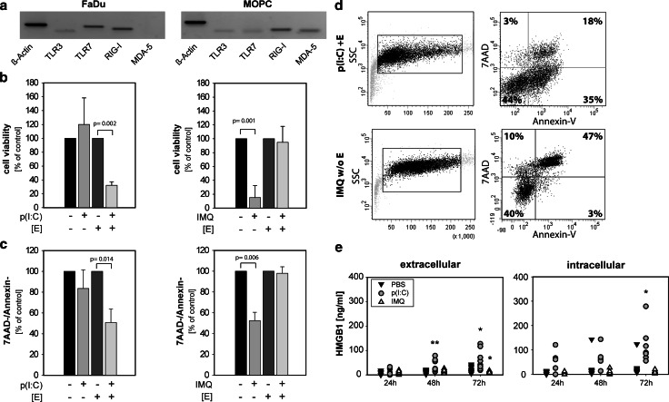
At the beginning of this experimental series, we analyzed gene expression of the receptors for poly(I:C) and imiquimod on the human cell line FaDu and the murine MOPC cells. Basal expression of TLR 3, TLR 7 and the cytoplasmic receptor RIG-I was detectable in both cell lines by PCR. MDA-5 was only detectable in MOPC cells (Fig. 1a). These data established the gene expression of endosomal and cytoplasmic receptors for poly(I:C) and imiquimod. To assess the effects on cell viability and death, the PRR agonists were delivered extracellularly via culture medium (endosomal pathway) or intracellularly via electroporation (cytosolic pathway). A MTT assay showed a significantly lower number of viable cells after cytosolic delivery of poly(I:C) compared to mock transfection (Fig. 1b). To discriminate whether this was a result of impaired cell division or enhanced cell death, annexin-V/7-AAD staining and flow cytometric analysis were conducted (Fig. 1c). After transfection of poly(I:C), 50 % of the cells were positive for annexin-V and 7-AAD suggesting a considerable induction of cell death. Extracellular poly(I:C) had no impact on cell division or cell death. In contrast, extracellular stimulation with imiquimod resulted in a significant reduction in cell viability and induced tumor cell death in 50 % of tumor cells. For imiquimod, cytosolic delivery showed no effect on tumor cell death. Flow cytometric analysis further revealed a high number of annexin-V-positive, 7AAD-negative cells after poly(I:C) treatment and conversely a high number of double-positive cells in imiquimod-treated cells (Fig. 1d). These data suggest that poly(I:C) induced signs of early apoptosis, while imiquimod triggered a more necrotic type of death. To further characterize the mode of cell death, release of HMGB1 as a surrogate marker for immunogenic cell death was analyzed. Only delivery of poly(I:C) resulted in a time-dependent release of HMGB1 by tumor cells (Fig. 1e). Cytosolic delivery showed a strong release which appeared already after 24 h and constantly increased over time, while extracellular delivery resulted in a delayed and lower HMGB1 release. No HMGB1 was detectable in the supernatant of imiquimod-treated FaDu cells, providing initial evidence for distinct modes of cell death induced by the two different synthetic PRR ligands.
Fig. 1.
Poly(I:C) induces cell death in HNSCC via intracellular delivery and imiquimod via extracellular delivery. a Qualitative PCR analysis of unstimulated FaDu and MOPC cells shows basal expression of TLR 3, TLR 7, RIG-I and MDA-5; ß-Actin was used as a loading control. Shown is one representative example of two. b FaDu cells were stimulated with poly(I:C) [p(I:C)] or imiquimod [IMQ] for 72 h. Stimuli were delivered extracellularly (endocytic pathway) or intracellularly (cytosolic pathway, [E] = electroporation). Cell viability was analyzed by MTT assay. Unstimulated tumor cells were set as 100 %. The mean values ± SD from three independent experiments are shown. P value (t test) is indicated. c Cells were stimulated as in (b). Dead cells were identified by PE-conjugated annexin-V and 7-AAD staining and analyzed by flow cytometry. Unstimulated tumor cells were set as 100 %. The mean values ± SD from three to six independent experiments are shown. P value (t test) is indicated. One representative FACS-analysis of stimulated FaDu cells is shown in (d). e FaDu cells were stimulated with PBS (filled square), poly(I:C) (filled circle) or imiquimod (open triangle). Supernatants were collected after 24, 48 and 72 h, and HMGB1 concentration was analyzed by ELISA. Scatter plots depict seven independent experiments. P value (t test) is indicated
Poly(I:C) induces cancer cell death and inhibits tumor growth in vivo
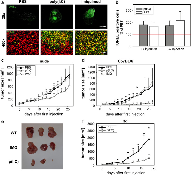
To further investigate the effect of both TLR ligands on tumor cell death and tumor growth in vivo, a xenograft tumor model was established. Human FaDu tumors were s.c. injected, and established tumors were treated by local injections of poly(I:C) and imiquimod. In frozen sections of treated tumors, the number of dead cells was determined by TUNEL staining (Fig. 2a). Increased cell death was clearly detectable after one or three treatments with poly(I:C) and imiquimod compared to control (Fig. 2b). TUNEL staining is visible in different regions throughout the tumor with distinct areas of strong positivity indicative of high cell death.
Fig. 2.
Poly(I:C) induces cell death in vivo and shows therapeutic effects in murine models. a and b 1 × 10⁶ FaDu cells were injected s.c. into nude mice. When tumor size had reached 5 mm in diameter, one or three intratumoral applications of poly(I:C), imiquimod or PBS were administered. Mice were killed 1 day after treatment. In frozen sections of FaDu tumors, cell death was determined using TUNEL kit. a Representative examples of TUNEL staining 1 day after last treatment are depicted. Green TUNEL, Red 7-AAD nuclear staining. Upper panel whole tumor overview for TUNEL staining. Lower panel colocalization of the TUNEL signal with 7AAD nuclear staining. b Quantification of dead cells in FaDu tumors was determined by calculating the area of TUNEL positive cells in relation to the entire tumor area. TUNEL positive cells in control PBS-treated mice were set as 100 %. c 5 × 10⁵ FaDu cells were injected s.c. into nude mice. When tumor size had reached 5 mm in diameter, intratumoral application of poly(I:C) (filled circle), imiquimod (open triangle) or PBS (filled square) was administered five times every other day (day 1, 3, 5, 7, 9) and tumor size was measured. The means + SEM of seven [poly(I:C)], six (imiquimod) and four (PBS) mice obtained from two independent experiments are shown. d 5 × 10⁵ MOPC cells were injected s.c. into immunocompetent C57Bl/6 mice. When tumor size had reached 5 mm in diameter, intratumoral application of poly(I:C) (filled circle), imiquimod (open triangle) or PBS (filled square) was administered five times every other day and tumor size was measured. The mean ± standard deviation of four [poly(I:C)] and three (imiquimod and PBS) mice per group are shown. P value (t test) is indicated. e 5 weeks after the first therapeutic injection, the experiment was terminated and tumors were dissected. Shown are three representative examples for each treatment group. f 5 × 10⁵ MOPC cells were injected s.c. into ‘triple d’ (3d) mice with defective endosomal TLR signaling. Application of poly(I:C) and PBS and tumor measurement was performed as described in (d). The mean ± SEM of two independent experiments are shown with a total number of seven to eight mice per group. P value (t test) is indicated
To analyze whether induction of cell death had an impact on tumor growth, a 9-day treatment using TLR ligands or PBS was performed. Interestingly, tumors treated with poly(I:C) showed reduced tumor growth compared to PBS control (Fig. 2c), while imiquimod did not affect tumor size. To further substantiate these findings, a similar experiment was performed in immunocompetent C57BL/6J mice using a syngeneic murine oropharyngeal cell line. Again, local intratumoral delivery of poly(I:C) significantly reduced tumor growth, while local injections of imiquimod did not change tumor growth kinetics (Fig. 2d, e). Interestingly, in immunocompetent mice the therapeutic effect of poly(I:C) was greater compared to nude mice, suggesting an additional role for T cell-mediated immunity in tumor control.
To distinguish TLR signaling on tumor cells from potential TLR signaling on host cells, MOPC cells were injected into ‘triple d’ (3d) mice with deficient endosomal TLR signaling. Host mice bear a mutation in the Unc93b1 protein, are unable to transport TLRs from the ER to the endosome and thus show an overall defect in endosomal TLR function [27]. Importantly, and similar to wild-type immunocompetent mice, the 9-day treatment with intratumoral poly(I:C) resulted in a significant tumor reduction compared to PBS treatment. These data show that TLR signaling on host cells is not required for the anti-tumor activity of poly(I:C) (Fig. 2f).
Poly(I:C)-treated tumor cells release pro-inflammatory cytokines
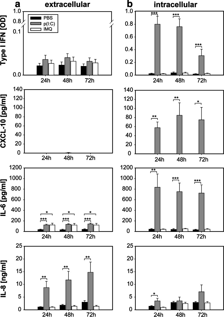
The results obtained so far prompted us to investigate whether, in addition to tumor cell killing itself, the induction of cell death may be associated with additional immunological consequences. To further explore these functional consequences, the immunobiology of TLR stimulated tumor cells was characterized in vitro. First, different cytokines were measured in the supernatants of tumor cells exposed to the TLR ligands. Poly(I:C) delivered via the endosomal pathway induced the release of high amounts of IL-8, but only minor secretion of type I IFN, IL-6 and CXCL-10 (Fig. 3a). In contrast, electroporation with poly(I:C) resulted in a strong and broad cytokine release including type I IFN, IL-6 and CXCL-10 (Fig. 3b). In sharp contrast to poly(I:C), stimulation with imiquimod did not result in the release of IFN, CXCL-10 and IL-8. Only secretion of IL-6 was slightly induced after extracellular delivery (Fig. 3a). The cytokines TNF-α, IL-10, IL-12 and IFN-γ were not detectable in any of the samples (data not shown).
Fig. 3.
Poly(I:C)-treated HNSCC release pro-inflammatory cytokines. FaDu cells were stimulated with poly(I:C) or imiquimod. Stimuli were delivered extracellularly (a) or intracellularly (b). Supernatants were collected after 24, 48 and 72 h, and concentration of human type I IFN, CXCL-10, IL-6 and IL-8 was analyzed. Data are mean ± SEM of seven to ten independent experiments. P value (t test) is indicated
These data suggest that both TLR ligands induce tumor cell death depending on the route of delivery: While induction of cell death by poly(I:C) required intracellular delivery to the cytosol via electroporation, tumor cell killing by imiquimod occurred after extracellular delivery of the compound. Of note, substantial induction of cytokines only occurred in poly(I:C)-stimulated tumor cells and was dependent on delivery via electroporation. For subsequent experiments, which aimed at the further comparative characterization of the immunological consequences of cell death, conditions (detailed in materials and methods) were chosen, which resulted in 50 % tumor cell viability for both compounds. To this end, poly(I:C) was delivered via the cytosolic pathway using electroporation and imiquimod via the endosomal pathway (addition to the culture medium).
Differential regulation of MHC class I on tumor cells
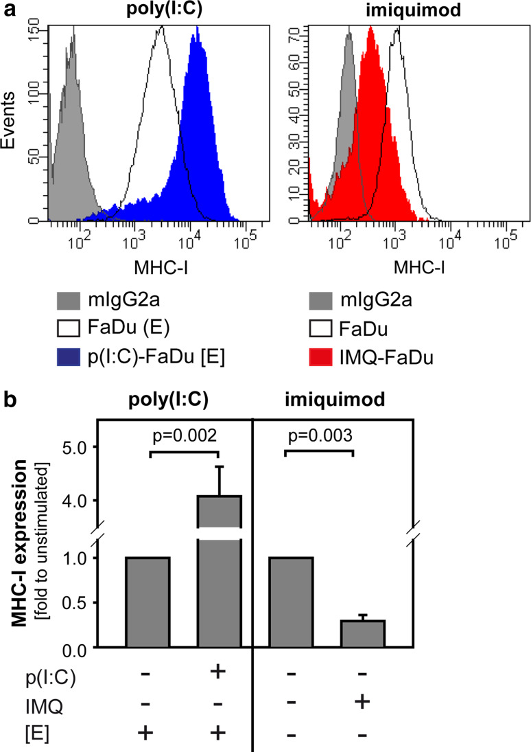
For recognition of tumor cells by natural killer (NK) cells or cytotoxic T cells, surface expression of major histocompatibility complex (MHC) molecules plays a pivotal role [28]. For that reason, MHC-I expression on FaDu cells after treatment with poly(I:C) or imiquimod was analyzed using flow cytometry: poly(I:C) increased MHC-I expression fourfold, while imiquimod reduced MHC-I expression by 75 % (Fig. 4). Co-staining with 7-AAD revealed that MHC-I expression was equally regulated in 7-AAD positive and negative cells (data not shown).
Fig. 4.
Poly(I:C) and imiquimod differentially regulate MHC-I expression on tumor cells. To induce 50 % cell death, FaDu cells were stimulated with cytosolic poly(I:C) (7 h + 72 h) or extracellular imiquimod (48 h + 48 h) as indicated in “Materials and methods” section. FaDu cells were stained with PE-conjugated anti-MHC-I antibody and analyzed by flow cytometry. a One representative experiment is depicted. b The mean ± SD of three to four independent experiments is shown. P value (t test) is indicated. [E] = electroporation
In summary, tumor cell death induced by cytosolic poly(I:C) has immunostimulatory potential via the release of pro-inflammatory molecules (HMGB1, type I IFN, CXCL-10, IL-6, IL-8) and up-regulation of MHC-I molecules on tumor cells. In contrast, imiquimod-treated tumor cells undergo a rather non-inflammatory cell death.
Poly(I:C)-induced tumor cell death modulates the activity of monocytes
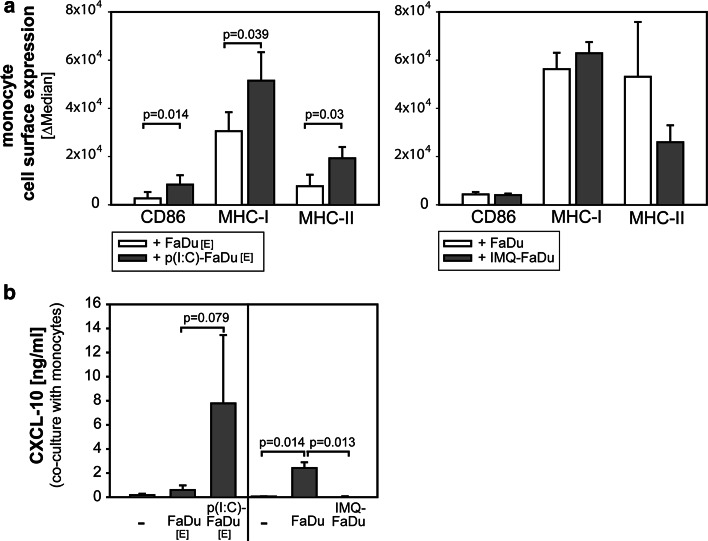
In the next set of experiments, the consequences of PRR-triggered tumor cell death on immune cell activation were analyzed. To this end, FaDu cells were pre-treated with poly(I:C) or imiquimod, washed and co-cultured with monocytes for 48 h. Poly(I:C)-treated FaDu cells shifted the phenotype of co-cultured monocytes toward a state of activation: Expression of the co-stimulatory molecule CD86, antigen-presenting molecules MHC-I and MHC-II on monocytes was significantly increased (Fig. 5a), while imiquimod-treated tumor cells did not alter the activation status of monocytes. In monocyte–tumor co-cultures, small amounts of CXCL-10 were produced (Fig. 5b). Treatment of tumor cells with poly(I:C) and subsequent co-incubation with monocytes strongly enhanced the concentration of CXCL-10 in the co-culture supernatant. In contrast, stimulation with imiquimod did not increase the production of CXCL-10 in tumor–monocyte co-cultures. In addition, also IL-10 was secreted in monocyte–FaDu co-cultures. Treatment with both TLR ligands reduced this IL-10 release (data not shown). TNFα and IL-12 were not detectable in the supernatants (data not shown).
Fig. 5.
Poly(I:C)-treated HNSCC cells activate monocytes. a FaDu cells were stimulated with cytosolic poly(I:C) or extracellular imiquimod as in Fig. 4 to achieve similar rates of cell death. Thereafter, CD14+ monocytes were added to FaDu cells in a 2:1 (monocytes to FaDu cell) ratio and co-culture was continued for another 48 h. Monocytes were harvested, stained with anti-CD86, anti-MHC-I and anti-MHC-II and analyzed by flow cytometry. b Concentration of CXCL-10 in supernatants from (a) was analyzed by ELISA. Cytokine concentration is corrected for the amount of CXCL-10 in FaDu supernatants alone. The mean ± SD three to four independent experiments is shown. P value (t test) is indicated
Poly(I:C) mediates CXCL-10-dependent migration of Th1 cells
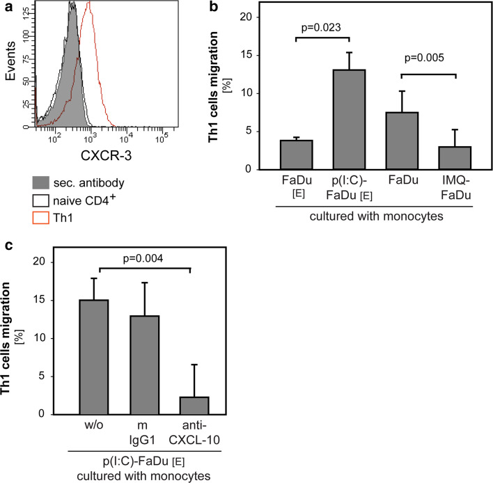
One major function of monocytes is to attract and activate T cells. Therefore, the indirect effects of poly(I:C) and imiquimod-stimulated tumor cells on the capacity of monocytes to recruit Th1 cells were analyzed. For generation of Th1 cells, CD4+ T cells from PBMC were isolated and polarized toward a Th1 phenotype using PHA, IL-2 and IL-12 over 10 days. Successful polarization was verified by CXCR-3 expression (Fig. 6a). In transwell assays, the migration of Th1 cells was significantly augmented, if FaDu cells had been treated with poly(I:C) and significantly decreased after stimulation with imiquimod (Fig. 6b). As supernatants of monocytes co-cultured with poly(I:C)-treated FaDu cells contained high amounts of the chemokine CXCL-10 (Fig. 5b), we tested whether this chemokine was responsible for Th1 recruitment. Adding an anti-CXCL-10 neutralizing antibody to the supernatant abolished Th1 migration. These data suggest that induction of CXCL-10 is a major mechanism involved in poly(I:C)-mediated Th1 cell recruitment (Fig. 6c).
Fig. 6.
Poly(I:C) indirectly induces chemotaxis of Th1 cells. a Polarization of human CD4+ T cells toward Th1 cells was verified by CXCR-3 surface expression using flow cytometry. One representative example is depicted. b Th1 cells were allowed to migrate for 3 h at 37 °C toward supernatants of monocytes co-cultured with FaDu cells. FaDu cells had been treated with poly(I:C) or imiquimod, if indicated. The mean ± SD of three independent experiments are shown. P value (t test) is indicated. c Th1 cells were allowed to migrate for 3 h at 37 °C toward supernatants of monocytes co-cultured with poly(I:C)-treated FaDu cells. Neutralizing anti-CXCL-10 antibody or isotype control antibody was added to the supernatants if indicated. The mean ± SD of three independent experiments are shown. P value (t test) is indicated
Discussion
The activation of certain immune cell subsets most prominently dendritic cells (DCs) by synthetic TLR ligands is well known [7, 29] and has been intensively explored for cancer therapy. More recently, expression of TLRs on tumor cells has been demonstrated yielding contradictory results with respect to tumor biology and progression. While some studies reported on protumorigenic effects [30], other studies provided evidence for the induction of tumor cell death after TLR triggering [17, 18]. Our study addresses a third mechanism, i.e., the modulation of tumor–immune cell interaction affected by PRR triggering on tumor cells.
In our study, we compared poly(I:C) and imiquimod, each applied to tumor cells by two different routes of delivery. We could show a high immunostimulatory potential of poly(I:C)-induced tumor cell death, if poly(I:C) was delivered directly into the cytosol. Poly(I:C), which mimics intracellular dsRNA, significantly enhanced MHC-I expression on tumor cells and stimulated the release of pro-inflammatory factors like HMGB1, type I IFN, IL-6, CXCL-10 and IL-8. This immunogenic cell death fosters monocyte activation and recruitment of Th1 cells in a CXCL-10 dependent manner in vitro. In two murine in vivo models, poly(I:C)-induced death was accompanied by reduced tumor growth. In contrast, local intratumoral injection of imiquimod also resulted in short-term induction of tumor cell death in vivo, but rather down-regulated MHC-I expression on tumor cells and did not trigger broad cytokine release in vitro. Induction of tumor cell death was not accompanied by delayed tumor growth in vivo, which is consistent with the absence of immune activation by this treatment.
Poly(I:C) and related compounds are well recognized as an activator of immune cells and increasing evidence suggests the induction of cell death in cancer cells [31]. However, little data is available regarding the interaction of RNA-treated tumor cells with immune cells. Initial studies suggested that pre-treatment of tumor cells with synthetic TLR ligands resulted in increased activity of immune cells [18, 29]. In agreement with our results, it was also shown that cytosolic poly(I:C) resulted in an enhanced expression of MHC-I molecules on human ovarian cancer and hepatoma cells [18, 32]. The increase in MHC expression counteracts down-regulation of MHC molecules as a major tumor–immune escape mechanism [33]. Additionally, poly(I:C)-treated leukemia cells were better phagocytized by DCs or lysed by NK cells [34]. Kovalcski et al. [29] reported that the treatment of melanoma cells with a combination of endosomal poly(I:C) and IFN-γ triggered DC maturation and Th1 differentiation. Immunogenic cell death of human ovarian cancer cells induced by cytosolic poly(I:C) led to myeloid cell maturation and activation or rather enhanced cytolytic activity of NK cells in gastric adenocarcinoma [18, 19].
It has been suggested that the presence of molecules such as type I IFN, pathogen-associated molecular pattern (PAMPs) or danger-associated molecular pattern (DAMPs) in the context of dying cells marks a type of inflammatory cell death [35, 36]. As such, HMGB1, a DAMP released by tumor cells after cytosolic delivery of poly(I:C) in our study, has been described as a key molecule involved in the mechanisms of immunogenic cells death as defined by Kroemer and Zitvogel. This type of cell death is induced by certain chemotherapeutics such as oxaliplatin and involves pre-apoptotic cell surface exposure of chaperones followed by post-apoptotic release of HMGB1 [37, 38]. Similarly, HMGB1 release after cytosolic delivery of poly(I:C) into HNSCC cells could play a pivotal role in activating monocytes to release pro-inflammatory cytokines such as CXCL-10 and to up-regulate expression of co-stimulatory molecules in our study.
The relative effects of direct tumor cell death versus immune activation may vary between different tumor types and may also be influenced by the tumor microenvironment. In our in vivo xenograft model, cell death was induced via both types of TLR ligands. Interestingly, cell death was already observed immediately after treatment. However, differences in tumor growth become apparent as late as 10–20 days after poly(I:C) treatment. These data are consistent with the hypothesis that at least a part of the anti-tumor activity of poly(I:C) is linked to late and indirect effects rather than direct pro-apoptotic activity [8, 9]. The greater therapeutic activity of poly(I:C) in immunocompetent versus nude mice additionally suggests the involvement of T cells.
It has been previously reported that TLR 3, a major poly(I:C) receptor, is expressed on HNSCC [39]. However, its role in HNSCC biology still remains somewhat controversial. For example, Pries et al. could demonstrate that TLR 3 inhibition in HNSCC cell lines resulted in down-regulation of the oncoprotein c-Myc and decreased cell proliferation [40] suggesting a protumoral effect of TLR 3. Others reported that TLR 3 is able to trigger cell death, when its expression is high or when the level of expression can be up-regulated [31, 41]. However, in none of these studies, a direct comparison of extracellular versus intracellular delivery of poly(I:C) or other TLR ligands has been performed. Interestingly, in our study, which directly compared both routes of delivery, extracellular poly(I:C), which is believed to primarily activate TLR 3 after endosomal uptake, failed to induce cell death in vitro and only induced a limited cytokine response. Thus, our data clearly show that different uptake mechanisms result in different functional responses to poly(I:C) in our HNSCC model. In contrast to poly(I:C), which can act via RIG-I-like receptors, no cytosolic receptor for imiquimod has yet been identified. Consequently, only extracellular delivery of imiquimod, and not electroporation, was able to elicit a cell biological response in our HNSCC model.
In summary, our study provides novel insight into the effects of synthetic TLR ligands on tumor–immune cross talk. We could show, to the best of our knowledge, for the first time that poly(I:C) treatment of HNSCC cells can induce immunomodulatory cell death and monocyte activation, resulting in a CXCL-10 dependent Th1 recruitment. Interestingly, imiquimod a TLR 7 ligand with proven therapeutic activity in the treatment of basal cell carcinoma and lentigo maligna [1, 2] showed no immune modulatory activity associated with its induction of cell death. Modulation of the tumor–immune cell interaction by immunomodulatory tumor cell death should be considered as an additional functional parameter in the design of cancer immunotherapies.
Acknowledgments
We thank Kirsten Bruderek and Petra Altenhoff for their excellent technical assistance, Carsten Kirschning (University Hospital, Essen) for providing the ‘triple d’ mice (3d) and John Lee (Sanford Research, University of South Dakota) for providing the MOPC cell line. Parts of this study were supported by a Grant from the Bartling Stiftung (to Stephan Lang and Sven Brandau).
Abbreviations
- DAMPs
Danger-associated molecular pattern
- DCs
Dendritic cells
- dsRNA
Double-stranded RNA
- FaDu SN
FaDu supernatants
- HMGB1
High-mobility-group-protein B1
- HNSCC
Head and neck squamous cell carcinoma
- IFN
Interferon
- IL
Interleukin
- MDA-5
Melanoma differentiation-associated protein 5
- MHC
Major histocompatibility complex
- MTT
3-(4,5-Dimethylthiazol-2-yl)-2,5-diphenyltetrazolium bromide
- NK
Natural killer
- PAMPs
Pathogen-associated molecular pattern
- PBMC
Peripheral blood mononuclear cells
- PRRs
Pattern recognition receptors
- RIG-I
Retinoic acid-inducible gene I
- ssRNA
Single-stranded RNA
- TLR
Toll-like receptor
Compliance with ethical standards
Conflict of interest
The authors declare that they have no conflict of interest.
References
- 1.Beutner KR, Geisse JK, Helman D, Fox TL, Ginkel A, Owens ML. Therapeutic response of basal cell carcinoma to the immune response modifier imiquimod 5% cream. J Am Acad Dermatol. 1999;41:1002–1007. doi: 10.1016/S0190-9622(99)70261-6. [DOI] [PubMed] [Google Scholar]
- 2.Fleming CJ, Bryden AM, Evans A, Dawe RS, Ibbotson SH. A pilot study of treatment of lentigo maligna with 5% imiquimod cream. Br J Dermatol. 2004;151:485–488. doi: 10.1111/j.1365-2133.2004.05983.x. [DOI] [PubMed] [Google Scholar]
- 3.Korman N, Moy R, Ling M, Matheson R, Smith S, McKane S, Lee JH. Dosing with 5% imiquimod cream 3 times per week for the treatment of actinic keratosis: results of two phase 3, randomized, double-blind, parallel-group, vehicle-controlled trials. Arch Dermatol. 2005;141:467–473. doi: 10.1001/archderm.141.4.467. [DOI] [PubMed] [Google Scholar]
- 4.Vacchelli E, Galluzzi L, Eggermont A, Fridman WH, Galon J, Sautes-Fridman C, Tartour E, Zitvogel L, Kroemer G. Trial watch: FDA-approved toll-like receptor agonists for cancer therapy. Oncoimmunology. 2012;1:894–907. doi: 10.4161/onci.20931. [DOI] [PMC free article] [PubMed] [Google Scholar]
- 5.Amos SM, Pegram HJ, Westwood JA, John LB, Devaud C, Clarke CJ, Restifo NP, Smyth MJ, Darcy PK, Kershaw MH. Adoptive immunotherapy combined with intratumoral TLR agonist delivery eradicates established melanoma in mice. Cancer Immunol Immunother. 2011;60:671–683. doi: 10.1007/s00262-011-0984-8. [DOI] [PMC free article] [PubMed] [Google Scholar]
- 6.Adams S. Toll-like receptor agonists in cancer therapy. Immunotherapy. 2009;1:949–964. doi: 10.2217/imt.09.70. [DOI] [PMC free article] [PubMed] [Google Scholar]
- 7.Hemmi H, Kaisho T, Takeuchi O, Sato S, Sanjo H, Hoshino K, Horiuchi T, Tomizawa H, Takeda K, Akira S. Small anti-viral compounds activate immune cells via the TLR7 MyD88-dependent signaling pathway. Nat Immunol. 2002;3:196–200. doi: 10.1038/ni758. [DOI] [PubMed] [Google Scholar]
- 8.Matsumoto M, Funami K, Tanabe M, Oshiumi H, Shingai M, Seto Y, Yamamoto A, Seya T. Subcellular localization of toll-like receptor 3 in human dendritic cells. J Immunol. 2003;171:3154–3162. doi: 10.4049/jimmunol.171.6.3154. [DOI] [PubMed] [Google Scholar]
- 9.Shime H, Matsumoto M, Oshiumi H, Tanaka S, Nakane A, Iwakura Y, Tahara H, Inoue N, Seya T. Toll-like receptor 3 signaling converts tumor-supporting myeloid cells to tumoricidal effectors. Proc Natl Acad Sci USA. 2012;109:2066–2071. doi: 10.1073/pnas.1113099109. [DOI] [PMC free article] [PubMed] [Google Scholar]
- 10.Alexopoulou L, Holt AC, Medzhitov R, Flavell RA. Recognition of double-stranded RNA and activation of NF-kappaB by toll-like receptor 3. Nature. 2001;413:732–738. doi: 10.1038/35099560. [DOI] [PubMed] [Google Scholar]
- 11.Gitlin L, Barchet W, Gilfillan S, Cella M, Beutler B, Flavell RA, Diamond MS, Colonna M. Essential role of mda-5 in type I IFN responses to polyriboinosinic:polyribocytidylic acid and encephalomyocarditis picornavirus. Proc Natl Acad Sci USA. 2006;103:8459–8464. doi: 10.1073/pnas.0603082103. [DOI] [PMC free article] [PubMed] [Google Scholar]
- 12.Yoneyama M, Kikuchi M, Natsukawa T, Shinobu N, Imaizumi T, Miyagishi M, Taira K, Akira S, Fujita T. The RNA helicase RIG-I has an essential function in double-stranded RNA-induced innate antiviral responses. Nat Immunol. 2004;5:730–737. doi: 10.1038/ni1087. [DOI] [PubMed] [Google Scholar]
- 13.Heil F, Hemmi H, Hochrein H, Ampenberger F, Kirschning C, Akira S, Lipford G, Wagner H, Bauer S. Species-specific recognition of single-stranded RNA via toll-like receptor 7 and 8. Science. 2004;303:1526–1529. doi: 10.1126/science.1093620. [DOI] [PubMed] [Google Scholar]
- 14.Goutagny N, Estornes Y, Hasan U, Lebecque S, Caux C. Targeting pattern recognition receptors in cancer immunotherapy. Target Oncol. 2012;7:29–54. doi: 10.1007/s11523-012-0213-1. [DOI] [PubMed] [Google Scholar]
- 15.Pinto A, Morello S, Sorrentino R. Lung cancer and toll-like receptors. Cancer Immunol Immunother. 2011;60:1211–1220. doi: 10.1007/s00262-011-1057-8. [DOI] [PMC free article] [PubMed] [Google Scholar]
- 16.Rydberg C, Mansson A, Uddman R, Riesbeck K, Cardell LO. Toll-like receptor agonists induce inflammation and cell death in a model of head and neck squamous cell carcinomas. Immunology. 2009;128:e600–e611. doi: 10.1111/j.1365-2567.2008.03041.x. [DOI] [PMC free article] [PubMed] [Google Scholar]
- 17.Schön M, Bong AB, Drewniok C, Herz J, Geilen CC, Reifenberger J, Benninghoff B, Slade HB, Gollnick H, Schön MP. Tumor-selective induction of apoptosis and the small-molecule immune response modifier imiquimod. J Natl Cancer Inst. 2003;95:1138–1149. doi: 10.1093/jnci/djg016. [DOI] [PubMed] [Google Scholar]
- 18.Kübler K, tho Pesch C, Gehrke N, Riemann S, Dassler J, Coch C, Landsberg J, Wimmenauer V, Pölcher M, Rudlowski C, Tüting T, Kuhn W, Hartmann G, Barchet W. Immunogenic cell death of human ovarian cancer cells induced by cytosolic poly(I:C) leads to myeloid cell maturation and activates NK cells. Eur J Immunol. 2011;41:3028–3039. doi: 10.1002/eji.201141555. [DOI] [PubMed] [Google Scholar]
- 19.Qu J, Hou Z, Han Q, Jiang W, Zhang C, Tian Z, Zhang J. Intracellular poly(I:C) initiated gastric adenocarcinoma cell apoptosis and subsequently ameliorated NK cell functions. J Interferon Cytokine Res. 2014;34:52–59. doi: 10.1089/jir.2012.0118. [DOI] [PMC free article] [PubMed] [Google Scholar]
- 20.Besch R, Poeck H, Hohenauer T, Senft D, Hacker G, Berking C, Hornung V, Endres S, Ruzicka T, Rothenfusser S, Hartmann G. Proapoptotic signaling induced by RIG-I and MDA-5 results in type I interferon-independent apoptosis in human melanoma cells. J Clin Invest. 2009;119:2399–2411. doi: 10.1172/JCI37155. [DOI] [PMC free article] [PubMed] [Google Scholar]
- 21.Schön MP, Wienrich BG, Drewniok C, Bong AB, Eberle J, Geilen CC, Gollnick H, Schön M. Death receptor-independent apoptosis in malignant melanoma induced by the small-molecule immune response modifier imiquimod. J Invest Dermatol. 2004;122:1266–1276. doi: 10.1111/j.0022-202X.2004.22528.x. [DOI] [PubMed] [Google Scholar]
- 22.Smith EB, Schwartz M, Kawamoto H, You X, Hwang D, Liu H, Scherr DS. Antitumor effects of imidazoquinolines in urothelial cell carcinoma of the bladder. J Urol. 2007;177:2347–2351. doi: 10.1016/j.juro.2007.01.112. [DOI] [PubMed] [Google Scholar]
- 23.Fukata M, Chen A, Vamadevan AS, Cohen J, Breglio K, Krishnareddy S, Hsu D, Xu R, Harpaz N, Dannenberg AJ, Subbaramaiah K, Cooper HS, Itzkowitz SH, Abreu MT. Toll-like receptor-4 promotes the development of colitis-associated colorectal tumors. Gastroenterology. 2007;133:1869–1881. doi: 10.1053/j.gastro.2007.09.008. [DOI] [PMC free article] [PubMed] [Google Scholar]
- 24.Kroemer G, Galluzzi L, Kepp O, Zitvogel L. Immunogenic cell death in cancer therapy. Annu Rev Immunol. 2013;31:51–72. doi: 10.1146/annurev-immunol-032712-100008. [DOI] [PubMed] [Google Scholar]
- 25.Yang H, Zhou H, Feng P, Zhou X, Wen H, Xie X, Shen H, Zhu X. Reduced expression of toll-like receptor 4 inhibits human breast cancer cells proliferation and inflammatory cytokines secretion. J Exp Clin Cancer Res. 2010;29:92. doi: 10.1186/1756-9966-29-92. [DOI] [PMC free article] [PubMed] [Google Scholar]
- 26.Williams R, Lee DW, Elzey BD, Anderson ME, Hostager BS, Lee JH. Preclinical models of HPV + and HPV − HNSCC in mice: an immune clearance of HPV + HNSCC. Head Neck. 2009;31:911–918. doi: 10.1002/hed.21040. [DOI] [PubMed] [Google Scholar]
- 27.Tabeta K, Hoebe K, Janssen EM, Du X, Georgel P, Crozat K, Mudd S, Mann N, Sovath S, Goode J, Shamel L, Herskovits AA, Portnoy DA, Cooke M, Tarantino LM, Wiltshire T, Steinberg BE, Grinstein S, Beutler B. The Unc93b1 mutation 3d disrupts exogenous antigen presentation and signaling via toll-like receptors 3, 7 and 9. Nat Immunol. 2006;7:156–164. doi: 10.1038/ni1297. [DOI] [PubMed] [Google Scholar]
- 28.Levitsky HI, Lazenby A, Hayashi RJ, Pardoll DM. In vivo priming of two distinct antitumor effector populations: the role of MHC class I expression. J Exp Med. 1994;179:1215–1224. doi: 10.1084/jem.179.4.1215. [DOI] [PMC free article] [PubMed] [Google Scholar]
- 29.Kovalcsik E, Lowe K, Fischer M, Dalgleish A, Bodman-Smith MD. Poly(I:C)-induced tumour cell death leads to DC maturation and Th1 activation. Cancer Immunol Immunother. 2011;60:1609–1624. doi: 10.1007/s00262-011-1058-7. [DOI] [PMC free article] [PubMed] [Google Scholar]
- 30.Basith S, Manavalan B, Yoo TH, Kim SG, Choi S. Roles of toll-like receptors in cancer: a double-edged sword for defense and offense. Arch Pharm Res. 2012;35:1297–1316. doi: 10.1007/s12272-012-0802-7. [DOI] [PubMed] [Google Scholar]
- 31.Cheng YS, Xu F. Anticancer function of polyinosinic–polycytidylic acid. Cancer Biol Ther. 2010;10:1219–1223. doi: 10.4161/cbt.10.12.13450. [DOI] [PubMed] [Google Scholar]
- 32.Peng S, Geng J, Sun R, Tian Z, Wei H. Polyinosinic–polycytidylic acid liposome induces human hepatoma cells apoptosis which correlates to the up-regulation of RIG-I like receptors. Cancer Sci. 2009;100:529–536. doi: 10.1111/j.1349-7006.2008.01062.x. [DOI] [PMC free article] [PubMed] [Google Scholar]
- 33.Tong CC, Kao J, Sikora AG. Recognizing and reversing the immunosuppressive tumor microenvironment of head and neck cancer. Immunol Res. 2012;54:266–274. doi: 10.1007/s12026-012-8306-6. [DOI] [PubMed] [Google Scholar]
- 34.Lion E, Anguille S, Berneman ZN, Smits EL, Van Tendeloo VF. Poly(I:C) enhances the susceptibility of leukemic cells to NK cell cytotoxicity and phagocytosis by DC. PLoS ONE. 2011;6:e20952. doi: 10.1371/journal.pone.0020952. [DOI] [PMC free article] [PubMed] [Google Scholar]
- 35.Kono H, Rock KL. How dying cells alert the immune system to danger. Nat Rev Immunol. 2008;8:279–289. doi: 10.1038/nri2215. [DOI] [PMC free article] [PubMed] [Google Scholar]
- 36.Rock KL, Kono H. The inflammatory response to cell death. Annu Rev Pathol. 2008;3:99–126. doi: 10.1146/annurev.pathmechdis.3.121806.151456. [DOI] [PMC free article] [PubMed] [Google Scholar]
- 37.Tesniere A, Panaretakis T, Kepp O, Apetoh L, Ghiringhelli F, Zitvogel L, Kroemer G. Molecular characteristics of immunogenic cancer cell death. Cell Death Differ. 2008;15:3–12. doi: 10.1038/sj.cdd.4402269. [DOI] [PubMed] [Google Scholar]
- 38.Tesniere A, Schlemmer F, Boige V, Kepp O, Martins I, Ghiringhelli F, Aymeric L, Michaud M, Apetoh L, Barault L, Mendiboure J, Pignon JP, Jooste V, van Endert P, Ducreux M, Zitvogel L, Piard F, Kroemer G. Immunogenic death of colon cancer cells treated with oxaliplatin. Oncogene. 2010;29:482–491. doi: 10.1038/onc.2009.356. [DOI] [PubMed] [Google Scholar]
- 39.Pries R, Hogrefe L, Xie L, Frenzel H, Brocks C, Ditz C, Wollenberg B. Induction of c-Myc-dependent cell proliferation through toll-like receptor 3 in head and neck cancer. Int J Mol Med. 2008;21:209–215. [PubMed] [Google Scholar]
- 40.Pries R, Wulff S, Wollenberg B. Toll-like receptor modulation in head and neck cancer. Crit Rev Immunol. 2008;28:201–213. doi: 10.1615/CritRevImmunol.v28.i3.20. [DOI] [PubMed] [Google Scholar]
- 41.Luo Q, Hu S, Yan M, Sun Z, Chen W, Chen F. Activation of toll-like receptor 3 induces apoptosis of oral squamous carcinoma cells in vitro and in vivo. Int J Biochem Cell Biol. 2012;44:1266–1275. doi: 10.1016/j.biocel.2012.04.025. [DOI] [PubMed] [Google Scholar]


