Abstract
Size and posttranslational modifications are obstacles in the recombinant expression of high-molecular-weight melanoma-associated antigen (HMW-MAA). Creating a tumor antigen mimic via the phage display technology may be a means to overcome this problem for vaccine design. In this study, we aimed to generate an immunogenic epitope mimic of HMW-MAA. Therefore we screened a linear 9mer phage display peptide library, using the anti-HMW-MAA monoclonal antibody (mAb) 225.28S. This antibody mediates antibody-dependent cellular cytotoxicity (ADCC) and has already been used for anti-idiotype therapy trials. Fifteen peptides were selected by mAb 225.28S in the biopanning procedure. They share a consensus sequence, but show only partial homology to the amino acid sequence of the HMW-MAA core protein, indicating mimicry with a conformational epitope. One mimotope was chosen to be fused to albumin binding protein (ABP) as an immunogenic carrier. Immunoassays with 225.28S indicated that the mimotope fusion protein was folded correctly. Subsequently, the fusion protein was tested for immunogenicity in BALB/c mice. The induced anti-mimotope antibodies recognized HMW-MAA of 518A2 human melanoma cells, whereas sera of mice immunized with the carrier ABP alone showed no reactivity. These anti-mimotope antibodies were capable of inducing specific lysis of 518A2 melanoma cells in ADCC assays with murine effector cells. In conclusion, the presented data indicate that mimotopes fused to an immunogenic carrier are suitable tools to elicit epitope-specific anti-melanoma immune responses.
Keywords: HMW-MAA, 225.28S, Mimotope, Phage display, Cancer vaccine
Introduction
The incidence of malignant melanoma has increased in recent years more than that of any other cancer. While thin melanomas are usually curable by excision surgery, no therapy is available for metastatic disease, because the tumor is resistant to standard radiotherapy and chemotherapy. As melanomas have been observed to be susceptible to immunological attack, immunotherapy is a possible alternative. High-molecular-weight melanoma-associated antigen (HMW-MAA) is considered to be an attractive target in this respect because of its high frequency of expression in patients with melanoma, as well as its restricted tissue distribution: it is expressed by over 90% of melanomas and nevi and by a low percentage of squamous and basal cell carcinomas, but is not detectable in normal tissues of ectodermal, mesodermal and endodermal origin [1]. The expression of HMW-MAA does neither depend on the synthesis of melanin nor on the primary, metastatic or recurrent nature of the lesion. Antibodies elicited against it might inhibit the functional properties of HMW-MAA and thus reduce or suppress the metastatic potential of HMW-MAA-bearing melanoma cells.
The expression of HMW-MAA by activated pericytes [2] adds another feature to this potential target: the effect of anti-HMW-MAA immunity on melanoma lesions may be mediated not only by a direct interaction with melanoma cells, but also by disturbance of the blood supply via anti-angiogenic mechanisms.
The monoclonal antibody (mAb) 225.28S is one of various monoclonal antibodies elicited against this immunodominant melanoma antigen and has been used in immunoscintigraphy in a large number of patients [3, 4]. We selected it for biopanning because prolongation of survival times was observed in patients undergoing multiple applications of the antibody as a diagnostic tool [4], suggesting that an immune response against the epitope recognized by 225.28S might be of clinical value. Indeed, it was demonstrated that the development of anti-HMW-MAA antibodies in patients immunized with anti-idiotypic antibodies derived from 225.28S was associated with a statistically significant prolongation of survival and with regression of metastatic lesions [5].
In the present study, we generated epitope mimics, so-called mimotopes, of the epitope recognized by mAb 225.28S on HMW-MAA. As we intended to apply the peptides for epitope-specific vaccination, we fused them to albumin binding protein (ABP), an immunogenic carrier, and analyzed antigenicity, immunogenicity, and anti-melanoma effects of this vaccine construct in BALB/c mice.
Materials and methods
Cell lines and lysate preparations
The human melanoma cell line 518A2 was grown in Dulbecco’s MEM (Gibco BRL, Inchinnan, UK) supplemented with 10% fetal calf serum, 1% glutamine, 1% penicillin/streptomycin, and 50 μg/ml gentamicin sulfate. The human mammary carcinoma cell line SK-BR-3 was used as a control. It was grown in McCoy’s medium (Gibco BRL), supplemented as described above. Cells were regularly harvested by incubation with 0.1% Na-EDTA in the respective medium at 37°C for 15 min and lysates prepared as outlined by Klinger et al. [6].
Monoclonal antibodies
The mAb 225.28S was raised against M21 melanoma cells [7] and is of isotype IgG2a [8]. A mouse IgG2a negative control mAb, X 0943, directed against Aspergillus niger glucose oxidase, was purchased from DAKO (Glostrup, Denmark).
Phage libraries and biopanning
Monoclonal antibody ligands were selected from a random phage library, pVIII9aa [9], expressing linear nanopeptides fused to pVIII of the filamentous phage fd. The library was kindly provided by IRBM (Istituto di Ricerche di Biologia Molecolare P. Angeletti SPA, Rome, Italy). Three rounds of biopanning were performed as outlined by Parmley and Smith [10]. Phage particles were amplified in Escherichia coli XL-1 Blue, precipitated from the bacterial culture supernatant with polyethylene glycol, and either immediately used for the next round of biopanning or stored at −20°C. For titer determination (colony-forming units, cfu), aliquots of the eluate or the amplificate were used to infect E. coli XL-1 Blue, and these plated in serial dilutions on LB agar plates containing carbenicillin (100 μg/ml).
Colony screening assay and DNA sequencing
After each round of biopanning, colony screenings for selection of specific phage clones were done according to Barbas and Lerner [11]. Immunoscreenings were performed with 225.28S and isotype control (2.5 μg/ml in PBS/casein). Bound antibody was detected by 125I-sheep anti-mouse Ig. Positive clones were amplified as described above. DNA sequencing was performed by the Sanger dideoxy method [12] using a Thermo Sequenase cycle sequencing kit (Amersham).
Phage sequence analysis and peptide alignments
Sequence comparisons were done by the AlignX module of the Vector NTI Suite software (InforMax Inc., Frederick, MD, USA), which uses a modified Clustal W algorithm [13] to create multiple sequence alignments.
Expression of fusion proteins in E. coli
For production of recombinant fusion proteins the vector pSB 411 (kindly provided by Prof. M. Suter, Dept. of Virology, University of Zurich, Switzerland) was used, as described by Baumann et al. [14]. The gene coding for the desired peptide is directionally inserted via two non-compatible SfiI-sites and fused to the ABP-6×His module, coding for ABP and a His-tag (6×His). All features besides the ABP-6×His module were derived from pAK 400 [15]. DNA sequences of clone K11, with insert RPSSNFNPL, flanked by SfiI restriction sites, were purchased as oligo-nucleotides (MWG-Biotech GmbH). The expression constructs for the fusion proteins were transformed into competent E. coli XL-1 cells and checked by DNA analysis.
E. coli XL-1 Blue cells were grown in Super-Broth medium at 37°C and induced with 0.1 M isopropyl-β-D-thiogalactopyranoside (IPTG) at an optical density of 0.5 at 600 nm. Expression took place overnight. Cells were harvested, resuspended in sonication buffer (50 mM NaH2PO4, pH 8, 300 mM NaCl) and lysed by sonication. After centrifugation at 10,000g for 30 min, the fusion proteins were enriched from supernatants using Ni-NTA agarose according to the standard purification protocol for native proteins as described by the manufacturer (Qiagen, Hilden, Germany).
SDS-PAGE and immunoblotting
The SDS-PAGE (6% homogenous gels for cell extracts, 12% homogenous gels for fusion proteins) was performed by the method of Laemmli [16] under non-reducing conditions. The electrophoretical transfer of proteins from gel to nitrocellulose sheets was performed according to the method of Towbin et al. [17]. After blocking with PBS containing 0.5% powdered milk, 0.05% NaN3 and 0.5% Tween 20, immunodetection of blotted proteins was done with antibodies (10 μg/ml) or pooled sera (1:100), respectively, diluted in blocking buffer. For titer determination experiments, sera were added in serial dilutions. Bound mAb was detected by 125I-sheep anti-mouse Ig (Amersham, Little Chalfont, IL, USA), 0.1 μCi/ml buffer. Blots were then washed, dried and exposed to Kodak Biomax-MS film at −70°C.
Immunization of BALB/c mice
Five-week-old female BALB/c mice (Institute for Laboratory Animal Science and Genetics, University of Vienna) were immunized intraperitoneally (i.p.) with fusion proteins (10 μg), using Al(OH)3 (Serva, Heidelberg, Germany) as an adjuvant. Two groups of mice (n=5) were immunized on days 0, 14, 28 and 126 either (1) with the K11-ABP fusion protein, or (2) with ABP alone. Blood samples from the tail vein were taken on day -1 (preimmune serum), and days 7, 21, 35 and 133 after immunization. Mice were treated according to European Union Rules of Animal Care, with permission no. 66009/147/PR-4-2001 from the Austrian Ministry of Science.
Fluorescence microscopy
518A2 cells were plated at low density on four-well Lab-Tek tissue culture chamber slides (Miles Laboratories Inc., Naperville, IL, USA), and grown overnight until half-confluent. They were then cooled to 4°C, washed with ice-cold PBS, fixed with 4% paraformaldehyde in PBS for 30 min, quenched with 50 mM NH4Cl in PBS and blocked with 1% BSA/PBS. Membranes were stained with a rabbit anti-placental alkaline phosphatase antibody (Signet Laboratories, Inc., Dedham, MA, USA), followed by Alexa 568-conjugated goat anti-rabbit IgG (Molecular Probes Inc., Eugene, OR, USA). HMW-MAA was detected with mAb 225.28S or with third immune sera (pooled from all mice in the group), followed by fluorescein isothiocyanate (FITC)-conjugated goat anti-mouse IgG (Caltag Laboratories, Burlingame, CA, USA). Nuclei were stained with 0.1 μg/ml Hoechst dye (Sigma) in PBS for 10 min. Cells were mounted in Mowiol mounting medium and viewed with a Zeiss Axioplan 2 (Carl Zeiss, Jena, Germany).
Antibody-dependent cellular cytotoxicity assay
A sample of 2×10⁵ 518A2 target cells was washed twice and resuspended in 2 ml medium. Ten microliters of Delfia BATDA reagent (bis (acetoxymethyl) 2,2′:6′,2′′-terpyridine-6,6′′-dicarboxylic acid, Wallac Oy) was added and the suspension incubated at 37°C for 20 min. Labeling was stopped by centrifugation and washing in PBS; subsequently, cells were resuspended in 2 ml medium. Murine effector cells were obtained by mashing the spleen of a BALB/c mouse and lysing the erythrocytes with ammonium chloride. Target cells were aliquoted into a 96-well tissue culture plate in a concentration of 10,000 cells/well. Ten micrograms of mAb or 10 μl of pooled murine sera, containing ~2 μg HMW-MAA-specific antibodies (as estimated from immonoblot titer determination experiments), was added to wells for antibody-dependent cellular cytotoxicity (ADCC) reactions and incubated at 37°C for 45 min. Effectors were added at a ratio of 50:1 for ADCC and NK activity assays. After a 2-h incubation at 37°C, 5% CO2, plates were centrifuged at 1,600 rpm for 5 min. A 20-μl sample of supernatant from each well was transferred to the wells of flat-bottomed microtiter plates containing 180 μl Europium solution (Wallac cytotoxicity test, Wallac Oy) and mixed at RT for 15 min. Fluorescence was measured with a time-resolved 1234 Delfia fluorometer (Wallac Oy). Controls included cells incubated with culture medium or mAb alone. Spontaneous release was defined as the amount released by cells incubated with medium alone. Maximum release was obtained by freezing and quickly thawing an aliquot of labeled target cells. The percentage of specific cytolysis was calculated according to the formula:
 |
Results
Selection and definition of mimotopes
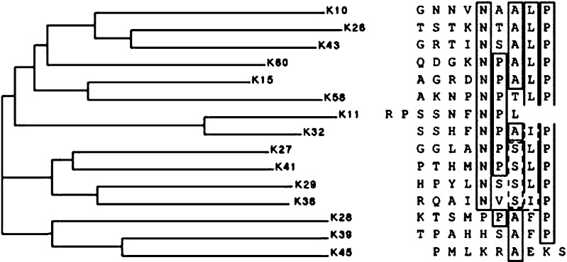
We performed three rounds of biopanning with 225.28S, screening a linear nanopeptide pVIII phage display library. Fifteen phage clones were specifically recognized in colony screenings, with clones K11 and K43 yielding the relatively highest reactivities. Most of the deduced amino acid sequences of the 15 different inserts shared a consensus motif between them, namely NPALP (Fig. 1).
Fig. 1.
Alignment and sequence of peptides selected by mAb 225.28S. The peptide sequences were subjected to the multiple alignment algorithm of the Vector NTI Suite, which produces a phylogenetic tree indicating the degree of similarities of the peptides to each other. Residues in boxes were found at a specific position four or more times
Characterization of mimotope fusion proteins
The peptide insert displayed by phage clone K11, RPSSNFNPL, showing the highest immunoreactivity with 225.28S, was chosen to be fused to ABP. The resulting fusion protein was specifically recognized by 225.28S at 25 kDa in immunoblot, ascertaining correct folding and display of the mimotope (Fig. 2, panel B). Loading of equal amounts of fusion protein to the gel was confirmed by an anti-His-tag mAb, detecting both K11-ABP and ABP (panel A, lane 1 and 2, respectively), whereas isotype and buffer controls were clear (panels C and D).
Fig. 2.

Specific recognition of the K11-ABP fusion protein by mAb 225.28S in immunoblot. K11-ABP (lane 1) and ABP (lane 2) fusion proteins were subjected to 12% SDS-PAGE and blotted onto a nitrocellulose membrane. Strips were incubated with A an anti-His tag antibody, B mAb 225.28S, C isotype control, and D buffer. Bound antibodies were detected by a radioactively (125I) labeled sheep anti-mouse Ig antibody
Immune response induced by peptide region K11
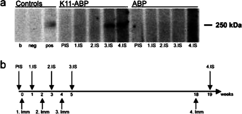
The immunogenicity of the mimotope-ABP fusion protein, as well as of ABP alone, was evaluated in a vaccination experiment (see immunization schedule in Fig. 3b), using BALB/c mice (n=5 per group). The antibodies induced were predominantly of the IgG1 subclass (data not shown). In both groups, antibodies against ABP with titers of 1:200,000 (as determined by immunoblot) were induced, indicating successful immunizations. Anti-HMW-MAA antibodies were detectable in the K11-ABP group after the third immunization. It was possible to boost this immune response after a prolonged interval, i.e. 14 weeks (Fig. 3b), with a titer of 1:10,000. This finding is indicative of induction of a memory response. The antibodies recognized the original antigen HMW-MAA at 250 kDa in an immunoblot of a cell lysate of 518A2 melanoma cells (Fig. 3a). In contrast, sera of mice of the ABP control group did not react with the molecule, indicating that the mimotope-induced immune response was specific.
Fig. 3.
Sera of mice immunized with K11-ABP recognize the HMW-MAA band at 250 kDa in an 518A2 cell lysate immunoblot. a Immunoblot. 518A2 membrane preparations were separated by 6% SDS-PAGE and proteins were transferred to a nitrocellulose membrane. Strips were incubated with controls or pooled sera as indicated (b buffer control, neg isotype control, pos 225.28S, PIS preimmune serum, IS immune serum). b Immunization schedule. BALB/c mice (n=5 per group) were immunized ip with 10 μg of the respective fusion protein at the indicated time points (Imm immunization). Blood samples were taken 1 week after each immunization
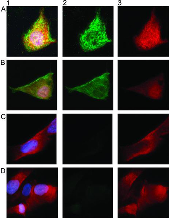
To demonstrate that the induced antibodies also recognized HMW-MAA on the cell surface, we performed immunofluorescence staining. MAb 225.28S was employed as a positive control (Fig. 4, panel A). Sera from mice of immunized with K11-ABP showed intense staining of 518A2 melanoma cells (panel B), whereas sera from mice immunized with ABP alone (panel C) and pre-immune sera (panel D) only caused background staining. For comparability, membranes and nuclei were stained in all panels in the same way.
Fig. 4.
Sera from mimotope-immunized mice recognize HMW-MAA in immunofluorescence staining of 518A2 melanoma cells. Panel A: 225.28S positive control. Panel B: Serum IgG from mice immunized with K11-ABP specifically recognize cell surface HMW-MAA. Serum IgG from mice immunized with ABP alone (panel C) and pre-immune sera (panel D) only show background staining. Column 1: A–D antibody staining (green), membrane staining (red) and nuclear staining (blue). Column 2: FITC channel. Column 3: Texas red channel
Functional analysis of the induced humoral immune response
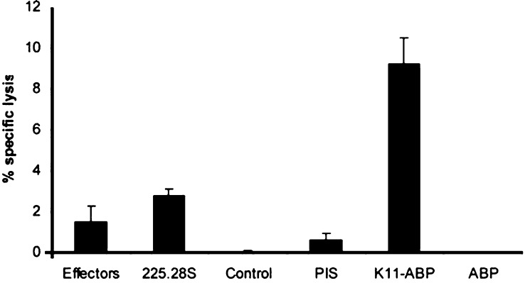
The functional properties of the induced antibodies were tested in ADCC assays. We employed murine effector cells, as murine IgG1, the main antibody subclass induced in this study, does not interact with human effector cells [18]. In the ADCC assay, serum antibodies of the K11-ABP group were capable of inducing a 9% specific lysis of the human melanoma target cells (518A2), whereas the direct lytic capacity of the original antibody 225.28S was much lower (Fig. 5). An ADCC assay using the control cell line SK-BR-3 only showed NK cell baseline activity (data not shown).
Fig. 5.
Sera of mice immunized with K11-ABP induce specific lysis of 518A2 melanoma cells in an ADCC assay. 518A2 target cells were labeled with a substance fluorescent at contact with europium. Following incubation with 10 μg mAb/10 μl pooled sera (equaling ~2 μg HMW-MAA-specific antibodies), murine effector cells were added at an effector-to-target ratio of 50:1. Aliquots of supernatants were mixed with europium solution, and fluorescence was measured in a time-resolved fluorometer. Percentage of specific lysis was calculated relative to maximum release, corrected by spontaneous release values. Column heights represent mean ± SD. Effectors: lytic activity of effector cells alone. Other columns: ADCC mediated by mAb/pooled sera as indicated on the x-axis
Discussion
The possibility of mimicking cancer antigens has opened new perspectives: mimics of cancer antigens, as opposed to vaccines directly derived from cancer cells, bear the same surface characteristics, but are not identical to the original antigen [19]. Therefore, they may have the potency of activating lymphocyte populations recognizing malignant cells that have escaped self-tolerance by virtue of their low affinity to the original antigen. These normally inactive lymphocytes might be turned into efficient effector cells by vaccination.
The phage display technique makes it possible to retrieve appropriate mimicking structures. With the use of phage displayed random peptide libraries, up to 109 different potentially mimicking structures can be screened simultaneously for the optimal peptide candidates. Additionally, the desired epitope can be rationally selected by the choice of the screening antibody. This is an important feature, as antibodies directed to different epitopes on the same target molecule can have opposed effects on tumor cell growth [20]. Obviously, for the design of cancer vaccines, growth-inhibitory or cell-destructive effects are desired. Furthermore, peptide mimics isolated from phage display libraries are expected to induce both humoral and cellular anti-HMW-MAA immunity [21].
In the present study, we selected the anti-HMW-MAA mAb 225.28S for biopanning. In addition to its favorable participation in ADCC in vitro [7], the antibody prolonged survival in patients undergoing multiple applications as a diagnostic measure [4]. Possibly, the observed effects are due to interference with the role of HMW-MAA in cell to cell contact or cell spreading [22, 23], in addition to antibody-mediated immunological mechanisms. Also patients immunized with anti-idiotypic antibodies derived from 225.28S, who developed an anti-HMW-MAA immune response, experienced a statistically significant prolongation of survival and regression of metastatic lesions [5]. These data led us to the hypothesis that the targeted induction of an immune response against the 225.28S epitope could be therapeutically relevant.
The selection of 225.28S binding phage clones was achieved by three rounds of biopanning. The 15 obtained sequences shared a consensus sequence—NPALP—between them, and showed homology to four regions in the primary sequence of HMW-MAA. As B cell/antibody epitopes are known to be largely conformational, we did not expect exact mapping of the 225.28S binding site. As region 4 maps to the intracellular region of the molecule, we speculate that the putative epitope of mAb 225.28S may be composed of polypeptide chain regions amino acid (aa) 1923–1933 and aa 1995–2000 in the third structural domain of the extracellular portion of the molecule. Among the identified phage clones, clone K11 was singled out to be fused to ABP, due to its good homology with HMW-MAA and highest immunoreactivity with 225.28S.
Theoretically, phage particles themselves would be ideal immunogens, as they carry sufficient CD4+ T cell epitopes to elicit immune responses [24]. We recently demonstrated that they recruit bystander T cells for the formation of a mimotope-specific humoral immune response [25]. Additionally, when displayed on phage, the mimotope is presented in exactly the same way as it was recognized by the selecting mAb [26]. However, while phage immunizations may hold promise for animal therapies, phage immunizations for humans are not feasible. Especially with applications via the oral route, there is a risk of bacteriophages infecting F pilus-positive E. coli residing in the intestinal flora. Furthermore, the transfer of antibiotic resistance with phage particles might harm the patient.
On the other hand, there are several advantages that make recombinant peptide mimics an attractive option for vaccination. They are easily defined and can be reliably manufactured in large scale. As peptides alone are too small to elicit an immune response [27], they have to be coupled or fused to an immunogenic carrier protein for successful vaccination. In this study, the streptococcal ABP was chosen because of its favorable characteristics: ABP is a bacterial protein and consequently highly immunogenic [28, 29]. Moreover, the selected expression vector possesses a domain that ensures correct mimotope conformation through hydrophobic tagging [14].
In a BALB/c immunization model, we were indeed able to show that the mimotope fusion protein is capable of eliciting anti-HMW-MAA antibodies. This was shown by sera from the mimotope-immunized mice recognizing HMW-MAA both in immunoblot and on the melanoma cell surface (immunofluorescence). Analyzed for their functional properties, the induced antibodies exhibited an ADCC potential, that was, although low in absolute numbers, higher than that of the original mAb 225.28S. This observation suggests that polyclonal antibodies directed to a specific epitope are more effective in eliciting ADCC reactions than a mAb aimed at the same structure, a phenomenon earlier described after immunizations with anti-idiotypic antibodies [30].
From the data presented in this study we conclude that peptide mimics are suitable candidates for the generation of epitope-specific cancer vaccines. We show ABP to be an adequate antigenic carrier molecule, as it conserves the conformation of the mimotope. The experimental data indicate that structural mimics of HMW-MAA B cell epitopes are useful to induce a humoral immune response directed to melanoma cells.
Acknowledgements
We thank Magdolna Vermes and Harald Kurz for excellent technical assistance. This work was supported by BioLife Science GmbH, Vienna, Austria, and by a grant of the Austrian National Bank, OeNB 8301. B. Hantusch was supported by grant P14339-B13 of the Austrian Science Fund.
References
- 1.Natali PG, Imai K, Wilson BS, Bigotti A, Cavaliere R, Pellegrino MA, Ferrone S. Structural properties and tissue distribution of the antigen recognized by the monoclonal antibody 653.40S to human melanoma cells. J Natl Cancer Inst. 1981;67:591–601. [PubMed] [Google Scholar]
- 2.Schlingemann RO, Rietveld FJ, de Waal RM, Ferrone S, Ruiter DJ. Expression of the high molecular weight melanoma-associated antigen by pericytes during angiogenesis in tumors and in healing wounds. Am J Pathol. 1990;136:1393–1405. [PMC free article] [PubMed] [Google Scholar]
- 3.Buraggi GL. Radioimmunodetection of malignant melanoma with the 225.28S monoclonal antibody to HMW-MAA. Nuklearmedizin. 1986;25:220–224. [PubMed] [Google Scholar]
- 4.Bender H, Grapow M, Schomburg A, Reinhold U, Biersack HJ. Effects of diagnostic application of monoclonal antibody on survival in melanoma patients. Hybridoma. 1997;16:65–68. doi: 10.1089/hyb.1997.16.65. [DOI] [PubMed] [Google Scholar]
- 5.Mittelman A, Chen ZJ, Liu CC, Hirai S, Ferrone S. Kinetics of the immune response and regression of metastatic lesions following development of humoral anti-high molecular weight-melanoma associated antigen immunity in three patients with advanced malignant melanoma immunized with mouse antiidiotypic monoclonal antibody MK2-23. Cancer Res. 1994;54:415–421. [PubMed] [Google Scholar]
- 6.Klinger M, Kudlacek O, Seidel MG, Freissmuth M, Sexl V. MAP kinase stimulation by cAMP does not require RAP1 but SRC family kinases. J Biol Chem. 2002;277:32490–32497. doi: 10.1074/jbc.M200556200. [DOI] [PubMed] [Google Scholar]
- 7.Imai K, Molinaro GA, Ferrone S. Monoclonal antibodies to human melanoma-associated antigens. Transplant Proc. 1980;12:380–383. [PubMed] [Google Scholar]
- 8.Wilson BS, Imai K, Natali PG, Ferrone S. Distribution and molecular characterization of a cell-surface and a cytoplasmic antigen detectable in human melanoma cells with monoclonal antibodies. Int J Cancer. 1981;28:293–300. doi: 10.1002/ijc.2910280307. [DOI] [PubMed] [Google Scholar]
- 9.Felici F, Luzzago A, Folgori A, Cortese R. Mimicking of discontinuous epitopes by phage-displayed peptides, II. Selection of clones recognized by a protective monoclonal antibody against the Bordetella pertussis toxin from phage peptide libraries. Gene. 1993;128:21–27. doi: 10.1016/0378-1119(93)90148-V. [DOI] [PubMed] [Google Scholar]
- 10.Parmley SF, Smith GP. Antibody-selectable filamentous fd phage vectors: affinity purification of target genes. Gene. 1988;73:305–318. doi: 10.1016/0378-1119(88)90495-7. [DOI] [PubMed] [Google Scholar]
- 11.Barbas CF III, Lerner RA (1991) Combinatorial immunoglobulin libraries on the surface of phage (phabs): rapid selection of antigen-specific Fabs. In: Methods: a companion to methods in enzymology. Academic, New York, pp 119–24
- 12.Sanger F, Nicklen S, Coulson AR. DNA sequencing with chain-terminating inhibitors. Proc Natl Acad Sci USA. 1977;74:5463–5467. doi: 10.1073/pnas.74.12.5463. [DOI] [PMC free article] [PubMed] [Google Scholar]
- 13.Thompson JD, Higgins DG, Gibson TJ. CLUSTAL W: improving the sensitivity of progressive multiple sequence alignment through sequence weighting, position-specific gap penalties and weight matrix choice. Nucleic Acids Res. 1994;22:4673–4680. doi: 10.1093/nar/22.22.4673. [DOI] [PMC free article] [PubMed] [Google Scholar]
- 14.Baumann S, Grob P, Stuart F, Pertlik D, Ackermann M, Suter M. Indirect immobilization of recombinant proteins to a solid phase using the albumin binding domain of streptococcal protein G and immobilized albumin. J Immunol Methods. 1998;221:95–106. doi: 10.1016/S0022-1759(98)00168-9. [DOI] [PubMed] [Google Scholar]
- 15.Krebber A, Bornhauser S, Burmester J, Honegger A, Willuda J, Bosshard HR, Pluckthun A. Reliable cloning of functional antibody variable domains from hybridomas and spleen cell repertoires employing a reengineered phage display system. J Immunol Methods. 1997;201:35–55. doi: 10.1016/S0022-1759(96)00208-6. [DOI] [PubMed] [Google Scholar]
- 16.Laemmli UK. Cleavage of structural proteins during the assembly of the head of bacteriophage T4. Nature. 1970;227:680–685. doi: 10.1038/227680a0. [DOI] [PubMed] [Google Scholar]
- 17.Towbin H, Staehelin T, Gordon J. Electrophoretic transfer of proteins from polyacrylamide gels to nitrocellulose sheets: procedure and some applications. Proc Natl Acad Sci USA. 1979;76:4350–4354. doi: 10.1073/pnas.76.9.4350. [DOI] [PMC free article] [PubMed] [Google Scholar]
- 18.Ravetch JV, Kinet JP. Fc receptors. Annu Rev Immunol. 1991;9:457–492. doi: 10.1146/annurev.iy.09.040191.002325. [DOI] [PubMed] [Google Scholar]
- 19.Riemer AB, Klinger M, Wagner S, Bernhaus A, Mazzucchelli L, Pehamberger H, Scheiner O, Zielinski CC, Jensen-Jarolim E. Generation of peptide mimics of the epitope recognized by trastuzumab on the oncogenic protein Her-2/neu. J Immunol. 2004;173:394–401. doi: 10.4049/jimmunol.173.1.394. [DOI] [PubMed] [Google Scholar]
- 20.Yip YL, Smith G, Koch J, Dubel S, Ward RL. Identification of epitope regions recognized by tumor inhibitory and stimulatory anti-ErbB-2 monoclonal antibodies: implications for vaccine design. J Immunol. 2001;166:5271–5278. doi: 10.4049/jimmunol.166.8.5271. [DOI] [PubMed] [Google Scholar]
- 21.Ferrone S, Wang X. Active specific immunotherapy of malignant melanoma and peptide mimics of the human high-molecular-weight melanoma-associated antigen. Recent Results Cancer Res. 2001;158:231–235. doi: 10.1007/978-3-642-59537-0_23. [DOI] [PubMed] [Google Scholar]
- 22.Garrigues HJ, Lark MW, Lara S, Hellstrom I, Hellstrom KE, Wight TN. The melanoma proteoglycan: restricted expression on microspikes, a specific microdomain of the cell surface. J Cell Biol. 1986;103:1699–1710. doi: 10.1083/jcb.103.5.1699. [DOI] [PMC free article] [PubMed] [Google Scholar]
- 23.Eisenmann KM, McCarthy JB, Simpson MA, Keely PJ, Guan JL, Tachibana K, Lim L, Manser E, Furcht LT, Iida J. Melanoma chondroitin sulphate proteoglycan regulates cell spreading through Cdc42, Ack-1 and p130cas. Nat Cell Biol. 1999;1:507–513. doi: 10.1038/70302. [DOI] [PubMed] [Google Scholar]
- 24.Meola A, Delmastro P, Monaci P, Luzzago A, Nicosia A, Felici F, Cortese R, Galfre G. Derivation of vaccines from mimotopes. Immunologic properties of human hepatitis B virus surface antigen mimotopes displayed on filamentous phage. J Immunol. 1995;154:3162–3172. [PubMed] [Google Scholar]
- 25.Schöll I, Wiedermann U, Forster-Waldl E, Ganglberger E, Baier K, Boltz-Nitulescu G, Scheiner O, Ebner C, Jensen-Jarolim E. Phage-displayed Bet mim 1, a mimotope of the major birch pollen allergen Bet v 1, induces B cell responses to the natural antigen using bystander T cell help. Clin Exp Allergy. 2002;32:1583–1588. doi: 10.1046/j.1365-2222.2002.01527.x. [DOI] [PubMed] [Google Scholar]
- 26.Jensen-Jarolim E, Leitner A, Kalchhauser H, Zurcher A, Ganglberger E, Bohle B, Scheiner O, Boltz-Nitulescu G, Breiteneder H. Peptide mimotopes displayed by phage inhibit antibody binding to bet v 1, the major birch pollen allergen, and induce specific IgG response in mice. FASEB J. 1998;12:1635–1642. doi: 10.1096/fasebj.12.15.1635. [DOI] [PubMed] [Google Scholar]
- 27.Partidos CD. Peptide mimotopes as candidate vaccines. Curr Opin Mol Ther. 2000;2:74–79. [PubMed] [Google Scholar]
- 28.Ganglberger E, Sponer B, Scholl I, Wiedermann U, Baumann S, Hafner C, Breiteneder H, Suter M, Boltz-Nitulescu G, Scheiner O, Jensen-Jarolim E. Monovalent fusion proteins of IgE mimotopes are safe for therapy of type I allergy. FASEB J. 2001;15:2524–2526. doi: 10.1096/fj.00-0888fje. [DOI] [PubMed] [Google Scholar]
- 29.Hantusch B, Untersmayr E, Schöll I, Krieger S, Wiedermann U, Ganglberger E, Suter M, Boltz-Nitulescu G, Scheiner O, Jensen-Jarolim E (2004) IgE-mimotopes are safe for immunizations when displayed in a monovalent manner. In: Allergy Frontiers and Futures, Bienenstock JB, Ring S, Togias AG (eds) Proceedings of the 24th Symposium of the Collegium Internationale Allergologicum: 292–297
- 30.Chattopadhyay P, Kaveri SV, Byars N, Starkey J, Ferrone S, Raychaudhuri S. Human high molecular weight-melanoma associated antigen mimicry by an anti-idiotypic antibody: characterization of the immunogenicity and the immune response to the mouse monoclonal antibody IMel-1. Cancer Res. 1991;51:6045–6051. [PubMed] [Google Scholar]


