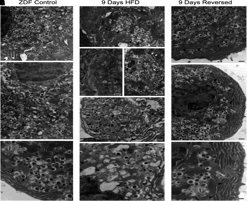
Figure 6.
Electron microscopy. Recovery from structural damage in ZDF rat caused by high-fat diet and its reversal to normal structure after return to 2 weeks of regular diet is shown. Control data are shown in A–C; 9-day data in D–H; and HFD reversal data in I–K. Significant alterations can be noted in β-cells from animals fed with HFD, including dysmorphic secretory vesicles (arrowheads) (D), disorganized Golgi apparatus (asterisks) (E), numerous autophagic bodies (black arrowheads) (F), increased cytosolic free ribosomes (white arrowheads) (F), and significantly enlarged endoplasmic reticulum cisternae (arrowheads) (G and H).

