Abstract
Background
Hyperbaric oxygen therapy (HBOT) has shown potential in salvaging compromised flaps, although its application has primarily been focused on local flaps rather than free flaps.
Case
In this case report, we present the successful use of HBOT in a 76-year-old man who underwent free flap reconstruction for calcaneal osteomyelitis. Despite undergoing 2 reoperations on the second and third days post reconstruction, no thrombosis was observed at the anastomotic site. Following the second reoperation, HBOT was promptly initiated and continued for a total of 9 sessions. Notably, after the sixth HBOT session, fresh bleeding occurred upon flap puncture. Eventually, the flap developed epidermal necrosis, which was conservatively treated.
Discussion
It is crucial to first rule out mechanical causes of compromised free flaps through surgical exploration, with HBOT serving as an adjunctive rather than a primary treatment option—-even considered as the last resort. Nevertheless, in cases where mechanical causes have been ruled out, HBOT may significantly enhance flap survival rates in compromised free flaps.
Keywords: Hyperbaric Oxygen Therapy, Free Flap, Salvage, Compromised Flap
Introduction
Hyperbaric oxygen therapy (HBOT) has emerged as a promising adjunctive therapy for salvaging compromised flaps.1 However, there is currently no standardized HBOT protocol, and its application has predominantly been observed in local flaps rather than free flaps.1,2 In this report, we present a case in which HBOT was utilized to salvage a compromised free flap in a patient who underwent 2 unsuccessful reoperations. Remarkably, the flap was able to avoid complete failure with the aid of HBOT.
Case
A 76-year-old man who had undergone surgery for calcaneal osteomyelitis caused by trauma 18 years prior developed a painful heel fistula. The patient had a history of hypertension and aortic valve replacement for aortic stenosis and was on warfarin treatment. He was diagnosed with calcaneal osteomyelitis recurrence using magnetic resonance imaging, and oral antibiotics were administered along with conservative treatment. However, he was referred to our department after 3 months due to a lack of improvement in his symptoms. A fistula with an exudate was observed within a 4 × 3 cm scar on the heel, accompanied by linear scars around it. Considering the complexity of reconstructing the affected area with a local flap, such as a perforator flap, we performed a computed tomography angiography to assess the maintenance of blood flow in the lower leg. Once satisfactory blood flow was confirmed, we planned for reconstruction using a free flap.
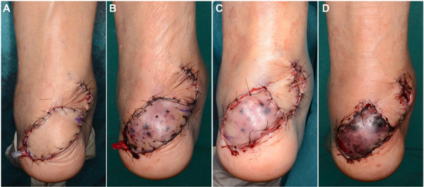
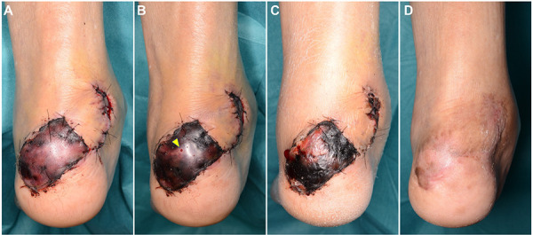
After excising the fistula-containing scar and debriding the sequestrum, the defect was covered with a medial sural artery perforator free flap (Figure 1A). The flap artery and vein were connected in an end-to-side fashion, with one anastomosis performed to the posterior tibial artery and another to the posterior tibial vein, using the technique described in a previous report.3 On postoperative day 2, as the color tone of the flap gradually deteriorated to resemble congestion (Figure 1B), a reoperation was performed; however, no evidence of thrombosis was observed at the anastomotic site of the artery and vein. After removing the flap part with compromised blood circulation, we suspected that the anastomotic site was overstrained, and a skin graft was subsequently placed immediately above the anastomotic site (Figure 1C). Prior to surgery, the patient was on warfarin treatment, and additional treatment with edoxaban was initiated. Despite these interventions, the color tone of the flap failed to improve and gradually transitioned into a congested color. On postoperative day 3 (the day after the first reoperation), the second reoperation was performed; however, no thrombosis was observed at the anastomotic site of the artery and vein, as was the case in the first reoperation (Figure 1D). HBOT was initiated immediately after the second reoperation. The sessions were 60 min long at an atmosphere absolute (ATA) of 2.0, and HBOT was continued for 9 sessions. After initiating HBOT, flap puncture resulted in minor congested bleeding only (Figure 2A). After the sixth HBOT, flap puncture resulted in fresh bleeding (Figure 2B). The flap eventually developed epidermal necrosis (Figure 2C), which was treated conservatively. The ulcer subsequently epithelialized, and no osteomyelitis or recurrence of the ulcer was observed 8 months after the free flap reconstruction (Figure 2D).
Figure 1.

Photographs captured before hyperbaric oxygen therapy (HBOT). (A) Immediately after free flap reconstruction. (B) Before the first reoperation (2 days after free flap reconstruction). (C) Immediately after the first reoperation. (D) Immediately after the second reoperation (3 days after free flap reconstruction).
Figure 2.

Photograph captured after HBOT. (A) After the 3rd HBOT (5 days after free flap reconstruction). (B) After the 6th HBOT (8 days after free flap reconstruction). Puncture of the flap showed fresh bleeding (yellow triangle). (C) After the ninth HBOT (19 days after free flap reconstruction). (D) Eight months after free flap reconstruction. HBOT, hyperbaric oxygen therapy
Discussion
HBOT has been approved for 14 indications, including indications for plastic surgery such as gas gangrene, acute traumatic ischemia, necrotizing soft tissue infections, and compromised grafts and flaps.4 Bringing flammable materials into the HBOT chamber is an absolute contraindication, and chronic lung disease and claustrophobia are relative contraindications.
HBOT can maximize the survival of compromised flaps, thereby reducing the need for additional surgery. In this case, despite 2 reoperations, the cause of compromised blood flow to the flap could not be determined, and the risk of flap failure was high. By initiating HBOT immediately after the second reoperation, total flap failure was successfully avoided, potentially preventing the development of epidermal necrosis in the compromised flap. In most studies, HBOT was initiated within 72 hours after surgery, with the efficacy decreasing to approximately 50% when initiated ≥ 48 hours after surgery.1,5 In this case report, although HBOT was initiated approximately 72 hours after the first free flap reconstruction, the postoperative outcome might have been further improved if HBOT had been initiated after the first reoperation (within 48 hours). However, it is possible to consider that HBOT was started within 48 hours after the color tone of the skin flap deteriorated. It may be difficult to initiate HBOT immediately after free flap reconstruction owing to the presence of closed drains and requirement of intravenous fluids. Therefore, further studies are required to determine the criteria for introducing HBOT for the treatment of compromised free flaps.
Furthermore, compromised free flaps should first be surgically assessed to rule out causes such as vascular thrombosis. The salvaging of compromised free flaps mostly involves surgical treatment and thrombolytic drugs and rarely HBOT.6,7 One reason for this is the unavailability of HBOT; only few hospitals have this available in an inpatient setting. Another reason may be the expectation of a limited therapeutic effect in events in which HBOT is performed on compromised free flaps with unknown causes for compromise. Furthermore, several reports have found no significant difference in free flap survival with or without HBOT.8,9 Therefore, HBOT should be applied to compromised free flaps with a risk of partial failure that can be successfully salvaged or are not compromised due to a mechanical cause, as this may have the same therapeutic effect that is observed in the treatment of conventional compromised local flaps. Venous thrombosis due to congestion is the most common cause of free flap failure in the lower extremity.10 Our patient had post-traumatic osteomyelitis and the flap congestion progressed even after 2 reoperations; thus, it is suspected that the recipient vein had chronic venous dysfunction. In this case report, the extent to which HBOT contributed to flap survival and reduced the extent of flap failure is unclear. However, considering the progress of the free flap after initiation of HBOT, it is highly likely that HBOT prevents complete flap failure and maximizes flap survival.
HBOT is considered to contribute to local tissue oxygenation by promoting angiogenesis and collagen synthesis and improving wound healing by reducing hypoxia, ischemic injury, cell death, and inflammation.11,12 In this case report, the flap was partially blackened before HBOT was initiated, suggesting that HBOT contributed to salvage tissues deeper than the epidermis (dermis and subcutaneous fat). HBOT was planned for 30 dives (2.0 ATA, 60 min) based on our institutional protocol but was terminated at 9 dives at the patient's request. According to the Undersea and Hyperbaric Medical Society guidelines, HBOT is given at a pressure of 2.0 to 2.5 ATA for 90 to 120 min.2 However, optimal HBOT sessions and session duration are patient-specific and there are no clear guidelines.1 Improvement in wound healing may reduce the number of planned dives; however, clear criteria guided by more dedicated research and prospective studies are needed.
In conclusion, HBOT is rarely performed on compromised free flaps, and mechanical causes should be ruled out by surgical exploration. HBOT treatment protocols for free flaps have not yet been established and should be used as an adjunct rather than a primary treatment, and even as the last resort. Nevertheless, HBOT may maximize flap survival in compromised free flaps in which mechanical causes are ruled out.
Acknowledgments
We would like to thank Editage (www.editage.com) for English language editing.
Ethics: Informed consent for the case report was obtained from the patient.
Disclosures: The authors disclose no relevant conflicts of interest or financial disclosures for this manuscript.
References
- 1.Yousef A, Solomon I, Hom DB. Can hyperbaric oxygen salvage a compromised local/regional skin flap? Laryngoscope. 2022;132(10):1892-1894. doi:10.1002/lary.30160 10.1002/lary.30160 [DOI] [PubMed] [Google Scholar]
- 2.Kleban S, Baynosa RC. The effect of hyperbaric oxygen on compromised grafts and flaps. Undersea Hyperb Med. 2020;47(4):635-648. doi:10.22462/10.12.2020.13 10.22462/10.12.2020.13 [DOI] [PubMed] [Google Scholar]
- 3.Shimbo K, Kawamoto H, Koshima I. Venous end-to-side anastomosis for free-flap reconstruction in the extremities: A case series and meta-analysis. J Plast Reconstr Aesthet Surg. 2023;83:4-11. doi:10.1016/j.bjps.2023.05.004 10.1016/j.bjps.2023.05.004 [DOI] [PubMed] [Google Scholar]
- 4.Moon RE, ed. Hyperbaric Oxygen Therapy Indications. 14th ed. Best Publishing Company; 2019. [Google Scholar]
- 5.Zhou YY, Liu W, Yang YJ, Lu GD. Use of hyperbaric oxygen on flaps and grafts in China: analysis of studies in the past 20 years. Undersea Hyperb Med. 2014;41(3):209-216. [PubMed] [Google Scholar]
- 6.Shen AY, Lonie S, Lim K, Farthing H, Hunter-Smith DJ, Rozen WM. Free flap monitoring, salvage, and failure timing: a systematic review. J Reconstr MicroSurg. 2021;37(3):300-308. doi:10.1055/s-0040-1722182 10.1055/s-0040-1722182 [DOI] [PubMed] [Google Scholar]
- 7.Brouwers K, Kruit AS, Hummelink S, Ulrich DJO. Management of free flap salvage using thrombolytic drugs: A systematic review. J Plast Reconstr Aesthet Surg. 2020;73(10):1806-1814. doi:10.1016/j.bjps.2020.05.057 10.1016/j.bjps.2020.05.057 [DOI] [PubMed] [Google Scholar]
- 8.Nolen D, Cannady SB, Wax MK, et al. Comparison of complications in free flap reconstruction for osteoradionecrosis in patients with or without hyperbaric oxygen therapy. Head Neck. 2014;36(12):1701-1704. doi:10.1002/hed.23520 10.1002/hed.23520 [DOI] [PubMed] [Google Scholar]
- 9.Vishwanath G. Hyperbaric oxygen therapy in free flap surgery: is it meaningful? Med J Armed Forces India. 2011;67(3):253-256. doi:10.1016/S0377-1237(11)60052-X 10.1016/S0377-1237(11)60052-X [DOI] [PMC free article] [PubMed] [Google Scholar]
- 10.Shimbo K, Kawamoto H, Koshima I. Selection of deep or superficial recipient vein in lower extremity reconstruction using free flap: a systematic review and meta-analysis. MicroSurg. 2022;42(7):732-739. doi:10.1002/micr.30946 10.1002/micr.30946 [DOI] [PubMed] [Google Scholar]
- 11.Perrins DJ. Influence of hyperbaric oxygen on the survival of split skin grafts. Lancet. 1967;1(7495):868-871. doi:10.1016/s0140-6736(67)91428-6 10.1016/S0140-6736(67)91428-6 [DOI] [PubMed] [Google Scholar]
- 12.Simman R, Bach K. Role of hyperbaric oxygen therapy in cosmetic and reconstructive surgery in ischemic soft tissue wounds: a case series. Eplasty. 2022;22:e61. [PMC free article] [PubMed] [Google Scholar]


