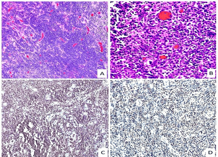
Figure 3. Histopathology of the thyroid specimens.
A: Large atypical lymphocytes grow between nonneoplastic thyroid follicles (H&E stain at 100x). B: Diffuse infiltrate of large lymphocytes with prominent nucleoli and numerous apoptotic bodies (H&E stain at 400x). C: Lymphocyte infiltrate positive for CD79a marker on the immunohistochemical stain. D: Lymphocyte infiltrate positive for Bcl-6 marker on the immunohistochemical stain.

