Abstract
African swine fever virus (ASFV) replicates in the cytoplasm of infected cells and contains genes encoding a number of enzymes needed for DNA synthesis, including a thymidine kinase (TK) gene. Recombinant TK gene deletion viruses were produced by using two highly pathogenic isolates of ASFV through homologous recombination with an ASFV p72 promoter–β-glucuronidase indicator cassette (p72GUS) flanked by ASFV sequences targeting the TK region. Attempts to isolate double-crossover TK gene deletion mutants on swine macrophages failed, suggesting a growth deficiency of TK− ASFV on macrophages. Two pathogenic ASFV isolates, ASFV Malawi and ASFV Haiti, partially adapted to Vero cells, were used successfully to construct TK deletion viruses on Vero cells. The selected viruses grew well on Vero cells, but both mutants exhibited a growth defect on swine macrophages at low multiplicities of infection (MOI), yielding 0.1 to 1.0% of wild-type levels. At high MOI, the macrophage growth defect was not apparent. The Malawi TK deletion mutant showed reduced virulence for swine, producing transient fevers, lower viremia titers, and reduced mortality. In contrast, 100% mortality was observed for swine inoculated with the TK+ revertant virus. Swine surviving TK− ASFV infection remained free of clinical signs of African swine fever following subsequent challenge with the parental pathogenic ASFV. The data indicate that the TK gene of ASFV is important for growth in swine macrophages in vitro and is a virus virulence factor in swine.
African swine fever virus (ASFV) is a large icosahedral enveloped DNA virus and is classified as the only member of a new virus family, the Asfaviridae (3, 11). The structure and replication of its 170- to 190-kbp genome are similar to those of the poxviruses, but the icosahedral morphology of the virus more closely resembles that of the iridoviruses (for reviews see references 9, 43, and 44). African swine fever (ASF) is recognized as an important disease of domestic swine and is characterized by high fevers, hemorrhage, shock, and 100% mortality for highly pathogenic isolates. ASF may range from an acute, highly lethal infection to a subclinical form, depending on contributing viral and host factors. The virus infects cells of the mononuclear-phagocytic system, including highly differentiated fixed-tissue macrophages and specific lineages of reticular cells in the spleen, lymph node, lung, kidney, and liver. These tissues show extensive damage with highly virulent strains of ASFV, and the ability of ASFV to replicate and induce cytopathology in these tissues in vivo appears to be a critical factor in ASFV virulence (8, 24, 25, 30, 32). The nature of viral factors responsible for the virulence and pathogenesis of ASFV remains poorly understood.
Like poxviruses and iridoviruses, ASFV replicates in the cytoplasm and encodes enzymes for transcription and DNA synthesis (17, 46), since enzymes of the host cell nucleus are unavailable. Also, ASFV encodes enzymes involved in synthesis of deoxynucleoside triphosphates: the thymidine kinase (TK) enzyme (1, 28, 38), the ribonucleotide reductase (RR) enzyme (2, 10), and the thymidylate kinase (TMPK) enzyme (47). These enzymes are also encoded by other large DNA viruses (29). Herpesviruses encode a single polypeptide with both TK and TMPK activities (39). These enzymes are involved in the salvage and de novo pathways of dTTP synthesis. The ASFV TK gene has been shown to be nonessential for the growth of ASFV in cultured hamster and monkey cells (27, 40).
The inactivation of the TK gene in poxviruses and herpesviruses showed the gene to be nonessential for growth in cultured cells (12, 21, 26, 36), but TK− viruses exhibited a reduction in virulence and pathogenicity in experimental animal hosts. A 104 to 105 reduction in the 50% lethal dose (LD50) was noted for mice injected with TK− vaccinia or ectromelia virus (4, 23), and TK− herpes simplex virus and marmoset herpesvirus showed similar losses of virulence in mice (13, 22). Cell culture conditions were found to be important, as serum-starved cultures showed reduced growth of TK−, but not of TK+, herpesviruses (13, 21). Also, TK− herpes simplex virus showed a reduced ability to infect highly differentiated neuronal tissues (13, 42). In similar fashion, highly differentiated murine macrophages, an important type of target cell in the pathogenesis of ectromelia virus infection in mice, failed to support the growth of TK− ectromelia virus (23).
In this study, we have constructed TK gene deletion mutants of pathogenic ASFV isolates in order to examine the role of this gene in viral pathogenesis and virulence. Experiments using primary swine macrophages failed to produce stable TK deletion mutants, suggesting that the ASFV TK gene might be important for viral replication in this cell type (31). Two pathogenic ASFV isolates, partially adapted to Vero cells, were used successfully to construct TK deletion viruses on Vero cells. The loss of TK from these viruses impaired their growth on swine macrophages in vitro and reduced their virulence in vivo. Thus, TK could be considered an important gene for growth in target macrophage cells and possibly in other tissues involved in the pathogenesis of lethal ASFV infections.
Construction of recombinant ASFV TK gene deletion mutants and TK-positive revertants.
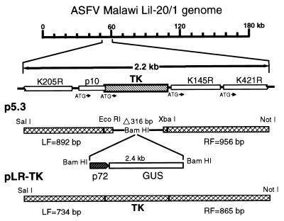
The pathogenic tissue culture-adapted ASFV Malawi LiL-20/1V (Vero Malawi) and ASFV Haiti H811 (H811) were obtained from the Plum Island Animal Disease Center ASFV reference collection and grown on Vero cells, and viral DNA was obtained (45). ASFV recombinant viruses were generated by homologous recombination between parental ASFV genomes and engineered recombination vectors as previously described (34, 48), except that Vero cells were used for transfection/infection and isolation of β-glucuronidase (GUS)-positive virus foci. The exchange vector (p5.3) was produced by the sequential cloning into plasmid pBluescript II KS (Stratagene) of PCR-derived DNA of left (892 bp) and right (956 bp) TK flanking sequences, deleting the central region of the TK gene (315 bp; codons 55 to 158) but leaving the N-terminal and C-terminal TK coding regions intact and inserting a 2.4-kb reporter cassette (p72GUS) consisting of the ASFV p72 gene promoter linked to the GUS gene (34) (Fig. 1). GUS-positive foci were picked from agarose overlays containing 100 μg of 5-bromo-4-chloro-3-indolyl-β-d-glucuronic acid (X-Gluc) per ml (48) and were plaque purified four to six times until PCR and Southern blot analysis (41) showed no evidence of contamination with the parental virus.
FIG. 1.
Diagram of the TK region of the Malawi LiL-20/1 genome, showing the placement and orientation of the TK gene and adjacent genes p10 (DNA binding protein 5-AR) (33), K205R, K145R, and K421R (Ba71V open reading frames) (46); structure of the TK gene deletion transfer vector, p5.3, showing insertion sites into the pBluescript multiple cloning site; and structure of plasmid pLR-TK, containing a 2,164-bp PCR fragment of the intact TK gene and flanking ASFV DNA.
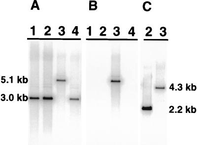
Virus stocks were amplified on Vero cells, and viral DNA was prepared. The structure of the TK deletion mutant (ASFV v5.3) was revealed by Southern blotting, yielding the predicted BglII 5.1-kb band representing the deletion of TK sequences and the insertion of the 2.4-kb p72GUS cassette (Fig. 2A and B). PCR analysis of v5.3 DNA with appropriate primer pairs revealed only a 1,250-bp recombinant-specific fragment (data not shown). These results indicated the isolation of a virus, derived from a double-crossover recombination, in which the p72GUS cassette replaced the central part of the TK gene. In similar fashion, a recombinant virus was produced by using the pathogenic, Vero-adapted H811 ASFV isolate and the TK deletion recombination vector (p5.3). This virus (ASFV vH53) yielded the predicted 4.3-kb BglII fragment, compared to the 2.2-kb uninterrupted TK fragment of the parental virus (Fig. 2C).
FIG. 2.
Southern blot analysis of parental, recombinant, and revertant ASFV DNA digested with BglII. ASFV Malawi (A and B) and ASFV Haiti (C) were probed with a TK probe (HindIII fragment of pLR-TK) (A and C) or a GUS probe (SmaI/SacI fragment of the p72GUS cassette) (34) (B). Lanes 1, swine spleen isolate; lanes 2, Vero-adapted isolates; (lanes 3, TK deletion mutant viruses; lanes 4, revertant virus.
The difficulty in isolating GUS-positive TK− recombinant viruses on macrophages and initial growth analysis of ASFV recombinant v5.3 on macrophages suggested a growth deficiency in macrophages. Therefore, a positive growth selection method was attempted in order to rescue TK+ revertants. A 2,164-bp fragment spanning the TK gene was amplified by using the left flank 5′-forward and the right flank 3′-reverse primer (described above) to construct the TK knockout vector and cloned into pBluescript, resulting in plasmid pLR-TK. This plasmid contained the complete TK gene with 734 bp of ASFV TK-flanking sequences upstream and 865 bp downstream (Fig. 1). Swine macrophage cultures (16, 34) were infected with ASFV v5.3, transfected with plasmid pLR-TK, blind passaged three times on swine macrophages, and purified by endpoint dilution, resulting in virus v5.3R. Virus stocks were made on swine macrophages, and viral DNA was prepared. Southern blot analysis showed the presence of the native 3.0-kb TK fragment and no reactivity with the GUS probe (Fig. 2A and B, lanes 4), indicating restoration of the native TK genotype.
TK is important for in vitro growth of ASFV on porcine macrophages.
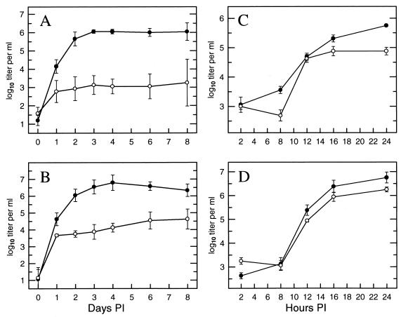
Growth properties of the Malawi (v5.3) and Haiti (vH53) TK deletion mutants were compared to those of the respective parental Vero-adapted viruses on swine macrophage and Vero cell cultures. At a low multiplicity of infection (MOI = 0.01), the parental isolates grew well on both Vero and macrophage cultures (data not shown). The TK deletion mutants grew well on Vero cells (data not shown) but grew poorly on macrophages; Malawi v5.3 and Haiti vH53 grew to 0.1 and 1% of parental levels, respectively (Fig. 3A and B). However, at high MOIs (10 to 20), the growth defect of the TK deletion mutants was not apparent (Fig. 3C and D).
FIG. 3.
Growth characteristics of ASFV Malawi and Haiti parental (closed circles) and recombinant (open circles) viruses on primary swine macrophages. Cells were infected at MOIs of 0.01 (A and B) and 10 to 20 (C and D) with the appropriate viruses (which were absorbed for 2 h at 37°C), rinsed twice with growth medium, and incubated. At the indicated times, cultures were harvested and lysates were titrated for total virus yield on Vero cells by the immunoperoxidase method (49). (A and C) Growth of ASFV Malawi (parent) and TK deletion mutant v5.3; (B and D) growth of ASFV Haiti H811 (parent) and TK deletion mutant vH53. Data represent the TCID50 titers ± standard errors of the means (14) assayed for two or three independent experiments.
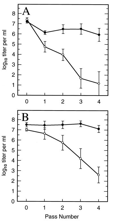
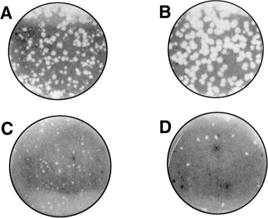
To confirm the macrophage growth deficiency of ASFV TK deletion mutants, high-titer Vero cell stocks of the mutant and parental viruses were serially passaged four times on macrophage cultures by using large inoculum volumes. Cultures were observed for cytopathic effect (CPE), and cell lysates were monitored for GUS expression (GUS-positive mutants) and titrated for ASFV. As expected, the parental ASFVs caused complete CPE through each passage and maintained high titers on macrophage cell passage (Fig. 4). Positive CPE and GUS expression were observed for ASFV v5.3 on the first passage, and trace levels were observed on the second passage; ASFV vH53 showed CPE on the first and second passages, and low levels of GUS were present through the third passage. Infectivity titers of the TK deletion mutants dropped successively at each passage to low levels, with somewhat higher titers of Haiti vH53 remaining (Fig. 4B). The viruses were also compared in a plaque assay on swine macrophages. Dilutions of virus were plated and overlayered as described above for isolation of recombinants, but without X-Gluc in the overlays. At 6 days, the cultures were fixed with formalin, the agar was removed, and the cells were stained with crystal violet. Figure 5 shows that for each virus, the plaque size was substantially reduced for the respective TK− mutants.
FIG. 4.
Virus titers obtained on successive passages of parental (closed circles) and TK deletion mutant (open circles) viruses on primary swine macrophages. (A) ASFV Malawi viruses; (B) ASFV Haiti H811 viruses. Cells in T-25 flasks were inoculated with 1 ml (passage [pass] 1) or 2 ml (passages 2, 3, and 4) of virus (which was absorbed for 2 h), rinsed twice with growth medium, and incubated for 2 to 4 days. Cultures were observed for CPE, and cells and medium were harvested at 2 days (passage 1) or 4 days (passages 2, 3, and 4); then culture lysates were examined for GUS expression, and TCID50 titers were determined by the immunoperoxidase method. Titers ± standard errors of the means are the results of three independent experiments.
FIG. 5.
Plaques formed on primary swine macrophages by parental Malawi (A) and Haiti (B) ASFV and by TK deletion mutants Malawi v5.3 (C) and Haiti vH53 (D).
Amounts of viral DNA synthesized by Vero Malawi and the ASFV TK− mutant v5.3 grown on Vero cell and macrophage cultures were examined by dot blot hybridization (41). Cells were infected at MOIs of 1 to 2, DNA was isolated from cells at 24 h postinfection (p.i.), and dilution sets of DNA were blotted onto Zeta Probe membranes (Bio-Rad) and probed with a 32P-labelled 2,164-bp TK region DNA fragment (SalI/NotI-cut DNA from pLR-TK). On macrophages, the parental ASFV Malawi consistently produced large amounts of viral DNA. For v5.3 no viral DNA was detected in two experiments, and in a third, a small amount of v5.3 DNA was present over background levels measured at 2 h p.i. In comparison, both the Vero-adapted ASFV Malawi and the TK− mutant v5.3 synthesized large amounts of viral DNA on Vero cells (data not shown).
Cellular levels of TK are very low in quiescent, nondividing cells but increase dramatically during cell division (37), and TK activity in primary macrophage cultures is very low, even during prolonged incubation in complete growth medium (31). With pools of nucleoside triphosphates low in macrophages, the loss of viral TK activity might be expected to have a significant effect on viral DNA synthesis and subsequent production of viral progeny. The TK, RR, and TMPK enzymes have all been implicated in attenuation of poxviruses (4, 7, 19) and herpesviruses (5, 20, 22). For ASFV, the levels of expression of these enzymes in different isolates of ASFV are not known, and each may contribute to viral replication in quiescent cells such as macrophages. Here, in virus growth experiments, the growth defect of the TK mutants was overcome at high MOIs. The levels of RR found in ASFV-infected Vero cells were shown to be proportional to the MOIs, and inhibition of DNA replication did not alter the levels of RR present (10). Thus, early transcription of RR at high MOIs could supplement the TK deficiency by providing an alternate source of deoxythymidylate.
In the growth curve experiments, the TK deletion mutants grew poorly at low MOIs compared to the parental viruses (Fig. 3A and B). However, since there was some increase in virus titers over the first several days, we cannot formally exclude the possibility that, after a single round of replication, subsequent poor growth was due to failure of the virus to be released and to infect adjacent cells. However, the nature of growth defects of TK deletion mutants of other DNA viruses is linked to nucleotide metabolism and subsequent viral DNA synthesis. Furthermore, the passage experiments performed here showed that despite harvest of virus from cell lysates and reapplication to new cells, virus titers diminished (Fig. 4). The results of the growth experiments and the diminished levels of DNA synthesized at low MOIs with v5.3 suggest that TK is important in macrophages for replication of viral DNA in the cytoplasm of these cells.
TK affects ASFV virulence for swine.
To assess the importance of the TK gene in viral virulence, two separate groups of four Yorkshire pigs were inoculated intramuscularly with 104 50% tissue culture infective doses (TCID50) of the revertant ASFV v5.3R or the TK− mutant ASFV v5.3 and were observed for clinical signs of ASF: fever, anorexia, lethargy, shivering, cyanosis, and recumbency. A dose of 104 TCID50 of pathogenic ASFV strains represents a challenge of 1,000 to 10,000 LD100s (34, 48). Results of the experiment are shown in Table 1. The animals inoculated with the revertant virus showed signs typical of acute ASF, with onset of fever at day 4 and all animals dying between days 9 and 13. The swine inoculated with the mutant virus v5.3 showed only transient fever responses, and one animal, which had the most persistent fever, died at day 15. Except for transient fevers, the remaining animals were clinically normal throughout the experiment. Maximum viremia titers in the revertant virus group reached 106 to 107 TCID50, whereas the titers for the mutant v5.3 group ranged from <103.5 to 106 TCID50, with viremia onset delayed 4 to 5 days. DNA from virus isolated at the peak of viremia from the recombinant virus group was examined by PCR and found to be that of the recombinant virus, with no wild-type virus DNA detected (data not shown). By using a virus isolation-PCR-blot detection method (6), virus was detected in only one animal of the ASFV v5.3 group at day 30 p.i. and none of the animals was positive at day 56 p.i. (data not shown). At day 56 p.i., the three surviving v5.3-infected swine were challenged intramuscularly with 104 TCID50 of the parental Vero-adapted Malawi virus. A fever response was noted in two of the three animals on days 4 to 6, and one animal remained febrile following this time. That animal developed an infected abscess at the inoculation site which advanced to a severe cellulitis (the animal was sacrificed on day 21). Maximum viremia titers ranged from undetectable levels to levels lower than 104 TCID50/ml, and all animals remained free of clinical symptoms for the experiment’s duration.
TABLE 1.
Swine survival, viremia, and fever response following infection with ASFV Malawi revertant and TK deletion mutant v5.3a
| Group | No. surviving | Days to death | Feverb
|
Viremia
|
||
|---|---|---|---|---|---|---|
| Days to onset | No. of days of fever | Days to onset | Maximum titer (log10 TCID50/ml) | |||
| Malawi revertant | 0/4 | 10.8 ± 0.9 | 4.3 ± 0.3 | 6.5 ± 1.0c | 4.0 ± 0.0 | 6.5 ± 0.2 |
| TK− v5.3 | 3/4 | 15.0 ± 0.0 | 9.3 ± 1.9 | 2.5 ± 1.0 | 9.3 ± 0.7 | 4.8 ± 0.6 |
Data represent mean values ± standard errors of the means.
Rectal temperature above 40°C.
Until death.
The results of the animal experiment indicate that loss of the TK gene reduced the virulence of ASFV for domestic swine and that swine that recovered from infection with the TK− ASFV v5.3 were protected from developing ASF on virulent virus challenge. Recently, another ASFV gene, 23-NL-S, was shown to be a virulence-associated gene for ASF (48). However, ASFV 23-NL-S was nonessential for growth in vitro in Vero cells and swine macrophages, but deletion of the gene significantly reduced virulence for swine, suggesting that the gene may serve as a host range gene in swine. Work with tissue culture-adapted isolates of ASFV established that TK was nonessential for growth in Vero cells (27, 40). However, the present report shows the TK gene to be important for growth on macrophages in vitro and to be a virulence factor in vivo. Thus, TK should be considered an essential gene for the target lymphoreticular tissues involved in the pathogenesis of lethal ASFV infections. However, it is not known at this time if loss of the TK gene affects the ability of the virus to infect lymphoreticular tissues and organs involved in the pathogenesis of lethal ASF (8, 24, 25, 30, 32) or if a reduction in virus load in swine may allow time for the development of host defenses.
The TK locus has been used to demonstrate the feasibility of constructing ASFV recombinant viruses (15, 27, 40), but these experiments used highly adapted, cell-cultured viruses no longer virulent for swine, so the role of TK in the virulence and pathogenesis of ASFV in swine could not be assessed. Several low-passage-number isolates of ASFV retaining their infectivity for swine have been engineered with chromogenic marker genes inserted into the TK locus, and their use in tropism, pathogenicity, and latency studies has been suggested (18). We have observed CPE, the expression of GUS driven by an ASFV late gene promoter, and positive hemadsorption (late expression of the CD2 gene) with TK deletion mutants in swine macrophages, but the virus grows poorly and shows reduced virulence in swine. The present work shows that the TK gene itself plays a significant role in virus virulence and that this site would not be suitable for the insertion of markers to monitor pathogenesis or as an insertion site for other viral genes to assess their role in virus virulence.
We have demonstrated that the selective macrophage growth advantage of TK+ ASFV may be used to engineer other ASFV gene deletions through TK+ rescue of the ASFV mutant v5.3 (35). Plasmids in which the intact TK gene with its upstream promoter sequence replaced a specific gene of interest were engineered. Preliminary experiments indicate that stable GUS+ TK+ recombinants which could be directly isolated through selective growth on macrophage cultures were produced. These results demonstrate the utility of using the TK gene as a selective factor in genome manipulations of pathogenic isolates of ASFV and also confirm that the growth defect of ASFV v5.3 in macrophages was directly due to the loss of the TK gene function.
Animals previously infected with the TK deletion mutant were resistant to challenge with virulent ASFV Malawi, although the level of attenuation of the TK deletion mutant would not render it suitable for use as a vaccine. Better understanding of the role of TK, RR, and TMPK enzymes in supplying pools of deoxynucleoside triphosphates for viral DNA replication may define how these viral enzymes support the replication of the virus in vivo. Modifications of the viral genes controlling DNA metabolism in infected cells may be a valuable component in the design of multiple-gene-deletion, attenuated live virus vaccines for ASFV.
Acknowledgments
We thank G. Kutish for assistance with sequence and statistical data analysis, T. Burrage for assistance in titration experiments, E. Kramer and R. Mireles for swine macrophage cell cultures, and the PIADC animal care staff for assistance with animal experiments.
REFERENCES
- 1.Blasco R, Lopez-Otin C, Munoz M, Bockamp E-O, Simon-Mateo C, Vinuela E. Sequence and evolutionary relationships of African swine fever virus thymidine kinase. Virology. 1990;178:301–304. doi: 10.1016/0042-6822(90)90409-K. [DOI] [PMC free article] [PubMed] [Google Scholar]
- 2.Boursnell M, Shaw K, Yanez R J, Vinuela E, Dixon L. The sequences of the ribonucleotide reductase genes from African swine fever virus show considerable homology with those of the orthopoxvirus, vaccinia virus. Virology. 1991;184:411–416. doi: 10.1016/0042-6822(91)90860-e. [DOI] [PubMed] [Google Scholar]
- 3.Brown F. The classification and nomenclature of viruses: summary of results of meetings of the International Committee on Taxonomy of Viruses in Sendai, September 1984. Intervirology. 1986;25:141–143. doi: 10.1159/000150091. [DOI] [PubMed] [Google Scholar]
- 4.Buller R M L, Smith G L, Cremer K, Notkins A L, Moss B. Decreased virulence of recombinant vaccinia virus expression vectors is associated with a thymidine kinase-negative phenotype. Nature. 1985;317:813–815. doi: 10.1038/317813a0. [DOI] [PubMed] [Google Scholar]
- 5.Cameron J M, McDougall I, Marsden H S, Preston V G, Ryan D M, Subak-Sharpe J H. Ribonucleotide reductase encoded by herpes simplex virus is a determinant of the pathogenicity of the virus in mice and a valid antiviral target. J Gen Virol. 1988;69:2607–2612. doi: 10.1099/0022-1317-69-10-2607. [DOI] [PubMed] [Google Scholar]
- 6.Carrillo C, Borca M V, Afonso C L, Onisk D V, Rock D L. Long-term persistent infection of swine monocytes/macrophages with African swine fever virus. J Virol. 1994;68:580–583. doi: 10.1128/jvi.68.1.580-583.1994. [DOI] [PMC free article] [PubMed] [Google Scholar]
- 7.Child S J, Palumbo G J, Buller R M, Hruby D E. Insertional inactivation of the large subunit of ribonucleotide reductase encoded by vaccinia virus is associated with reduced virulence in vivo. Virology. 1990;174:625–629. doi: 10.1016/0042-6822(90)90119-c. [DOI] [PubMed] [Google Scholar]
- 8.Colgrove G S, Haelterman E O, Coggins L. Pathogenesis of African swine fever in young pigs. Am J Vet Res. 1969;30:1343–1359. [PubMed] [Google Scholar]
- 9.Costa J V. African swine fever virus. In: Darai G, editor. Molecular biology of iridoviruses. Boston, Mass: Kluwer Academic Publishers; 1990. pp. 247–270. [Google Scholar]
- 10.Cunha C V, Costa J V. Induction of ribonucleotide reductase activity in cells infected with African swine fever virus. Virology. 1992;187:73–83. doi: 10.1016/0042-6822(92)90296-2. [DOI] [PubMed] [Google Scholar]
- 11.Dixon, L. K., D. L. Rock, and E. Vinuela. 1995. African swine fever-like viruses. Arch. Virol. 10(Suppl.):92–94.
- 12.Dubbs D R, Kit S. Mutant strains of herpes simplex deficient in thymidine kinase-inducing activity. Virology. 1964;22:493–502. doi: 10.1016/0042-6822(64)90070-4. [DOI] [PubMed] [Google Scholar]
- 13.Field H J, Wildy P. The pathogenicity of thymidine kinase-deficient mutants of herpes simplex virus in mice. J Hyg Camb. 1978;81:267–277. doi: 10.1017/s0022172400025109. [DOI] [PMC free article] [PubMed] [Google Scholar]
- 14.Finney D J. Statistical methods in biological assays. 2nd ed. New York, N.Y: Hafner Publishing Co.; 1984. pp. 524–533. [Google Scholar]
- 15.Garcia R, Almazan F, Rodriguez J M, Alonso M, Vinuela E, Rodriguez J F. Vectors for the genetic manipulation of African swine fever virus. J Biotechnol. 1995;40:121–131. doi: 10.1016/0168-1656(95)00037-q. [DOI] [PubMed] [Google Scholar]
- 16.Genovesi E V, Villinger F, Gerstner D J, Whyard T C, Knudsen R C. Effect of macrophage-specific colony-stimulating factor (CSF-1) on swine monocyte/macrophage susceptibility to in vivo infection by African swine fever virus. Vet Microbiol. 1990;25:153–176. doi: 10.1016/0378-1135(90)90074-6. [DOI] [PubMed] [Google Scholar]
- 17.Goebel S J, Johnson G P, Perkus M E, Davis S W, Winslow J P, Paoletti E. The complete DNA sequence of vaccinia virus. Virology. 1990;179:247–266. doi: 10.1016/0042-6822(90)90294-2. [DOI] [PubMed] [Google Scholar]
- 18.Gomez-Puertas P, Rodriguez F, Ortega A, Oviedo J M, Alonso C, Escribano J M. Improvement of African swine fever virus neutralization assay using recombinant viruses expressing chromogenic marker genes. J Virol Methods. 1995;55:271–279. doi: 10.1016/0166-0934(95)00055-y. [DOI] [PubMed] [Google Scholar]
- 19.Hughes S J, Johnston L H, de Carlos A, Smith G L. Vaccinia virus encodes an active thymidylate kinase that complements a cdc8 mutant of Saccharomyces cerevisiae. J Biol Chem. 1991;266:20103–20109. [PubMed] [Google Scholar]
- 20.Idowu A D, Fraser-Smith E B, Poffenberger K L, Herman R C. Deletion of the herpes simplex virus type 1 ribonucleotide reductase gene alters virulence and latency in vivo. Antivir Res. 1992;17:145–156. doi: 10.1016/0166-3542(92)90048-a. [DOI] [PubMed] [Google Scholar]
- 21.Jamieson A T, Gentry G A, Subak-Sharpe J H. Induction of both thymidine and deoxycytidine kinase activity by herpes viruses. J Gen Virol. 1974;24:465–480. doi: 10.1099/0022-1317-24-3-465. [DOI] [PubMed] [Google Scholar]
- 22.Kit S, Qavi H, Dubbs D R, Otsuka H. Attenuated marmoset herpesvirus isolated from recombinants of virulent marmoset herpesvirus and hybrid plasmids. J Med Virol. 1983;12:25–36. doi: 10.1002/jmv.1890120104. [DOI] [PubMed] [Google Scholar]
- 23.Kochneva G V, Urmanov I H, Ryabchikova E I, Streltsov V V, Serpinsky O I. Fine mechanisms of ectromelia virus thymidine kinase-negative mutants avirulence. Virus Res. 1994;34:49–61. doi: 10.1016/0168-1702(94)90118-x. [DOI] [PubMed] [Google Scholar]
- 24.Konno S, Taylor W D, Dardiri A H. Acute African swine fever. Proliferative phase in lymphoreticular tissue and the reticuloendothelial system. Cornell Vet. 1971;61:71–84. [PubMed] [Google Scholar]
- 25.Konno S, Taylor W D, Hess W R, Heuschele W P. Liver pathology in African swine fever. Cornell Vet. 1971;61:125–150. [PubMed] [Google Scholar]
- 26.Mackett M, Smith G L, Moss B. Vaccinia virus: a selectable eukaryotic cloning and expression vector. Proc Natl Acad Sci USA. 1982;79:7415–7419. doi: 10.1073/pnas.79.23.7415. [DOI] [PMC free article] [PubMed] [Google Scholar]
- 27.Martin Hernandez A M, Camacho A, Prieto J, Menendez del Campo A M, Tabares E. Isolation and characterization of TK-deficient mutants of African swine fever virus. Virus Res. 1995;36:67–75. doi: 10.1016/0168-1702(94)00098-w. [DOI] [PubMed] [Google Scholar]
- 28.Martin Hernandez A M, Tabares E. Expression and characterization of the thymidine kinase gene of African swine fever virus. J Virol. 1991;65:1046–1052. doi: 10.1128/jvi.65.2.1046-1052.1991. [DOI] [PMC free article] [PubMed] [Google Scholar]
- 29.McGeoch D J, Dalrymple M A, Davison A J, Dolan A, Frame M C, McNab D, Perry L J, Scott J E, Taylor P. The complete DNA sequence of the long unique region in the genome of herpes simplex virus type 1. J Gen Virol. 1988;69:1531–1574. doi: 10.1099/0022-1317-69-7-1531. [DOI] [PubMed] [Google Scholar]
- 30.Mebus C A. African swine fever. Adv Virus Res. 1988;35:251–269. doi: 10.1016/s0065-3527(08)60714-9. [DOI] [PubMed] [Google Scholar]
- 31.Moore, D. M. Unpublished data.
- 32.Moulton J, Coggins L. Comparison of the lesions in acute and chronic African swine fever. Cornell Vet. 1968;58:364–388. [PubMed] [Google Scholar]
- 33.Neilan J G, Lu Z, Kutish G F, Sussman M D, Roberts P C, Yozawa T, Rock D L. An African swine fever virus gene with similarity to bacterial DNA binding proteins, bacterial integration host factors, and the Bacillus phage SP01 transcription factor, TF1. Nucleic Acids Res. 1993;21:1496. doi: 10.1093/nar/21.6.1496. [DOI] [PMC free article] [PubMed] [Google Scholar]
- 34.Neilan J G, Lu Z, Kutish G F, Zsak L, Burrage T G, Borca M V, Carrillo C, Rock D L. A BIR motif containing gene of African swine fever virus, 4CL, is nonessential for growth in vitro and viral virulence. Virology. 1997;230:252–264. doi: 10.1006/viro.1997.8481. [DOI] [PubMed] [Google Scholar]
- 35.Neilan, J. G., and D. M. Moore. 1997. Unpublished data.
- 36.Panicali D, Paoletti E. Construction of poxviruses as cloning vectors: insertion of the thymidine kinase gene from herpes simplex virus into the DNA of infectious vaccinia virus. Proc Natl Acad Sci USA. 1982;79:4927–4931. doi: 10.1073/pnas.79.16.4927. [DOI] [PMC free article] [PubMed] [Google Scholar]
- 37.Pelka-Fleischer R, Ruppelt W, Wilmanns W, Sauer H, Schalhorn A. Relation between cell cycle stage and the activity of DNA-synthesizing enzymes in cultured human lymphoblasts: investigations on cell fractions enriched according to cell cycle stages by way of centrifugal elutriation. Leukemia. 1987;1:182–187. [PubMed] [Google Scholar]
- 38.Polatnick J, Hess W. Altered thymidine kinase activity in culture cells inoculated with African swine fever virus. Am J Vet Res. 1970;31:1609–1613. [PubMed] [Google Scholar]
- 39.Robertson G R, Whalley J M. Evolution of the herpes thymidine kinase: identification and comparison of the equine herpesvirus 1 thymidine kinase gene reveals similarity to a cell-encoded thymidylate kinase. Nucleic Acids Res. 1988;16:11303–11317. doi: 10.1093/nar/16.23.11303. [DOI] [PMC free article] [PubMed] [Google Scholar]
- 40.Rodriguez J M, Almazan F, Vinuela E, Rodriguez J F. Genetic manipulation of African swine fever virus: construction of recombinant viruses expressing the β-galactosidase gene. Virology. 1992;188:67–76. doi: 10.1016/0042-6822(92)90735-8. [DOI] [PubMed] [Google Scholar]
- 41.Sambrook J, Fritsch E F, Maniatis T. Molecular cloning: a laboratory manual. 2nd ed. Cold Spring Harbor, N.Y: Cold Spring Harbor Laboratory; 1989. [Google Scholar]
- 42.Tenser R B, Rellel S, Dunstan M E. Herpes simplex virus thymidine kinase expression in trigeminal ganglion infection: correlation of enzyme activity with ganglion virus titer and evidence of in vivo complementation. Virology. 1981;112:328–341. doi: 10.1016/0042-6822(81)90638-3. [DOI] [PubMed] [Google Scholar]
- 43.Vinuela E. African swine fever. Curr Top Microbiol Immunol. 1985;116:151–170. doi: 10.1007/978-3-642-70280-8_8. [DOI] [PubMed] [Google Scholar]
- 44.Vineula E. Molecular biology of African swine fever virus. In: Becker Y, editor. Developments in veterinary virology. Boston, Mass: Martinus Nijhoff; 1987. pp. 31–49. [Google Scholar]
- 45.Wesley R D, Tuthill A E. Genome relatedness among African swine fever virus field isolates by restriction endonuclease analysis. Prev Vet Med. 1984;2:53–62. [Google Scholar]
- 46.Yanez R J, Rodriguez J M, Nogal M L, Yuste L, Enriquez C, Rodriguez J F, Vinuela E. Analysis of the complete nucleotide sequence of African swine fever virus. Virology. 1995;208:249–278. doi: 10.1006/viro.1995.1149. [DOI] [PubMed] [Google Scholar]
- 47.Yanez R J, Rodriguez J M, Rodriguez J F, Salas M L, Vinuela E. African swine fever virus thymidylate kinase gene: sequence and transcriptional mapping. J Gen Virol. 1993;74:1633–1638. doi: 10.1099/0022-1317-74-8-1633. [DOI] [PubMed] [Google Scholar]
- 48.Zsak L, Lu Z, Kutish G F, Neilan J G, Rock D L. An African swine fever virus virulence-associated gene NL-S with similarity to the herpes simplex virus ICP34.5 gene. J Virol. 1996;70:8865–8871. doi: 10.1128/jvi.70.12.8865-8871.1996. [DOI] [PMC free article] [PubMed] [Google Scholar]
- 49.Zsak L, Onisk D V, Afonso C L, Rock D L. Virulent African swine fever virus isolates are neutralized by swine immune serum and by monoclonal antibodies recognizing a 72-kDa viral protein. Virology. 1993;196:596–602. doi: 10.1006/viro.1993.1515. [DOI] [PubMed] [Google Scholar]


