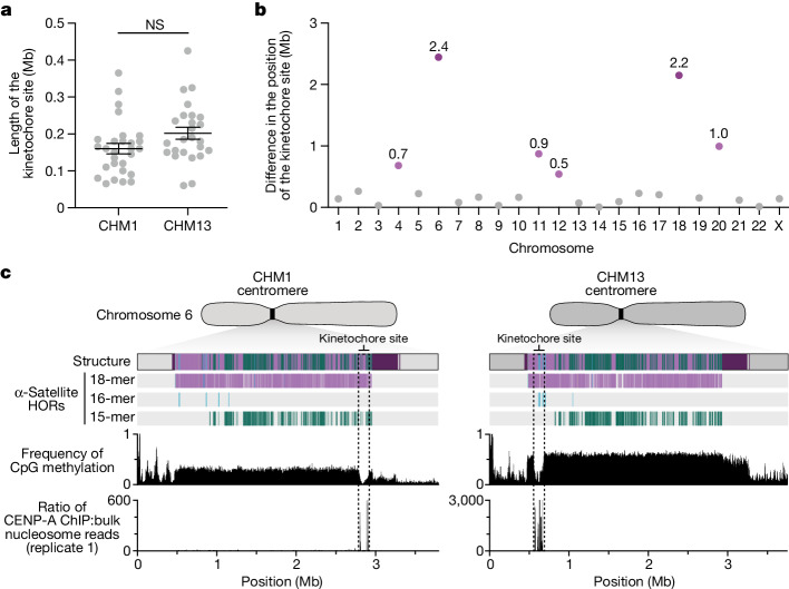
Fig. 4. Variation at the site of the kinetochore among two sets of human centromeres.
a, Comparison of the length of the kinetochore site, marked by hypomethylated DNA and CENP-A-containing chromatin, between the CHM1 and CHM13 centromeres. n = 28 and 25 kinetochore sites for the CHM1 and CHM13 centromeres, respectively. Data are mean ± s.e.m. Statistical analysis was performed using a two-sided Kolmogorov–Smirnov test; NS, not significant. b, The difference in the position of the kinetochore among the CHM1 and CHM13 centromeres. c, Comparison of the CHM1 and CHM13 chromosome 6 centromeres, which differ in kinetochore position by 2.4 Mb.

