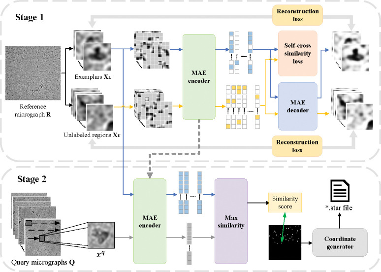
Figure 2:
Overview of the two-stage cryoMAE framework: stage 1 illustrates the training phase with a mix of labeled particle and unlabeled regions, employing reconstruction loss and self-cross similarity loss. Stage 2 depicts the particle picking process, where the trained MAE encoder assesses query micrographs, leveraging latent feature comparisons to identify particle positions accurately.

