

Summary
Cancer is often wrongly considered to be a modern disease in many popular medical venues. Cancers have been known to humanity since ancient times. In fact, its antiquity can be identified through the application of palaeopathological methodologies. The present perspective demonstrates by means of a historical and palaeopathological analysis how oncological manifestations were present long before the emergence of anatomically modern humans and addresses the epidemiological transition from ancient times to the contemporary world. The final section of the article examines breast cancer and its identification in ancient human remains.
Keywords: Breast cancer, Cancer, Epidemiology, Global health, History of medicine, Palaeopathology, Public health
Introduction
According to World Health Organization (WHO) statistics cancer represents “the second leading cause of death globally, accounting for an estimated 9.6 million deaths, or 1 in 6 deaths, in 2018” [1]. Additionally, the WHO identifies oncological manifestations in the lung, prostate gland, colorectal intestine, stomach, and liver as the most common types of cancer in males, whereas the breast, colorectal, lung, neck and thyroid are the most affected anatomical sites in females [1]. According to Siegel et al. “140,000 new cancer cases and 611,720 cancer deaths are projected to occur in the United States” in 2024 [2].
Such figures demonstrate cancer prevalence and the global impact it exerts on global health. For this reason, medicine should not only examine cancer as a modern phenomenon since a much broader perspective on it can be gathered by studying its antiquity and evolutionary trajectory. In addition, by not adopting such an approach, it is also possible to avoid the risk of considering it – the exclusive product of our contemporary societies and lifestyles. A similar interpretative problem could happen with other chronic diseases that are more prevalent today than in the past. The discipline that allows scientists to investigate the antiquity of diseases is called palaeopathology (from Greek: παλαιός = ancient, πάθος = suffering, disease, λόγος = study, research). Palaeopathology utilises both invasive and non-invasive methodologies and techniques also adopted by contemporary medicine (such as histology, radiology, genetics) that offer a complete picture of the clinical presentation of diseases in ancient populations, which are still present in the modern world [3, 4].
CANCER: FROM MYTHOLOGY TO MUMMY RESEARCH
With reference to oncologic pathology, the retrospective diagnostic approach has made it possible to show how wrong it was to perceive it as a relatively recent disease and, almost exclusively, linked to contemporary lifestyles and pollution. Before analysing this issue, it is necessary, nevertheless, to recall some linguistic aspects by offering a historical perspective, in order to remedy the superficial and unconscious use of such terms as “neoplasm”, “tumour” and “cancer”.
Neoplasia (from the Greek νέος and πλάσις) means a new uncontrolled abnormal growth of tissue; tumour (from the Latin “tumor”) originally meant as any kind of tumefaction caused by a tumour process or and inflammatory process, in the first meaning used nowadays interchangeably with “neoplasm”); cancer (in Latin “cancer”, in Greek καρκίνος, literally meaning “crab”) refers to malignant tumours, characterised by high aggressiveness [5].
In Greek mythology the crab Καρκίνος (Fig. 1) was sent by goddess Hera to aid the Hydra of Lerna, a monster representing the second labour to which Heracles had been subjected, who was hated by Hera.
Although defeated in the fight, the crustacean was rewarded by the goddess for his loyalty and was made as a constellation, the fourth sign of the Zodiac [5]. This term was already used by the ancient physicians Hippocrates (ca. 460-377 BC), Galen (AD 129-201) and Paul of Aegina (7th century AD), who wondered about the reason for such an identification between the word “crab” and the pathology it indicated. They went so far as to speculate either the similarity between the vascular turgidity in a tumour-affected breast and the crab’s legs or, metaphorically, the manner in which the disease vigorously and remorselessly grips the body, just as a crab does when it grabs its prey [6].
Hippocrates postulated an early scientific theory on cancer, although previous civilisations may have already developed their own theories on cancer by that time. He assumed that this disease was correlated to an excess of black bile. He believed that cancers, and more broadly any disease, developed when the balance in the four “body humours” (blood, phlegm, yellow bile, and black bile) were in disharmony. For example, the dominance of black bile correlated with the development of cancer.
Afterwards, a Greek physician who trained medicine in Rome, Claudius Galen (AD 129-c. 216) further developed and supported Hippocratic theory, proposing that black bile caused incurable cancer, whereas yellow bile caused curable cancer [7].
Hippocrates further argued and used the word carcinoma, comparing the disease to a crab (ĸαρκίνος) that adheres to its surroundings with its claws [8]. Also, the physician Aulus Cornelius Celsus (25 BC-AD 50), later translated this word and he used the term “cancer”, the Latin word for “crab” and Galen described tumours using the Greek term for swelling, “oncos” [9].
Again following WHO statistics, in the current Western world, cardiovascular disease occupies the first position among the causes of death from disease and is followed by cancer, the number of deaths caused by infectious diseases (especially pulmonary) having greatly decreased nowadays, since they are largely controlled through the administration of antibiotics or even preventable by means of vaccination [10], although a resurge in vaccine-preventable infections cannot be ruled out due to the widespread social phenomenon of vaccine hesitancy [11].
Until the end of the 19th century AD, cancer-related deaths occupied the third position in mortality prevalence. The recent surge of various types of cancer can be interpreted in the light of a positive Darwinian selection favouring this nosological category, which is typically associated with the more mature ages of human life [3], together with a higher diagnostic sensitivity due to the advances of medicine and public prevention campaigns. Increasing prevalence of cancer should not, however, be considered as a disease of modernity.
According to science, cancer has been present for hundreds of millions of years and seems to have emerged in multicellular life forms. Even before the dinosaurs, in Dinichthys (a placoderm, an extinct vertebrate like modern fish that lived 340 mln years ago) a cavitation was identified on the inner surface of the jaw, the potential outcome of an oncological process [9]. Another ancient vertebrate, the fossil fish Phanerosteon mirabile (ca. 300 mln years ago) was characterised by the presence of an osteoma, while in the marine reptile Mosasaurus (ca. 75-65 mln years ago) the first case of a tumour, namely an osteoma, in a vertebra was identified [12, 13]. Nonetheless, it is worth mentioning Rotschild’s stance on the Mosasaurus osteoma, in that the scientist was convinced that this was not an osteoma but a non-neoplastic hamartoma [14].
How, therefore, can we answer the now long-standing question Did cancer exist in antiquity? The answer can only be an emphatic “yes” according to Faltas [15]. Such a statement is corroborated by an impressive body of evidence from different regions of the world and all time periods [15].
Some of the earliest evidence of cancer is found among fossilised bone tumours in human mummies in ancient Egypt, and references to the same has been found in ancient manuscripts.
Since the earliest civilisations, cancer has indeed been present and recorded: let us take, for instance, the case of Ancient Egypt where the evidence from literary sources (including the Ebers Surgical Papyrus) already indicating the existence of the pathology, has been since the 1970s complemented by a robust body of novel information stemming from the anthropological study of the mortal remains of that ancient population.
According to the anthropologist Eugen Strouhal (1931-2016), one of the founders of the field of palaeopathology, the previous discrepancy between historical data and “field” evidence can be explained through the following points:
insufficient analysis and description of tumour cases reported in the literature;
prevalence of the description of cranial tumour lesions related to the preference of old-school anthropologists for the privileged study of the human skull;
rarity, in past generations of scholars, of palaeopathological training that allows for the detection of tumours on bone;
necessity to begin the search for tumours as early as the time of archaeological excavation in necropolis [16]. Among the types of cancer found in Ancient Egypt are numerous malignant conditions that are well known by contemporary medical research: osteosarcoma, multiple myeloma, osteolytic metastatic carcinoma, mixed metastatic carcinoma, multiple myeloma, nasopharyngeal carcinoma, primary osteolytic tumours of the maxillary sinus, and ovarian cystadenocarcinoma [16, 17].
In addition, it is relevant to underline how a major limitation is constituted by the fact that many ancient human remains are found skeletonised and only a minimal part of them retains soft tissues at all: even if embalmed or mummified, it does not always happen that they preserve all their soft tissues, yet only part of them.
Among the benign tumoural forms, at the cranial level, it has only recently been possible to demonstrate the actual existence of frontal sinus osteoma in Ancient Egypt, a condition previously only hypothesised on the basis of an unreasoned observation by Strouhal, of a mummy housed in the Musée d’Éthnographie de Neuchâtel (Switzerland) belonging to a male individual who lived between 664 and 332 BC [18]. The same condition was later confirmed in a more recent ancient Egyptian skull [19]. This very pneumatised anatomical district also represents the venue of the recent discovery of another form of osteoma, osteoid osteoma, considered to date not only very rare in that anatomical site but also believed not to have existed in antiquity at all, in the skull of an individual found in the Roman necropolis of Pianotta di Calatabiano (Fiumefreddo, Catania) radiocarbon dated to AD 418-536. In this case, due to the poor state of preservation of the skeletal find, it was opted for a verification of the radiological datum using histology, which demonstrated the typical tissue structure consisting of a central nest surrounded by an area of bony sclerosis [20]. Another interesting case is represented by the evidence of an osteochondroma in distal podal phalanx from the prehistoric hypogeum of Calaforno (Ragusa, Sicily), as demonstrated by a combination of morphological and palaeoradiological methods [21].
Additional evidence of other cancer types, which combined presentations at the skeletal and soft tissue levels, was extensively highlighted in mummified or embalmed bodies by Nerlich and Bianucci [22].
Undoubtedly, the process of carcinogenesis, both in ancient times and in our contemporary society, is intricately interconnected with a combination of diverse elements encompassing environmental, climatic, social, and hereditary influences.
Historically, the most notable risk factors were linked to the inhalation of polycyclic hydrocarbons emitted from the fumes generated by the prevalent practice of smoking food for cooking and preservation purposes. Additionally, the presence of radon gas, a naturally occurring radioactive gas predominantly found in caves and underground areas, further contributed to the potential development of cancer [23].
Moreover, the contamination of crops by toxins produced by plants and microorganisms served as an additional source of risk [23]. Furthermore, the damaging effects of ultraviolet radiation and specific viruses, such as papillomavirus, in conjunction with an individual’s genetic predisposition and the consumption of certain medications, all played meaningful roles within the intricate network of factors associated with carcinogenesis [23].
BREAST CANCER IN ANTIQUITY: WHAT IS THE EVIDENCE?
Regarding breast pathology, unfortunately, no definitive data on mummies could be found to date due to the poor preservation of mammary tissues in ancient human remains. Speculations about the gynecomastia of Egyptian pharaoh Tutankhamun (ca. 1341-1323 BC), based only on the morphology of artistic depictions and not on the biological data, can be dismissed in the words of Harrison, who was able to study the famous mummy:
The distortionate expressionism of Egyptian art in the XVIIIth Dynasty which was introduced by Akhenaten and displayed in his own monuments to the most marked degree, rubbed off on some, but not all, representations of his descendants and the pharaohs Smenkhkare and Tutankhamun [24].
In truth, gynaecomastia has only recently been demonstrated in a mummy through the multidisciplinary study of the body of the Finnish vicar Rungius (ca. AD 1560-1629) [25], while statuary examples from the Hellenistic period have recently been offered [26]. A further example of mammary pathology is the proposed diagnosis of hyaline fibroadenoma in the mummy of Mary of Aragon (AD 1503-1568) [27].
As for breast cancer, on the other hand, suggestive descriptions are found in the Ancient Egyptian Edwin Smith Papyrus (3000-2500 BC), while in Greece Hippocrates and Galen theorised that the aetiology of the disease resided in an excess of black bile, according to the humoral theory of the time [7, 28]. Moreover, in some recent studies based on the iconodiagnostic approach, the presence of breast cancer has been hypothesised in Renaissance works of art, including Michelangelo’s masterpiece La Notte in the New Sacristy of the Church of San Lorenzo in Florence [29] and Michele di Rodolfo del Ghirlandaio’s work La Notte in Florence [30]. Additionally, Nerlich and colleagues [31], adopting the same approach, reported a possible case of right breast cancer in Rembrandt’s work Woman Sitting Half-Dressed beside a Stove (AD 1658).
Despite sporadic remarks from secondary sources, there remains a significant lack of thorough presentation of palaeopathology in biological remains. Nevertheless, the methodical examination of mummies presents an opportunity to address this deficiency in the coming years, offering invaluable knowledge about the well-being and ailments of past civilisations.
Conclusions
To summarise, in order to effectively tackle the complex and ever-evolving challenges faced in oncology and modern medicine as a whole, it is crucial to engage in a thorough and contemplative analysis. This analysis should be enhanced by various branches of medicine, specifically those with an archaeological focus, as they can provide unparalleled insights into the historical context and evolutionary factors that influence diseases. By incorporating these multidisciplinary insights, we can attain a holistic and profound comprehension of the intricate nature of illnesses, ultimately leading to the development of more efficient and effective approaches for prevention, diagnosis, and treatment. Through this comprehensive and collaborative approach, we can strive towards improving the overall well-being and health outcomes for individuals and communities alike.
Funding
This research received no external funding.
Informed consent statement
Not applicable.
Data availability statement
Not applicable.
Conflict of interest statement
The authors declare that the research was conducted in the absence of any commercial or financial relationships that could be construed as a potential conflict of interest.
Authors’ contributions
FMG, VP: designed the study. FMG: conceived the manuscript. FMG, VP, EV, MM: drafted the manuscript.MV, EV, MM: revised the manuscript. MV, VP, EV, FMG, MM: performed a search of the literature. MM, VP: critically revised the manuscript. FMG, EV: conceptualization, and methodology. MV, VP, EV: investigation and data curation. FMG, VP: original draft preparation. EV, MM, FMG: review. MM: editing. All authors have read and approved the latest version of the paper for publication.
Figures and tables
Fig. 1.
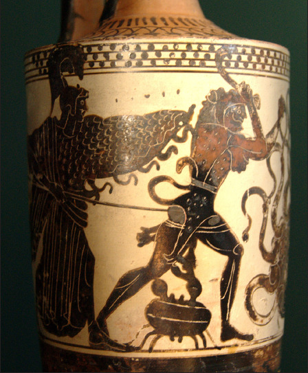
Heracles attacked by the Crab and the Lernaean Hydra. White-ground Attic lekythos, ca. 500-475 BC (Wikipedia Commons- Public domain).
References
- [1].WHO- Cancer. Available at: https://www.who.int/health-topics/cancer#tab=tab_1 (Accessed on: 19/03/2024).
- [2].Siegel RL, Giaquinto AN, Jemal A. Cancer statistics, 2024. CA Cancer J Clin 2024;74:12-49. https://doi.org/10.3322/caac.21820. 10.3322/caac.21820 [DOI] [PubMed] [Google Scholar]
- [3].Rühli FJ, Galassi FM, Haeusler M. Palaeopathology: Current challenges and medical impact. Clin Anat 2016;29:816-22. https://doi.org/10.1002/ca.22709. 10.1002/ca.22709 [DOI] [PubMed] [Google Scholar]
- [4].Grauer AL. A century of paleopathology. Am J Phys Anthropol 2018;165:904-914. https://doi.org/10.1002/ajpa.23366. 10.1002/ajpa.23366 [DOI] [PubMed] [Google Scholar]
- [5].Andrews T. Dictionary of Nature Myths: Legends of the Earth, Sea, and Sky. Oxford: Oxford University Press; 2000, p. 30. [Google Scholar]
- [6].Wagener DJTh. The History of Oncology. Houten: Springer; 2009, p. 14. [Google Scholar]
- [7].Di Lonardo A, Nasi S, Pulciani S. Cancer: we should not forget the past. J Cancer 2015;6:29-39. https://doi.org/10.7150/jca.10336. 10.7150/jca.10336 [DOI] [PMC free article] [PubMed] [Google Scholar]
- [8].Hajdu SI. A note from history: landmarks in history of cancer, Part 1. Cancer 2011;117:1097-102. https://doi.org/10.1002/cncr.25553. 10.1002/cncr.25553 [DOI] [PubMed] [Google Scholar]
- [9].Mitrus I, Bryndza E, Sochanik A, Szala S. Evolving models of tumor origin and progression. Tumour Biol 2012;33:911-7. https://doi.org/10.1007/s13277-012-0389-0. 10.1007/s13277-012-0389-0 [DOI] [PMC free article] [PubMed] [Google Scholar]
- [10].WHO- The top 10 causes of death. Available at: https://www.who.int/news-room/fact-sheets/detail/the-top-10-causes-of-death (Accessed on: 19/03/2024).
- [11].Orsini D, Bianucci R, Galassi FM, Lippi D, Martini M. Vaccine hesitancy, misinformation in the era of COVID-19: lessons from the past. Ethics Med Public Health 2022;24:100812. https://doi.org/10.1016/j.jemep.2022.100812. 10.1016/j.jemep.2022.100812 [DOI] [PMC free article] [PubMed] [Google Scholar]
- [12].Capasso LL. Antiquity of cancer. Int J Cancer 2005;113:2-13. https://doi.org/10.1002/ijc.20610. 10.1002/ijc.20610 [DOI] [PubMed] [Google Scholar]
- [13].Moodie RL. Paleontological evidences of the antiquity of disease. Sci Mon 1918;9:265-281. [Google Scholar]
- [14].Rothschild BM, Schultze H-P, Pellegrini R. Herpetological Osteopathology: Annotated Bibliography of Amphibians and Reptiles. New York: Springer; 2012. [Google Scholar]
- [15].Faltas B. Cancer is an ancient disease: the case for better palaeoepidemiological and molecular studies. Nat Rev Cancer 2011;11:76; author reply 76. https://doi.org/10.1038/nrc2914-c1. 10.1038/nrc2914-c1 [DOI] [PubMed] [Google Scholar]
- [16].Strouhal E. Tumors in the remains of ancient Egyptians. Am J Phys Anthropol 1976;45:613-20. https://doi.org/10.1002/ajpa.1330450328. 10.1002/ajpa.1330450328 [DOI] [PubMed] [Google Scholar]
- [17].Giuffra V, Ciranni R, Fornaciari G. I tumori maligni nell’Antico Egitto e in Nubia. Egitto e Vicino Oriente 2004;27:81-93. [Google Scholar]
- [18].Seiler R, Öhrström LM, Eppenberger P, Gascho D, Rühli FJ, Galassi FM. The earliest known case of frontal sinus osteoma in man. Clin Anat 2019;32:105-9. https://doi.org/10.1002/ca.23301. 10.1002/ca.23301 [DOI] [PubMed] [Google Scholar]
- [19].Galassi FM, Varotto E, Angelici D, Picchi D. Further Paleoradiological Evidence of Frontal Sinus Osteoma in Ancient Egypt. J Craniofac Surg 2020;31:604-5. https://doi.org/10.1097/SCS.0000000000006240. 10.1097/SCS.0000000000006240 [DOI] [PubMed] [Google Scholar]
- [20].Varotto E, Magro MT, Brancato R, Lubritto C, Memeo L, Galassi FM. Unique Osteoid Osteoma of the Frontal Sinus from the Late Roman Empire. J Craniofac Surg 2019;30:965-6. https://doi.org/10.1097/SCS.0000000000005312. 10.1097/SCS.0000000000005312 [DOI] [PubMed] [Google Scholar]
- [21].Varotto E, Militello PM, Platania E, Sferrazza P, Galassi FM. Paleopathological study of a podal osteochondroma from the prehistoric Hypogeum of Calaforno (Sicily). Clin Anat 2021;34:19-23. https://doi.org/10.1002/ca.23603. 10.1002/ca.23603 [DOI] [PubMed] [Google Scholar]
- [22].Nerlich AG, Bianucci R. Paleo-Oncology and Mummies. In: Shin DH, Bianucci R, eds. The Handbook of Mummy Studies. New Frontiers in Scientific and Cultural Perspectives. Singapore: Springer; 2021, pp. 131-46. [Google Scholar]
- [23].Fornaciari G, Giuffra V. Lezioni di Paleopatologia. Genova: ECIG 2009, pp. 302-5. [Google Scholar]
- [24].Harrison RG. Tutankhamun postmortem. Lancet 1973;1:259. [PubMed] [Google Scholar]
- [25].Väre T, Galassi FM, Niinimäki J, Junno JA. Potential case of gynecomastia in mummified remains of an early modern period Northern Finnish Vicar. Clin Anat 2018;31(5):641-644. https://doi.org/10.1002/ca.23187. 10.1002/ca.23187 [DOI] [PubMed] [Google Scholar]
- [26].Galassi FM, Cossarizza A, Varotto E. Superior vena cava syndrome and gynecomastia in antiquity: paleodermatologic considerations on aging in the past. Clin Dermatol 2023;41:309-11. https://doi.org/10.1016/j.clindermatol.2023.05.001. 10.1016/j.clindermatol.2023.05.001 [DOI] [PubMed] [Google Scholar]
- [27].Ventura L, Gaeta R, Giuffra V, Mercurio C, Pistoia ML, Ciccozzi A, Castagna M, Fornaciari G. Breast pathology in ancient human remains. An approach to mummified mammary gland by modern investigation methods. Pathologica 2014;106:216-8. [Google Scholar]
- [28].Retief FP, Cilliers L. Breast cancer in antiquity. S Afr Med J 2011;101:513-5. [PubMed] [Google Scholar]
- [29].Stark JJ, Nelson JK. The breasts of “Night”: Michelangelo as oncologist. N Engl J Med 2000;343:1577-8. https://doi.org/10.1056/NEJM200011233432118. 10.1056/NEJM200011233432118 [DOI] [PubMed] [Google Scholar]
- [30].Bianucci R, Perciaccante A, Charlier P, Appenzeller O, Lippi D. Earliest evidence of malignant breast cancer in Renaissance paintings. Lancet Oncol 2018;19:166-7. https://doi.org/10.1016/S1470-2045(18)30035-4. 10.1016/S1470-2045(18)30035-4 [DOI] [PubMed] [Google Scholar]
- [31].Nerlich AG, DeWaal JC, Donell ST, Bianucci R. Breast cancer in Woman Sitting Half-Dressed beside a stove (1658) by Rembrandt van Rijn. Breast 2022;64:134-5. https://doi.org/10.1016/j.breast.2022.06.001. 10.1016/j.breast.2022.06.001 [DOI] [PMC free article] [PubMed] [Google Scholar]
Associated Data
This section collects any data citations, data availability statements, or supplementary materials included in this article.
Data Availability Statement
Not applicable.