Abstract
Background
Karst caves serve as natural laboratories, providing organisms with extreme and constant conditions that promote isolation, resulting in a genetic relationship and living environment that is significantly different from those outside the cave. However, research on cave creatures, especially Opiliones, remains scarce, with most studies focused on water, soil, and cave sediments.
Results
The structure of symbiotic bacteria in different caves were compared, revealing significant differences. Based on the alpha and beta diversity, symbiotic bacteria abundance and diversity in the cave were similar, but the structure of symbiotic bacteria differed inside and outside the cave. Microorganisms in the cave play an important role in material cycling and energy flow, particularly in the nitrogen cycle. Although microbial diversity varies inside and outside the cave, Opiliones in Beijing caves and Hainan Island exhibited a strong similarity, indicating that the two environments share commonalities.
Conclusions
The karst cave environment possesses high microbial diversity and there are noticeable differences among different caves. Different habitats lead to significant differences in the symbiotic bacteria in Opiliones inside and outside the cave, and cave microorganisms have made efforts to adapt to extreme environments. The similarity in symbiotic bacteria community structure suggests a potential similarity in host environments, providing an explanation for the appearance of Sinonychia martensi in caves in the north.
Supplementary Information
The online version contains supplementary material available at 10.1186/s12862-024-02248-9.
Keywords: Symbiotic bacteria, Opiliones, Karst cave, Ecology
Introduction
Symbiotic bacteria play an important role in the life activities of many organisms, including insects and animals [1]. These bacteria reside in the host’s intestinal tract, epidermis and various organs, and jointly regulate each other’s life activities. The number of symbiotic bacteria even far exceeds the number of cells of the organism itself, and the diversity and the functions of symbiotic bacteria are vast, ranging from digestion and absorption [2–4] to detoxification, metabolism [5, 6], controlling the growth and reproduction of the host [7, 8], and adaptation to the environment [9–11].
Karst caves are a unique environment characterized by low air mobility, long-term darkness, high humidity, malnutrition, and isolation from the external environment. Researchers have studied microbial communities in small habitats such as cave rock walls [12], sediments [13], water and air [14]. The dominant phyla in these communities were Proteobacteria, Actinobacteria, and Firmicutes, although their relative abundance varied greatly among different caves [14]. Cave-dwelling organisms are also isolated from the outside world for a long time, which leads to the development of unique structures and habits. As a result, the cave-dwelling creatures differ from their counterparts outside the cave, both in their living environment and genetic relationships [15, 16]. In recent years, there have been a few studies on cave symbiotic bacteria. One study compared the symbiotic bacteria of Oculina patagonica obtained from bleached, cave and healthy corals and found a significant change in bacterial population in the absence of light [17]. Another study investigated cave calcium bodies in terrestrial isopod crustaceans Titanethes albus and Hyloniscus riparius [18].
Most studies on symbiotic bacteria of arachnids focus on the effects of endosymbiont on spiders [19, 20], mites [21], pseudoscorpions [22, 23] and other arachnids. However, relatively few studies have examined the entire bacterial community. One study of Marpiss magister found that endosymbionts are not the only microflora in spiders, and there are other bacterial groups in their bacterial communities [24]. Opiliones (harvestmen, shepherd spiders, daddy longlegs, etc.) are one of the most abundant and the oldest Arachnida. They are often widely distributed in ecosystems, independent Cardinium groups have been found in them. Some scholars use Opiliones as a control while studying Wolbachia and Cardinium infection of Araneae [25–28]. So far, studies on the symbiotic bacteria of Opiliones have mainly focused on single endosymbiont. To the best of our knowledge, no reports have been found on Opiliones-related bacteria based on different habitats and their effects on the growth, evolution and reproduction of the host. Opiliones have become an ideal subject for studying biological habitats due to their limited activity and narrow distribution areas [29, 30].
In this study, high-throughput technology (16S ribosomal RNA [rRNA] gene sequencing method) was used to analyze the relative abundance and diversity of symbiotic bacteria in different habitats of Opiliones and other arachnids, provide a basis for revealing the special material cycle in the cave environment, especially the nitrogen cycle. A variety of symbiotic bacteria have reproductive regulation and even co-evolution on the host. The comparison of symbiotic bacteria in Opiliones from different karst caves and habitats provided insights into the role of bacteria in the development and evolution of Opiliones and their distribution of the host.
Materials and methods
Sample collection
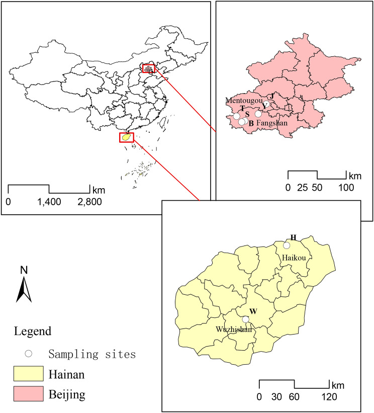
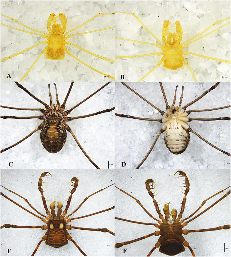
During the period from October 2021 to July 2022, five species of Opiliones were collected in Beijing and Hainan Island: Sinonychia martensi (Arachnida: Opiliones: Laniatores), Euepedanus flavimaculatus (Arachnida: Opiliones: Laniatores), Plistobunus columnarius (Arachnida: Opiliones: Laniatores), Himalphalangium palpalis (Arachnida: Opiliones: Eupnoi), Homolophus serrulatus (Arachnida: Opiliones: Eupnoi). Additionally, other Arachnida, including Pimoa clavata (Arachnida: Araneae), Spelaeochthonius sp. (Arachnida: Pseudoscorpionida) and Draconarius sp. (Arachnida: Araneae) were collected from various sections of Siyu Cave in Fangshan District (Fig. 1; Table 1). All samples were identified using traditional morphological classification (Fig. 2) and preserved in 95% ethanol.
Fig. 1.
Sampling sites in Beijing and Hainan Island. The location number corresponds to the first letters of the sample number
Table 1.
Samples were collected from different areas and caves
| Family | Genus | Species | Site | Latitude and longitude | Habitat | Group |
|---|---|---|---|---|---|---|
| Epedanidae | Euepedanus | E. flavimaculatus |
Haikou City, Hainan Province |
20°0′43.44″N, 110°18′56.57″E |
Forest, bush | HOF |
| Plistobunus | P. columnarius | Wuzhishan City, Hainan Province |
18°52′8.54″N, 109°40′57.71″E |
Forest, bush | WOC | |
| Cladonychiidae | Sinonychia | S. martensi |
Bianfu Cave, Beijing (Fangshan) |
39°42’23.94"N, 115°43’04.63"E |
The deep zone of the cave | BOM |
|
Siyu Cave, Beijing (Fangshan) |
39°41′1689″N, 115°40′4.83″E |
The deep zone of the cave | SOM | |||
|
You Cave Beijing (Fangshan) |
39°48′47.62″N, 115°55′21.19″E |
The deep zone of the cave | YOM | |||
| Sclerosomatidae | Himalphalangium | H. palpalis | Tangshang Village, Beijing (Fangshan) |
39°46′42.71″N, 115°35′20.57″E |
Forest, bush | TOP |
| Homolophus | H. serrulatus | Jingxi Ancient Road, Beijing (Mentougou) |
39°58′0.573″N, 116°2′59.07″E |
Forest, bush | JOS | |
| Tangshang Village, Beijing (Fangshan) |
39°46′42.71″N, 115°35′20.57″E |
Forest, bush | TOS | |||
| Pseudotyrannochthoniidae | Spelaeochthonius | Spelaeochthonius sp. |
Siyu Cave, Beijing (Fangshan) |
39°41′1689″N, 115°40′4.8252″E |
The deep zone of the cave | SPS |
| Pimoidae | Pimoa | P. clavata |
Siyu Cave, Beijing (Fangshan) |
39°41′1689″N, 115°40′4.8252″E |
The deep zone of the cave | SAC |
| Agelenidae | Draconarius | Draconarius sp. |
Siyu Cave, Beijing (Fangshan) |
39°41′1689″N, 115°40′4.8252″E |
Within 5 m of the entrance | SAS |
Note The deep zone of the cave: completely dark, high humidity and constant temperature. With the exception of S. martensi, P. clavata and Spelaeochthonius sp., other species were collected outside the cave. The three letters of the group name represent the site, order and species, respectively. For example, SOM corresponds to the Siyu Cave, Opiliones and martensi
Fig. 2.
Photographs of some Opiliones. (A, B) Sinonychia martensi, Laniatores; (C, D) Homolophus serrulatus, Eupnoi; (E, F) Euepedanus flavimaculatus, Laniatores. (A, C, E) dorsal view, (B, D, F) ventral view
Sample processing and DNA extraction
The collected samples were thoroughly rinsed with sterile water 2–3 times, followed by sequential sterilization using 75% ethanol and hypochlorite, and finally washed again with sterile water. The processed samples were then stored at a temperature of -80℃ until DNA extraction. The extracted DNA was quantified using Nanodrop, and the quality of the extracted DNA was verified by running it through 1.2% agarose gel electrophoresis. The obtained DNA was stored at -20℃ for future use.
Amplicon and library preparation
The 16S rRNA gene library construction and sequencing were performed by Personalbio Technology Company (Shanghai, China). The V3-V4 regions of the 16S rRNA gene were amplified using the primer pair 338 F/806R (5′-ACTCCTACGGGAGGCAGCA-3′ and 5′-GGACTACHVGGGTWTCTAAT-3′). PCR products were quantified using the PicoGreen dsDNA Assay Kit (Invitrogen, Carlsbad, USA), and samples were mixed according to the corresponding proportion based on the fluorescence quantitative results. The sequencing library was prepared using the TruSeq Nano DNA LT Library Prep Kit of the Illumina platform (Illumina, USA), and before sequencing, the library was checked. The qualified sequencing libraries were diluted, mixed, and denatured for computer sequencing.
Data processing and analysis
The original sequences were quality screened and divided into libraries and samples according to index and Barcode information, and the barcode sequences were removed. The primer fragments were removed using qiime cutadapt trim-paired sequence, and unmatched primers were discarded. The qiime dada2 denoise-paired was used for quality control, denoising, splicing and de-chimerism. After denoising, all libraries, the ASVs feature sequence and the ASV table were merged, and singletons ASVs (ASV with a total sequence of only 1 in all samples) were removed [31, 32]. Annotation information was obtained using different algorithms and parameters in different databases, including the Silva database (http://www.arb-silva.de), nt database (http://ftp://ftp.ncbi.nih.gov/blast/db/), nr database (http://ftp://ftp.ncbi.nih.gov/blast/db/), based on the feature sequence of ASVs [33].
The composition and relative abundance of bacterial groups in each sample at different taxonomic levels were obtained by QIIME2 software (2019.4) [34]. The richness and diversity of bacteria in different samples were analyzed using alpha diversity indices (including Chao1 richness estimator, Observed_species, Shannon diversity index, and Simpson index) [35]. Beta diversity analysis was performed to analyze differences in bacterial community composition structure among different samples, and beta diversity was evaluated by PCoA and Non-metric multidimensional scaling (NMDS) based on the weighted Bray-Curtis distance algorithm [35, 36]. According to the abundance data of ASVs in all samples, the Wenn diagram was made according to the grouping of samples and calculating the relationship between each set. Indicator groups in different habitats were obtained by the LEfSe (the least discriminant analysis effect size) method with the LDA (linear discriminant analysis) threshold set to 4.0 [37]. The similarity between samples was shown in the form of hierarchical tree, and the clustering effect was measured by the branch length of the clustering tree. The heat map was used to analyze the species composition and displayed the species abundance distribution trend of each sample.
PICRUSt2 (Phylogenetic Investigation of Communities by Reconstruction of Unobserved States) was utilized to predict the functional abundance of samples based on the sequence abundance of marker genes in these samples [38]. FAPROTAX (Functional Annotation of Prokaryotic Taxa) is a functional annotation database specifically designed for prokaryotic taxa, enabling the functional prediction of bacterial communities [39]. It can be employed to evaluate bacterial metabolism and other ecologically related functions in cave environments, particularly focusing on the cycling function of sulfur, carbon, hydrogen and nitrogen.
Results
Diversity of the bacteria in Opiliones among different karst caves
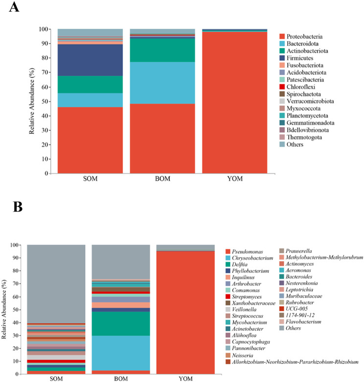
A study was conducted to investigate the diversity of bacteria in Sinonychia martensi, a species of harvestmen found in karst caves in Beijing. The results revealed significant differences in community structure and relative abundance of bacteria among different caves. In S. martensi inhabiting caves, the dominant phylum was Proteobacteria, with a much higher relative abundance compared to other bacteria. Proteobacteria accounted for an extremely high relative abundance of 98.06% in the samples from the You Cave (YOM) (Fig. 3a, Table S1). The relative abundance of Firmicutes in the Siyu Cave was 21.90%, second only to Proteobacteria, compared to the Bianfu Cave and You Cave. The three most abundance phyla accounted for more than 93% of the total microflora of S. martensi from the Bianfu Cave (BOM): Proteobacteria (48.10%), Bacteroidetes (29.01%), Actinobacteria (16.22%). At the genus level, Pseudomonas was the dominant bacteria in S. martensi from the You Cave (YOM), with a relative abundance as high as 95.17% (Fig. 3b, Table S1). The relative abundance of Chryseobacterium and Delftia in the samples from the Bianfu Cave (BOM) was 27.00% and 18.37%, respectively.
Fig. 3.
Relative abundance of S. martensi in Siyu, Bianfu and You Cave. (A) The relative abundance of phylum level in S. martensi. It shows the top 15. (B) Relative abundance of genus level in S. martensi. It shows the top 30. The relative abundance of other species merged and classified as Others
The alpha diversity of bacteria in different caves was assessed by Chao 1, Observed_species, Shannon and Simpson indices (Fig. 4a). A Kruskal–Wallis test revealed a significant difference in alpha diversities among the caves (P ≤ 0.05). According to the Dunn’s post hoc test, the diversity and richness of S. martensi from the Siyu Cave (SOM) were significantly higher than those from You Cave (YOM) (P < 0.05). The beta diversity was evaluated by principal coordinate analysis (PCoA) based on the weighted Bray-Curtisdistance algorithm, showing that Opiliones samples were loosely clustered (Fig. 4b). In the first dimension, the community composition of the Opiliones samples from Siyu Cave (SOM) was similar to that of the Bianfu Cave (BOM). It was more similar for symbiotic bacteria from You Cave (YOM) and Bianfu Cave (BOM) in the second dimension. These findings suggest that caves, as unique and enclosed habitats, harbor distinct symbiotic bacteria across different caves
Fig. 4.
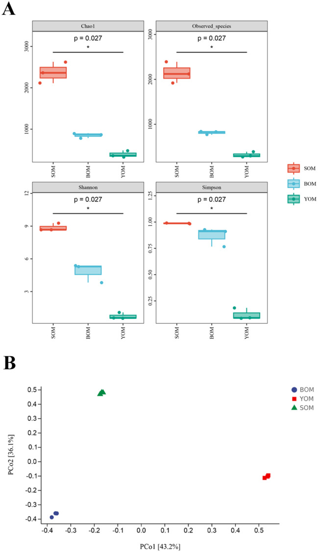
Comparison of microbial diversity in different cave samples. (A) Alpha-diversity analysis of S. martensi microbiome. Species richness and diversity were measured by Shannon, Simpson, Chao1 and Observed_species. Each panel corresponds to an alpha-diversity index, which is identified in the gray area at its top. In each panel, the abscissa is the grouping label, and the ordinate is the value of the corresponding alpha diversity index. The number under the diversity index label is the P value of the Kruskal-Wallis test. (B) Principal coordinate analysis (PCoA) of microbiome based on Bray-Curtis distances
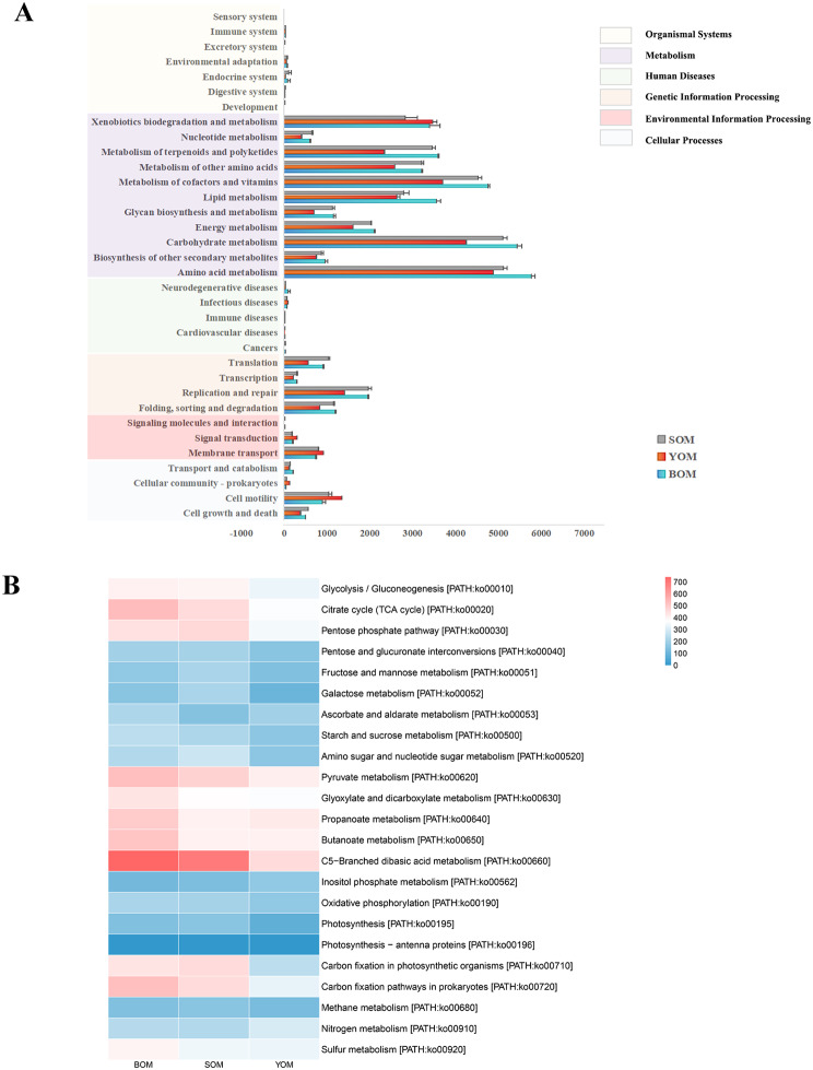
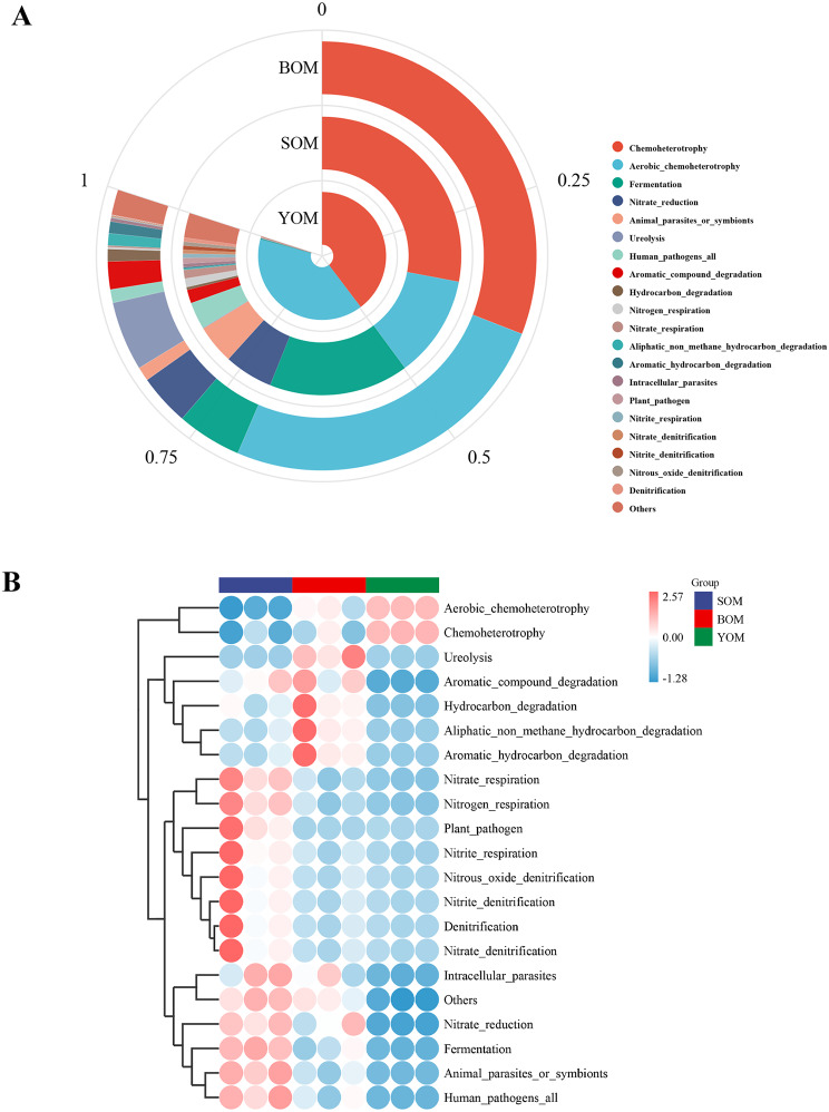
Caves represent unique habitats with distinct energy flows and material circulations compared to the outside environment, and each creature has adapted unique behaviors to survive. To understand the function of bacteria in different caves, PICRUSt2 software [18] was used to predict and analyze the symbionts. The KEGG (Kyoto Encyclopedia of Genes and Genomes) database was used to identify six functional modules, including cellular processes, environmental information processing, genetic information processing, human diseases, metabolism, and organismal systems. The metabolism module was found to be the most abundant functional module in all samples (Fig. 5a), which might include the glycolysis pathway, pentose phosphate pathway, nitrogen metabolism, sulfur metabolism, methane metabolism and multiple carbon sequestration pathways (Fig. 5b). The relative abundance of pathways was in different caves and samples. The COG (clusters of orthologs groups) database was used to predict key enzymes in multiple reaction pathways (Table S2), including Hexokinase [EC:2.7.1.1], glyceraldehyde 3-phosphate dehydrogenase [EC:1.2.1.12] and pyruvate kinase [EC:2.7.1.40] involved in the glycolysis pathway. Glucose-6-phosphate isomerase [EC:5.3.1.9] is involved in the pentose phosphate pathway. Nitrite reductase [EC1.7.2.1] catalyzes the reduction of nitrite (NIT), a crucial enzyme in the nitrogen cycle in nature. Chemoheterotrophy and aerobic chemoheterotrophy were found to be the most abundant functions based on the predictions made by FAPROTAX analyses (Fig. 6a). Among the top 20 predicted functions, nine were related to the nitrogen cycle, including nitrate reduction, nitrogen respiration, nitrate respiration, ureolysis, nitrification, nitrite respiration, nitrate denitrification, nitrite denitrification and nitrous oxide denitrification. Specifically, the SOM samples showed high abundance in the functions related to the nitrogen cycle and the BOM samples were positively correlated with the degradation function of organic compounds (Fig. 6b). These findings indicate that different caves employ different strategies in the nutrient cycle
Fig. 5.
Predicted bacterial function in cave samples using PICRUSt2. (A) Predicted function of bacteria among the five samples. The first level of the KEGG pathway was represented by different colors. (B) The second level of the KEGG pathway was shown in the heatmap. It included only carbohydrate metabolism and energy metabolism pathways
Fig. 6.
Predicted bacterial function in cave samples using FAPROTAX. It showed the top 20 functions predicted. (A) Abundance of the main metabolisms in the whole set of cave samples. (B) Heatmap of the FAPROTAX analysis revealed the differences among samples
Differences in the symbiotic bacteria between Opiliones inside and outside the cave
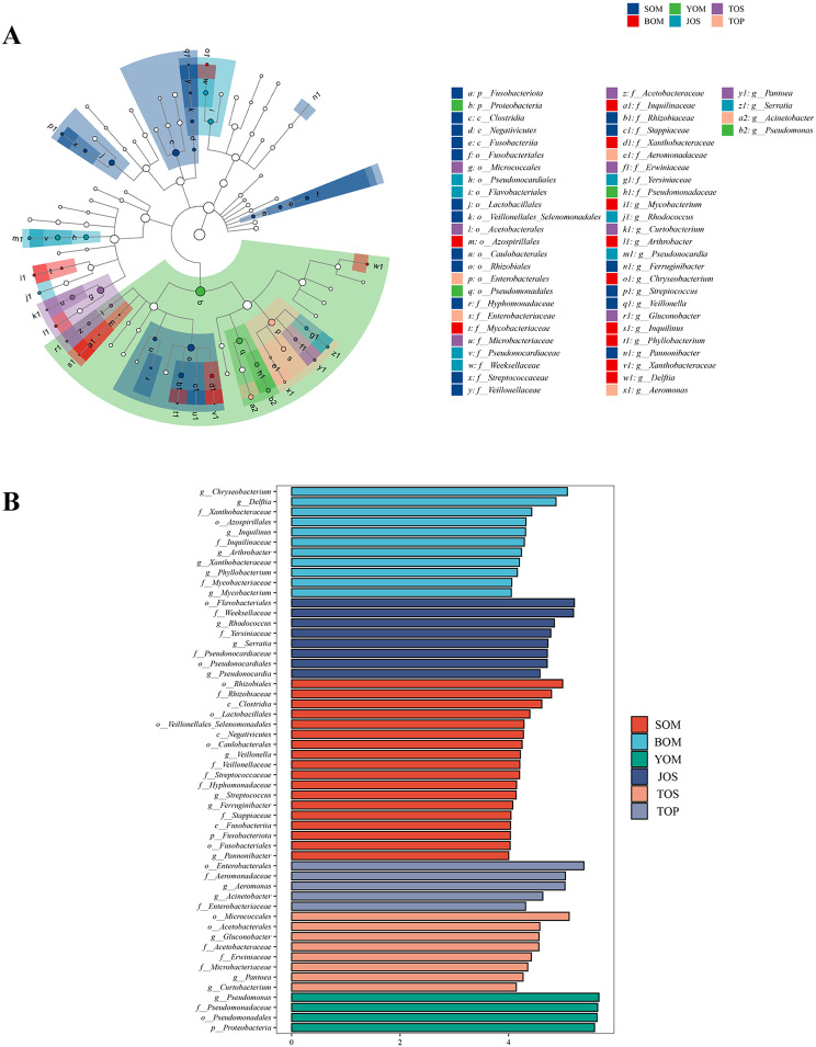
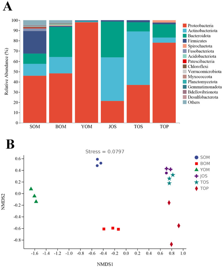
The sequencing results of Himalphalangium palpalis and Homolophus serrulatus outside the caves and S. martensi inside the cave revealed differences in the bacteria structure of symbiotic bacteria inside and outside the cave were different. LEfSe analysis was performed to compare bacterial communities to find the specialized bacterial groups within each type of the sample (Fig. 7a). The cladogram showed that 2 phyla, 3 classes, 12 orders, 17 families, and 20 genera were significantly variable across the five samples. When the LDA value was set to 4, the number of differentially abundant bacterial groups in the five samples were 11 (BOM), 8 (JOS),18 (SOM), 5 (TOP), 8 (TOS)and 4 (YOM), respectively (Fig. 7b). At the phylum level, Proteobacteria was the main dominant group in cave Opiliones, accounting for 98.06%, 48.10% and 45.80% in the three caves, respectively (Fig. 8a, Table S3). In contrast, Actinobacteria and Proteobacteria were differentially distributed in samples outside the cave, with Actinobacteria having the highest relative abundance in Homolophus serrulatus (TOS and JOS), and Proteobacteria being dominant in Himalphalangium palpalis (TOP). The NMDS plots based on Bray-Curtis distances clearly distinguished the Opiliones inside the cave from those outside the cave (Fig. 8b).
Fig. 7.
Results of LEfSe analysis. (A) Cladograms indicating the phylogenetic distribution of bacterial lineages associated with Opiliones inside and outside the cave. (B) Indicator bacterial group significantly differentiated across the three sample types with LDA values was 4
Fig. 8.
Diversity of symbiotic bacteria from Opiliones inside and outside caves in Beijing. (A) Relative abundance of Opiliones inside and outside caves. It showed the top 15 phyla and the relative abundance of other species merged and classified as Others. (B) Non-metric multidimensional scaling (NMDS) plots of microbiome difference at ASV level based on Bray-Curtis distances. The stress was 0.0797
Effect of cave environment on symbiotic bacteria of Arachnida
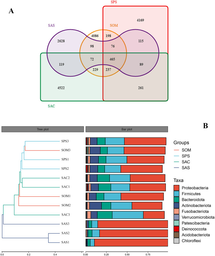
A study was conducted to investigate the effect of the cave environment on the symbiotic bacteria of three different Arachnida species: S. martensi (Opiliones), P. clavata (Araneae) and Spelaeochthonius sp. (Pseudoscorpionida) inside the Siyu Cave. The results showed that the bacteria structure in the samples was similar. Proteobacteria, Firmicutes, Bacteroidetes and Actinobacteria were consistently prevalent phyla among all species inside the cave, accounting for a comparable proportion (Figure S1a, Table S4). Draconarius sp. (Arachnida: Araneae) collected outside the cave differs from that inside. The relative abundance of Proteobacteria in the SAS was 85.10%, which was significantly higher than that inside the cave. At the genus level, the relative abundance of Wolbachia and Rickettsia in SAS was 49.52% and 23.82% respectively, whereas other bacteria were less than 1.5% (Figure S1b, Table S4). The diversity and richness of the symbiotic bacteria inside the cave were higher than those outside the cave (Figure S1c). The Venn diagram revealed that there were differences in ASVs among the four samples. A total of 465 ASVs were shared among all four samples and the cave samples shared 237 ASVs. The number of ASVs unique to spiders outside the Siyu Cave (SAS) was 2628 (Fig. 9a). The members of SAS were loosely clustered, which was confirmed in the hierarchical clustering diagram based on Bray-Curtis distance level and histograms of species composition (Fig. 9b).
Fig. 9.
Comparative microbial diversity of Arachnida in Siyu Cave using bate-diversity analysis. (A) Venn diagram of Siyu Cave samples. (B) Hierarchical clustering analysis of the similarity between samples of Siyu Cave. The hierarchical clustering analysis was utilized to demonstrate the similarity between the samples, represented by a hierarchical tree in the left panel. The samples were clustered based on their similarity, with shorter branch lengths indicating greater similarity. The right panel shows the top 10 phyla in a stacked histogram
Some similarities in environment between karst caves and Hainan Island
We conducted a comparison of symbiotic diversity between Opiliones from Hainan Island and those inside Beijing caves. To reflect their similarity, we utilized Opiliones outside the Beijing caves as a control group. At the phylum level, Proteobacteria was the dominant bacteria across all samples, with relative abundances of 98.06%, 92.36% and 78.18% in YOM, HOF and TOP, respectively (Figure S2a, Table S5). However, Actinobacteria was the main dominant bacteria in JOS and TOS, with lower levels in other classifications. Firmicutes emerged as the secondary dominant bacteria in HOF, WOC and SOM, with the relative abundance of 6.31%, 20.17%, and 21.90% respectively.
The taxonomic alpha-diversities measured by Simpson indicated significant differences in the diversity of symbiotic bacteria among different groups (P < 0.05). Furthermore, Shannon, Chao1 and Observed_species indicated significant differences in diversity and richness (P ≤ 0.01). Notably, Chao1 and Observed_species indicated differences between HOF and SOM, and significant diversity differences were observed between YOM and TOS (Figure S2b).
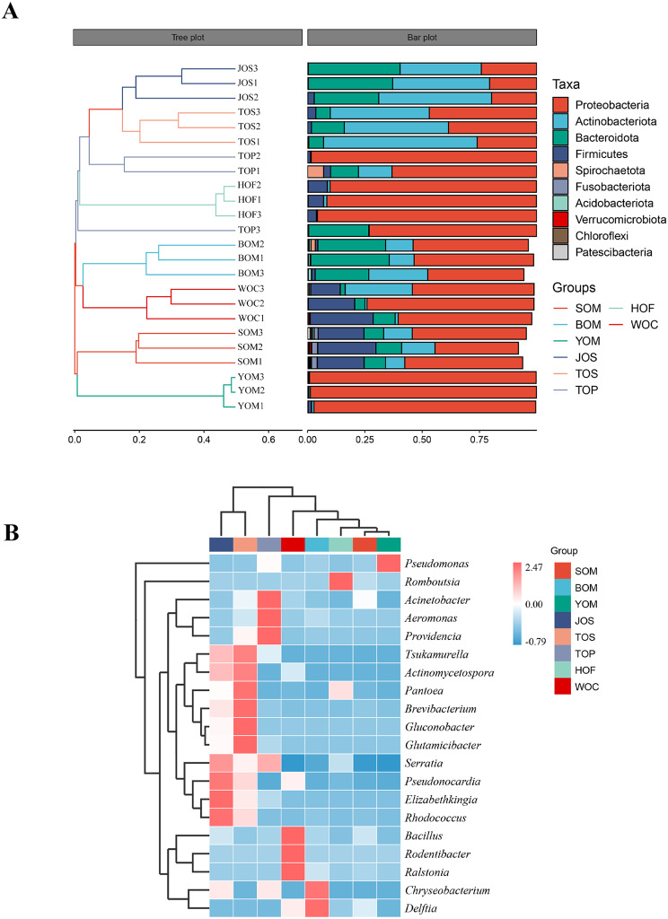
The population structure and alpha-diversity analyses revealed similarities between Opiliones from Hainan Island and those inhabiting the Beijing cave. Therefore, beta analysis methods, indicated distinct symbiont structures among Opiliones living in different regions, with symbiotic bacteria in Opiliones from Hainan Island and the Beijing caves showing greater similarities. Cluster analysis based on the Bray-Curtis distance level grouped BOM and WOC together (Fig. 10a). Similarly, JOS, TOP and TOS were clustered into a group, except that TOP3 was independent of them. Additionally, WOC, BOM, HOF, YOM and SOM clustered together in the heat map of UPGMA clustering based on the Euclidean distance of species composition data (Fig. 10b). Specifically, WOC and BOM were positively correlated with Bacillus, Rodentibacter, Ralstonia and Chryseobacterium, Delftia respectively, and clustered together. Pseudomonas, which belonged to YOM, also clustered with the aforementioned groups. The results confirmed that the symbiotic bacteria of Opiliones from Hainan Island and Beijing caves were more similar than those outside the cave.
Fig. 10.
Comparative microbial diversity of Opiliones in Beijing caves and Hainan using bate-diversity analysis. (A) Hierarchical clustering analysis of the similarity between samples in the form of the hierarchical tree. (B) Heatmap of the microbial composition and relative abundance of Opiliones
Discussion
Karst caves are unique habitats that are characterized by specific environmental conditions, such as almost constant temperature and humidity, permanent darkness and limited energy and nutrient sources [40]. These conditions exert a profound influence on the diversity and composition of cave ecosystems [41]. In addition, caves are mostly located in remote areas and are less disturbed by human activities, which makes them ideal locations for studying the underground biosphere. However, the cave environment is limited by many factors, such as restricted access to nutrition, low temperatures, unique air pressure conditions, connectivity with groundwater, variations in light and humidity, limited surface interaction, anthropogenic or animal influences and geological mineral compositions [42–44]. So each cave is unique in its biological, chemical and physical characteristics, and these factors cause differences within and between caves [45]. These disparities elucidate the variations in symbiotic diversity observed among Opiliones inhabiting different caves and those residing outside cave systems in this study.
Karst caves are relatively closed environments, there are obvious differences among different caves. The cave habitat serves as a critical factor influencing the survival and community structure of microorganisms, with these organisms occupying diverse niches in space and time. It is worth noting that each cave has a unique microbial community, which emphasizes the important role of the cave niche in shaping microbial diversity [46]. Moreover, the cave environment exerts a strong impact on the organisms inhabiting the cave, with up to five distinct zones identified in caves, including the entrance, twilight, transition, deep, and stagnant air zones, each harboring a distinct biological community. The transition zone, characterized by complete darkness and variable abiotic environment [16], and the deep zone, completely dark with high humidity and constant temperature, are of particular interest. S. martensi (Fig. 2a, b) observed in this study is found, which is adapted to the completely lightless deep zone of the cave, displays body coloration varying from yellowish white to pale yellow, greatly reduced ocular size, and complete absence of eyes and retinae, indicating long-term adaptation to the cave environment [47]. Although the Opiliones from Siyu Cave, Bianfu Cave and You Cave share the same appearance and molecule, their different environments lead to distinct symbiotic bacteria structures (Fig. 3a, b). Furthermore, significant differences were observed in symbiotic diversity among Opiliones from the three caves (Fig. 4a, b).
The unique environment of karst caves, characterized by darkness and nutrient limitation, results in distinct energy flow and material circulation compared to the external environment. Predictive functional analysis using PICRUSt2 revealed that despite variations among different cave environments, symbiotic bacteria in Opiliones from three caves exhibit similar functions, focusing on metabolic activities such as the synthesis and metabolism of various organic compounds (Fig. 5a). Symbiotic bacteria rely on the host for various life activities, including even simple metabolic functions. The results suggest that S. martensi and its symbiotic bacteria play direct or indirect roles in the material cycle within the cave, particularly in the carbon cycle, which involves crucial metabolic processes like carbon fixation, methane metabolism, and carbon degradation. Annotation by KEGG and COG databases indicated that most symbiotic bacteria in Opiliones participate in diverse carbon fixation pathways, such as the glycolysis pathway, tricarboxylic acid cycle, and pentose phosphate pathway, facilitated by relevant enzymes (Fig. 5b, Table S2). Additionally, Opiliones are omnivores, with their diet primarily comprising invertebrates in caves, along with plants and fungi [48, 49]. Following the ingestion of animal and plant residues and other organic matter into the Opiliones’ intestinal tract, enzymatic decomposition within the digestive tract, coupled with interactions with intestinal symbiotic bacteria, facilitates the return of organic matter to the cave environment in the form of feces. This process enhances the utilization of other plants and microorganisms within the cave ecosystem.
Nitrogen is a vital element in organisms, playing a crucial role in many biochemical cycles within cave ecosystems. The symbiotic relationship between S. martensi and its associated bacteria contributes significantly to the nitrogen cycle within caves. Firstly, they actively participate in biological nitrogen fixation, a process where nitrogen-fixing bacteria convert atmospheric nitrogen into biologically available ammonia. The prevalence of Proteobacteria in the samples suggests their key role in the nitrogen cycle within the cave environment [50]. Several genera of Pseudomonadales, known for their association with nitrogen fixation, particularly Pseudomonas, are abundant in Opiliones samples from the You Cave (YOM) [51, 52]. Additionally, Actinobacteria, which are abundant in all samples, also contribute to the nitrogen cycle in various ecosystems [53, 54]. The presence of nitrogenase molybdenum-iron (MoFe) protein and other nitrogenases further supports biological nitrogen fixation (Table S2).
Secondly, S. martensi and its symbiotic bacteria facilitate denitrification, a critical aspect of the nitrogen cycle. Predictive analysis using FAPROTAX revealed that denitrification accounts for a significant proportion of nitrogen-related functions (Fig. 6a). Denitrification establishes a loop between atmospheric and ecological water bodies, with the process involving key enzymes such as nitrate reductase (Nar), nitrite reductase (Nir), nitric oxide reductase (Nor), and nitrous oxide reductase (Nos). These enzymes are present in the symbiotic bacteria of Opiliones from all three caves (Table S2). Aerobic denitrifying bacteria such as Comamonas, Acinetobacter, and Pseudomonas, known to facilitate denitrification, exhibit high abundance in Opiliones samples from various caves, particularly Pseudomonas [55, 56] (Fig. 3b).
The karst cave represents a terrestrial oligotrophic extreme underground ecosystem characterized by limited sources of total organic carbon and nitrogen. Within this environment, Opiliones and their symbiotic bacteria play integral roles in the unique energy and material cycles. Operating amidst constant darkness and low energy availability, they contribute significantly to the ecosystem’s functioning. These findings underscore the importance of investigating microbial communities within cave ecosystems to gain insights into their ecological processes and biogeochemical cycles. Understanding the dynamics of these symbiotic relationships and their impact on nutrient cycling sheds light on the intricate balance of life in these remote and often overlooked environments.
Many differences between the environment inside and outside the cave, and the community composition of symbiotic bacteria is also obviously different. The environment inside the cave remains relatively constant. This makes it worthwhile to study the unique community structure of symbiotic bacteria in comparison to the Opiliones living outside the cave, especially in the north where there are obvious changes in the four seasons. We conducted a comparison between S. martensi inside the cave and the Himalphalangium palpalis and Homolophus serrulatus outside the cave and found that there was a significant difference between them. At the phylum level, although most of the samples had Proteobacteria, Actinobacteria and Bacteroidetes as the dominant group, the abundance of symbiotic bacteria varied across different environmental samples (Fig. 8a). Comparing the Opiliones inside and outside of the cave, and a significant difference was found not only in the composition of the community but also in the species of dominant bacteria (Fig. 8a, b).
Two potential explanations may account for this phenomenon. First, the genetic relationship between S. martensi and Himalphalangium palpalis, Homolophus serrulatus is relatively distant. S. martensi belongs to the Laniatores group, characterized by robust tentacles (Fig. 2a, b, e, f) and a diet primarily consisting of other insects, while Himalphalangium palpalis and Homolophus serrulatus belong to the Eupnoi group, lacking strong tentacles (Fig. 2c, d) and capable of feeding on fungi, plants and animals. Previous studies have shown that genetic relationships can have a great influence on the structure of symbiotic bacteria in animals. In addition to the co-evolution of primary endosymbionts with the host [57, 58], there are also symbiotic bacteria that are vertically transmitted from parents and give priority to transmission to closely related hosts [19]. Genes can regulate the body structure of different species, creating distinct internal environments and resulting in different dominant phyla. Secondly, differences in cave environment compared to outside conditions also contribute to this disparity. The cave environment remains constant, rarely disturbed by external temperature, and the temperature is maintained at more than ten degrees Celsius all year round. Opiliones inhabiting caves are thus unaffected by cold winters and remain active throughout the year. In contrast, Beijing experiences distinct four seasons, with temperature dropping significantly below zero in winter. The breeding, feeding and activities of Opiliones living outside Beijing caves are greatly limited by climate change.
Based on the two points mentioned above, which has more influence? We collected different samples (harvestmen, spiders and pseudoscorpions) from Siyu Cave and compared them. According to the results, we found that Proteobacteria, Actinobacteria, Firmicutes, and Bacteroidetes were the main constituent phyla of the species in the cave, and they each accounted for a similar proportion (Figure S1a, b). Although they belong to different orders and have completely different feeding methods (spiders inject digestive juices into their prey, while harvestmen chew directly), there was no significant difference in the structure of symbiotic bacteria (Figure S1c).
Afterward, we studied the spiders outside the Siyu Cave (SAS). To reduce the impact of the external environment and geographical location, we collected samples near the entrance, where there is light and climate change, significantly differs from that in the cave. From inside the cave to the outside, there were obvious changes in the structure of microflora in spiders from different environments (Figure S1a, b). Compared with the spiders in the cave (SAC), the proportion of Proteobacteria in the symbiotic bacteria outside the cave (SAS) increased significantly at the phylum level, and at the genus level, Wolbachia and Rickettsia became the dominant flora. Both the diversity and richness of symbiotic bacteria in cave spiders were much higher than those outside the cave (Figure S1c). Although the spiders, harvestmen and pseudoscorpions in the same cave environment have obvious differences in genetic relationship, their symbiotic bacteria are highly similar. Compared with harvestmen and pseudoscorpions, spiders inside and outside the cave are significantly closer in genetic relationship and feeding methods, but there are significant differences in the community structure of symbiotic bacteria. Therefore, compared with heredity, the effect of the cave environment on symbiotic bacteria is more obvious.
Symbiotic bacteria are not fixed, and there are many factors affecting them, among which the environment plays an important role. Most of the microbes found in organisms are acquired from the surrounding environment during the growth process, either directly or indirectly. Significant seasonal fluctuations of microbial communities are observed in wild mice [59] and hibernating ground squirrels (Ictidomys tridecemlineatus and Urocitellus parryii) every year [60, 61]. Organisms residing in deep-sea hydrothermal regions characterized by high pressure, hypoxia, high levels of toxic substances, sulfides, and heavy metals, as well as being dark, are doubly symbiotic with two gammaproteobacterial endosymbionts: a sulfur oxidizer and a methane oxidizer [62, 63]. High temperatures can denature proteins, causing physiological and developmental problems [10]. Endosymbiosis with heat shock tolerance can reduce the impact of high temperature on the survival and development of the host [11]. Various factors such as season, altitude, diet and photoperiod impact the diversity of animal microbial communities [9]. The community composition of larvae of Aedes aegypti, Aedes albopictus and Culex quinquefasciatus from different areas varied greatly, while strong similarities were found among larvae of different species developing in the same site [64]. These findings, along with previous studies, suggest that the environment exerts a more significant influence on symbiotic bacteria than heredity. However, in this study, arachnids were the subjects, and their genetic relationships were relatively close. If we take more closely related animals from different phyla or classes as samples, it remains uncertain whether the environment would still play such a significant role. Furthermore, due to residing in the cave for an extended period, Araneae, Opiliones and Pseudoscorpionida have undergone convergent evolution to some extent, exhibiting similar morphological features, such as the lack of eyes and a light body color. Could this similarity also affect the symbiotic structure of these three groups, making them more alike? Further studies are necessary to verify this hypothesis.
It is indeed interesting that the symbiotic bacteria richness of cave organisms surpasses found that outside the cave despite the challenging environment. This suggests that the cave environment may provide a unique niche for symbiotic bacteria to thrive. The caves in this study are located in the north of the Taihang Mountains, within the center of the North China Craton (NCC). This area is known for its abundance of mineral deposits [65, 66]. Phyllobacterium, previously isolated from plant roots in mines, exhibits the capability to oxidize Mn (II) [67]. The abundance of Phyllobacterium in cave species (Table S5) may be due to its tolerance to heavy metals [68, 69], which are often present in mineral-rich cave environments. The joint action of nutrition, rock and other factors may contribute to the richness of symbiotic bacteria in caves [70, 71], and may also explain the observed difference in symbiotic bacteria between cave and non-cave organisms. Further studies are needed to fully understand the mechanisms underlying these observations.
Sinonychia martensi found in Beijing caves in this study is a highly adapted species to cave life, and the first superfamily Travunioidea discovered in China [72]. Currently, the Travunioidea are distributed across the Holarctic, in the temperate regions of central Europe, southern British Columbia, eastern and western United States, Japan, and South Korea [73]. Although Sinonychia is closely related to Speleonychia and shares some morphological similarities, it is considered a new genus. Interestingly, Speleonychia, closely related to Sinonychia, is mainly located in the Pacific Northwest of North America, across the Pacific Ocean from the Sinonychia [72]. The Laniatores, the order to which Sinonychia belongs, are mainly distributed in the tropical and temperate regions of the southern hemisphere [74] with more common occurrences in Hainan and Sichuan, situated in the south of China [75–77]. Therefore, it raises the question of why S. martensi, the only Travunioidea found in China, appears in caves in the north.
Two main hypotheses are generally proposed to explain the shift from surface to underground habitats. The first hypothesis suggests that the colonization of underground habitats is the result of the active expansion and colonization by a founder [78], rather than accidental stranding and persistence in lightless areas [79, 80]. The second hypothesis proposes that species may be forced to adapt to cave life due to environmental changes that make the surface uninhabitable [81–83]. The Opiliones in this study exhibit obvious characteristics of cave life, indicating that they have been forced to move underground and have lived at the bottom of the cave for many years. Therefore, S. martensi, being the first of its kind found in China, likely originated from the same southern family with a warm and humid climate rather than migrating all the way to the north.
Some similarities in symbiotic bacteria of Opiliones from Hainan Island and karst caves suggest that karst caves can serve as an alternative habitat for species thriving in warm and humid climates. However, does this imply that the cave environment is comparable to its natural habitat? Hainan Island, a typical tropical region in China, is home to a significant number of Laniatores species. In this study, E. flavimaculatus and P. columnarius were collected from Haikou City and Wuzhishan City of Hainan Island respectively, and compared with S. martensi from Beijing caves. To provide a clearer comparison, Himalphalangium palpalis and Homolophus serrulatus, also found outside the cave in Beijing, were included in the study. The results showed that the symbiotic structure of S. martensi was more similar to that of Opiliones from Hainan Island compared to Opiliones outside the cave in Beijing. The relative abundance of Proteobacteria in Homolophus serrulatus was only 21.44% and 37.07%, whereas it was the highest in other Opiliones (Figure S2a, Table S5). The relative abundance of Firmicutes in SOM, HOF, and WOC was 21.90%, 6.30% and 20.17% respectively, ranking second. Cluster analysis of symbiotic bacteria in each sample revealed that the species composition of Opiliones from Hainan Island and Beijing caves were more similar (Fig. 10a, b). Therefore, the study suggests that the environment of the caves in Beijing may be similar to that in Hainan Island, and provides insights into the origin of S. martensi.
The cave can be thought of as an independent “small world” with constant temperature and humidity, permanent darkness and limited food resources. Unlike terrestrial vertebrates, which have high diffusivity, terrestrial cave invertebrates are usually confined to small geographical areas and are the main components of cave fauna [40]. S. martensi has lived for a long time in isolated caves, with limited interaction with external biota, particularly from the south to the north. In addition, the northern climate is not fixed, with the onset of the monsoon climate from the Oligocene-Miocene boundary [84], East Asia has experienced seasonality of lower average temperature and humidity in winter [85]. Due to the relatively stable microclimate, caves may serve as a refuge for numerous organisms, and their surface counterparts tend to become extinct under adverse climatic conditions, which means some local cave biota remain [86]. This also explains why this study was unable to collect any other Sinonychia or even Laniatores closely related to S. martensi outside the cave from Beijing, only other harvestmen of the Eupnoi. A spider study suggested that climate change during the middle Miocene period facilitated the development of underground lifestyles in the middle latitudes of Asia [87]. The seasonality may have contributed to the extinction of surface species, and caves with relatively stable temperatures and humidity may have provided an ideal refuge for local species. In addition to climate change, human-made environmental destruction has impacted the distribution of S. martensi. With the decrease in temperature and humidity, reduction in surface trees, and destruction of the ecosystem, some Opiliones on the surface sought refuge underground and adapted to the stable cave environment, leading to changes in their morphology. With the passage of time and adaptation to the caves, their posture changed. The other part disappeared from the surface forever. Conversely, Laniatores, which are still found in the tropics and subtropics, have managed to survive until today.
Conclusions
This study focused on the Opiliones in karst caves in Beijing, comparing them to those found outside the caves as well as to other organisms such as harvestmen, spiders and pseudoscorpions within the same cave. The results showed that the environment plays a more important role in determining symbiotic bacteria diversity than heredity. S. martensi, a unique species belonging to the Travunioidea and first discovered in China, adds to the mystery of caves that are both geographically isolated and have a unique environment. The symbiotic bacteria structure also reflects the characteristics of the host environment and the symbiotic community composition of Opiliones in caves in Beijing is similar to those found in Hainan Island, suggesting that the cave environment may resemble that found in the tropics. This finding explains the occurrence of S. martensi in caves in the north, and helps explore the role of symbiotic bacteria in host distribution and evolution, and in explaining the biological characteristics of the host.
Electronic supplementary material
Below is the link to the electronic supplementary material.
Acknowledgements
We thank Zhao JJ (Hebei University) for her help in collecting these specimens during the fieldwork. This work was supported by the Natural Science Foundation of Hebei Province (No. C2021201030).
Abbreviations
- PCR
Polymerase Chain Reaction
- ASVs
Amplicon Sequence Variants
- PCoA
Principal Coordinate Analysis
- NMDS
Non-Metric Multidimensional Scaling
Author contributions
Author contributions are as fellows. C.Z., L.Z. and F.Z. designed the project and experiments; R.X., L.Z. analyzed results; R.X. and S.Z. performed the experimental works; L.Z. and R.X. wrote the manuscript. All authors read and approved the final manuscript.
Funding
This work was supported by the Natural Science Foundation of Hebei Province (No. C2021201030).
Data availability
The sequence data generated and analyzed in this study are available at NCBI (https://www.ncbi.nlm.nih.gov) under accession numbers (PRJNA985231).
Declarations
Ethical approval and consent to participate
Not applicable.
Consent for publication
Not applicable.
Competing interests
The authors declare that they have no competing interests.
Footnotes
Publisher’s Note
Springer Nature remains neutral with regard to jurisdictional claims in published maps and institutional affiliations.
Likun Zhao and Ruoyi Xiao contributed equally to this work.
Contributor Information
Chao Zhang, Email: Opiliones@163.com.
Feng Zhang, Email: dudu06042001@163.com.
References
- 1.Moran NA, McCutcheon JP, Nakabachi A. Genomics and evolution of heritable bacterial symbionts. Annu Rev Genet. 2008;42(1):165–90. doi: 10.1146/annurev.genet.41.110306.130119. [DOI] [PubMed] [Google Scholar]
- 2.Hongoh Y, Sharma VK, Prakash T, Noda S, Taylor TD, Kudo T, et al. Complete genome of the uncultured Termite Group 1 bacteria in a single host protist cell. Proc Natl Acad Sci USA. 2008;105(14):5555–60. doi: 10.1073/pnas.0801389105. [DOI] [PMC free article] [PubMed] [Google Scholar]
- 3.Watanabe H, Tokuda G. Cellulolytic systems in insects. Annu Rev Entomol. 2010;55(1):609–32. doi: 10.1146/annurev-ento-112408-085319. [DOI] [PubMed] [Google Scholar]
- 4.Sabree ZL, Kambhampati S, Moran NA. Nitrogen recycling and nutritional provisioning by Blattabacterium, the cockroach endosymbiont. Proc Natl Acad Sci USA. 2009;106(46):19521–6. doi: 10.1073/pnas.0907504106. [DOI] [PMC free article] [PubMed] [Google Scholar]
- 5.Engel P, Martinson VG, Moran NA. Functional diversity within the simple gut microbiota of the honey bee. Proc Natl Acad Sci USA. 2012;109(27):11002–7. doi: 10.1073/pnas.1202970109. [DOI] [PMC free article] [PubMed] [Google Scholar]
- 6.Kikuchi Y, Hayatsu M, Hosokawa T, Nagayama A, Tago K, Fukatsu T. Symbiont-mediated insecticide resistance. Proc Natl Acad Sci USA. 2012;109(22):8618–22. doi: 10.1073/pnas.1200231109. [DOI] [PMC free article] [PubMed] [Google Scholar]
- 7.Duron O, Bouchon D, Boutin S, Bellamy L, Zhou L, Engelstädter J, et al. The diversity of reproductive parasites among arthropods: Wolbachia do not walk alone. BMC Biol. 2008;6(1):27. doi: 10.1186/1741-7007-6-27. [DOI] [PMC free article] [PubMed] [Google Scholar]
- 8.Odden JP, Eng W, Lee K, Donelick H, Hiefield M, Steach J, et al. Novel host-bacterial symbioses revealed: characterization of Wolbachia in arthropods of Western North America. Western North Am Naturalist. 2019;79(4):534. doi: 10.3398/064.079.0407. [DOI] [Google Scholar]
- 9.Khakisahneh S, Zhang X-Y, Nouri Z, Wang D-H. Gut microbiota and host thermoregulation in response to ambient temperature fluctuations. mSystems. 2020;5(5):e00514–20. doi: 10.1128/mSystems.00514-20. [DOI] [PMC free article] [PubMed] [Google Scholar]
- 10.Feder ME, Hofmann GE. Heat-shock proteins, molecular chaperones, and the stress response: evolutionary and ecological physiology. Annu Rev Physiol. 1999;61(1):243–82. doi: 10.1146/annurev.physiol.61.1.243. [DOI] [PubMed] [Google Scholar]
- 11.Harmon JP, Moran NA, Ives AR. Species response to environmental change: impacts of food web interactions and evolution. Science. 2009;323(5919):1347–50. doi: 10.1126/science.1167396. [DOI] [PubMed] [Google Scholar]
- 12.Gulecal-Pektas Y. Bacterial diversity and composition in Oylat Cave (Turkey) with combined Sanger/pyrosequencing approach. Pol J Microbiol. 2016;65(1):69–75. doi: 10.5604/17331331.1197277. [DOI] [PubMed] [Google Scholar]
- 13.De Mandal S, Chatterjee R, Kumar NS. Dominant bacterial phyla in caves and their predicted functional roles in C and N cycle. BMC Microbiol. 2017;17(1):90. doi: 10.1186/s12866-017-1002-x. [DOI] [PMC free article] [PubMed] [Google Scholar]
- 14.Zhu H-Z, Zhang Z-F, Zhou N, Jiang C-Y, Wang B-J, Cai L, et al. Diversity, distribution and co-occurrence patterns of bacterial communities in a karst cave system. Front Microbiol. 2019;10:1726. doi: 10.3389/fmicb.2019.01726. [DOI] [PMC free article] [PubMed] [Google Scholar]
- 15.Protas M, Jeffery WR. Evolution and development in cave animals: from fish to crustaceans. Wiley Interdiscip Rev Dev Biol. 2012;1(6):823–45. doi: 10.1002/wdev.61. [DOI] [PMC free article] [PubMed] [Google Scholar]
- 16.Gabriel CR, Northup DE. Microbial ecology: caves as an extreme habitat. In: Cheeptham N, editor. Cave microbiomes: a Novel Resource for Drug Discovery. New York, NY: Springer New York; 2013. pp. 85–108. [Google Scholar]
- 17.Koren O, Rosenberg E. Bacteria Associated with the bleached and cave coral Oculina patagonica. Microb Ecol. 2008;55(3):523–9. doi: 10.1007/s00248-007-9297-z. [DOI] [PubMed] [Google Scholar]
- 18.Kostanjšek R, Vittori M, Srot V, van Aken PA, Štrus J. Polyphosphate-accumulating bacterial community colonizing the calcium bodies of terrestrial isopod crustaceans Titanethes albus and Hyloniscus riparius. FEMS Microbiol Ecol. 2017;93(6). [DOI] [PubMed]
- 19.Baldo L, Ayoub NA, Hayashi CY, Russell JA, Stahlhut JK, Werren JH. Insight into the routes of Wolbachia invasion: high levels of horizontal transfer in the spider genus Agelenopsis revealed by Wolbachia strain and mitochondrial DNA diversity: Wolbachia strain shuffling within Agelenopsis. Mol Ecol. 2007;17(2):557–69. doi: 10.1111/j.1365-294X.2007.03608.x. [DOI] [PubMed] [Google Scholar]
- 20.Perlman SJ, Magnus SA, Copley CR. Pervasive associations between Cybaeus spiders and the bacterial symbiont Cardinium. J Invertebr Pathol. 2010;103(3):150–5. doi: 10.1016/j.jip.2009.12.009. [DOI] [PubMed] [Google Scholar]
- 21.Gotoh T, Noda H, Ito S. Cardinium symbionts cause cytoplasmic incompatibility in spider mites. Heredity. 2007;98(1):13–20. doi: 10.1038/sj.hdy.6800881. [DOI] [PubMed] [Google Scholar]
- 22.Zeh DW, Zeh JA, Bonilla MM. Wolbachia, sex ratio bias and apparent male killing in the harlequin beetle riding pseudoscorpion. Heredity. 2005;95(1):41–9. doi: 10.1038/sj.hdy.6800666. [DOI] [PubMed] [Google Scholar]
- 23.Koop JL, Zeh DW, Bonilla MM, Zeh JA. Reproductive compensation favours male-killing Wolbachia in a live-bearing host. Proc R Soc B. 2009;276(1675):4021–8. doi: 10.1098/rspb.2009.1230. [DOI] [PMC free article] [PubMed] [Google Scholar]
- 24.Zhang L, Zhang G, Yun Y, Peng Y. Bacterial community of a spider, Marpiss Magister (Salticidae) 3 Biotech. 2017;7(6):371. doi: 10.1007/s13205-017-0994-0. [DOI] [PMC free article] [PubMed] [Google Scholar]
- 25.Chang J, Masters A, Avery A, Werren JH. A divergent cardinium found in daddy long-legs (Arachnida: Opiliones) J Invertebr Pathol. 2010;105(3):220–7. doi: 10.1016/j.jip.2010.05.017. [DOI] [PubMed] [Google Scholar]
- 26.Stouthamer CM, Kelly SE, Mann E, Schmitz-Esser S, Hunter MS. Development of a multi-locus sequence typing system helps reveal the evolution of Cardinium Hertigii, a reproductive manipulator symbiont of insects. BMC Microbiol. 2019;19(1):266. doi: 10.1186/s12866-019-1638-9. [DOI] [PMC free article] [PubMed] [Google Scholar]
- 27.Martin OY, Goodacre SL. Widespread infections by the bacterial endosymbiont cardinium in Arachnids. J Arachnology. 2009;37(1):106–8. doi: 10.1636/SH08-05.1. [DOI] [Google Scholar]
- 28.Duron O, Hurst GDD, Hornett EA, Josling JA, Engelstädter J. High incidence of the maternally inherited bacterium Cardinium in spiders. Mol Ecol. 2008;17(6):1427–37. doi: 10.1111/j.1365-294X.2008.03689.x. [DOI] [PubMed] [Google Scholar]
- 29.Bragagnolo C, Pinto-da-Rocha R, Antunes M, Clouse RM. Phylogenetics and phylogeography of a long-legged harvestman (Arachnida: Opiliones) in the Brazilian Atlantic Rain Forest reveals poor dispersal, low diversity and extensive mitochondrial introgression. Invert Syst. 2015;29(4):386. doi: 10.1071/IS15009. [DOI] [Google Scholar]
- 30.Clouse RM, Sharma PP, Stuart JC, Davis LR, Giribet G, Boyer SL, et al. Phylogeography of the harvestman genus Metasiro (Arthropoda, Arachnida, Opiliones) reveals a potential solution to the Pangean paradox. Org Divers Evol. 2016;16(1):167–84. doi: 10.1007/s13127-015-0233-7. [DOI] [Google Scholar]
- 31.Callahan BJ, McMurdie PJ, Rosen MJ, Han AW, Johnson AJA, Holmes SP. DADA2: high-resolution sample inference from Illumina amplicon data. Nat Methods. 2016;13(7):581–3. doi: 10.1038/nmeth.3869. [DOI] [PMC free article] [PubMed] [Google Scholar]
- 32.Bokulich NA, Kaehler BD, Rideout JR, Dillon M, Bolyen E, Knight R, et al. Optimizing taxonomic classification of marker-gene amplicon sequences with QIIME 2’s q2-feature-classifier plugin. Microbiome. 2018;6(1):90. doi: 10.1186/s40168-018-0470-z. [DOI] [PMC free article] [PubMed] [Google Scholar]
- 33.Quast C, Pruesse E, Yilmaz P, Gerken J, Schweer T, Yarza P, Peplies J, Glöckner FO. The SILVA ribosomal RNA gene database project: improved data processing and web-based tools. Nucleic Acids Res. 2013;41(Database issue):D590–6. doi: 10.1093/nar/gks1219. [DOI] [PMC free article] [PubMed] [Google Scholar]
- 34.Bolyen E, Rideout JR, Dillon MR, Bokulich NA, Abnet CC, Al-Ghalith GA, et al. Reproducible, interactive, scalable and extensible microbiome data science using QIIME 2. Nat Biotechnol. 2019;37(8):852–7. doi: 10.1038/s41587-019-0209-9. [DOI] [PMC free article] [PubMed] [Google Scholar]
- 35.Zhou Z, Wu H, Li D, Zeng W, Huang J, Wu Z. Comparison of gut microbiome in the Chinese mud snail (Cipangopaludina chinensis) and the invasive golden apple snail (Pomacea canaliculata). 2022;10:e13245. [DOI] [PMC free article] [PubMed]
- 36.Ramette A. Multivariate analyses in microbial ecology: multivariate analyses in microbial ecology. FEMS Microbiol Ecol. 2007;62(2):142–60. doi: 10.1111/j.1574-6941.2007.00375.x. [DOI] [PMC free article] [PubMed] [Google Scholar]
- 37.Segata N, Izard J, Waldron L, Gevers D, Miropolsky L, Garrett WS, et al. Metagenomic biomarker discovery and explanation. Genome Biol. 2011;12(6):R60. doi: 10.1186/gb-2011-12-6-r60. [DOI] [PMC free article] [PubMed] [Google Scholar]
- 38.Douglas GM, Maffei VJ, Zaneveld J, Yurgel SN, Brown JR, Taylor CM et al. PICRUSt2: An improved and customizable approach for metagenome inference. Bioinformatics.
- 39.Louca S, Parfrey LW, Doebeli M. Decoupling function and taxonomy in the global ocean microbiome. Science. 2016;353(6305):1272–7. doi: 10.1126/science.aaf4507. [DOI] [PubMed] [Google Scholar]
- 40.Clements R, Sodhi NS, Schilthuizen M, Ng PKL. Limestone karsts of Southeast Asia: imperiled arks of biodiversity. Bioscience. 2006;56(9):733. doi: 10.1641/0006-3568(2006)56[733:LKOSAI]2.0.CO;2. [DOI] [Google Scholar]
- 41.Poulson TL, White WB. The cave environment. Science. 1969;165:971–81. doi: 10.1126/science.165.3897.971. [DOI] [PubMed] [Google Scholar]
- 42.Engel AS. Karst ecosystems. Encyclopedia of earth sciences series. Dordrecht: Springer; 2011. pp. 521–31. [Google Scholar]
- 43.Barton HA. 4. Starving artists: bacterial oligotrophic heterotrophy in caves. In: Summers Engel A, editor. Microbial Life of Cave systems. DE GRUYTER; 2015. pp. 79–104.
- 44.Cheeptham N. Advances and challenges in studying cave microbial diversity. In: Cheeptham N, editor. Cave microbiomes: a Novel Resource for Drug Discovery. New York, NY: Springer New York; 2013. pp. 1–34. [Google Scholar]
- 45.Onac B, Forti P. Minerogenetic mechanisms occurring in the cave environment: an overview. IJS. 2011;40(2):79–98. doi: 10.5038/1827-806X.40.2.1. [DOI] [Google Scholar]
- 46.Kajan K, Cukrov N, Cukrov N, Bishop-Pierce R, Orlić S. Microeukaryotic and prokaryotic diversity of Anchialine caves from Eastern Adriatic Sea Islands. Microb Ecol. 2022;83(2):257–70. doi: 10.1007/s00248-021-01760-5. [DOI] [PMC free article] [PubMed] [Google Scholar]
- 47.Rétaux S, Casane D. Evolution of eye development in the darkness of caves: adaptation, drift, or both? EvoDevo. 2013;4(1):26. doi: 10.1186/2041-9139-4-26. [DOI] [PMC free article] [PubMed] [Google Scholar]
- 48.Powell EC, Painting CJ, Hickey AJ, Machado G, Holwell GI. Diet, predators, and defensive behaviors of New Zealand harvestmen (Opiliones: Neopilionidae). J Arachnology. 2021;49(1).
- 49.Santos FH, Gnaspini P. Notes on the foraging behavior of the Brazilian cave harvestman Goniosoma Spelaeum (Opiliones, Gonyleptidae) J Arachnol. 2002;30(1):177. doi: 10.1636/0161-8202(2002)030[0177:NOTFBO]2.0.CO;2. [DOI] [Google Scholar]
- 50.Nelson MB, Martiny AC, Martiny JBH. Global biogeography of microbial nitrogen-cycling traits in soil. Proc Natl Acad Sci USA. 2016;113(29):8033–40. doi: 10.1073/pnas.1601070113. [DOI] [PMC free article] [PubMed] [Google Scholar]
- 51.Kuklinsky-Sobral J, Araujo WL, Mendes R, Geraldi IO, Pizzirani-Kleiner AA, Azevedo JL. Isolation and characterization of soybean-associated bacteria and their potential for plant growth promotion. Environ Microbiol. 2004;6(12):1244–51. doi: 10.1111/j.1462-2920.2004.00658.x. [DOI] [PubMed] [Google Scholar]
- 52.Kuypers MMM, Marchant HK, Kartal B. The microbial nitrogen-cycling network. Nat Rev Microbiol. 2018;16(5):263–76. doi: 10.1038/nrmicro.2018.9. [DOI] [PubMed] [Google Scholar]
- 53.Zhang B, Wu X, Tai X, Sun L, Wu M, Zhang W, et al. Variation in actinobacterial community composition and potential function in different soil ecosystems belonging to the arid Heihe River Basin of Northwest China. Front Microbiol. 2019;10:2209. doi: 10.3389/fmicb.2019.02209. [DOI] [PMC free article] [PubMed] [Google Scholar]
- 54.Ronoh RC, Budambula NLM, Mwirichia RK, Boga HI. Isolation and characterization of actinobacteria from Lake Magadi, Kenya. Afr J Microbiol Res. 2013;7:4200–6. [Google Scholar]
- 55.Chen Q, Ni J. Heterotrophic nitrification–aerobic denitrification by novel isolated bacteria. J Ind Microbiol Biotechnol. 2011;38(9):1305–10. doi: 10.1007/s10295-010-0911-6. [DOI] [PubMed] [Google Scholar]
- 56.Kou L, Huang T, Zhang H, Wen G, Li N, Wang C, et al. Mix-cultured aerobic denitrifying bacterial communities reduce nitrate: novel insights in micro-polluted water treatment at lower temperature. Sci Total Environ. 2021;796:148910. doi: 10.1016/j.scitotenv.2021.148910. [DOI] [PubMed] [Google Scholar]
- 57.Baumann P, Baumann L, Lai C-Y, Rouhbakhsh D, Moran NA, Clark MA. Genetics, physiology, and evolutionary relationships of the genus Buchnera: intracellular symbionts of aphids. Annu Rev Microbiol. 1995;49:55–94. doi: 10.1146/annurev.mi.49.100195.000415. [DOI] [PubMed] [Google Scholar]
- 58.Santos-Garcia D, Vargas-Chavez C, Moya A, Latorre A, Silva FJ. Genome evolution in the primary endosymbiont of whiteflies sheds light on their divergence. Genome Biol Evol. 2015;7(3):873–88. doi: 10.1093/gbe/evv038. [DOI] [PMC free article] [PubMed] [Google Scholar]
- 59.Maurice CF, Knowles CL, Ladau S, Pollard J, Fenton KS, Pedersen A. Marked seasonal variation in the wild mouse gut microbiota. ISME J. 2015;9(11):2423–34. doi: 10.1038/ismej.2015.53. [DOI] [PMC free article] [PubMed] [Google Scholar]
- 60.Carey HV, Walters WA, Knight R. Seasonal restructuring of the ground squirrel gut microbiota over the annual hibernation cycle. Am J Physiology-Regulatory Integr Comp Physiol. 2013;304(1):R33–42. doi: 10.1152/ajpregu.00387.2012. [DOI] [PMC free article] [PubMed] [Google Scholar]
- 61.Stevenson TJ, Duddleston KN, Buck CL. Effects of season and host physiological state on the diversity, density, and activity of the arctic ground squirrel cecal microbiota. Appl Environ Microbiol. 2014;80(18):5611–22. doi: 10.1128/AEM.01537-14. [DOI] [PMC free article] [PubMed] [Google Scholar]
- 62.Orcutt BN, Sylvan JB, Knab NJ, Edwards KJ. Microbial ecology of the dark ocean above, at, and below the seafloor. Microbiol Mol Biol Rev. 2011;75(2):361–422. doi: 10.1128/MMBR.00039-10. [DOI] [PMC free article] [PubMed] [Google Scholar]
- 63.Lan Y, Sun J, Chen C, Sun Y, Zhou Y, Yang Y, et al. Hologenome analysis reveals dual symbiosis in the deep-sea hydrothermal vent snail Gigantopelta aegis. Nat Commun. 2021;12(1):1165. doi: 10.1038/s41467-021-21450-7. [DOI] [PMC free article] [PubMed] [Google Scholar]
- 64.Coon KL, Brown MR, Strand MR. Mosquitoes host communities of bacteria that are essential for development but vary greatly between local habitats. Mol Ecol. 2016;25(22):5806–26. doi: 10.1111/mec.13877. [DOI] [PMC free article] [PubMed] [Google Scholar]
- 65.Gao Y, Santosh M, Wei R, Ma G, Chen Z, Wu J. Origin of high Sr/Y magmas from the northern Taihang Mountains: implications for mesozoic porphyry copper mineralization in the North China Craton. J Asian Earth Sci. 2013;78:143–59. doi: 10.1016/j.jseaes.2012.10.040. [DOI] [Google Scholar]
- 66.Xu H, Song Y, Ye K, Zhang J, Wang H. Petrogenesis of mafic dykes and high-Mg adakitic enclaves in the late mesozoic Fangshan low-Mg adakitic pluton, North China Craton. J Asian Earth Sci. 2012;54–55:143–61. doi: 10.1016/j.jseaes.2012.04.008. [DOI] [Google Scholar]
- 67.Newsome L, Bacon CGD, Song H, Luo Y, Sherman DM, Lloyd JR. Natural attenuation of lead by microbial manganese oxides in a karst aquifer. Sci Total Environ. 2021;754:142312. doi: 10.1016/j.scitotenv.2020.142312. [DOI] [PubMed] [Google Scholar]
- 68.Ma Y, Rajkumar M, Luo Y, Freitas H. Phytoextraction of heavy metal polluted soils using Sedum plumbizincicola inoculated with metal mobilizing Phyllobacterium myrsinacearum RC6b. Chemosphere. 2013;93(7):1386–92. doi: 10.1016/j.chemosphere.2013.06.077. [DOI] [PubMed] [Google Scholar]
- 69.Zappelini C, Alvarez-Lopez V, Capelli N, Guyeux C, Chalot M. Streptomyces dominate the soil under Betula trees that have naturally colonized a red gypsum landfill. Front Microbiol. 2018;9:1772. doi: 10.3389/fmicb.2018.01772. [DOI] [PMC free article] [PubMed] [Google Scholar]
- 70.Nicolosi G, Galdenzi S, Messina MA, Miller AZ, Petralia S, Sarbu SM, et al. Sulfidic habitats in the gypsum karst system of Monte Conca (Italy) host a chemoautotrophically supported invertebrate community. IJERPH. 2022;19(5):2671. doi: 10.3390/ijerph19052671. [DOI] [PMC free article] [PubMed] [Google Scholar]
- 71.Adetutu EM, Ball AS. Microbial diversity and activity in caves. Microbiol Aust. 2014;35(4):192. doi: 10.1071/MA14062. [DOI] [Google Scholar]
- 72.Zhang C, Derkarabetian S. First record of Travunioidea (Arachnida: Opiliones: Laniatores) from China, with the description of a new monotypic genus from a cave. Zootaxa. 2021;4984(1). [DOI] [PubMed]
- 73.Derkarabetian S, Starrett J, Tsurusaki N, Ubick D, Castillo S, Hedin M. A stable phylogenomic classification of Travunioidea (Arachnida, Opiliones, Laniatores) based on sequence capture of ultraconserved elements. ZK. 2018;760:1–36. doi: 10.3897/zookeys.760.24937. [DOI] [PMC free article] [PubMed] [Google Scholar]
- 74.Kury A. Annotated catalogue of the Laniatores of the New World (Arachnida, Opiliones) Revista Iberica De Aracnología. 2003;1:1–337. [Google Scholar]
- 75.Zhang C, Zhang F. A new Tithaeus species from Hainan Island, China (Arachnida, Opiliones, Laniatores, Epedanidae), with a key to the Chinese species. ZK. 2010;67:65–72. doi: 10.3897/zookeys.67.705. [DOI] [PMC free article] [PubMed] [Google Scholar]
- 76.Lian W-G, Zhang C, Zhang F. Review of the genus Plistobunus Pocock, 1903, with description of a new species from Hainan Island, China (Opiliones, Laniatores, Epedanidae) ZK. 2011;112:39–52. doi: 10.3897/zookeys.112.1110. [DOI] [PMC free article] [PubMed] [Google Scholar]
- 77.Schwendinger P, Martens J. A taxonomic revision of the family Oncopodidae V. Gnomulus from Vietnam and China, with the description of five new species (Opiliones, Laniatores) Rev Suisse Zool. 2006;113:595–615. doi: 10.5962/bhl.part.80365. [DOI] [Google Scholar]
- 78.Rouch R, Danielopol D. The origin of the subterranean freshwater fauna, between the refugium paradigm and the model of active colonization [L’origine de la faune aquatique souterraine, entre le paradigme du refuge et le modele de la colonisation active] Stygologia. 1987;3:345–72. [Google Scholar]
- 79.Kane TC, Culver DC, Jones RT. Genetic structure of morphologically differentiated populations of the amphipod Gammarus minus. Evolution. 1992;46(1):272–8. doi: 10.2307/2409822. [DOI] [PubMed] [Google Scholar]
- 80.Kano Y, Kase T. Genetic exchange between anchialine cave populations by means of larval dispersal: the case of a new gastropod species Neritilia Cavernicola. Zool Scripta. 2004;33(5):423–37. doi: 10.1111/j.0300-3256.2004.00159.x. [DOI] [Google Scholar]
- 81.Leys R, Watts CHS, Cooper SJB, Humphreys WF. Evolution of subterranean diving beetles (Coleoptera: Dytiscidae: Hydroporini, Bidessini) in the arid zone of Australia. Evolution. 2003;57(12):2819–34. doi: 10.1111/j.0014-3820.2003.tb01523.x. [DOI] [PubMed] [Google Scholar]
- 82.Barr TC. Speciation in cave faunas. Annu Rev Ecol Syst. 1985;16:313–37. doi: 10.1146/annurev.es.16.110185.001525. [DOI] [Google Scholar]
- 83.Bryson RW, Prendini L, Savary WE, Pearman PB. Caves as microrefugia: pleistocene phylogeography of the troglophilic north American scorpion Pseudouroctonus reddelli. BMC Evol Biol. 2014;14(1):9. doi: 10.1186/1471-2148-14-9. [DOI] [PMC free article] [PubMed] [Google Scholar]
- 84.Ding W, Hou D, Gan J, Wu P, Zhang M, George SC. Palaeovegetation variation in response to the late Oligocene-early Miocene East Asian summer monsoon in the Ying-Qiong Basin, South China Sea. Palaeogeography, Palaeoclimatology, Palaeoecology. 2021;567:110205.
- 85.Li X-Q, Xiang X-G, Jabbour F, Hagen O, Ortiz R, del Soltis C. Biotic colonization of subtropical east Asian caves through time. Proc Natl Acad Sci USA. 2022;119(34):e2207199119. doi: 10.1073/pnas.2207199119. [DOI] [PMC free article] [PubMed] [Google Scholar]
- 86.Barr TC. Cave Ecology and the evolution of troglobites. In: Dobzhansky T, Hecht MK, Steere WC, editors. Evolutionary Biology. Boston, MA: Springer US; 1968. pp. 35–102. [Google Scholar]
- 87.Ballarin F, Li S. Diversification in tropics and subtropics following the mid-miocene climate change: a case study of the spider genus Nesticella. Glob Change Biol. 2018;24(2):e577–91. doi: 10.1111/gcb.13958. [DOI] [PubMed] [Google Scholar]
Associated Data
This section collects any data citations, data availability statements, or supplementary materials included in this article.
Supplementary Materials
Data Availability Statement
The sequence data generated and analyzed in this study are available at NCBI (https://www.ncbi.nlm.nih.gov) under accession numbers (PRJNA985231).


