



Abstract
Thymic carcinomas are rare malignancies that in general arise in the prevascular (anterior) mediastinum. These tumors are usually invasive, often present at advanced stages, and typically behave aggressively. Studies are hampered by the paucity of these tumors, the large variety of carcinoma subtypes, and the lack of unique morphologic and immunophenotypic features. Despite these challenges, advances in diagnostic imaging, surgical approaches, systemic therapies, and radiation therapy techniques have been made. The World Health Organization classification of thymic epithelial tumors has been updated in 2021 and the 8th tumor nodal metastasis staging by the American Joint Committee on Cancer /Union for International Cancer Control included thymic carcinomas in 2017. Molecular alterations that provide more insight into the pathogenesis of these tumors and that potentially permit use of novel targeted therapies are increasingly being identified. New approaches to radiation therapy, chemotherapy, and immunotherapy are under evaluation. International societies including the International Thymic Malignancy Interest Group, European Society of Thoracic Surgeons, and Japanese, Chinese, and Korean thymic associations have been critical in organizing and conducting multi-institutional clinical studies. Herein we review contemporary multidisciplinary perspectives in diagnosis and management of thymic carcinoma.
Keywords: Thymic carcinoma, ITMIG, International Thymic Malignancy Interest Group, TNM staging, WHO
Introduction
Thymic carcinomas are rare malignancies that usually occur in the prevascular (anterior) mediastinum.1, 2 These tumors are typically locally invasive and present at advanced stages. Because of their paucity, clinical studies are limited to case reports and small series. These constraints have not allowed for larger scale prospective studies or clinical trials solely recruiting patients with thymic carcinoma. Consequently, a standardized diagnostic evaluation and approach to treatment is lacking and clinical management can be challenging.3 Large studies of this rare disease are feasible only with a dedicated multi-institutional effort. A recent example of such a study was The Cancer Genomic Atlas (TCGA) Program for Thymic Epithelial Tumors (TET) that evaluated tumor samples derived from 117 patients, including 10 individuals with thymic carcinoma, from 20 institutions.4 This study discovered important molecular alterations in TET that built the foundation for multiple subsequent studies.
The International Thymic Malignancy Interest Group (ITMIG) is comprised of an international collaboration of radiologists, thoracic surgeons, pathologists, medical oncologists, radiation oncologists, and neurologists with an interest in basic, clinical, and translational research on TET. This multidisciplinary effort has led to large-scale international data harvesting allowing for clinical research, standardization of clinical pathways, and providing data substrate for tumor nodal metastasis (TNM) staging.5–8 In addition, the 2021 WHO classification of TET provides a more in-depth review of the subject histologic subtypes.9 Furthermore, the 8th American Joint Committee on Cancer (AJCC)/Union for International Cancer Control (UICC) TNM staging manual, which was published in 2017, is a valuable resource that now also includes thymic carcinomas.10 However, as some of the clinical and trial protocols are still using the Masaoka-Koga staging11, some literature still reports on that staging system. The relationship between Masaoka-Koga staging and TNM staging is illustrated in Figure 1.
Figure 1:
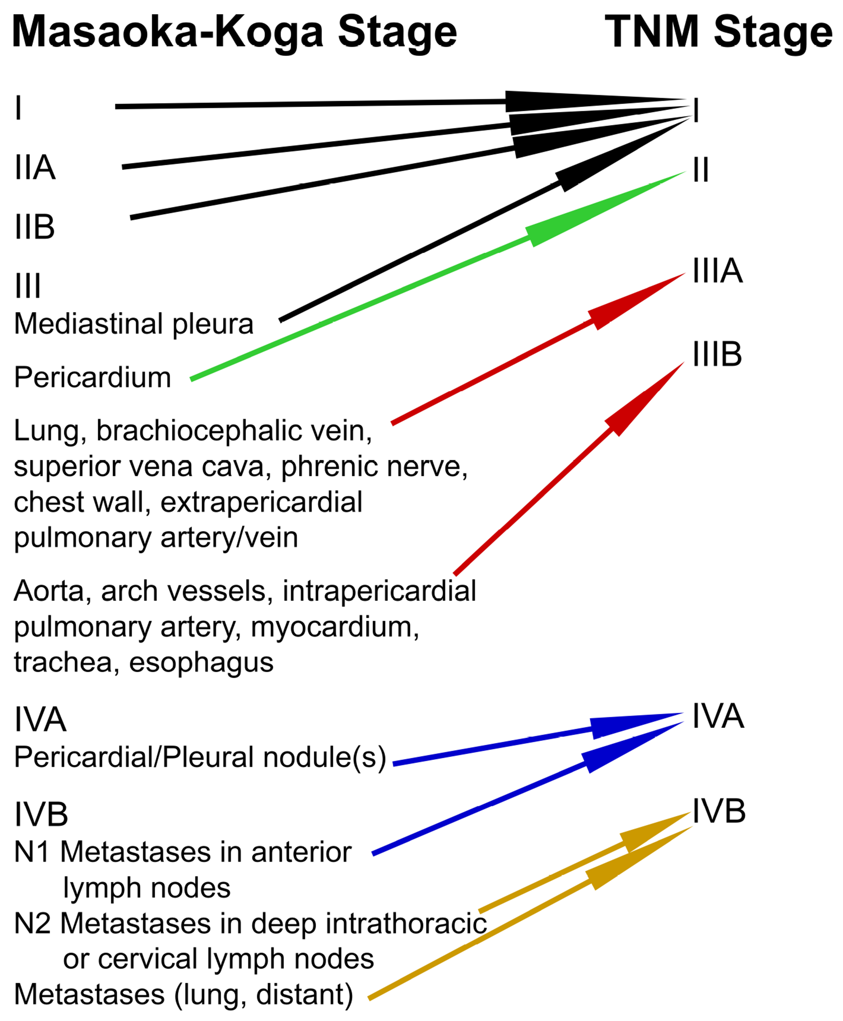
Relationship between Masaoka Koga staging and TNM staging.
This manuscript is a multidisciplinary update in diagnosis and management of thymic carcinoma and a review of recent developments.
Epidemiology and Clinical Features of Thymic Carcinomas
The incidence of thymic carcinomas is reported to be between 0.07 and 0.38/100,000/year. This result was confirmed by a study from Japan published in 2020 that utilized a national database of hospital-based cancer registries that yielded an incidence rate of 0.29 thymic carcinomas per 100,000 person-years.1 An ITMIG-initiated multi-institutional radiology database study also published in 2020 found that thymic carcinoma comprise 5.6% of all solitary mediastinal lesions.2 In addition, this study showed that in the prevascular mediastinum, thymic carcinomas are the fourth most common solitary lesions after thymomas, benign cysts, and lymphomas, and account for 7.5% of all solitary lesions in that compartment.2 The disease occurs in patients with a median age of 54 to 65.5 years old, although the reported age range is broad (12 to 96 years old).1, 2, 12–17 Patients are more commonly male.1, 16–18
Thymic carcinomas are usually large and infiltrative at time of diagnosis, with a mean or median tumor size of 5.4 to 8.7 cm (range, 2.1 to 19 cm).12–15, 17, 19–21 Therefore, patients often present with symptoms of mass effect on surrounding structures in the prevascular or possible visceral (middle) mediastinum, such as bronchi and superior vena cava (SVC), leading to dyspnea, chest pain, and SVC syndrome among others.15 In addition, at the time of diagnosis, thymic carcinomas frequently present at an advanced stage, with most patients presenting at TNM stage III12, IVA or IVB18. That was confirmed in a study published in 2020 based on data from the national cancer database that showed that 68% of patients with thymic carcinoma had regional or distant disease.17 Nodal metastases occur in approximately one-third of patients at diagnosis.12 Some patients also present with symptoms from metastatic spread.15 In many patients, the tumor may be found incidentally.15 Although there are a few reports of paraneoplastic syndromes occurring with thymic carcinomas5, 18–20, this should raise suspicion of an overlooked thymoma component and prompt further investigation.
Imaging
Computed tomography (CT) is the primary imaging modality for initial diagnostic evaluation of thymic carcinoma, although magnetic resonance imaging (MRI) is equally appropriate for initial diagnosis.22 Although a recent study 23 has shown once more that MRI is superior to CT in differentiating thymic hyperplasia and thymic cysts from thymic neoplasms, due to the small cohort of thymic carcinoma patients, superiority of MRI versus CT in differentiating thymic carcinomas from lymphomas/germ cell tumors was not proven.
High-risk imaging features such as local invasion and lymphadenopathy should prompt imaging evaluation for extra-thoracic systemic disease. Fluorodeoxyglucose (FDG) positron emission tomography (PET) can be helpful in evaluating for metastatic disease, however its ability to differentiate histologic differences is not fully established.[21] Thymic carcinomas may display FDG uptake, similar to lymphoma and non-seminomatous germ cell tumors, and the FDG-avidity is significantly higher than that of thymomas (Figures 2A&B).24
Figure 2.
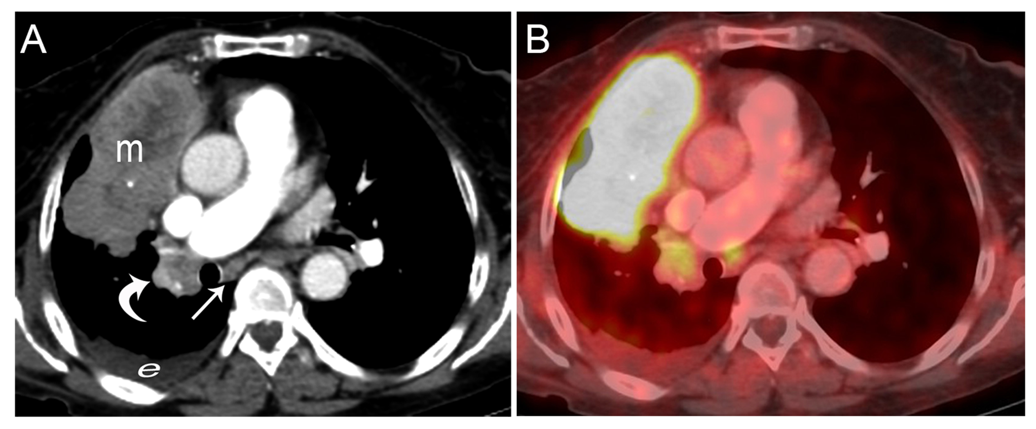
Thymic squamous cell carcinoma in a 28-year-old woman. A. CT imaging from an FDG PET-CT study shows an 11 cm prevascular necrotic appearing mass (m) with a small right pleural effusion (e), right hilar (curved arrow) and a subcarinal smaller than 1 cm lymph node (straight arrow). B. Fused image shows the mass is markedly FDG avid with an SUVmax of 12.2. The lymph nodes have an SUVmax of up to 4. Thymic carcinoma is typically markedly FDG avid. This FDG uptake is useful in identifying metastatic disease which may be overlooked with morphological imaging (CT or MRI), as seen with the subcarinal lymph node which was smaller than 1cm yet showed FDG uptake and was proven to be due to metastatic disease, N2, upstaging the patient to stage IVb.
Imaging findings on CT, PET and MRI are an adjunct to the clinical findings and can aid in decision-making. Suggestive imaging features including local invasion, lymphadenopathy, or metastatic disease can guide the diagnostic approach in biopsy and decision to administer neoadjuvant therapy if indicated.25
Investigative studies assessing newer MRI techniques for diagnosing thymic carcinoma, including dynamic contrast-enhanced MRI, radiomics analysis and diffusion-weighted imaging, show promising early results. These methods are not yet used in routine clinical practice due to lack of standardization of analysis, image acquisition, and interobserver variability, preventing the use of reliable threshold values.
For staging of thymic carcinoma, CT is the imaging modality of choice. The use of intravenous contrast is imperative for visualization of direct vascular involvement and aids in identifying nodal and distant metastatic disease. Unlike thymoma, because of the avid FDG uptake in thymic carcinoma, the use of FDG PET-CT has been shown to be beneficial in staging of thymic carcinoma and identifying nodal and pleural metastatic disease overlooked by CT.26
Assessment of response to treatment is routinely performed with contrast enhanced chest and abdominal CT because thymic carcinoma is more likely to recur at distant sites than locally as has been shown in a publication in 2020 by Rappaport et al.27 In the assessment of treatment response, the tumor is measured according to a thymic disease modification to Response Evaluation Criteria in Solid Tumors (RECIST) version 1.1.28 ITMIG recommends surveillance imaging following treatment to be performed more frequently for patients with thymic carcinoma than for those with thymoma: every 3 months for year 1, every 6 months for year 2, and then annually for the next 3 years, followed by an annual chest radiograph alternating with CT for 5 additional years at minimum.29, 30 Due to the limited sensitivity of chest radiography to detect recurrence/progression, some centers alternate CT with MRI, as MRI is comparable with CT for surveillance.23, 31 MRI is useful as a problem solving tool when postoperative fluid collections may mimic tumor recurrence in the surgical field.31 Metabolic response on an FDG-PET scan with >30% decrease in FDG uptake after treatment correlated with less than one third of tumor cell viability in a study by Fukumoto et al published in 2017 32; however, in a prospective study published in 2013 by Thomas et al it did not correlate with survival.33
The lack of large-scale prospective studies and standardization of newer imaging techniques with MRI and PET-CT limit the ability to perform multi-institutional studies. Hopefully, future standardization and improvements in automatic computer segmentation with the implementation of artificial intelligence (AI) to aid image interpretation will enable better diagnosis, staging, prognostication, and evaluation of response to treatment of thymic carcinoma.
Pathology
Advances in thymic carcinoma pathology in the last 5 years concern histological, nomenclatural, and molecular aspects and are mostly covered in the new WHO classification (2021).9, 34 Staging of thymic carcinomas should now follow the first AJCC/UICC-approved TNM staging system for TET (TNM Classification of Malignant Tumours, 8th edition, 2017), replacing the Masaoka-Koga classification.35
In the WHO 2021 classification, thymic carcinoma includes the following new subtypes (Table 1): 1) Micronodular thymic carcinoma with lymphoid hyperplasia (Figures 3A–F), a provisional, histologically distinct, apparently less aggressive subtype of thymic squamous cell carcinoma carrying the risk of confusion with “Micronodular thymoma with lymphoid stroma”; 2) Hyalinizing clear cell carcinoma (Figures 4A–C) that mimics its salivary gland counterpart in terms of histology and EWSR1 translocation; and 3) Thymic sebaceous carcinoma (Figures 4D–F) that can harbor a potentially actionable FGFR2 amplification and is listed with other rare thymic carcinomas under the new “basket” term, Thymic carcinoma not otherwise specified (NOS).
Table 1:
Pathologic classification of thymic carcinomas, taking new entities/variants and new nomenclature into account (WHO 2021)9, 34
| Squamous cell carcinoma |
| Micronodular thymic carcinoma with lymphoid hyperplasia (provisional subtype)a |
| Basaloid carcinoma |
| Lymphoepithelial carcinomab |
| NUT carcinoma of the thorax |
| Clear cell carcinoma |
| Hyalinizing clear cell carcinoma (subtype)a |
| Low-grade papillary adenocarcinomac |
| Mucoepidermoid carcinoma |
| Thymic carcinoma with adenoid cystic carcinoma-like features |
| Enteric-type adenocarcinomaa,d |
| Adenocarcinoma NOSa,e,f |
| Adenosquamous carcinoma |
| Sarcomatoid carcinoma (including carcinosarcoma) |
| Undifferentiated carcinoma |
| Thymic carcinoma NOSa |
| Hepatoid carcinoma |
| Rhabdoid carcinoma |
| Undifferentiated large cell carcinoma with Castleman disease-like reaction |
| Sebaceous carcinomaa |
New carcinoma (sub-) type, variant or tumor group introduced by the WHO in 2021;
previously called ‘lymphoepithelioma-like carcinoma’;
replacing the ambiguous previous term ‘papillary carcinoma’;
Thymic carcinoma subtype delineated through immunohistochemical positivity for at least one enteric differentiation marker (CK20, CDX2, MUC2);
Including poorly differentiated papillary carcinoma;
NOS, not otherwise specified
Figure 3:
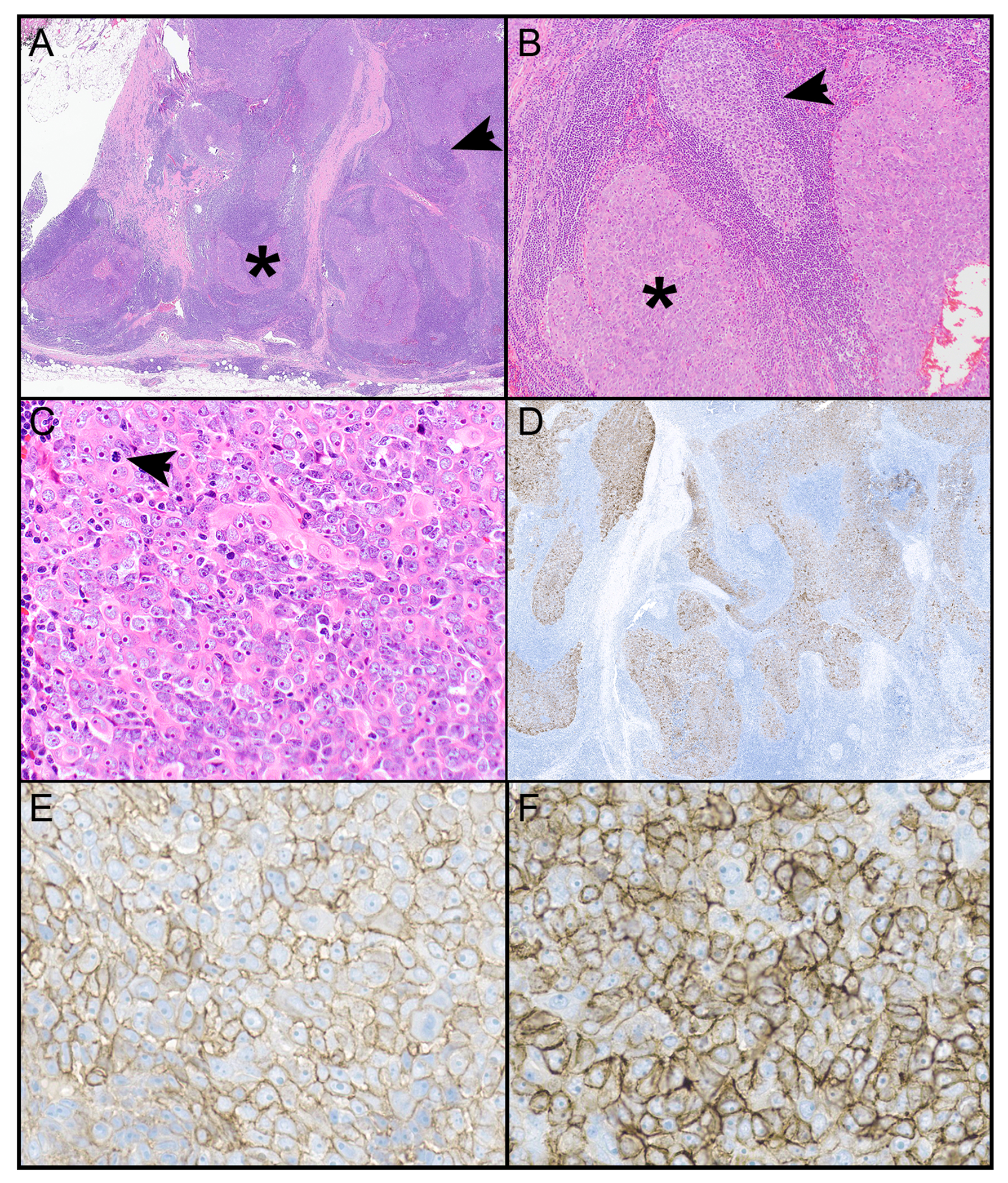
Micronodular thymic carcinoma with lymphoid hyperplasia. A. A circumscribed lesion is comprised of scattered nodular areas (*) in a background of lymphoid follicles with germinal centers (arrow). B. Lymphoid follicles with germinal centers (arrow) are in between paler cellular nodules (*). C. The nodules are comprised of neoplastic cells that are characterized by a polygonal shape, prominent nucleoli and occasional mitotic figures (arrow). The neoplastic cells are positive for keratin (pankeratin) which also highlights the nodular arrangement of the neoplastic cells (D), CD5 (E), and CD117 (F).
Magnification, H&E x 1.25 (A), x 10 (B), x 40 (C), pankeratin scanning power (D), CD5 x 400 (E), CD117 x 400 (F).
Figure 4:
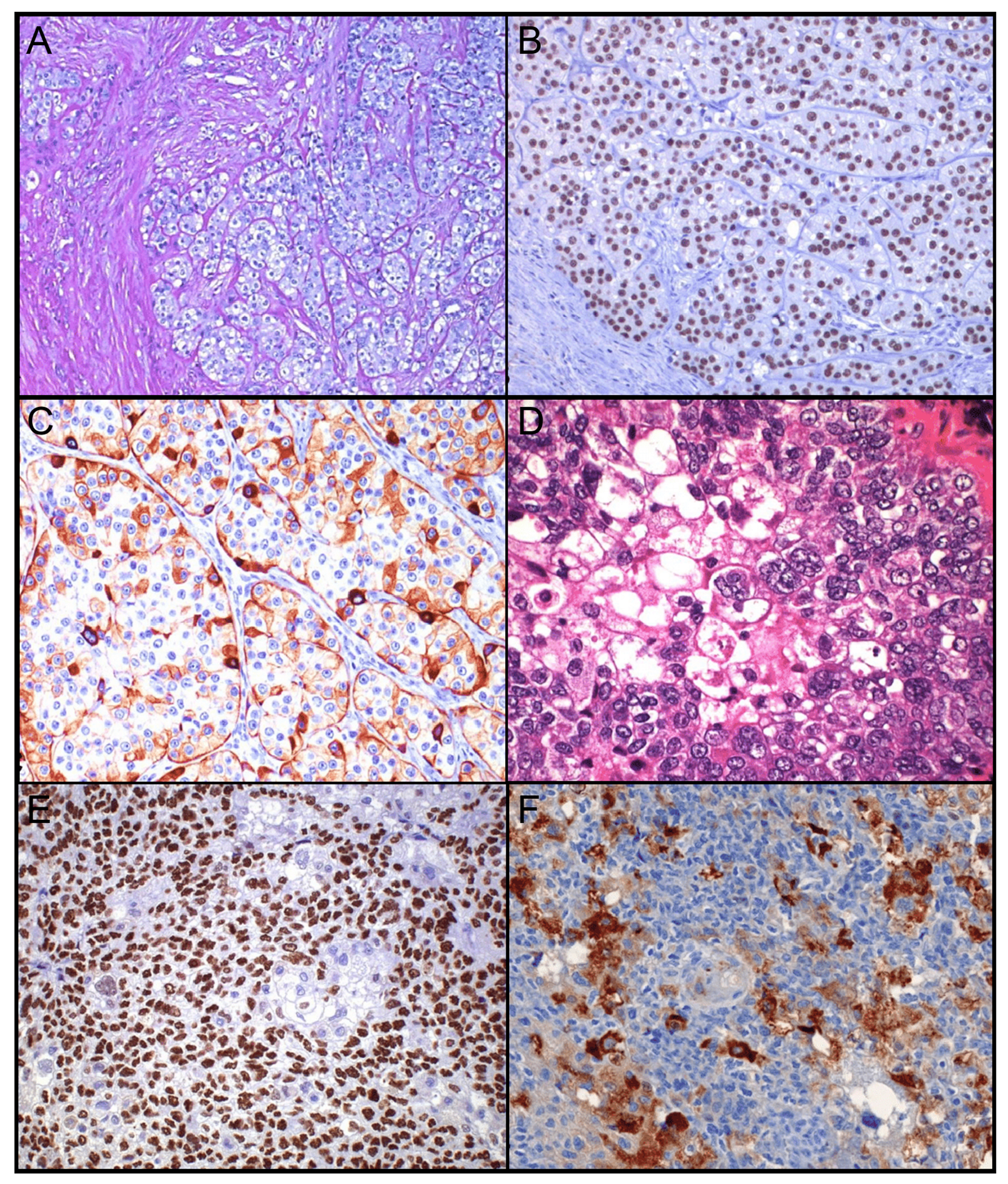
Thymic carcinoma entities described during the last 5 years: A.-C. Hyalinizing clear cell carcinoma A. Nests of tumor cells with clear cytoplasm in hyalinized stroma. B. P40 staining labels all tumor cell nuclei. C. CK5/6 stain reveals heterogeneous positivity of tumor cells (“mosaic pattern”). D.-F. Sebaceous carcinoma D. Epidermoid tumor cells and clear sebocytes. E. P40 staining of epidermoid cell nuclei but not sebocyte nuclei. F. Adipophilin staining restricted to sebocytes. Androgen receptor was also positive in the sebaceous carcinoma (not shown).
Magnification, H&E x 50 (A), x 200 (D), p40 x 100 (B, E), CK5/6 x 100 (C), adipophilin x 100 (F)
New immunohistochemical markers show diagnostic and prognostic associations: expression of cancer testis antigens (CTAs, e.g. MAGE-A, NY-ESO-1, SAGE, GAGE7)36 and PRAME (preferentially expressed antigen in melanoma) are more frequent in thymic carcinomas than thymomas, with PRAME showing the highest thymic carcinoma specificity37. Higher expression of some CTAs and GAD1 (the latter in relation to GAD1 hypermethylation) is a negative prognostic marker for thymic carcinomas.36, 38
The multi-omics TCGA project revealed a distinct molecular pathogenesis of almost all thymic carcinomas.4 The identification of only two thymic carcinomas arising from type B3 thymomas among 368 thymic carcinomas in an independent cohort39 supports this conclusion. Specifically, the most common abnormality in thymic carcinomas, loss of chromosome 16q, was lacking in thymomas, and the tumor mutation burden was higher in thymic carcinomas.4 On the other hand, thymic carcinomas, like thymomas, lacked recurrently mutated actionable genes. This finding is in agreement with observations from larger cohorts in which only rare thymic carcinomas showed diverse activating and potentially targetable mutations in the KIT, PDGFRA36, CDKN2A, FGFR340 and various PI3K subunit genes.41
Another new observation is microsatellite instability in rare thymic carcinomas due to variable inactivating MLH1 mutations that may increase the likelihood of response to PD-1/PD-L1 pathway blockade with immune checkpoint inhibitors (ICIs).4, 42 Indeed, PD-L1 expression is commonly high in tumor cells of thymic squamous cell carcinoma and thymic lymphoepithelial carcinomas and had some predictive value in two PD-1-targeting ICI clinical trials.43, 44 However, correlation of PD-LI expression and clinical activity of ICIs requires further investigation.
Finally, 80% of thymic squamous cell carcinomas, including all KIT(+) cases, were found to be POU2F3-positive “tuft cell-like cancers” that share a unique gene expression profile not only with normal chemosensory tuft cells45, but also with 20% of small cell lung cancers46 and a smaller subset of non-small cell lung cancers.45 Since almost all thymic and pulmonary tuft cell-like cancers lack targetable mutations, this hints at a currently unknown carcinogenic mechanism in tuft cell-like cancers, including thymic squamous cell carcinoma.45
Whole genome sequencing and single cell sequencing, non-coding RNAs, and the establishment of relevant models for preclinical testing are potential future studies to enhance our understanding of the pathogenesis of thymic carcinomas and to find potential therapeutically targetable molecular alterations.
Surgery
Surgical resection maintains a central role in the management of early stage and locally advanced thymic carcinomas. In patients who undergo resection as part of their multimodal therapy, 5-year overall survival ranges from 50% to 75% and complete resection rates range from 58 to 78%.47–49 The traditional oncologic principles of resection have been upheld by contemporary data. The landscape of surgical techniques, however, has significantly evolved over the last decade, with higher utilization of minimally invasive techniques for early-stage tumors. This can have important ramifications in case of incidentally discovered and resected thymic carcinomas.
In Masaoka-Koga stage I to III thymic carcinomas (TNM stage I-IIIA) local control is of paramount importance. Complete resection (R0) and postoperative radiation therapy have been shown to improve disease-free and overall survival in most series.50 The ITMIG database reported improved overall survival in patients with completely resected tumors.50 Similarly, a report from the Japanese Association for Research of the Thymus (JART) showed improved overall survival after R0 resection.51 More than half of these patients had Masaoka stage III to IV disease, and the majority also received postoperative radiation therapy (PORT). Interestingly, in this Japanese series as well as in the ITMIG database report, even early-stage patients were administered PORT.50, 51 A series of 203 TETs from the European Society of Thoracic Surgery (ESTS) database showed that complete resection was achieved in 158 (77.8%) patients and these patients had 3-, 5-, and 10-year survival rates of 79%, 75%, and 63%, respectively.49 Adjuvant therapy was performed in 68 (33.5%) patients including radiation therapy (N=60), chemotherapy (N=5) or chemoradiation (N=3).
It is important to note that even after complete resection, thymic carcinomas carry high local and systemic recurrence rates.52 The more aggressive biology and higher propensity to metastasize are exemplified by the high rate of mediastinal nodal metastases. This has been highlighted by data published in 2020 from Japan1 and in a prospective study performed by the Chinese Alliance for Research in Thymoma (ChART) published in 2018 of thymectomy with systematic nodal sampling.53 In the ChART study, among 275 patients with TET, 5.5% exhibited nodal metastases, however, amongst 24 patients with thymic carcinoma, 6 (25%) had nodal metastases.53
Controversy exists around the extent of nodal sampling and if this should be performed for every thymic tumor resection. Although there is no level 1 evidence that shows improved survival with systematic nodal sampling, extrapolation from other tumors such as early-stage lung cancer suggests that improved pathologic staging better allows for stage-appropriate use of multimodal therapy, which may in turn lead to improved disease control and survival.54 Appropriate systematic nodal sampling should be considered during resection for thymic carcinoma, as this allows for more accurate pathologic staging and should include prethymic, and anterior perithymic nodes (level N1) plus deep intrathoracic or cervical nodes (level N2) according to the IASLC/ITMIG staging system which defines anterior and deep regions based on boundaries that outline the peripheral extent of surgical dissection in all planes.55
In the last decade, minimally invasive surgical techniques, including video assisted thoracoscopic and robotic approaches, are being increasingly utilized, particularly in resection of early-stage disease.56 There are no strict criteria to guide preoperative biopsy or surgical approach. In general, larger or locally invasive tumors can be biopsied while smaller tumors can be resected without a biopsy. Known thymic carcinoma or intra-operative concern for thymic carcinoma should heighten the vigilance to achieving clear resection margins.57 Data comparing video-assisted thoracic surgery (VATS), robotic-assisted thoracic surgery (RATS) and open thoracotomy are only few and not conclusive. In experienced hands RATS allows for a more precise dissection compared to VATS. However, minimally invasive surgery should be performed with caution even in patients with Masaoka stage II thymoma, especially if they compress the innominate vein.58, 59
Although these approaches are attractive due to potentially less pain and morbidity and faster recovery, oncologic principles have to be upheld. This can be a challenge in the case of early-stage tumors that are being resected without a diagnosis. For example, a thymomectomy, as is sometimes undertaken for suspected early stage thymoma, may result in either incomplete resection or close surgical margin of an incidental thymic carcinoma. Intra-operative frozen section can sometimes differentiate thymoma from thymic carcinoma, which may change the planned extent of resection. A systematic approach of wide resection and nodal sampling, including both N1 and N2 stations for all tumors can be followed to avoid incomplete resection or understaging.
The surgical approach should be chosen after careful imaging evaluation, usually CT, ideally with intravenous contrast, and/or MRI, and FDG PET-CT, when indicated. Extent of disease and the extirpation it requires for complete resection should dictate the surgical approach. In case of intra-operative concern for local invasion, the surgical approach should be appropriately modified (bilateral or open approach) to enhance and optimize the chances for complete resection through a safe operation.
For locally advanced tumors including those with oligometastatic pleural and or pericardial disease, well planned resection in combination with radiation therapy delivered using newer techniques can allow for pulmonary preservation while allowing excellent local control. A SEER analysis based on 311 stage IVA thymic carcinoma patients enrolled from 1973 to 2015 showed surgical treatment could be beneficial.52 Patient selection and multidisciplinary care are central components of these complex multimodal treatment plans.
Medical Oncology
Thymic carcinomas can exhibit aggressive clinical behavior with a predisposition toward disease recurrence after definitive therapy. Approximately one-third of patients with newly diagnosed thymic carcinoma present with metastatic disease that requires systemic therapy.60 Five-year survival of patients with TNM stage III or IV thymic carcinoma ranges from 67% for individuals who have undergone complete surgical resection to 24% for patients with inoperable disease.61
Platinum-based combination chemotherapy is used for frontline treatment of inoperable thymic carcinoma (TNM stage IVB). Commonly used regimens include a combination of cisplatin, doxorubicin and cyclophosphamide, cisplatin with etoposide, or carboplatin with paclitaxel.60, 62 A retrospective analysis of the efficacy of first-line combination chemotherapy in Japanese patients with advanced thymic carcinoma showed response rates of 12% to 67%, median progression-free survival (mPFS) of 6 to 31 months, and median overall survival in the range of 17 to 52 months, with no significant differences between various chemotherapy combinations.63
Despite ongoing efforts at drug development, treatment options for recurrent thymic carcinoma are limited. Gemcitabine, 5-fluorouracil/capecitabine, taxanes, etoposide and ifosfamide have modest clinical activity when used as monotherapy.62 Gemcitabine in combination with capecitabine has a response rate of 38% and mPFS of 6 months in pretreated patients.64 In a phase II trial, single-agent amrubicin demonstrated a response rate of 11% and mPFS of 7 months in patients with previously treated thymic carcinoma.65
Development of targeted therapy for the treatment of thymic carcinoma has been challenging due to the paucity of actionable genomic alterations.4, 62 Mutations in the KIT gene, a potentially actionable oncogenic driver alteration, is observed in less than 10% of thymic carcinomas.66 However, the multikinase inhibitors, sunitinib and lenvatinib, and the mechanistic target of rapamycin inhibitor, everolimus have shown response rates of 17% to 38% and mPFS of 6 to 9 months in patients with recurrent thymic carcinoma, even in the absence of known genomic targets, and have entered routine clinical practice within the past 5 years.67–69 Limited success of biologic therapies to date has spurred the search for newer targets for treatment of thymic carcinomas. Mesothelin, a tumor differentiation antigen, is frequently expressed by thymic carcinomas.70 In preclinical studies, anetumab ravtansine, an antibody-drug conjugate targeting mesothelin, has shown the ability to inhibit mesothelin-expressing thymic carcinoma.71 These studies support the evaluation of mesothelin-targeted therapies in patients with thymic carcinoma.
Recent advances in immunotherapy for TETs include PD-1-directed immune checkpoint inhibition with pembrolizumab, which is associated with response rates of 19% to 23% in patients with recurrent thymic carcinoma.44, 72 A subset of patients experience durable benefit with a median duration of response of 10 to 36 months. However, due to underlying defects in immune self-tolerance, patients are at increased risk for the development of immune-related adverse events (irAEs), with 15% of patients treated with pembrolizumab and 13% of patients receiving nivolumab experiencing severe irAEs.44, 72, 73 Other forms of immunotherapy, including combination immunotherapy, are currently under investigation for treatment of recurrent thymic carcinoma.
Guidelines are lacking for selection of the optimal treatment option for recurrent thymic carcinoma. Recurrent tumors harboring KIT mutations can be treated with KIT-directed therapy. For tumors without actionable targets, patient preference and an assessment of potential benefits and risks associated with various drugs influence treatment selection. Immunotherapy should not be offered to patients with paraneoplastic autoimmune disease due to the high risk of development of immune-mediated toxicity. Participation in clinical trials should be encouraged whenever possible.
Platinum-based combination chemotherapy is also used as part of multimodality treatment for locally advanced thymic carcinomas. Neoadjuvant chemotherapy is recommended for treatment of unresectable TNM stage III and IVA disease.60, 74 Adjuvant chemotherapy can be considered in carefully selected patients at high risk for disease recurrence, including individuals with higher stages of the disease, after R1 resection or after resection of recurrent solitary metastases or ipsilateral pleural metastases.60 The benefit of post-operative chemotherapy might be limited in patients who have received neoadjuvant chemotherapy, and clinical judgment should be used to select patients most likely to benefit from additional postoperative chemotherapy.
Ongoing efforts to develop newer targeted and immune therapies for treatment of thymic carcinomas will inevitably lead to questions about combining and/or sequencing these treatment options to optimize response, survival and tolerability in patients with thymic carcinoma. Strategies to combine chemotherapy with immunotherapy and/or antiangiogenics are under evaluation. Further research is needed to define the role of immunotherapy and targeted therapies as a component of multimodality treatment for early-stage and locally advanced thymic carcinomas. The role of these drugs for maintenance therapy in patients with advanced or metastatic thymic carcinoma also needs to be studied in future clinical trials.
Radiation Oncology
The role of radiation therapy in patients with thymic carcinoma has typically been in the context of three settings: 1) Adjuvant therapy to reduce the risk of recurrence after surgical resection; 2) Definitive therapy in inoperable patients; and 3) Palliative treatment to manage or prevent symptoms caused by the tumor. Recent data in thymic carcinoma and advancements in radiation technologies have expanded the role of adjuvant radiation therapy to patients with earlier stage disease and allowed for new potential indications of radiation therapy, such as hemithoracic intensity-modulated pleural radiation therapy (IMPRINT) for patients with pleural metastases.
Adjuvant radiation therapy has typically been recommended for patients with positive margins after surgical resection (Figures 5A–C) and for patients with completely resected Masaoka Stage III-IVA thymic malignancies. Adjuvant radiation therapy has historically been considered controversial in patients with Masaoka Stage II disease. There have been 4 recent publications from the past 5 to 6 years, however, demonstrating the benefit of adjuvant radiation therapy in patients with Masaoka Stage II thymic carcinoma.51, 75–77 Omasa et al. demonstrated that adjuvant radiation therapy results in an improvement in relapse-free survival in patients with stage II-III thymic carcinoma on multivariable analysis (HR 0.48, 95%CI: 0.30-0.78, p=0.003).51 Recent data from the ITMIG, ESTS, and National Cancer Institute (NCI) databases demonstrated that this improvement in relapse-free survival translates to an overall survival benefit.75–78 Considering these recent data, patients with positive margins and patients with completely resected Masaoka stage II-IVA (TNM stage TIb-T4, N0-2, M0-1a) disease should be strongly considered for adjuvant radiation therapy. It is important to note that the TNM staging system collapsed patients with Masaoka-Koga stages I and II and some with stage III into TNM stage group I (Figure 1). Currently available data on the role of and indication for adjuvant radiation therapy are based on the Masaoka-Koga staging system, and it remains unclear how to apply the indication to the TNM staging system at this point.
Figure 5:
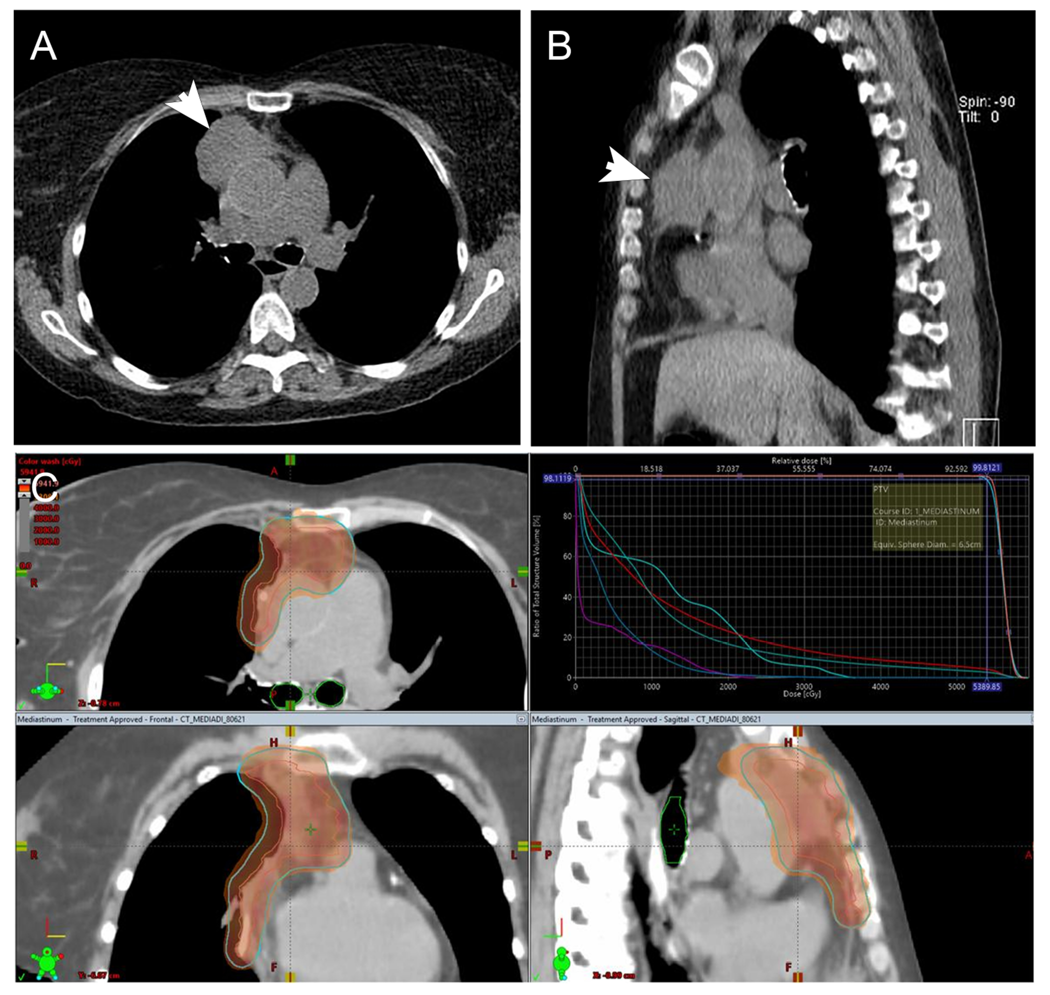
A 62-year-old woman with TNM stage II thymic carcinoma (arrows on non-contrast enhanced CT chest; A, axial slices, B, sagittal slices) underwent wedge resection of the right upper lobe lung and pericardectomy (R1 resection with positive anterior and posterior margins and invasion of the pericardium) (pT2N0M0). No adjuvant chemotherapy was given due to stage IV chronic kidney disease. She was going to be treated with post-operative radiotherapy for R1 resection. C. Volumetric modulated arc therapy radiation treatment plan to 5400 cGy in 30 fractions. The red line indicates gross tumor volumes; the blue line is the planning target and the color wash overlay is the 100% isodose level.
Recent advancements in radiation technology, including the use of IMPRINT, image-guided radiation therapy and proton therapy, have likely contributed to the recent advantages seen with adjuvant radiation therapy in patients with thymic carcinoma. These technological advancements have resulted in less exposure of radiation to normal organs and improved the risk-to-benefit ratio of radiotherapy. The development of the revolutionary technique of IMPRINT is an innovative strategy to manage pleural disease in patients with malignant pleural mesothelioma.79 Because the pleura is a common site for recurrence and progression in patients who present with earlier stage disease, IMPRINT can potentially be translated to patients with pleural metastases from TET, including thymic carcinomas, as a novel potential option to control pleural disease.80, 81 The risk of toxicities, particularly radiation pneumonitis, with IMPRINT in the setting of pleural metastases from TET is unknown. Furthermore, the prognosis and survival of patients with TET, including thymic carcinomas, is significantly longer than patients with malignant pleural mesothelioma.82 Taken together, the role of IMPRINT for management of TET remains to be determined and currently is not advisable outside of a clinical trial.
Over the past 5 years, proton therapy has emerged as a particularly beneficial treatment modality for patients with thymic malignancies.83–87 Given the typical tumor location in the anterior mediastinum, patients can frequently benefit from the physical properties of proton therapy to limit dose to the surrounding normal tissues, particularly the heart, esophagus and lungs, and potentially decrease the risks of long-term toxicities from radiotherapy, including the risks of major cardiac events85 and secondary malignancies.87
As thymic carcinoma is a radiosensitive tumor and as the technology of radiation therapy continues to advance, the indications for radiation therapy to improve outcomes in patients with thymic carcinoma are likely to expand.
Clinical Outcomes in Thymic Carcinomas
Studies published within the last 5 years have confirmed that increasing age at the time of diagnosis, no surgical treatment, and higher tumor stage correlate with worse overall survival in patients with thymic carcinomas.16, 17 Among surgically treated patients, younger age at diagnosis and localized disease are associated with better overall survival.16 Incomplete resection has been identified as an independent predictor of worse overall survival.17 The median overall survival of patients with thymic carcinoma ranges from 5.6 to 8.4 years, with 5-year overall survival rates of 52 to 64% and disease-free survival of 41%.14, 17, 88 Metastases most commonly occur in the pleura and lung and have also been described in liver, brain, extrathoracic lymph node, and adrenal gland but can also occur in other organs.14, 88
Summary
Recent advances in imaging, pathologic diagnostics, surgical modalities, radiation techniques, and options for systemic therapy of thymic carcinomas are promising and aid individualized management of patients. Large, multi-institutional, and ideally, prospective studies are required to further enhance standardization of diagnosis and management of these patients. The inclusion of newer diagnostic techniques and advanced treatment options, and the work of societies such as ITMIG is likely to facilitate these efforts.
Acknowledgments:
This research was funded in part by the Intramural Research Program of the National Institutes of Health (NIH), National Cancer Institute (NCI), Center for Cancer Research (Dr. Rajan), and through the NIH/NCI Cancer Center Support Grant P30 CA008748 (Drs. Rimner, Shepherd, Simone).
Disclosures:
Dr. Edith Marom received honoraria for lectures from Merck Sharp & Dohme and Boehringer Ingelheim. Dr. Andre Moreira received consulting fees from Hoffman-La Roche and participated on a data safety monitoring board or advisory board of Olympus corporation. Dr. Andrew Nicholson received consultation fees from Merck, Boehringer Ingelheim, Pfizer, Novartis, AstraZeneca, Bristol Myer Squib, Roche, Abbvie, Oncologica, and Takeda UK, a research grant from Pfizer, and payment for provision of educational materials from up-to-date, European Society of Oncology, and Liberum. Dr. Roden received honorarium for contributing educational material to up-to-date. Dr. Annemarie Shepherd received support for attending meetings and travel from Memorial Sloan Kettering Cancer Center. Dr. Charles Simone received a honorarium from Varian Medical Systems, is the chair of the NRG Oncology Particle Therapy Work Group and the President of the Board of Directors of the Proton Collaborative Group. Dr. Malgorzata Szolkowska received honoraria for lectures from Boehringer Ingelheim, AstraZeneca Pharma Poland, Roche Polska, and MSD Polska, is the Secretary of ITMIG, Chair of a working group of the ESP, and member of the main revisory board of the Polish Society of Pathology. Dr. Andreas Rimner has grant funding for investigator-initiated trials to Memorial Sloan Kettering Cancer Center from Varian Medical Systems, AstraZeneca, Merck, Pfizer, Boehringer Ingelheim, received consultation fees from Boehringer Ingelheim, AstraZeneca, Merck, Cybrexa, MoreHealth, received honorarium from ResearchToPractice, travel support from Philips/Elekta and is scientific advisory board member for KEYLYNK-012 and KEYLYNK-013 studies for Merck, is Vice President of ITMIG, board member of IMIG, Chair of the lung track of the Annual ASTRO meeting, and member of the Board of examiners of the American Board of Radiology.
Footnotes
Publisher's Disclaimer: This is a PDF file of an unedited manuscript that has been accepted for publication. As a service to our customers we are providing this early version of the manuscript. The manuscript will undergo copyediting, typesetting, and review of the resulting proof before it is published in its final form. Please note that during the production process errors may be discovered which could affect the content, and all legal disclaimers that apply to the journal pertain.
References
- [1].Koizumi T, Otsuki K, Tanaka Y, et al. : National incidence and initial therapy for thymic carcinoma in Japan: based on analysis of hospital-based cancer registry data, 2009-2015. Jpn J Clin Oncol 2020, 50:434–9. [DOI] [PubMed] [Google Scholar]
- [2].Roden AC, Fang W, Shen Y, et al. : Distribution of Mediastinal Lesions Across Multi-Institutional, International, Radiology Databases. J Thorac Oncol 2020, 15:568–79. [DOI] [PubMed] [Google Scholar]
- [3].Shepherd A, Riely G, Detterbeck F, et al. : Thymic Carcinoma Management Patterns among International Thymic Malignancy Interest Group (ITMIG) Physicians with Consensus from the Thymic Carcinoma Working Group. J Thorac Oncol 2017, 12:745–51. [DOI] [PMC free article] [PubMed] [Google Scholar]
- [4].Radovich M, Pickering CR, Felau I, et al. : The Integrated Genomic Landscape of Thymic Epithelial Tumors. Cancer Cell 2018, 33:244–58 e10. [DOI] [PMC free article] [PubMed] [Google Scholar]
- [5].Padda SK, Yao X, Antonicelli A, et al. : Paraneoplastic Syndromes and Thymic Malignancies: An Examination of the International Thymic Malignancy Interest Group Retrospective Database. J Thorac Oncol 2018, 13:436–46. [DOI] [PMC free article] [PubMed] [Google Scholar]
- [6].Korst RJ, Fernando S, Catlin AC, et al. : Positron Emission Tomography in Thymic Tumors: Analysis Using a Prospective Research Database. Ann Thorac Surg 2017, 104:1815–20. [DOI] [PMC free article] [PubMed] [Google Scholar]
- [7].Carter BW, Benveniste MF, Madan R, et al. : ITMIG Classification of Mediastinal Compartments and Multidisciplinary Approach to Mediastinal Masses. Radiographics 2017, 37:413–36. [DOI] [PubMed] [Google Scholar]
- [8].Fang W, Yao X, Antonicelli A, et al. : Comparison of surgical approach and extent of resection for Masaoka-Koga Stage I and II thymic tumours in Europe, North America and Asia: an International Thymic Malignancy Interest Group retrospective database analysis. Eur J Cardiothorac Surg 2017, 52:26–32. [DOI] [PMC free article] [PubMed] [Google Scholar]
- [9].Chan J, Marx A, Detterbeck F, Marino M, Marom EM, Ströbel P, Rajan A: Thymic carcinoma. WHO Classification of Tumours Thoracic Tumours 5 ed. Lyon (France): International Agency for Research on Cancer, 2021. [Google Scholar]
- [10].Amin MB, American Joint Committee on Cancer, American Cancer Society: AJCC cancer staging manual. Eight edition / editor-in-chief, Mahul B. Amin MD, FCAP; editors, Stephen B. Edge MD, FACS et al. ; Chicago IL: American Joint Committee on Cancer, Springer, 2017. [Google Scholar]
- [11].Koga K, Matsuno Y, Noguchi M, Mukai K, Asamura H, Goya T, Shimosato Y: A review of 79 thymomas: modification of staging system and reappraisal of conventional division into invasive and non-invasive thymoma. Pathol Int 1994, 44:359–67. [DOI] [PubMed] [Google Scholar]
- [12].Roden AC, Yi ES, Cassivi SD, Jenkins SM, Garces YI, Aubry MC: Clinicopathological features of thymic carcinomas and the impact of histopathological agreement on prognostical studies. European Journal of Cardio-Thoracic Surgery 2013, 43:1131–9. [DOI] [PubMed] [Google Scholar]
- [13].Sakane T, Murase T, Okuda K, et al. : A mutation analysis of the EGFR pathway genes, RAS, EGFR, PIK3CA, AKT1 and BRAF, and TP53 gene in thymic carcinoma and thymoma type A/B3. Histopathology 2019, 75:755–66. [DOI] [PubMed] [Google Scholar]
- [14].Hishida T, Nomura S, Yano M, et al. : Long-term outcome and prognostic factors of surgically treated thymic carcinoma: results of 306 cases from a Japanese Nationwide Database Study. Eur J Cardiothorac Surg 2016, 49:835–41. [DOI] [PubMed] [Google Scholar]
- [15].Chalabreysse L, Etienne-Mastroianni B, Adeleine P, Cordier JF, Greenland T, Thivolet-Bejui F: Thymic carcinoma: a clinicopathological and immunohistological study of 19 cases. Histopathology 2004, 44:367–74. [DOI] [PubMed] [Google Scholar]
- [16].Wu J, Wang Z, Jing C, Hu Y, Yang B, Hu Y: The incidence and prognosis of thymic squamous cell carcinoma: A Surveillance, Epidemiology, and End Results Program population-based study. Medicine (Baltimore) 2021, 100:e25331. [DOI] [PMC free article] [PubMed] [Google Scholar]
- [17].Bakhos CT, Salami AC, Kaiser LR, Petrov RV, Abbas AE: Thymic Neuroendocrine Tumors and Thymic Carcinoma: Demographics, Treatment, and Survival. Innovations (Phila) 2020, 15:468–74. [DOI] [PubMed] [Google Scholar]
- [18].Litvak AM, Woo K, Hayes S, et al. : Clinical characteristics and outcomes for patients with thymic carcinoma: evaluation of Masaoka staging. J Thorac Oncol 2014, 9:1810–5. [DOI] [PMC free article] [PubMed] [Google Scholar]
- [19].Liu HC, Hsu WH, Chen YJ, Chan YJ, Wu YC, Huang BS, Huang MH: Primary thymic carcinoma. Ann Thorac Surg 2002, 73:1076–81. [DOI] [PubMed] [Google Scholar]
- [20].Hosaka Y, Tsuchida M, Toyabe S, Umezu H, Eimoto T, Hayashi J: Masaoka stage and histologic grade predict prognosis in patients with thymic carcinoma. Ann Thorac Surg 2010, 89:912–7. [DOI] [PubMed] [Google Scholar]
- [21].Okereke IC, Kesler KA, Freeman RK, et al. : Thymic carcinoma: outcomes after surgical resection. Ann Thorac Surg 2012, 93:1668–73. [DOI] [PubMed] [Google Scholar]
- [22].Expert Panel on Thoracic I, Ackman JB, Chung JH: ACR Appropriateness Criteria(R) Imaging of Mediastinal Masses. J Am Coll Radiol 2021, 18:S37–S51. [DOI] [PubMed] [Google Scholar]
- [23].Li HR, Gao J, Jin C, Jiang JH, Ding JY: Comparison between CT and MRI in the Diagnostic Accuracy of Thymic Masses. J Cancer 2019, 10:3208–13. [DOI] [PMC free article] [PubMed] [Google Scholar]
- [24].Benveniste MF, Moran CA, Mawlawi O, Fox PS, Swisher SG, Munden RF, Marom EM: FDG PET-CT aids in the preoperative assessment of patients with newly diagnosed thymic epithelial malignancies. J Thorac Oncol 2013, 8:502–10. [DOI] [PMC free article] [PubMed] [Google Scholar]
- [25].Sonnett JR: FDG PET-CT – How Do I Use It? How Does It Really Help Me – the Surgeon’s View. The International Thymic Malignancy Interest Group 2020 Webinar Sseries, 2020. [Google Scholar]
- [26].Sung YM, Lee KS, Kim BT, Choi JY, Shim YM, Yi CA: 18F-FDG PET/CT of thymic epithelial tumors: usefulness for distinguishing and staging tumor subgroups. J Nucl Med 2006, 47:1628–34. [PubMed] [Google Scholar]
- [27].Rappaport JMP, Huang J, Ahmad U: Thymic Malignancy-Updates in Staging and Management. Surg Oncol Clin N Am 2020, 29:581–601. [DOI] [PubMed] [Google Scholar]
- [28].Benveniste MF, Korst RJ, Raj an A, Detterbeck FC, Marom EM, International Thymic Malignancy Interest G: A practical guide from the International Thymic Malignancy Interest Group (ITMIG) regarding the radiographic assessment of treatment response of thymic epithelial tumors using modified RECIST criteria. J Thorac Oncol 2014, 9:S119–24. [DOI] [PubMed] [Google Scholar]
- [29].Huang J, Detterbeck FC, Wang Z, Loehrer PJ Sr.: Standard outcome measures for thymic malignancies. Journal of thoracic oncology : official publication of the International Association for the Study of Lung Cancer 2010, 5:2017–23. [DOI] [PubMed] [Google Scholar]
- [30].Systematic Treatment General Reporting Guidelines in Reporting Guidelines for Thymic Malignancies Treated with Chemotherapy. ITMIG Medical Oncologist Kit: International Thymic Malignancy Interest Group, https://itmig.org/education-and-research/itmig-standards-and-definitions/itmig-medical-oncologist-kit/
- [31].Kerpel A, Beytelman A, Ofek E, Marom EM: Magnetic Resonance Imaging for the Follow-up of Treated Thymic Epithelial Malignancies. J Thorac Imaging 2019, 34:345–50. [DOI] [PubMed] [Google Scholar]
- [32].Fukumoto K, Fukui T, Okasaka T, et al. : The Role of (18)F-fluorodeoxyglucose Positron Emission Tomography-Computed Tomography for Predicting Pathologic Response After Induction Therapy for Thymic Epithelial Tumors. World J Surg 2017, 41:1828–33. [DOI] [PubMed] [Google Scholar]
- [33].Thomas A, Mena E, Kurdziel K, et al. : 18F-fluorodeoxyglucose positron emission tomography in the management of patients with thymic epithelial tumors. Clin Cancer Res 2013, 19:1487–93. [DOI] [PMC free article] [PubMed] [Google Scholar]
- [34].Marx A, Chan JKC, Chalabreysse L, et al. : The 2021 World Health Organization Classification of Tumors of the Thymus and Mediastinum: What’s new in thymic epithelial, germ cell and mesenchymal tumors? J Thorac Oncol 2021. [DOI] [PubMed] [Google Scholar]
- [35].Ruffini E, Fang W, Guerrera F, et al. : The International Association for the Study of Lung Cancer Thymic Tumors Staging Project: The Impact of the Eighth Edition of the Union for International Cancer Control and American Joint Committee on Cancer TNM Stage Classification of Thymic Tumors. J Thorac Oncol 2020, 15:436–47. [DOI] [PubMed] [Google Scholar]
- [36].Sakane T, Sakamoto Y, Masaki A, Murase T, Okuda K, Nakanishi R, Inagaki H: Mutation Profile of Thymic Carcinoma and Thymic Neuroendocrine Tumor by Targeted Next-generation Sequencing. Clin Lung Cancer 2021, 22:92–9 e4. [DOI] [PubMed] [Google Scholar]
- [37].Taniguchi Y, Ishida M, Saito T, et al. : Preferentially expressed antigen in melanoma as a novel diagnostic marker differentiating thymic squamous cell carcinoma from thymoma. Sci Rep 2020, 10:12286. [DOI] [PMC free article] [PubMed] [Google Scholar]
- [38].Soejima S, Kondo K, Tsuboi M, et al. : GAD1 expression and its methylation as indicators of malignant behavior in thymic epithelial tumors. Oncol Lett 2021, 21:483. [DOI] [PMC free article] [PubMed] [Google Scholar]
- [39].Massoth LR, Hung YP, Dias-Santagata D, et al. : Pan-Cancer Landscape Analysis Reveals Recurrent KMT2A-MAML2 Gene Fusion in Aggressive Histologic Subtypes of Thymoma. JCO Precis Oncol 2020, 4. [DOI] [PMC free article] [PubMed] [Google Scholar]
- [40].Asselta R, Di Tommaso L, Perrino M, et al. : Mutation profile and immunoscore signature in thymic carcinomas: An exploratory study and review of the literature. Thorac Cancer 2021, 12:1271–8. [DOI] [PMC free article] [PubMed] [Google Scholar]
- [41].Alberobello AT, Wang Y, Beerkens FJ, et al. : PI3K as a Potential Therapeutic Target in Thymic Epithelial Tumors. J Thorac Oncol 2016, 11:1345–56. [DOI] [PMC free article] [PubMed] [Google Scholar]
- [42].Repetto M, Conforti F, Pirola S, et al. : Thymic carcinoma with Lynch syndrome or microsatellite instability, a rare entity responsive to immunotherapy. Eur J Cancer 2021, 153:162–7. [DOI] [PubMed] [Google Scholar]
- [43].Giaccone G, Kim C, Thompson J, et al. : Pembrolizumab in patients with thymic carcinoma: a single-arm, single-centre, phase 2 study. Lancet Oncol 2018, 19:347–55. [DOI] [PMC free article] [PubMed] [Google Scholar]
- [44].Cho J, Kim HS, Ku BM, et al. : Pembrolizumab for Patients With Refractory or Relapsed Thymic Epithelial Tumor: An Open-Label Phase II Trial. J Clin Oncol 2019, 37:2162–70. [DOI] [PubMed] [Google Scholar]
- [45].Yamada Y, Simon-Keller K, Belharazem-Vitacolonnna D, et al. : A Tuft Cell-Like Signature Is Highly Prevalent in Thymic Squamous Cell Carcinoma and Delineates New Molecular Subsets Among the Major Lung Cancer Histotypes. J Thorac Oncol 2021, 16:1003–16. [DOI] [PubMed] [Google Scholar]
- [46].Huang YH, Klingbeil O, He XY, et al. : POU2F3 is a master regulator of a tuft cell-like variant of small cell lung cancer. Genes Dev 2018, 32:915–28. [DOI] [PMC free article] [PubMed] [Google Scholar]
- [47].Li W, Miao Z, Liu X, Zhang Q, Sun L, Li P, Liu W, Zhang L: Thymic carcinoma patients with myasthenia gravis exhibit better prognoses. Int J Clin Oncol 2016, 21:75–80. [DOI] [PubMed] [Google Scholar]
- [48].Fu H, Gu ZT, Fang WT, et al. : Long-Term Survival After Surgical Treatment of Thymic Carcinoma: A Retrospective Analysis from the Chinese Alliance for Research of Thymoma Database. Ann Surg Oncol 2016, 23:619–25. [DOI] [PubMed] [Google Scholar]
- [49].Lococo F, Nachira D, Chiappetta M, et al. : Does Myasthenia Gravis Affect Long-Term Survival in Thymic Carcinoma Patients? A Retrospective Analysis from the ESTS Database. Eur J Cardiothorac Surg 2021, In Press. [Google Scholar]
- [50].Ahmad U, Yao X, Detterbeck F, et al. : Thymic carcinoma outcomes and prognosis: results of an international analysis. The Journal of thoracic and cardiovascular surgery 2015, 149:95–100, 1 e1-2. [DOI] [PubMed] [Google Scholar]
- [51].Omasa M, Date H, Sozu T, et al. : Postoperative radiotherapy is effective for thymic carcinoma but not for thymoma in stage II and III thymic epithelial tumors: the Japanese Association for Research on the Thymus Database Study. Cancer 2015, 121:1008–16. [DOI] [PubMed] [Google Scholar]
- [52].Ye C, Bao M, Li H, Liu X, Jiang G, Wang W: Surgery in Masaoka stage IV thymic carcinoma: a propensity-matched study based on the SEER database. J Thorac Dis 2020, 12:659–71. [DOI] [PMC free article] [PubMed] [Google Scholar]
- [53].Fang W, Wang Y, Pang L, et al. : Lymph node metastasis in thymic malignancies: A Chinese multicenter prospective observational study. J Thorac Cardiovasc Surg 2018, 156:824–33 e1. [DOI] [PubMed] [Google Scholar]
- [54].Drevet G, Collaud S, Tronc F, Girard N, Maury JM: Optimal management of thymic malignancies: current perspectives. Cancer Manag Res 2019, 11:6803–14. [DOI] [PMC free article] [PubMed] [Google Scholar]
- [55].Carter BW, Benveniste MF, Madan R, et al. : IASLC/ITMIG Staging System and Lymph Node Map for Thymic Epithelial Neoplasms. Radiographics 2017, 37:758–76. [DOI] [PubMed] [Google Scholar]
- [56].Ruffini E, Filosso PL, Guerrera F, Lausi P, Lyberis P, Oliaro A: Optimal surgical approach to thymic malignancies: New trends challenging old dogmas. Lung Cancer 2018, 118:161–70. [DOI] [PubMed] [Google Scholar]
- [57].Burt BM, Yao X, Shrager J, et al. : Determinants of Complete Resection of Thymoma by Minimally Invasive and Open Thymectomy: Analysis of an International Registry. J Thorac Oncol 2017, 12:129–36. [DOI] [PMC free article] [PubMed] [Google Scholar]
- [58].Wang GW, Tao T, Li CK, Li QC, Duan GX, Sang HW, Dong HJ, Wang ZY: Comparison between thoracoscopic and open approaches in thymoma resection. J Thorac Dis 2019, 11:4159–68. [DOI] [PMC free article] [PubMed] [Google Scholar]
- [59].Raveglia F, Bertolaccini L, Solli P, Minervini F, Scarci M: VATS thymectomy: oncological results and comparison between minimally invasive strategies. Shanghai Chest 2018, 2. [Google Scholar]
- [60].Girard N, Ruffini E, Marx A, Faivre-Finn C, Peters S, Committee EG: Thymic epithelial tumours: ESMO Clinical Practice Guidelines for diagnosis, treatment and follow-up. Ann Oncol 2015, 26 Suppl 5:v40–55. [DOI] [PubMed] [Google Scholar]
- [61].Kondo K, Monden Y: Therapy for thymic epithelial tumors: a clinical study of 1,320 patients from Japan. Ann Thorac Surg 2003, 76:878–84; discussion 84-5. [DOI] [PubMed] [Google Scholar]
- [62].Berghmans T, Durieux V, Holbrechts S, et al. Systemic treatments for thymoma and thymic carcinoma: A systematic review. Lung Cancer 2018, 126:25–31. [DOI] [PubMed] [Google Scholar]
- [63].Ko R, Shukuya T, Okuma Y, et al. : Prognostic Factors and Efficacy of First-Line Chemotherapy in Patients with Advanced Thymic Carcinoma: A Retrospective Analysis of 286 Patients from NEJ023 Study. Oncologist 2018, 23:1210–7. [DOI] [PMC free article] [PubMed] [Google Scholar]
- [64].Palmieri G, Buonerba C, Ottaviano M, et al. : Capecitabine plus gemcitabine in thymic epithelial tumors: final analysis of a Phase II trial. Future Oncol 2014, 10:2141–7. [DOI] [PubMed] [Google Scholar]
- [65].Hellyer JA, Gubens MA, Cunanan KM, et al. : Phase II trial of single agent amrubicin in patients with previously treated advanced thymic malignancies. Lung Cancer 2019, 137:71–5. [DOI] [PubMed] [Google Scholar]
- [66].Tateo V, Manuzzi L, Parisi C, De Giglio A, Campana D, Pantaleo MA, Lamberti G: An Overview on Molecular Characterization of Thymic Tumors: Old and New Targets for Clinical Advances. Pharmaceuticals (Basel) 2021, 14:316. [DOI] [PMC free article] [PubMed] [Google Scholar]
- [67].Thomas A, Rajan A, Berman A, et al. : Sunitinib in patients with chemotherapy-refractory thymoma and thymic carcinoma: an open-label phase 2 trial. Lancet Oncol 2015, 16:177–86. [DOI] [PMC free article] [PubMed] [Google Scholar]
- [68].Sato J, Satouchi M, Itoh S, et al. : Lenvatinib in patients with advanced or metastatic thymic carcinoma (REMORA): a multicentre, phase 2 trial. Lancet Oncol 2020, 21:843–50. [DOI] [PubMed] [Google Scholar]
- [69].Zucali PA, De Pas T, Palmieri G, et al. : Phase II Study of Everolimus in Patients With Thymoma and Thymic Carcinoma Previously Treated With Cisplatin-Based Chemotherapy. J Clin Oncol 2018, 36:342–9. [DOI] [PubMed] [Google Scholar]
- [70].Thomas A, Chen Y, Berman A, et al. : Expression of mesothelin in thymic carcinoma and its potential therapeutic significance. Lung Cancer 2016, 101:104–10. [DOI] [PMC free article] [PubMed] [Google Scholar]
- [71].Chen V, Umemura S, Han Y, et al. : Exploiting mesothelin in thymic carcinoma as a drug delivery target for anetumab ravtansine. Br J Cancer 2021. [DOI] [PMC free article] [PubMed] [Google Scholar]
- [72].Giaccone G, Kim C: Durable Response in Patients With Thymic Carcinoma Treated With Pembrolizumab After Prolonged Follow-Up. J Thorac Oncol 2021, 16:483–5. [DOI] [PubMed] [Google Scholar]
- [73].Katsuya Y, Horinouchi H, Seto T, et al. : Single-arm, multicentre, phase II trial of nivolumab for unresectable or recurrent thymic carcinoma: PRIMER study. Eur J Cancer 2019, 113:78–86. [DOI] [PubMed] [Google Scholar]
- [74].Patel DC, Shrager JB, Padda SK: The role of induction therapy for thymic malignancies: a narrative review. . Mediastinum 2020, 4:36. [DOI] [PMC free article] [PubMed] [Google Scholar]
- [75].Ahmad U, Yao X, Detterbeck F, et al. : Thymic carcinoma outcomes and prognosis: results of an international analysis. J Thorac Cardiovasc Surg 2015, 149:95–100, 1 e1-2. [DOI] [PubMed] [Google Scholar]
- [76].Ruffini E, Detterbeck F, Van Raemdonck D, et al. : Thymic carcinoma: a cohort study of patients from the European society of thoracic surgeons database. J Thorac Oncol 2014, 9:541–8. [DOI] [PubMed] [Google Scholar]
- [77].Jackson MW, Palma DA, Camidge DR, et al. : The Impact of Postoperative Radiotherapy for Thymoma and Thymic Carcinoma. J Thorac Oncol 2017, 12:734–44. [DOI] [PubMed] [Google Scholar]
- [78].Rimner A: Postoperative radiotherapy: not all thymic malignancies are created equal. Cancer 2015, 121:972–4. [DOI] [PubMed] [Google Scholar]
- [79].Rimner A, Zauderer MG, Gomez DR, et al. : Phase II Study of Hemithoracic Intensity-Modulated Pleural Radiation Therapy (IMPRINT) As Part of Lung-Sparing Multimodality Therapy in Patients With Malignant Pleural Mesothelioma. J Clin Oncol 2016, 34:2761–8. [DOI] [PMC free article] [PubMed] [Google Scholar]
- [80].Kamel MK, Stiles BM, Ghaly G, et al. : Predictors of Pleural Implants in Patients With Thymic Tumors. Ann Thorac Surg 2016, 102:1647–52. [DOI] [PubMed] [Google Scholar]
- [81].Rimner A, Gomez DR, Wu AJ, et al. : Failure patterns relative to radiation treatment fields for stage II-IV thymoma. J Thorac Oncol 2014, 9:403–9. [DOI] [PubMed] [Google Scholar]
- [82].Choe G, Ghanie A, Riely G, Rimner A, et al. : Long-term, disease-specific outcomes of thymic malignancies presenting with de novo pleural metastasis. J Thorac Cardiovasc Surg 2020, 159:705–14 e1. [DOI] [PMC free article] [PubMed] [Google Scholar]
- [83].Vogel J, Berman AT, Lin L, et al. : Prospective study of proton beam radiation therapy for adjuvant and definitive treatment of thymoma and thymic carcinoma: Early response and toxicity assessment. Radiotherapy and oncology. Radiother Oncol 2016, 118:504–9. [DOI] [PubMed] [Google Scholar]
- [84].Parikh RR, Rhome R, Hug E, Tsai H, Cahlon O, Chon B, Goenka A: Adjuvant Proton Beam Therapy in the Management of Thymoma: A Dosimetric Comparison and Acute Toxicities. Clin Lung Cancer 2016, 17:362–6. [DOI] [PubMed] [Google Scholar]
- [85].Vogel J, Lin L, Simone CB 2nd, Berman AT: Risk of major cardiac events following adjuvant proton versus photon radiation therapy for patients with thymic malignancies. Acta Oncol 2017, 56:1060–4. [DOI] [PubMed] [Google Scholar]
- [86].Mercado CE, Hartsell WF, Simone CB, et al. : Proton therapy for thymic malignancies: multi-institutional patterns-of-care and early clinical outcomes from the proton collaborative group and the university of Florida prospective registries. Acta Oncol 2019, 58:1036–40. [DOI] [PubMed] [Google Scholar]
- [87].Vogel J, Lin L, Litzky LA, Berman AT, Simone CB 2nd: Predicted Rate of Secondary Malignancies Following Adjuvant Proton Versus Photon Radiation Therapy for Thymoma. Int J Radiat Oncol Biol Phys 2017, 99:427–33. [DOI] [PubMed] [Google Scholar]
- [88].Zhao Y, Gu H, Fan L, Han K, Yang J, Zhao H: Comparison of clinical features and survival between thymic carcinoma and thymic carcinoid patients. Eur J Cardiothorac Surg 2017, 52:33–8. [DOI] [PubMed] [Google Scholar]