Abstract
Leukemias are among the most prevalent types of cancer worldwide. Bone marrow mesenchymal stem cells (MSCs) participate in the development of a suitable niche for hematopoietic stem cells, and are involved in the development of diseases such as leukemias, to a yet unknown extent. Here we described the effect of secretome of bone marrow MSCs obtained from healthy donors and from patients with acute myeloid leukemia (AML) on leukemic cell lineages, sensitive (K562) or resistant (K562-Lucena) to chemotherapy drugs. Cell proliferation, viability and death were evaluated, together with cell cycle, cytokine production and gene expression of ABC transporters and cyclins. The secretome of healthy MSCs decreased proliferation and viability of both K562 and K562-Lucena cells; moreover, an increase in apoptosis and necrosis rates was observed, together with the activation of caspase 3/7, cell cycle arrest in G0/G1 phase and changes in expression of several ABC proteins and cyclins D1 and D2. These effects were not observed using the secretome of MSCs derived from AML patients. In conclusion, the secretome of healthy MSCs have the capacity to inhibit the development of leukemia cells, at least in the studied conditions. However, MSCs from AML patients seem to have lost this capacity, and could therefore contribute to the development of leukemia.
Keywords: leukemia, bone marrow, mesenchymal stem cells, secretome, cell death, cytokines, ABC transporters
1. Introduction
Leukemias are one of the most prevalent types of malignant disorders of the hematopoietic cells, characterized by abnormal proliferation and increased lifespan of myeloid or lymphoid cells [1]. Acute myeloid leukemia (AML) is characterized by impaired differentiation and uncontrolled clonal expansion of myeloid progenitors/precursors, resulting in bone marrow (BM) failure and impaired normal hematopoiesis [2]. Moreover, only 40–50% of AML patients can be cured using conventional chemotherapy [3,4].
Hematopoietic stem cells (HSCs) reside in the bone marrow niche, a specialized microenvironment that contains complex and diverse cell populations [5], including the mesenchymal stem cells (MSCs) [6]. These are multipotent cells that contribute substantially to the BM niche [7,8], supporting hematopoiesis as well [9], playing an essential role by regulating HSC proliferation and differentiation. A defect in AML MSCs was shown to lead to dysfunctional crosstalk between the BM niche and HSCs, potentially resulting in AML initiation or propagation [10]. Specific changes in MSCs have also been demonstrated to initiate leukemia in mice [11,12,13]. In addition, it is well accepted that bone marrow MSCs promote leukemia cell resistance to chemotherapy [14].
MSCs may exert their effects on bone marrow cells and surrounding niche via release of extracellular vesicles (EV) such as exosomes or microvesicles or by soluble factors that are responsible for paracrine signaling [15]. MSCs secretome plays crucial roles in maintaining BM homeostasis, such as promoting endogenous repair and tissue regeneration, and contributes to the pathogenesis of diseases as well. It has the potential to alter several aspects of tumor biology, including the proliferative capacity [16,17]. In fact, MSC secretome was shown to inhibit the proliferation of leukemic cells and arrest the cycle of these cells in G0/G1 phase [18]. Exosomes have also been described as modulating ABC transporters [19]. In cancer, ABC transporters are deeply involved in tumor cell resistance to chemotherapy by regulating the flow of anticancer agents into the cancer cells [20].
Here we evaluated the effects of the secretome of BM MSCs derived from acute myeloid leukemia patients and from healthy donors on two leukemia cell lines: (1) K562, a cell lineage established from the pleural effusion of a patient in blast crisis of chronic myeloid leukemia [21]; (2) K562-Lucena, a multidrug resistance (MDR) cell selected by the treatment of K562 cells with increasing concentration of vincristine [22]. Cell proliferation, viability and death were evaluated, together with cell cycle and modulation of cytokine and ABC transporter profile.
2. Results
2.1. Cell Death Assays
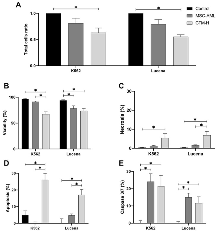
Total cell count ratio decreased in K562 transwell cultured with MSC-H when compared with the control (p = 0.015), but not when compared with transwell culture with MSC-AML (Figure 1A). Lucena cells exhibited the same behavior (MSC-AML vs. control, p < 0.001) (Figure 1A).
Figure 1.
Total cell ratio (A), viability (B), necrosis (C), apoptosis (D) and caspase 3/7 activity (E) in K562 and Lucena cells after transwell culture by 48 h with bone marrow-derived mesenchymal stem cells from patients with acute myeloid leukemia (MSC-AML) or from healthy donors (MSC-H). Data are expressed as mean ± SEM from at least three independent experiments in duplicate. (*) p values ≤ 0.05.
Cell viability decreased in both K562 and Lucena cells transwell cultured with MSC-AML or MSC-H (Figure 1B).
Cell death was also affected by coculturing K562 or Lucena cells with MSC-H. No necrosis was observed in control cells. However, the level of necrosis in K562 and Lucena cells was increased by coculturing with MSC-H (p = 0.045 and 0.003, respectively), but not with MSC-AML (Figure 1C).
K562 and Lucena cells showed low apoptosis rates. Again, apoptosis levels increased when these cells were transwell cultured with MSC-H (p < 0.001), but not with MSC-AML (Figure 1D).
Caspase 3/7 activation rates were very low in both K562 and Lucena cells. Interestingly, caspase 3/7 activation rates increased in K562 cells transwell cultured with MSC-AML (p = 0.002) and MSC-H (p = 0.005). The same was observed in Lucena cells transwell cultured with MSC-AML (p = 0.003) and MSC-H (p = 0.017) (Figure 1E).
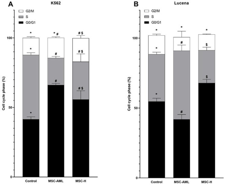
Figure 2 shows the cell cycle pattern of K562 and Lucena cells transwell cultured with MSC-AML or MSC-H for 48 h. In K562 cells, when compared with control cells, G0/G1 phase increased in both MSC-AML (p < 0.001) and MSC-H (p = 0.023) transwell cultures. The S phase decreases in both MSC-AML (p < 0.001) and MSC-H (p = 0.001) transwell cultures. G2/M phase showed alterations in MSC-H (p = 0.036) transwell culture, but not in MSC-AML. In Lucena cells, we observed a different behavior. G0/G1 phase decreased in MSC-AML transwell culture (p = 0.001) and increased in MSC-H transwell culture (p = 0.008). The S phase increased in MSC-AML transwell culture (p = 0.002) and decreased in MSC-H transwell culture (p = 0.028). No changes were observed in the G2/M phase.
Figure 2.
Cell cycle phases of K562 (A) and Lucena (B) cells after coculturing with mesenchymal stem cells from acute myeloid leukemia patients (MSC-AML) or mesenchymal stem cells from healthy donors (MSC-H) for 48 h. Data are expressed as mean ± SEM from 3 independent experiments in duplicate. Significant p values were considered as ≤0.050. Different symbols (*, # or $) in same cell cycle phases indicate statistical significancy.
2.2. Molecular Biology Assays
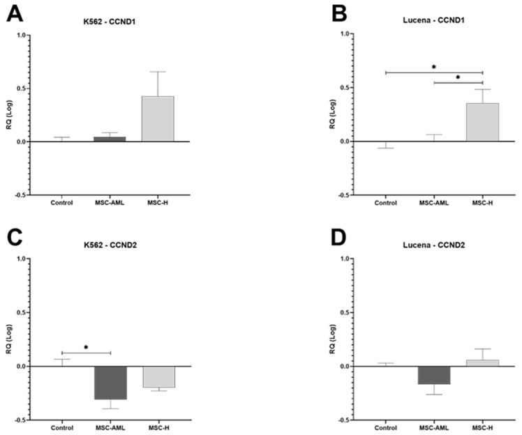
CCND1 gene expression in K562 control cells did not change by coculturing with MSC-AML or MSC-H (Figure 3A). CCND1 expression in Lucena cells, however, increased only after coculturing with MSC-H (p = 0.017) (Figure 3B).
Figure 3.
Gene expression of cyclin D1 (CCND1) and cyclin D2 (CCND2) in K562 and Lucena cells after transwell culture by 48 h with bone marrow-derived mesenchymal stem cells from patients with acute myeloid leukemia (MSC-AML) or from healthy donors (MSC-H). (A) K562 gene expression of CCND1; (B) Lucena gene expression of CCND1; (C) K562 gene expression of CCND2; (D) Lucena gene expression of CCND2. Data are expressed as mean ± SEM from 3 independent experiments in duplicate. (*) p ≤ 0.05.
CCND2 gene expression in K562 cells transwell cultured with MSC-AML decreased (p = 0.011) but not after MSC-H transwell culture (Figure 3C). CCND2 gene expression in Lucena cells was not affected by coculturing with MSC-AML or MSC-H (Figure 3D).
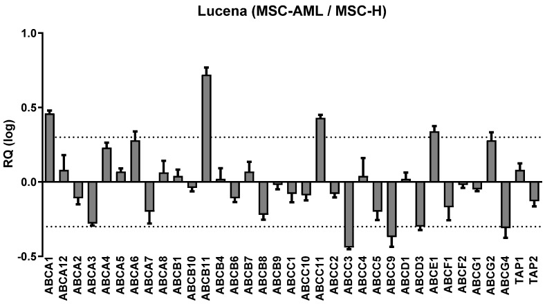
Figure 4 shows the gene expression of ABC transporters in Lucena cells transwell cultured with MSC-H, compared to coculturing with MSC-AML. ABCA1, ABCB11, ABCC11 and ABCE1 genes which were overexpressed, while ABCC3, ABCC9, ABCD3 and ABCG4 gene expression was downregulated.
Figure 4.
ABC transporters gene expression of Lucena cells transwell cultured with mesenchymal stem cells from healthy donors (MSC-H) compared with Lucena cells transwell cultured with mesenchymal stem cells from acute myeloid leukemia patients (MSC-AML). Results were obtained from a pool of cDNA samples from 3 independent experiments. Data are expressed as mean ± SEM. Dotted lines are the cutline to overexpression (RQ Log = 0.30) or inhibition (RQ Log = −0.30) of ABC transporter gene expressions. RQ: relative quantification. Dotted lines represent the significance limits of RQ (relative quantification) changed by at least two-fold: more than 2 (RQ log = 0.30) or less then 0,5 (RQ log = −0.30).
2.3. Cytokine Array Assay
Table 1 shows that, from the 39 cytokines tested, only IL-8 was detected in K562 and Lucena cells. In MSC-AML and MSC-H, 6 were observed: CCL2/MCP-1, CXCL12/SDF-1, IL-6, IL-8, MIF and Serpin E1/PAI-1.
Table 1.
Cytokine detection in K562, Lucena, MSC-AML and MSC-H.
| Cytokine | K562 | Lucena | MSC-AML | MSC-H |
|---|---|---|---|---|
| CCL2/MCP-1 | - | - | 24.03 ± 0.44 | 4.41 ± 0.16 |
| CXCL12/SDF-1 | - | - | 4.16 ± 3.09 | 4.15 ± 0.71 |
| IL-6 | - | - | 7.87 ± 4.73 | 20.60 ± 0.54 |
| IL-8 | 4.45 ± 0.09 | 3.30 ± 0.50 | 8.67 ± 0.36 | 3.23 ± 0.21 |
| MIF | - | - | 2.93 ± 1.22 | 2.72 ± 0.45 |
| Serpin E1/PAI-1 | - | - | 39.28 ± 5.27 | 39.11 ± 2.71 |
MSC-AML: mesenchymal stem cells from acute myeloid leukemia patients; MSC-H: mesenchymal stem cells from healthy donors; CCL2: C-C motif chemokine ligand 2; MCP-1: monocyte chemoattractant protein 1; CXCL12: C-X-C motif chemokine 12; SDF-1: stromal cell-derived factor 1; IL-6: interleukin 6; IL-8: interleukin 8; MIF: macrophage migration inhibitory factor; PAI-1: plasminogen activator inhibitor-1; -: not detected. Data are shown as pixels (mean ± SEM).
Table 2 shows the cytokines detected in K562 and Lucena cells transwell cultured with MSC-AML or MSC-H. In K562 cells, there was an increase in CCL2/MCP-1, and a decrease in IL-6 and IL-8, in cells transwell cultured with MSC-H compared with those transwell cultured with MSC-AML. In Lucena cells, there was an increase in CCL2/MCP-1, IL-6, IL-8 and Serpin E1/PAI-1, and a decrease in CXCL12/SDF-1 in cells transwell cultured with MSC-H compared with those transwell cultured with MSC-AML.
Table 2.
Cytokine production in K562 and Lucena cells transwell cultured with MSC-AML and MSC-H.
| Cytokine | K562 | Lucena | ||||
|---|---|---|---|---|---|---|
| MSC-AML | MSC-H | p | MSC-AML | MSC-H | p | |
| CCL2/MCP-1 | 22.8 ± 2.6 | 113.5 ± 11.4 | 0.016 | 17.5 ± 0.7 | 78.5 ± 5.0 | 0.007 |
| CXCL12/SDF-1 | 5.1 ± 1.1 | 8.5 ± 0.1 | 0.082 | 4.4 ± 0.1 | 3.1 ± 0.1 | 0.012 |
| IL-6 | 83.1 ± 2.8 | 65.4 ± 1.1 | 0.027 | 84.5 ± 6.3 | 128.5 ± 1.5 | 0.021 |
| IL-8 | 28.7 ± 2.4 | 15.8 ± 0.1 | 0.033 | 23.9 ± 2.3 | 43.0 ± 2.2 | 0.027 |
| MIF | 27.5 ± 1.8 | 32.8 ± 1.8 | 0.167 | 22.6 ± 1.1 | 20.8 ± 1.1 | 0.369 |
| Serpin E1/PAI-1 | 172.9 ± 5.1 | 188.7 ± 20.9 | 0.540 | 136.8 ± 2.6 | 182.1 ± 2.3 | 0.006 |
MSC-AML: mesenchymal stem cells from acute myeloid leukemia patients; MSC-H: mesenchymal stem cells from healthy donors; CCL2: C-C motif chemokine ligand 2; MCP-1: monocyte chemoattractant protein 1; CXCL12: C-X-C motif chemokine 12; SDF-1: stromal cell-derived factor 1; IL-6: interleukin 6; IL-8: interleukin 8; MIF: macrophage migration inhibitory factor; PAI-1: plasminogen activator inhibitor-1. Data are shown as pixels (mean ± SEM).
3. Discussion
The secretome of MSCs have a paracrine action promoted by structures, such as extracellular vesicles, and soluble factors, such as cytokines [23]. Relatively very few studies have been done on the effects of MSCs and their secretome on leukemia cell proliferation and death. Human umbilical cord blood-derived MSCs were described to inhibit the K562 cell proliferation, together with alterations in cell cycle [24]. Human Wharton’s jelly stem cell secretions were shown to be capable of decreasing the number of K562 cells in vitro by inducing cell cycle arrest and increasing apoptosis [25]. On the other hand, exosomes from human umbilical cord MSCs were described to have no effect on K562 cell viability and apoptosis [26].
Here we have shown that MSC-H secretome acts on leukemic cell lineage K562 by reducing the cell number and viability, while increasing cell death by necrosis and apoptosis. Moreover, the same was observed with the MDR cell lineage Lucena, showing that MSC-H could act on both sensible and resistant leukemic cells. Maybe more importantly, MCS-AML was not able to promote apoptosis or necrosis in both leukemic K562 and Lucena cells.
An increase in caspase 3/7 activation in K562 and Lucena cells transwell cultured with MSC-AML or MSC-H was observed. Activation of caspase 3/7, as expected, was related with increased apoptosis levels in K562 and Lucena cells transwell cultured with MSC-H. However, K562 and Lucena cells transwell cultured with MSC-AML have also increased caspase 3/7 activation with no changes in apoptosis levels. It is known that some cells can survive to caspase activation when the stimulus is transitory or sublethal [27]. This process is called anastasis and can protect cells from permanent damage after exposure to a damaging agent such as chemotherapy and even radiotherapy [28]. Thus, it is possible that MSC-AML secretome has some factors different to that produced by MSC-H that leads leukemic cells to apoptosis. In this way, MSC-AML would be protecting leukemic cells from death or loss of the propriety to combat these cells.
Transwell culture of K562 or Lucena cells with MSC-H promoted cell cycle arrest in the G0/G1 phase and a decrease in the S phase. These findings could be related to increased levels of apoptosis and a decrease in cell proliferation, as described. Anticancer effects of several drugs have been described to promote G0/G1 cell cycle arrest and apoptosis such as 2,4-dinitrobenzenesulfonamide derivative in acute leukemia cells [29], TH-39 in K562 cells [30], Mere 15 in K562 cells [31] and thio-Cl-IB-MECA in HL-60 cells [32].
D-Cyclins 1 and 2 (CCND1, CCND2) are key elements in the control of cell cycle progression from G1 to S phases [33,34]. It has been described that CCND1 overexpression is related with the arresting of cell proliferation [35]. Moreover, overexpression of CCND2 has been associated with high proliferation of leukemic cells (K562 and Lucena) [36], and downregulation of CCND2 with the arresting of cell proliferation [37].
We have found overexpression of CCND1 in Lucena cells transwell cultured with MSC-H. In addition, MSC-AML promotes no changes in CCND1 expression in K562 and Lucena cells. We found also a significative downregulation of CCND2 expression in K562 cells transwell cultured with MSC-AML. It is possible that CCND1 overregulation is the mechanism involved in Lucena proliferation arrest, while CCND2 downregulation is involved in K562 proliferation arrest.
In humans, ABC transporters are expressed in several tissues. Besides their important physiological roles, ABC transporters participate in the process of tumor cell resistance to chemotherapy by regulating the flow of anticancer agents into the cancer cells [20].
Here we have found that some ABC transporter expressions were changed in Lucena cells after coculturing with MSC-H. Overexpression was observed in four ABC transporters: ABCA1, ABCB11, ABCC11 and ABCE1. Under expression was observed in four other ABC transporters: ABCC3, ABCC9, ABCD3 and ABCG4.
ABCA1 deficiency can accelerate myeloproliferative disorder [38]; therefore, the overexpression of ABCA1 could be involved in the growth suppression of leukemic cells transwell cultured with MSC-H [39]. ABCC11 is considered a drug efflux pump for nucleotide analogs [40,41], although, ABCC11 can be associated with fluoropyrimidine resistance in chronic lymphocytic leukemia [40]. In addition, ABCE1 may play a role in the biological processes of leukemic cells participating in the MDR phenotype of these cells such as the resistance to adriamycin in K562 cells [42].
An overexpression of ABCC3 was found in patients in blast crisis of chronic myeloid leukemia with disease recurrence [43], and ABCC3 can promote imatinib efflux being responsible for the failure in imatinib-target treatments [44]. ABCC3 is considered a potential therapeutic target in human cancers [45] and its downregulation here described may explain in part our results. Trojani et al. (2019) described that ABCD3 is significantly under-expressed in the chronic phase of chronic myeloid leukemia in patients after 12 months of treatment with nilotinib [46]. The mechanism of drug resistance by ABCG4 probably involves alterations in the pH value around cancer cells [47]. The under-expression of ABCC3, ABCC9, ABCD3 and ABCG4 in Lucena cells transwell cultured with MSC-H could inhibit some resistance mechanisms developed by these cells and support the action MSC-H secretome in the attempt to eliminate these leukemic cells.
In both K562 and Lucena cells transwell cultured with MSC-AML and MSC-H, the production of six distinct cytokines was observed.
CCL2/MCP-1, CXCL12/SDF1, IL-6, IL-8, MIF and PAI-1/serpin E1. We have observed a significant elevation in the levels of CCL2/MCP-1 in both MSC-H transwell cultures (K562 and Lucena) when compared with MSC-AML. Little is known about the role of CCL2 in leukemia biology [48]; however, high levels of CCL2 can be associated with a better prognosis in some tumor types such as melanoma [49,50] and pancreatic cancer [51]. In addition, alterations in CCL2/MCP-1 production in leukemic cells can suppress cell proliferation arresting cells in G1 phase of cell cycle mediated by CCND1 [52].
CXCL12/SDF-1, produced by K562 cells, was not affected by coculturing with MSC-AML and MSC-H. Nonetheless, there was a significant decrease in CXCL12/SDF-1 levels in the transwell culture of MSC-H with Lucena cells compared with MSC-AML transwell culture. CXCL12 produced by bone marrow MSCs is a ligand to CXC chemokine receptor 4 (CXCR4), which is highly present on leukemic cells, and is responsible for growth-promoting and anti-apoptotic signals in these cells [53]. In addition, the CXCL12 decrease can lead leukemic cells to be more sensitive to treatment by getting these cells out of the quiescent state [54].
IL-6 also can participate as a cell growth factor, as in malignant plasma cells [55]. Thus, the inhibition of IL-6 can eliminate sensitive IL-6 clones of malignance cells creating free tumor environment niches, although favoring the proliferation of other malignant clone cells whose growth is triggered by other growth factors [55].
IL-8 production by BM-MSC can be stimulated by acute myeloid leukemia cells exosomes, contributing to the drug resistance of these cells [56]. Wu at al. (2021) described that both IL-6 and IL-8 levels are increased in resistant acute myeloid leukemia cells [57]. Thus, IL-8 could promote proliferation and resistance in acute myeloid leukemia cells, while IL-6 predicts survival rate and regulates the drug resistance in these cells [57].
Interestingly, we have a found significant reduction in IL-6 and IL-8 levels in the transwell culture of MSC-H with K562 cells compared with the transwell culture with MSC-AML. Nonetheless, we have observed increased IL-6 and IL-8 levels in Lucena cells transwell cultured with MSC-H in comparison with the transwell culture with MSC-AML.
MIF is secreted by several cells, including MSC [58] and K562 cells [59]. Liu et al. (2020) described that MIF is present in exosomes of bone marrow MSCs, promoting paracrine actions, acting on the maintenance of MSC survival and rejuvenation [60].
Serpin E1/PAI-1 is a negative regulator of MSC survival [61], and curiously is significantly reduced in the Lucena/MSC-AML transwell culture.
Bone marrow MSCs and their secretome are key components in leukemia bone marrow microenvironment, playing critical roles in leukemia progression [62]. In addition, it is known that there is a modulation on cytokine profiles by bone marrow MSCs in leukemia that favors leukemia maintenance and development [63]. Here we have shown that transwell culturing leukemic cells (K562 and Lucena) with MSC-H and MSC-AML promotes alterations on cytokine profiles. How these changes would affect the behavior of leukemia cells remains to be investigated.
4. Materials and Methods
This study was conducted in accordance with the Declaration of Helsinki (1975, revised in 2013), and the protocol was approved by the Ethics Committee of the Institution (CAAE: 24060619.0.0000.0068 of 27 November 2019).
4.1. Cell Culture and Transwell Culture
4.1.1. Cell Lineages Culture
Two human leukemic cell lineages were used: K562 (ATCC CCL-243), a cell line derived from chronic myeloid leukemia (in blast crisis) and vincristine sensible; and K562-Lucena (Lucena) (BCRJ Code 0127), a MDR cell lineage kindly donated by Dr Vivian Rumjanek. Cells were grown in 182 cm2 culture flasks (Santa Cruz Biotechnology, Dallas, TX, USA) with Dulbecco’s Modified Eagle Medium (DMEM) (Sigma-Aldrich, Saint Louis, MO, USA) supplemented with 10% heat-inactivated fetal bovine serum (FBS) (Vitrocell, Waldkirch, Germany) and 1% antibiotics (streptomycin (100 µg/mL) and penicillin (100 UI/mL), Sigma-Aldrich, Germany), incubated at 37 °C in a 5% CO2 atmosphere (Thermo Scientific™ Forma™ Series II, Thermo Fisher Scientific, Marietta, OH, USA), until experiments were performed. MDR phenotype of Lucena cells was maintained by the addition of vincristine (60 nM; Sigma-Aldrich, Deisenhofen, Germany). Both leukemic cells were adapted to grow in DMEM medium, rather than in RPMI, by at least 20 consecutive passages with this medium. No changes in proliferation or death rates were observed. This was a necessary step because the culture of MSCs is in DMEM media. Cell lineages were evaluated by STR profile.
Human bone marrow MSCs were obtained from 3 acute myeloid leukemia patients (MSC-AML) and from 3 healthy donors (MSC-H), after written informed consent. These cells were from the laboratory biorepository and were cultured as described above for K562 cells. These cells were characterized as MSCs as previously described [64].
4.1.2. Transwell Cultures
MSC-AML or MSC-H were seeded (2.0 × 10⁵ cells/well) on 6-well flat bottom polystyrene microplates (Corning, Kennebunk, ME, USA) and incubated at 37 °C in a 5% CO2 atmosphere for 24 h. Medium was then removed and 3 mL of DMEM supplemented with 10% FBS, and 1% antibiotics was added in each well. PET membrane with 0.4 µm pores (Corning® Costar® Transwell®, Kennebunk, ME, USA) was inserted in each well and 1.0 × 10⁶ cells (K562 or Lucena), suspended in 1.0 mL of the same medium, were added inside each transwell and cultured for 48 h in the same described conditions. Control group refers to K562 or Lucena cells seeded in the same conditions with no addition of MSCs.
4.2. Cell Death Assays
4.2.1. Total Cell Count and Cell Viability Assay
K562 or Lucena cells (1.0 × 10⁴) from the above experiments were transferred to 96-well black flat bottom polystyrene microplates. PBS was added to complete 100 µL volume before incubation with 0.1 µg/mL Hoechst 33342 (Molecular Probes, Eugene, OR, USA) and 100.0 µg of propidium iodide (PI) (Molecular Probes, Eugene, OR, USA) for 15 min. The ImageXpress Micro High Content Screening System (Molecular Devices, San José, CA, USA) was used to determine the total cell count and the number of live and dead cells. Five sites per well and 2 wells per condition were acquired. Cell Scoring MetaXpress software (version 5.0, Molecular Devices, San José, CA, USA) was used to analyze the number of cells and the viability. Total cell ratio was obtained by dividing transwell cultured cell count values by their respective cell control count values.
4.2.2. Apoptosis and Necrosis
1.0 × 10⁴ of K562 or Lucena cells (control or transwell cultured with MSC-AML or MSC-H) were transferred to a 96-well plate and the volume was completed to 100 µL with PBS. The Annexin V: FITC Apoptosis Detection Kit I (BD Biosciences, San Jose, CA, USA) was used to determine the percentage of apoptotic and necrotic cells as described by the manufacturer. The nuclei were counterstained with 0.1 µg/mL Hoechst 33342. Apoptosis was determined using an ImageXpress and the MetaXpress software Five sites per well and two wells per condition were acquired. Cells stained with Hoechst 33342 were considered as living cells. An apoptotic process was defined by the presence of Hoechst 33342 and Annexin V or Annexin V/PI. A necrotic process was defined by the presence of Hoechst 33342 and PI.
4.2.3. Detection of Caspase 3/7 Activity
A total of 1.0 × 10⁴ of K562 or Lucena cells (control or transwell cultured with MSC-AML or MSC-H) were transferred to a 96-well plate and the volume was completed to 100 µL with PBS. Caspase 3/7 activity was measured after transferring using CellEvent Caspase 3/7 Green (Invitrogen Life Technologies, Carlsbad, CA, USA) as described by the manufacturer. The nuclei were counterstained with 0.1 µg/mL Hoechst 33342. Fluorogenic substrates were determined using the ImageXpress and the MetaXpress software. Five sites per well and two wells per condition were acquired.
4.2.4. Cell Cycle Analysis
A total of 1.0 × 10⁶ K562 or Lucena, cells (control or transwell cultured with MSC-AML or MSC-H) were collected and fixed with cold 70% ethanol and stored at −20 °C until experiments were performed. Cells were washed, resuspended in PBS and incubated at 37 °C for 45 min with 100 µg/mL RNAse and 20 µg/mL PI. Flow cytometric analysis was performed in FACSCantoTM II flow cytometer (Becton Dickinson, San Jose, CA, USA) and the percentage of DNA content in the different cell cycle phases was determined using FlowJoTM software v10.5.3 (Becton Dickinson, Ashland, OR, USA).
4.3. Molecular Biology Assays
A total of 1.0 × 10⁶ of K562 or Lucena cells (control or transwell cultured with MSC-AML or MSC-H) were washed with PBS, 1.0 mL of TRI Reagent® (Sigma-Aldrich, St. Louis, MO, USA) was added and RNA was extracted as described by the manufacturer. RNA was resuspended in 20 µL of DEPC-treated water and spectrophotometrically quantified with NanoDrop 1000 Spectrophotometer (Thermo Fisher Scientific, Wilmington, DE, USA). For cDNA synthesis, 1 µg of RNA was incubated with RQ1 RNase-Free Dnase (Promega, Madison, WI, USA) as described by the manufacturer. This step was followed by cDNA synthesis, using the High-Capacity cDNA Reverse Transcription kit (Applied Biosystem, Waltham, MA, USA) as described by manufacturer.
4.3.1. Cyclin D1 (CCND1) and Cyclin D2 (CCND2) Gene Expression
The 7500 Fast Real-Time PCR System (Applied Biosystems, Waltham, MA, USA) was used to evaluate the expression of genes CCND1 and CCND2 from K562 and Lucena cells. TaqMan hydrolysable probes for CCND1 (Hs00765553_m1) and CCND2 (Hs00153380_m1) and TaqMan Universal Master Mix were purchased from Applied Biosystems. The expression of mRNA was normalized to glyceraldehyde-3-phosphate dehydrogenase (GAPDH, 402869, Thermo Fisher, Waltham, MA, USA), using the comparative cycle threshold (CT); duplicates did not exceed a 0.5 CT value. Tests were performed using the 2−ΔΔCT method [65].
4.3.2. ABC Transporter Gene Expression
The expression of 44 ABC transporters was measured in cDNA samples from Lucena cells transwell cultured with MSC-AML (reference) or MSC-H, using the TaqMan® Array 96 Well FAST Plate Human ABC Transporters (Applied Biosystems, Life Technologies Corporation, Pleasanton, CA, USA) and the 7500 v.2.0.5 software. Significance was achieved when RQ (relative quantification) was changed by at least two-fold: more than 2 (RQ log = 0.30) or less then 0.5 (RQ log = −0.30) [66].
4.4. Cytokine Array Assay
The supernatants from isolated cultures of K562, Lucena, MSC-AML and MSC-H or transwell cultures of K562 or Lucena cells with MSC-AML or MSC-H were collected, filtered through 0.22 µm membrane and stored at −80 °C until analysis was performed. The production of 36 cytokines was evaluated in a pool of supernatant of each condition using the semiquantitative kit Proteome Profiler Antibody Arrays Human Cytokine Array (R&D Systems, Minneapolis, NE, USA), according with the manufacturer instructions. The images were obtained with ImageQuant LAS 4000 (GE Healthcare Bio-Sciences AB, Uppsala, Sweden) adjusted to chemiluminescence, after 10 min of exposure, and analyzed in Image Quant TL 8.1 software (GE Healthcare Bio-Science, Piscataway, NJ, USA) in array analysis mode. Data were normalized by dividing values by the mean of positive controls and multiplying by 1000.
4.5. Statistical Analysis
Results are expressed as mean ± SEM from at least three independent experiments of each sample. Means were compared using ANOVA followed by the Tukey post-hoc test or by Student’s t test when adequate. Analysis was performed in GraphPad Prism version 9.3.0 (GraphPad Software, La Jolla, CA, USA). p values ≤ 0.05 were considered significant.
5. Conclusions
Leukemic cells are influenced by the secretome profiles of bone marrow mesenchymal stem cells, creating a niche which is somehow favorable for disease progression.
Healthy MSC secretome can inhibit cell growth, reduce cell number and viability, while increasing cell death by necrosis and apoptosis in the leukemic cell lines K562 and Lucena, showing that MSC-H could act on both sensible and resistant leukemic cells. These effects are aligned with alterations in ABC transporters expression, cell cycle arrest in G0/G1 phase and decrease in S phase, and alterations in cytokine profile. Maybe more importantly, the MCS-AML secretome was not able to promote apoptosis or necrosis in both leukemic K562 and Lucena cells, showing that MSC-AML may be permissive to leukemia development.
Acknowledgments
Vivian Rumjanek, from the Federal University of Rio de Janeiro, Brazil, for kindly providing the K562-Lucena; National Institute of Science and Technology Complex Fluids (INCT-FCx); National Institute of Science and Technology Regenerative Medicine (INCT-Regenera).
Author Contributions
D.L. and S.P.B.—conceived the idea of this study; F.A.d.F., D.L. and S.P.B.—designed the research; P.N.G., L.A.d.P.C.L., M.K.D. and J.P.—collected clinical data and samples and followed the patients; F.A.d.F., D.L. and J.S.-S.—performed the experiments; F.A.d.F., D.L. and C.O.R.—performed statistical analyses; F.A.d.F., D.L. and S.P.B.—analyzed the results; F.A.d.F.—wrote and prepared the original draft; D.L.—writing, review and editing; S.P.B.—review and editing, supervision of the study and project administration. All authors have read and agreed to the published version of the manuscript.
Institutional Review Board Statement
This study was conducted in accordance with the Declaration of Helsinki (1975, revised in 2013), and the protocol was approved by the Ethics Committee of the Faculdade de Medicina da Universidade de Sao Paulo (FMUSP) (CAAE: 24060619.0.0000.0068 of 27 November 2019).
Informed Consent Statement
All patients gave written informed consent.
Data Availability Statement
The datasets generated during the current study are not publicly available but are available from the corresponding author on reasonable request.
Conflicts of Interest
The authors declare no conflicts of interest.
Funding Statement
This work was supported in part by grants from Coordination of Superior Level Staff Improvement (CAPES) (88882.328134/2019-01).
Footnotes
Disclaimer/Publisher’s Note: The statements, opinions and data contained in all publications are solely those of the individual author(s) and contributor(s) and not of MDPI and/or the editor(s). MDPI and/or the editor(s) disclaim responsibility for any injury to people or property resulting from any ideas, methods, instructions or products referred to in the content.
References
- 1.Burke V.P., Startzell J.M. The leukemias. Oral Maxillofac. Surg. Clin. N. Am. 2008;20:597–608. doi: 10.1016/j.coms.2008.06.011. [DOI] [PubMed] [Google Scholar]
- 2.Castelli G., Pelosi E., Testa U. Emerging therapies for acute myelogenus leukemia patients targeting apoptosis and mitochondrial metabolism. Cancers. 2019;11:260. doi: 10.3390/cancers11020260. [DOI] [PMC free article] [PubMed] [Google Scholar]
- 3.Döhner H., Weisdorf D.J., Bloomfield C.D. Acute myeloid leukemia. N. Engl. J. Med. 2015;373:1136–1152. doi: 10.1056/NEJMra1406184. [DOI] [PubMed] [Google Scholar]
- 4.Brunetti L., Gundry M.C., Goodell M.A. New insights into the biology of acute myeloid leukemia with mutated npm1. Int. J. Hematol. 2019;110:150–160. doi: 10.1007/s12185-018-02578-7. [DOI] [PubMed] [Google Scholar]
- 5.Boulais P.E., Frenette P.S. Making sense of hematopoietic stem cell niches. Blood. 2015;125:2621–2629. doi: 10.1182/blood-2014-09-570192. [DOI] [PMC free article] [PubMed] [Google Scholar]
- 6.Reichert C.O., de Freitas F.A., Levy D., Bydlowski S.P. Oxysterols and mesenchymal stem cell biology. Vitam. Horm. 2021;116:409–436. doi: 10.1016/bs.vh.2021.02.004. [DOI] [PubMed] [Google Scholar]
- 7.Frenette P.S., Pinho S., Lucas D., Scheiermann C. Mesenchymal stem cell: Keystone of the hematopoietic stem cell niche and a stepping-stone for regenerative medicine. Annu. Rev. Immunol. 2013;31:285–316. doi: 10.1146/annurev-immunol-032712-095919. [DOI] [PubMed] [Google Scholar]
- 8.Méndez-Ferrer S., Michurina T.V., Ferraro F., Mazloom A.R., Macarthur B.D., Lira S.A., Scadden D.T., Ma’ayan A., Enikolopov G.N., Frenette P.S. Mesenchymal and haematopoietic stem cells form a unique bone marrow niche. Nature. 2010;466:829–834. doi: 10.1038/nature09262. [DOI] [PMC free article] [PubMed] [Google Scholar]
- 9.Xia C., Wang T., Cheng H., Dong Y., Weng Q., Sun G., Zhou P., Wang K., Liu X., Geng Y., et al. Mesenchymal stem cells suppress leukemia via macrophage-mediated functional restoration of bone marrow microenvironment. Leukemia. 2020;34:2375–2383. doi: 10.1038/s41375-020-0775-3. [DOI] [PMC free article] [PubMed] [Google Scholar]
- 10.Corradi G., Baldazzi C., Očadlíková D., Marconi G., Parisi S., Testoni N., Finelli C., Cavo M., Curti A., Ciciarello M. Mesenchymal stromal cells from myelodysplastic and acute myeloid leukemia patients display in vitro reduced proliferative potential and similar capacity to support leukemia cell survival. Stem Cell Res. Ther. 2018;9:271. doi: 10.1186/s13287-018-1013-z. [DOI] [PMC free article] [PubMed] [Google Scholar]
- 11.de Alvarenga E.C., Silva W.N., Vasconcellos R., Paredes-Gamero E.J., Mintz A., Birbrair A. Promyelocytic leukemia protein in mesenchymal stem cells is essential for leukemia progression. Ann. Hematol. 2018;97:1749–1755. doi: 10.1007/s00277-018-3463-x. [DOI] [PMC free article] [PubMed] [Google Scholar]
- 12.Yehudai-Resheff S., Attias-Turgeman S., Sabbah R., Gabay T., Musallam R., Fridman-Dror A., Zuckerman T. Abnormal morphological and functional nature of bone marrow stromal cells provides preferential support for survival of acute myeloid leukemia cells. Int. J. Cancer. 2019;144:2279–2289. doi: 10.1002/ijc.32063. [DOI] [PubMed] [Google Scholar]
- 13.Raaijmakers M.H., Mukherjee S., Guo S., Zhang S., Kobayashi T., Schoonmaker J.A., Ebert B.L., Al-Shahrour F., Hasserjian R.P., Scadden E.O., et al. Bone progenitor dysfunction induces myelodysplasia and secondary leukaemia. Nature. 2010;464:852–857. doi: 10.1038/nature08851. [DOI] [PMC free article] [PubMed] [Google Scholar]
- 14.Walkley C.R., Olsen G.H., Dworkin S., Fabb S.A., Swann J., McArthur G.A., Westmoreland S.V., Chambon P., Scadden D.T., Purton L.E. A microenvironment-induced myeloproliferative syndrome caused by retinoic acid receptor gamma deficiency. Cell. 2007;129:1097–1110. doi: 10.1016/j.cell.2007.05.014. [DOI] [PMC free article] [PubMed] [Google Scholar]
- 15.Wang L., Zhang H., Rodriguez S., Cao L., Parish J., Mumaw C., Zollman A., Kamoka M.M., Mu J., Chen D.Z., et al. Notch-dependent repression of mir-155 in the bone marrow niche regulates hematopoiesis in an nf-κb-dependent manner. Cell Stem Cell. 2014;15:51–65. doi: 10.1016/j.stem.2014.04.021. [DOI] [PMC free article] [PubMed] [Google Scholar]
- 16.Madel R.J., Börger V., Dittrich R., Bremer M., Tertel T., Phuong N.N.T., Baba H.A., Kordelas L., Staubach S., Stein F., et al. Independent human mesenchymal stromal cell-derived extracellular vesicle preparations differentially attenuate symptoms in an advanced murine graft-versus-host disease model. Cytotherapy. 2023;25:821–836. doi: 10.1016/j.jcyt.2023.03.008. [DOI] [PubMed] [Google Scholar]
- 17.Yates A.G., Pink R.C., Erdbrügger U., Siljander P.R., Dellar E.R., Pantazi P., Akbar N., Cooke W.R., Vatish M., Dias-Neto E., et al. In sickness and in health: The functional role of extracellular vesicles in physiology and pathology in vivo: Part i: Health and normal physiology: Part i: Health and normal physiology. J. Extracell. Vesicles. 2022;11:e12151. doi: 10.1002/jev2.12151. [DOI] [PMC free article] [PubMed] [Google Scholar]
- 18.Yates A.G., Pink R.C., Erdbrügger U., Siljander P.R., Dellar E.R., Pantazi P., Akbar N., Cooke W.R., Vatish M., Dias-Neto E., et al. In sickness and in health: The functional role of extracellular vesicles in physiology and pathology in vivo: Part ii: Pathology: Part ii: Pathology. J. Extracell. Vesicles. 2022;11:e12190. doi: 10.1002/jev2.12190. [DOI] [PMC free article] [PubMed] [Google Scholar]
- 19.Zanetti S.R., Romecin P.A., Vinyoles M., Juan M., Fuster J.L., Cámos M., Querol S., Delgado M., Menendez P. Bone marrow msc from pediatric patients with b-all highly immunosuppress t-cell responses but do not compromise cd19-car t-cell activity. J. Immunother. Cancer. 2020;8:e001419. doi: 10.1136/jitc-2020-001419. [DOI] [PMC free article] [PubMed] [Google Scholar]
- 20.Liang W., Chen X., Zhang S., Fang J., Chen M., Xu Y., Chen X. Mesenchymal stem cells as a double-edged sword in tumor growth: Focusing on msc-derived cytokines. Cell. Mol. Biol. Lett. 2021;26:3. doi: 10.1186/s11658-020-00246-5. [DOI] [PMC free article] [PubMed] [Google Scholar]
- 21.Baharaghdam S., Yousefi M., Movasaghpour A., Solali S., Talebi M., Ahani-Nahayati M., Lotfimehr H., Shamsasanjan K. Effects of hypoxia on biology of human leukemia t-cell line (molt-4 cells) co-cultured with bone marrow mesenchymal stem cells. Avicenna J. Med. Biotechnol. 2018;10:62–68. [PMC free article] [PubMed] [Google Scholar]
- 22.Ruiz-Aparicio P.F., Uribe G.I., Linares-Ballesteros A., Vernot J.P. Sensitization to drug treatment in precursor b-cell acute lymphoblastic leukemia is not achieved by stromal nf-κb inhibition of cell adhesion but by stromal pkc-dependent inhibition of abc transporters activity. Molecules. 2021;26:5366. doi: 10.3390/molecules26175366. [DOI] [PMC free article] [PubMed] [Google Scholar]
- 23.Paz J.L., Levy D., Oliveira B.A., de Melo T.C., de Freitas F.A., Reichert C.O., Rodrigues A., Pereira J., Bydlowski S.P. 7-ketocholesterol promotes oxiapoptophagy in bone marrow mesenchymal stem cell from patients with acute myeloid leukemia. Cells. 2019;8:482. doi: 10.3390/cells8050482. [DOI] [PMC free article] [PubMed] [Google Scholar]
- 24.Livak K.J., Schmittgen T.D. Analysis of relative gene expression data using real-time quantitative pcr and the 2(-delta delta c(t)) method. Methods. 2001;25:402–408. doi: 10.1006/meth.2001.1262. [DOI] [PubMed] [Google Scholar]
- 25.Liu X. Abc family transporters. Adv. Exp. Med. Biol. 2019;1141:13–100. doi: 10.1007/978-981-13-7647-4_2. [DOI] [PubMed] [Google Scholar]
- 26.Stutzbach L. Perk Genetic Variation and Function in Progressive Supranuclear Palsy. University of Pennsylvania Penn Libraries; Philadelphia, PA, USA: 2015. [Google Scholar]
- 27.Maacha S., Sidahmed H., Jacob S., Gentilcore G., Calzone R., Grivel J.C., Cugno C. Paracrine mechanisms of mesenchymal stromal cells in angiogenesis. Stem Cells Int. 2020;2020:4356359. doi: 10.1155/2020/4356359. [DOI] [PMC free article] [PubMed] [Google Scholar]
- 28.Fonseka M., Ramasamy R., Tan B.C., Seow H.F. Human umbilical cord blood-derived mesenchymal stem cells (hucb-msc) inhibit the proliferation of k562 (human erythromyeloblastoid leukaemic cell line) Cell Biol. Int. 2012;36:793–801. doi: 10.1042/CBI20110595. [DOI] [PubMed] [Google Scholar]
- 29.Huwaikem M.A.H., Kalamegam G., Alrefaei G., Ahmed F., Kadam R., Qadah T., Sait K.H.W., Pushparaj P.N. Human wharton’s jelly stem cell secretions inhibit human leukemic cell line k562 in vitro by inducing cell cycle arrest and apoptosis. Front. Cell Dev. Biol. 2021;9:614988. doi: 10.3389/fcell.2021.614988. [DOI] [PMC free article] [PubMed] [Google Scholar]
- 30.Liu Y., Song B., Wei Y., Chen F., Chi Y., Fan H., Liu N., Li Z., Han Z., Ma F. Exosomes from mesenchymal stromal cells enhance imatinib-induced apoptosis in human leukemia cells via activation of caspase signaling pathway. Cytotherapy. 2018;20:181–188. doi: 10.1016/j.jcyt.2017.11.006. [DOI] [PubMed] [Google Scholar]
- 31.Tang H.L., Tang H.M., Mak K.H., Hu S., Wang S.S., Wong K.M., Wong C.S., Wu H.Y., Law H.T., Liu K., et al. Cell survival, DNA damage, and oncogenic transformation after a transient and reversible apoptotic response. Mol. Biol. Cell. 2012;23:2240–2252. doi: 10.1091/mbc.e11-11-0926. [DOI] [PMC free article] [PubMed] [Google Scholar]
- 32.Ding A.X., Sun G., Argaw Y.G., Wong J.O., Easwaran S., Montell D.J. Casexpress reveals widespread and diverse patterns of cell survival of caspase-3 activation during development in vivo. eLife. 2016;5:e10936. doi: 10.7554/eLife.10936. [DOI] [PMC free article] [PubMed] [Google Scholar]
- 33.Alves Almeida P., Schmitz de Souza L.F., Franzoni Maioral M., Otto Walter L., Fischer Duarte B., Mattos Santos-Pirath Í., Bauer Speer D., Sens L., Tizziani T., Sena de Oliveira A., et al. Cell cycle arrest and apoptosis induction by a new 2,4-dinitrobenzenesulfonamide derivative in acute leukemia cells. J. Pharm. Pharm. Sci. A Publ. Can. Soc. Pharm. Sci. Soc. Can. Des Sci. Pharm. 2021;24:23–36. doi: 10.18433/jpps31349. [DOI] [PubMed] [Google Scholar]
- 34.Zhu Y., Wei W., Ye T., Liu Z., Liu L., Luo Y., Zhang L., Gao C., Wang N., Yu L. Small molecule th-39 potentially targets hec1/nek2 interaction and exhibits antitumor efficacy in k562 cells via g0/g1 cell cycle arrest and apoptosis induction. Cell. Physiol. Biochem. Int. J. Exp. Cell. Physiol. Biochem. Pharmacol. 2016;40:297–308. doi: 10.1159/000452546. [DOI] [PubMed] [Google Scholar]
- 35.Liu M., Zhao X., Zhao J., Xiao L., Liu H., Wang C., Cheng L., Wu N., Lin X. Induction of apoptosis, g₀/g₁ phase arrest and microtubule disassembly in k562 leukemia cells by mere15, a novel polypeptide from meretrix meretrix linnaeus. Mar. Drugs. 2012;10:2596–2607. doi: 10.3390/md10112596. [DOI] [PMC free article] [PubMed] [Google Scholar]
- 36.Lee E.J., Min H.Y., Chung H.J., Park E.J., Shin D.H., Jeong L.S., Lee S.K. A novel adenosine analog, thio-cl-ib-meca, induces g0/g1 cell cycle arrest and apoptosis in human promyelocytic leukemia hl-60 cells. Biochem. Pharmacol. 2005;70:918–924. doi: 10.1016/j.bcp.2005.06.017. [DOI] [PubMed] [Google Scholar]
- 37.Qie S., Diehl J.A. Cyclin d1, cancer progression, and opportunities in cancer treatment. J. Mol. Med. 2016;94:1313–1326. doi: 10.1007/s00109-016-1475-3. [DOI] [PMC free article] [PubMed] [Google Scholar]
- 38.Poon R.Y. Cell cycle control: A system of interlinking oscillators. Methods Mol. Biol. 2016;1342:3–19. doi: 10.1007/978-1-4939-2957-3_1. [DOI] [PubMed] [Google Scholar]
- 39.Wilhide C.C., Van Dang C., Dipersio J., Kenedy A.A., Bray P.F. Overexpression of cyclin d1 in the dami megakaryocytic cell line causes growth arrest. Blood. 1995;86:294–304. doi: 10.1182/blood.V86.1.294.bloodjournal861294. [DOI] [PubMed] [Google Scholar]
- 40.Song J.M., Xu D., Fan E.J., Xu S.R., Li D., Zhao C.H. cyclin d2 expression in chronic myelogenous leukemia. Zhonghua Xue Ye Xue Za Zhi Zhonghua Xueyexue Zazhi. 2004;25:103–105. [PubMed] [Google Scholar]
- 41.Chen B.B., Glasser J.R., Coon T.A., Zou C., Miller H.L., Fenton M., McDyer J.F., Boyiadzis M., Mallampalli R.K. F-box protein fbxl2 targets cyclin d2 for ubiquitination and degradation to inhibit leukemic cell proliferation. Blood. 2012;119:3132–3141. doi: 10.1182/blood-2011-06-358911. [DOI] [PMC free article] [PubMed] [Google Scholar]
- 42.Luciani M.F., Denizot F., Savary S., Mattei M.G., Chimini G. Cloning of two novel abc transporters mapping on human chromosome 9. Genomics. 1994;21:150–159. doi: 10.1006/geno.1994.1237. [DOI] [PubMed] [Google Scholar]
- 43.Schmitz G., Langmann T. Structure, function and regulation of the abc1 gene product. Curr. Opin. Lipidol. 2001;12:129–140. doi: 10.1097/00041433-200104000-00006. [DOI] [PubMed] [Google Scholar]
- 44.Stieger B., Meier Y., Meier P.J. The bile salt export pump. Pflug. Arch. Eur. J. Physiol. 2007;453:611–620. doi: 10.1007/s00424-006-0152-8. [DOI] [PubMed] [Google Scholar]
- 45.Tian Y., Tian X., Han X., Chen Y., Song C.Y., Jiang W.J., Tian D.L. Abce1 plays an essential role in lung cancer progression and metastasis. Tumour Biol. J. Int. Soc. Oncodevelopmental Biol. Med. 2016;37:8375–8382. doi: 10.1007/s13277-015-4713-3. [DOI] [PubMed] [Google Scholar]
- 46.Hirohashi T., Suzuki H., Takikawa H., Sugiyama Y. Atp-dependent transport of bile salts by rat multidrug resistance-associated protein 3 (mrp3) J. Biol. Chem. 2000;275:2905–2910. doi: 10.1074/jbc.275.4.2905. [DOI] [PubMed] [Google Scholar]
- 47.Balaji S.A., Udupa N., Chamallamudi M.R., Gupta V., Rangarajan A. Role of the drug transporter abcc3 in breast cancer chemoresistance. PLoS ONE. 2016;11:e0155013. doi: 10.1371/journal.pone.0155013. [DOI] [PMC free article] [PubMed] [Google Scholar]
- 48.Lagas J.S., Fan L., Wagenaar E., Vlaming M.L., van Tellingen O., Beijnen J.H., Schinkel A.H. P-glycoprotein (p-gp/abcb1), abcc2, and abcc3 determine the pharmacokinetics of etoposide. Clin. Cancer Res. Off. J. Am. Assoc. Cancer Res. 2010;16:130–140. doi: 10.1158/1078-0432.CCR-09-1321. [DOI] [PubMed] [Google Scholar]
- 49.Bryan J., Muñoz A., Zhang X., Düfer M., Drews G., Krippeit-Drews P., Aguilar-Bryan L. Abcc8 and abcc9: Abc transporters that regulate k+ channels. Pflug. Arch. Eur. J. Physiol. 2007;453:703–718. doi: 10.1007/s00424-006-0116-z. [DOI] [PubMed] [Google Scholar]
- 50.Kawaguchi K., Morita M. Abc transporter subfamily d: Distinct differences in behavior between abcd1-3 and abcd4 in subcellular localization, function, and human disease. BioMed Res. Int. 2016;2016:6786245. doi: 10.1155/2016/6786245. [DOI] [PMC free article] [PubMed] [Google Scholar]
- 51.Zhang Y., Zhang Y., Wang J., Yang J., Yang G. Abnormal expression of abcd3 is an independent prognostic factor for colorectal cancer. Oncol. Lett. 2020;19:3567–3577. doi: 10.3892/ol.2020.11463. [DOI] [PMC free article] [PubMed] [Google Scholar]
- 52.Yang A., Alrosan A.Z., Sharpe L.J., Brown A.J., Callaghan R., Gelissen I.C. Regulation of abcg4 transporter expression by sterols and lxr ligands. Biochim. Et Biophys. Acta (BBA)-Gen. Subj. 2021;1865:129769. doi: 10.1016/j.bbagen.2020.129769. [DOI] [PubMed] [Google Scholar]
- 53.Viaud M., Abdel-Wahab O., Gall J., Ivanov S., Guinamard R., Sore S., Merlin J., Ayrault M., Guilbaud E., Jacquel A., et al. Abca1 exerts tumor-suppressor function in myeloproliferative neoplasms. Cell Rep. 2020;30:3397–3410.e3395. doi: 10.1016/j.celrep.2020.02.056. [DOI] [PMC free article] [PubMed] [Google Scholar]
- 54.Wang L., Luo Q., Zeng S., Lou Y., Li X., Hu M., Lu L., Liu Z. Disordered farnesoid x receptor signaling is associated with liver carcinogenesis in abcb11-deficient mice. J. Pathol. 2021;255:412–424. doi: 10.1002/path.5780. [DOI] [PubMed] [Google Scholar]
- 55.Pastor-Anglada M., Molina-Arcas M., Casado F.J., Bellosillo B., Colomer D., Gil J. Nucleoside transporters in chronic lymphocytic leukaemia. Leukemia. 2004;18:385–393. doi: 10.1038/sj.leu.2403271. [DOI] [PubMed] [Google Scholar]
- 56.Guo Y., Kotova E., Chen Z.S., Lee K., Hopper-Borge E., Belinsky M.G., Kruh G.D. Mrp8, atp-binding cassette c11 (abcc11), is a cyclic nucleotide efflux pump and a resistance factor for fluoropyrimidines 2′,3′-dideoxycytidine and 9′-(2′-phosphonylmethoxyethyl)adenine. J. Biol. Chem. 2003;278:29509–29514. doi: 10.1074/jbc.M304059200. [DOI] [PubMed] [Google Scholar]
- 57.Wuxiao Z., Wang H., Su Q., Zhou H., Hu M., Tao S., Xu L., Chen Y., Hao X. Microrna-145 promotes the apoptosis of leukemic stem cells and enhances drug-resistant k562/adm cell sensitivity to adriamycin via the regulation of abce1. Int. J. Mol. Med. 2020;46:1289–1300. doi: 10.3892/ijmm.2020.4675. [DOI] [PMC free article] [PubMed] [Google Scholar]
- 58.Radich J.P., Dai H., Mao M., Oehler V., Schelter J., Druker B., Sawyers C., Shah N., Stock W., Willman C.L., et al. Gene expression changes associated with progression and response in chronic myeloid leukemia. Proc. Natl. Acad. Sci. USA. 2006;103:2794–2799. doi: 10.1073/pnas.0510423103. [DOI] [PMC free article] [PubMed] [Google Scholar]
- 59.Giannoudis A., Davies A., Harris R.J., Lucas C.M., Pirmohamed M., Clark R.E. The clinical significance of abcc3 as an imatinib transporter in chronic myeloid leukaemia. Leukemia. 2014;28:1360–1363. doi: 10.1038/leu.2014.38. [DOI] [PubMed] [Google Scholar]
- 60.Seborova K., Kloudova-Spalenkova A., Koucka K., Holy P., Ehrlichova M., Wang C., Ojima I., Voleska I., Daniel P., Balusikova K., et al. The role of trip6, abcc3 and cps1 expression in resistance of ovarian cancer to taxanes. Int. J. Mol. Sci. 2021;23:73. doi: 10.3390/ijms23010073. [DOI] [PMC free article] [PubMed] [Google Scholar]
- 61.De Grouw E.P., Raaijmakers M.H., Boezeman J.B., van der Reijden B.A., van de Locht L.T., de Witte T.J., Jansen J.H., Raymakers R.A. Preferential expression of a high number of atp binding cassette transporters in both normal and leukemic cd34+cd38- cells. Leukemia. 2006;20:750–754. doi: 10.1038/sj.leu.2404131. [DOI] [PubMed] [Google Scholar]
- 62.Trojani A., Pungolino E., Dal Molin A., Lodola M., Rossi G., D’Adda M., Perego A., Elena C., Turrini M., Borin L., et al. Nilotinib interferes with cell cycle, abc transporters and jak-stat signaling pathway in cd34+/lin- cells of patients with chronic phase chronic myeloid leukemia after 12 months of treatment. PLoS ONE. 2019;14:e0218444. doi: 10.1371/journal.pone.0218444. [DOI] [PMC free article] [PubMed] [Google Scholar]
- 63.Zhang Z., Li X., Wang X., Zhou Y., Xu H., Wang J., Huang L., Tian Y., Cheng Q. The expression of abcg4, v-atpase and clinic significance of their correlation with nsclc. Zhongguo Fei Ai Za Zhi Chin. J. Lung Cancer. 2008;11:691–695. doi: 10.3779/j.issn.1009-3419.2008.05.023. [DOI] [PubMed] [Google Scholar]
- 64.Cole S.P., Bhardwaj G., Gerlach J.H., Mackie J.E., Grant C.E., Almquist K.C., Stewart A.J., Kurz E.U., Duncan A.M., Deeley R.G. Overexpression of a transporter gene in a multidrug-resistant human lung cancer cell line. Science. 1992;258:1650–1654. doi: 10.1126/science.1360704. [DOI] [PubMed] [Google Scholar]
- 65.Andersson L.C., Nilsson K., Gahmberg C.G. K562—A human erythroleukemic cell line. Int. J. Cancer. 1979;23:143–147. doi: 10.1002/ijc.2910230202. [DOI] [PubMed] [Google Scholar]
- 66.Rumjanek V.M., Trindade G.S., Wagner-Souza K., de-Oliveira M.C., Marques-Santos L.F., Maia R.C., Capella M.A. Multidrug resistance in tumour cells: Characterization of the multidrug resistant cell line k562-lucena 1. An. Da Acad. Bras. De Cienc. 2001;73:57–69. doi: 10.1590/S0001-37652001000100007. [DOI] [PubMed] [Google Scholar]
Associated Data
This section collects any data citations, data availability statements, or supplementary materials included in this article.
Data Availability Statement
The datasets generated during the current study are not publicly available but are available from the corresponding author on reasonable request.


