Abstract
Introduction
Artificial intelligence (AI) is computer-generated intelligence, as opposed to the natural intelligence of humans and some animals. Kaplan and Haenlein define AI as “the ability of a system to correctly interpret external data, to learn from such data and use what it has learned to achieve specific goals and tasks through a flexible adaptation”. The term “artificial intelligence” is used colloquially to describe machines that mimic the “cognitive” functions that people associate with other human minds. One of the areas where technological advances have brought significant changes is orthodontics, especially in terms of diagnosis and orthodontic prediction.
The aim of this study is to conduct a comparative analysis between the results obtained by using the complete algorithms that define Artificial Intelligence and the simple algorithms of classical medical software, used in the detection of the position and shape of teeth in various orthodontic anomalies.
Methods
A group of 45 patients with maxillary-dento anomalies Angle Class I (DDM with crowding and deviation of the superior inter-incisive line) was studied. Two types of algorithms were used in the study group: modern type I algorithms and simple algorithms used in classical software to detect the position of the frontal teeth. Through the symmetrical points of the face the facial axes were determined, and after the detection of the contour of each tooth the incisional curve was calculated. The median line was analyzed against the vertical axis of the face, and the incisional curve towards the horizontal axis.
Results
The study shows that AI algorithms offer an increased level of tooth position detection, compared to traditional softwares. Complex algorithms, specific to Artificial Intelligence, showed superior detection, and more stability in the analysis.
Conclusion
Technological evolution and the development of machine learning capabilities have opened new perspectives in guiding orthodontic treatments through artificial intelligence (AI).
Keywords: orthodontics, diagnostic, algorithms, median line, artificial intelligence
Introduction
Artificial intelligence has multiple applications in medicine, including health care management: scheduling patients through various systems, the simulation of an orthodontic diagnosis and even therapy (robotics in surgery). Researchers classified artificial intelligence into two subtypes: Machine learning and Deep learning [1,2].
Machine Learning is a branch of artificial intelligence science that aims to give machines the ability to “learn”. This is done by using algorithms that identify models based on the data received, so that machines can make decisions and make predictions, i.e. become “intelligent” [3,4]. This way it is no longer necessary for the car to be programmed specifically for each action. As Arthur Samuel stated in 1959, machine learning is “science that gives computers the ability to learn without being explicitly programmed”. Current machine learning methods are becoming increasingly sophisticated, and they are being integrated into a number of complex medical applications such as genome analysis, in an effort to prevent disease, diagnosing depression based on speech patterns or identifying people with suicidal tendencies [5].
By using technologies such as artificial intelligence (AI), orthodontists are able to improve the efficiency and accuracy of the diagnostic process, opening new horizons for patient care. Orthodontic diagnosis is an important aspect in planning and implementing orthodontic treatment. Traditionally, this stage involved manual evaluation of dental models, X-rays and other diagnostic images. However, with technological advancements, systems based on artificial intelligence have begun to offer innovative solutions for optimizing this process [6].
Artificial intelligence, defined as the ability of a machine to simulate human thought processes, becomes a valuable partner in medical and orthodontic diagnosis. Advanced machine learning algorithms and deep neural networks have been trained to recognize complex patterns from radiological images, dental photographs, and 3D jaw scans. This allows for a quick and accurate analysis of malocclusions, tooth position and nearby bone structures.
Deep Learning is a branch of machine learning science that represents the most advanced field of artificial intelligence. It has the main objective of giving machines the opportunity to learn and think as much as possible like humans. Deep learning requires a complex architecture that mimics the neural networks of the human brain to give meaning to patterns even when details are missing, the available data are insufficient or when they may create confusion. Learning requires an algorithm to adjust these weights based on training data; a simple algorithm (on the principle of “connects what triggers together”) is to increase the share of the connection between two connected neurons [7].
Artificial neural networks were inspired by the architecture of neurons in the human brain. A simple „neuron” N supports inputs from multiple neurons, each of which, when activated, throws a weighted “vote” for or against activation of neuron N.
The network forms „concepts” that are distributed in a subnet of shared neurons that tend to activate together. Neurons have a continuous spectrum of activation; moreover, they can process nonlinear inputs. Modern neural networks can learn both continuous functions and, surprisingly, digital logical operations [8].
The aim of this study is to conduct a comparative analysis between the results obtained by using the algorithms provided by Artificial Intelligence and the simple algorithms of classical medical software, used in the evaluation of the position of the teeth for the diagnosis of a dento-maxillary anomaly.
Methods
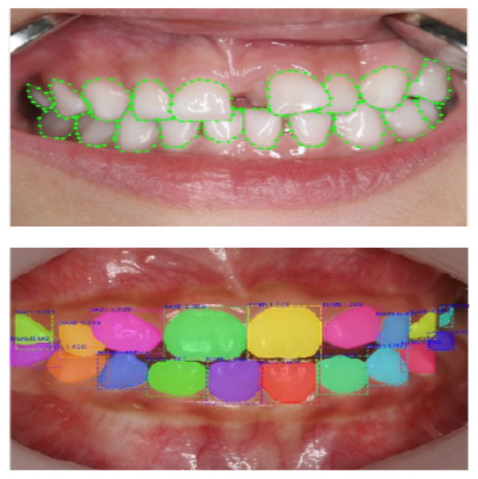
The study was carried out at Hack Dental SRL, Targoviste, Romania with the help of ORTHO AI software, our own conception design, with two versions, one using simple algorithms, specific to classical software, and the second using algorithms specific to Artificial Intelligence. The field of Artificial Intelligence that participates in the detection of objects (symmetrical points) Deep Learning is realized with various neural networks. As a type of network, convolutional and recurrent networks (CNN and RNN) are used, which are indicated for the detection of movements (Figure 1).
Figure 1.
Tooth Detection and Segmentation with R-CNN Mask.
Machine learning algorithms (machine learning) and image processing techniques were used to identify and locate anatomical tooth shapes in the frontal area.
In the modern version of AI, an Artificial Neural Network has been created, in which a high number of dental forms are implemented, (normal and pathological), which helps the application to recognize the contours of teeth in normal or malposition. The detected information compared to those in the artificial neural network is processed through rapid learning of the Deep Learning domain, which elaborates the forms of teeth that will be detected later [8].
In the classic version of the software, the detection of the contour of teeth is based only on the detection of the color difference of the teeth, processing the shapes and contours of the teeth, without the ability of software self-deduction.
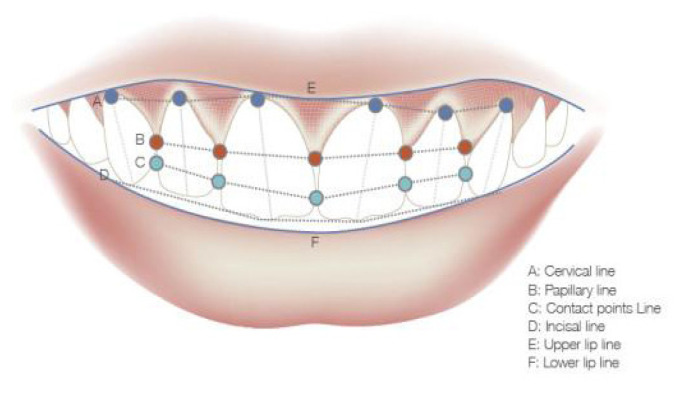
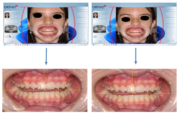
After detecting the shapes and contours of the teeth, each incisional edge relates to the horizontal axis, and by calculating the number of pixels existing between them, the ratio between the distances of each pair of teeth will be displayed, calculating the symmetry of the incisional curve [9]. By detecting the central incisors, according to the form that defines them, the point of contact between them is highlighted, thus establishing the inter-incisor line. This is related to the vertical facial axis detected by the software application (Figure 2).
Figure 2.
The detection of dental points with AI software.
The study included a group of 45 patients (30 female and 15 male) aged 18–35 years, with dento-maxillary anomalies Class I Angle (DDM with crowding), which signed the informed consent. We used the following inclusion criteria:
- all permanent teeth erupted on the arch except for permanent 3rd molars, presenting first class Angle
- a harmonious profile
- no previous orthodontic treatment.
Exclusion criteria:
- orthodontic treatment in patient history.
- absence of teeth in the front area
- class II or III malocclusion
The patients were divided into 3 subgroups of 15 patients, evaluated by 3 doctors: group A - dr. Marius Hack, (MH) lot B - dr. Ludmila Hack, (LH) and lot C - dr. Daniela Stan. (DS) In each patient, both software versions were used, in order to be able to compare as concretely as possible the results of the detection by the two types of algorithms, classical and AI modern one.
Each dental office/each doctor had a video camera installed at the dental unit, with optical zoom suitable for dental treatments (30x optical zoom) connected to a computer on which the ORTHO AI software application was installed with both versions: one based on specific AI algorithms and a version based on classical algorithms.
By detecting the teeth of patients, both software versions process the contour of the teeth, and highlight the midline given by the mesial faces of the central incisors, and the incisal edges, creating the incisor curve. Thus, by referring to the facial axes determined by the symmetrical points of the face, one can diagnose the deviation of the median line or of the incisional curve for each patient.
The difference lies in the accuracy of detecting the contour of the front teeth through both software versions and processing the information, showing the degree of computer error that automatically leads to safety or uncertainty in the use of digital techniques in the field of orthodontics, diagnostics, treatment guidance and verification (Figure 3).
Figure 3.
Difference by processing AI and classical algorithms.
Results
The study group is not homogeneous, with a higher percentage distribution for the female sex (66.6%), women being more responsive to modern AI assessments and smile aesthetics, the dento-facial harmony.
Each case analyzed was awarded a score of 1–10 for each verification method, depending on the certainty of the detection of dental contours and the correctness of data processing.
These software applications are designed to provide a personalized approach and accurate guidance of orthodontic treatments, optimizing their effectiveness. Following the study we obtained the following results for each of the 3 doctors, results recorded in the following tables. Table I shows the results obtained by doctor MH (patients belonging to group A).
Table I.
The results for patients in group A.
| Group A Patients | Safety Detection | Correct Resulted | ||
|---|---|---|---|---|
| Classical algorithms | AI algorithms | Classical algorithms | AI algorithms | |
| 1 | 6 | 9 | 5 | 8 |
| 2 | 5 | 10 | 5 | 9 |
| 3 | 5 | 7 | 6 | 10 |
| 4 | 6 | 10 | 5 | 10 |
| 5 | 6 | 9 | 6 | 9 |
| 6 | 6 | 10 | 5 | 9 |
| 7 | 7 | 8 | 5 | 10 |
| 8 | 6 | 9 | 5 | 9 |
| 9 | 6 | 10 | 6 | 9 |
| 10 | 6 | 10 | 6 | 9 |
| 11 | 7 | 7 | 6 | 10 |
| 12 | 6 | 7 | 5 | 10 |
| 13 | 7 | 9 | 6 | 8 |
| 14 | 5 | 10 | 5 | 10 |
| 15 | 5 | 10 | 5 | 10 |
| Total | 89 | 135 | 81 | 140 |
- AI algorithm detection certainty was better than the classical method, with a ratio of 135 to 89 points.
Correctness of the result, data processing was better by the AI method, compared to the classical method by simple algorithms, with a ratio of 140 to 81 points. Table II shows the results obtained by doctor LH (patients belonging to group B).
Table II.
The results for patients in group B.
| Group B Patients | Safety Detection | Correct Resulted | ||
|---|---|---|---|---|
| Classical algorithms | AI algorithms | Classical algorithms | AI algorithms | |
| 1 | 8 | 10 | 7 | 9 |
| 2 | 6 | 9 | 7 | 10 |
| 3 | 7 | 9 | 6 | 10 |
| 4 | 7 | 10 | 7 | 10 |
| 5 | 7 | 10 | 8 | 10 |
| 6 | 6 | 10 | 6 | 9 |
| 7 | 8 | 10 | 7 | 10 |
| 8 | 6 | 9 | 5 | 9 |
| 9 | 5 | 10 | 6 | 10 |
| 10 | 5 | 10 | 5 | 10 |
| 11 | 6 | 9 | 5 | 9 |
| 12 | 6 | 10 | 6 | 10 |
| 13 | 8 | 9 | 8 | 10 |
| 14 | 7 | 10 | 6 | 10 |
| 15 | 6 | 10 | 7 | 10 |
| Total | 98 | 145 | 96 | 146 |
- The certainty of detection by AI algorithms was better than the classical method, with a ratio of 145 to 98 points.
The correctness of the result, of the data processing is superior by the AI method, compared to the classical method by simple algorithms, with a ratio of 146 to 96 points. Table III shows the results obtained by doctor DS (patients belonging to group C.
Table III.
The results for patients in group C.
| Group C Patients | Safety Detection | Correct Resulted | ||
|---|---|---|---|---|
| Classical algorithms | AI algorithms | Classical algorithms | AI algorithms | |
| 1 | 6 | 10 | 6 | 10 |
| 2 | 6 | 10 | 6 | 10 |
| 3 | 6 | 10 | 6 | 10 |
| 4 | 7 | 10 | 6 | 10 |
| 5 | 6 | 8 | 5 | 10 |
| 6 | 6 | 9 | 6 | 9 |
| 7 | 6 | 7 | 6 | 10 |
| 8 | 5 | 9 | 6 | 10 |
| 9 | 5 | 10 | 6 | 10 |
| 10 | 6 | 10 | 6 | 10 |
| 11 | 6 | 10 | 5 | 10 |
| 12 | 7 | 8 | 7 | 10 |
| 13 | 7 | 9 | 7 | 9 |
| 14 | 6 | 10 | 6 | 10 |
| 15 | 6 | 10 | 7 | 10 |
| Total | 91 | 140 | 91 | 148 |
- AI algorithm detection certainty was better than the classical method, with a ratio of 140 to 91 points.
Correctness of the result, data processing is better by the AI method, compared to the classical method by simple algorithms, with a ratio of 148 to 91 points. Table IV presents the final results obtained by doctors (patients in groups A; B and C).
Table IV.
Final results for patients in groups A, B and C.
| Patients | Accuracy Detection | Correct Resulted | ||
|---|---|---|---|---|
| Classical algorithms | AI algorithms | Classical algorithms | AI algorithms | |
| Group A | 89 | 135 | 81 | 140 |
| Group B | 98 | 145 | 96 | 146 |
| Group C | 91 | 140 | 91 | 148 |
| Total | 187 | 280 | 177 | 286 |
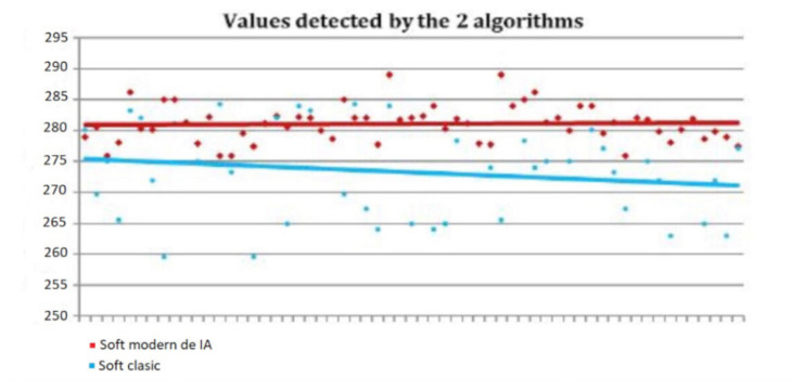
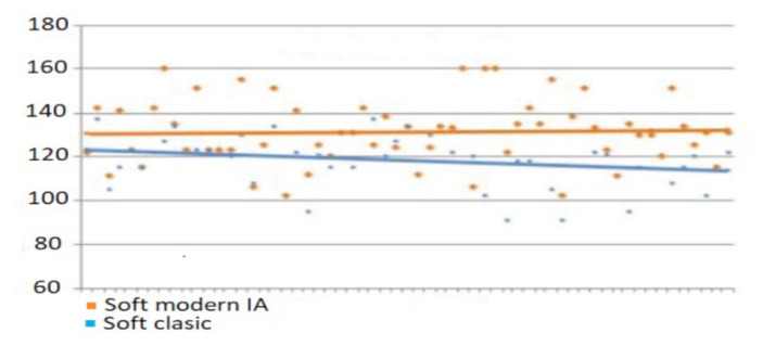
The certainty of detection by AI algorithms has been shown to be better than the classical method, with a ratio of 280 points compared to 187 points. Complex algorithms, specific to Artificial Intelligence, showed superior detection, and more stable in the analysis of peace.
Regarding the correctness of data processing, results were shown to be better by the AI method, compared to the classical method by simple algorithms, with a ratio of 286 to 177 points (Figure 4).
Figure 4.
Distribution of the values with the 2 software applications concerning certainty detection.
Processing the information, obviously led to more accurate results, and there a detection was approximately equal, the AI-specific algorithms again showed superiority, developing more real outlines by analogy with those in the database, thanks to Artificial Neural Networks and Deep Learning (Figure 5).
Figure 5.
Distribution of the values with the 2 software applications concerning correctness detection.
Discussion
Orthodontic treatments are an important stage for achieving a beautiful and healthy smile. In recent years, technological developments and the development of machine learning capabilities have opened new perspectives in guiding orthodontic diagnosis and treatment through artificial intelligence (AI) software. These software applications are designed to provide a personalized approach and accurate guidance of orthodontic treatments, optimizing their effectiveness.
In the digital age we live in, the use of artificial intelligence (AI) brings multiple advantages and benefits in a variety of fields. Deep neural networks have gained fame for their ability to process visual information, becoming a key component of many computer vision applications. Among the key problems that neural networks can solve is the detection and location of objects in images, as we have done in the patients studied, as showed Roberts Jacob in the study published in 2016 [10].
In addition to the obvious benefits of speeding up the diagnostic process, integrating artificial intelligence into orthodontics also brings advantages in personalizing treatment. AI-based systems can provide detailed information about the anatomical variability of the shape and color of teeth, thus allowing orthodontists to develop personalized treatment plans tailored to the individual needs of each patient. Through this study, it was possible to compare which class of algorithms brings more certainty in orthodontic diagnosis of dental contours in teeth in normoposition or malposition, similar to the studies of Huston C, B. [11,12].
In addition to these detection algorithms, other digital techniques are also used in Orthodontics: Procust Superimposition and EDMA Technique for the evaluation of the matrix distance of different contours on the face or skull.
Procrustes Superimposition measures, visualizes and tests the significance of quantitative differences, and quality of morphology. Each aspect is represented by a series of landmark coordinates, forming an image called “configuration” which is morphometric. Configurations are first brought to the same size. The Procust superimposition algorithm translates the configurations that overlap the centers and rotates them iteratively to minimize the square (sectoral) differences between their landmarks (Auffray et al., 1999). This is essentially the position that fits you maximally. After superimposition, the main configuration, called “consensus” is calculated [13,14].
Euclidean distance matrix analysis (EDMA) was introduced by Lele and Richtsmeier in 1991, it quantitatively compared the biological forms, using coordinated landmark data, mathematically locating the morphological differences between two aspect. The numerical product of EDMA is a series of Euclidean distance reports between two average aspects. All the Euclidean distances between the reference pairs for the denominator and the morphology numerator are calculated and an average aspect matrix is generated for each morphology. Important clinical reports can be presented by lines representing relevant distances between the landmarks so EDMA can be used to identify regions of shape changes between cephalograms due to growth or orthodontic treatment [15].
According to our study, the evaluation of the obtained results revealed the superiority of the algorithms specific to the artificial intelligence compared to the classical algorithms. AI algorithms were able to identify complex patterns and correlations in patients’ data, thus providing a solid basis for customizing orthodontic treatments. It not only optimized outcomes but also enhanced patient satisfaction through more accurate and tailored approaches to individual needs.
Other authors
Mew proposes certain suggestions for predicting and monitoring facial growth and dento-facial aesthetics using methods by which these traits can be indexed, objectively appreciated using digital software. The usefulness of these measurements is found in growth forecasts as well as in monitoring growth before, during and after the end of orthodontic treatment [16].
Ferrario and Gallagher conducted a modified computer analysis of a “network” diagram that allows for a quick and independent quantification of facial, soft tissue, and soft tissue sizes and shapes in a three-dimensional space. Diagram analysis – network, which is an application of transformation grids developed by D’Arcy Thompson, allows for an overall assessment of the hard and soft tissue structures of orthodontics patients, going beyond the fragmented approach obtained from conventional cephalometric analyses. Since its introduction, several changes have been presented, and the computer application now allows both a qualitative illustration and a rapid quantitative evaluation of the patient’s facial status [17,18].
Burns states: “the psychological aspects of the image representation that the patient has about himself and his body play a decisive role in aesthetics”. The oral sphere is the primary site of many emotional conflicts. In this context, each patient should be given individualized treatment. Since people associate beauty with success, happiness, socialization, aesthetic motives can be the main factor for seeking treatment, and personality, motivations, desires, expectations, self-estimation, self-esteem, the ability to accept changes and cooperation are important elements for the successful completion of orthodontic intervention [19,20].
The limits of this study are given by the small number of cases evaluated and by the fact that doctors belong to the Hack Clinic, know the software and perhaps there was a certain level of subjectivity involved. Shorter training of doctors in software use and communication skills with patients would be another limitation of this study, considering the reduced time from acquisition to implementation of the program.
Conclusions
Technological evolution and the development of machine learning capabilities have opened new perspectives in guiding orthodontic treatments through artificial intelligence (AI) software. One of the main advantages of AI algorithms is their ability to quickly and accurately analyze a significant amount of complex data, including static or dynamic images of patients. This approach speeds up the diagnostic process, allowing for faster and personalized treatment plans in correlation with the patient’s desire. An ethical and legal approach is required in the implementation of artificial intelligence in dental practice. Protecting patient data privacy and ensuring compliance with industry rules and regulations are key issues for sustainable and responsible integration of this technology. The disadvantages of this technique are represented by higher costs and training of medical personnel in facial analysis with artificial intelligence algorithms, disadvantages that will be solved in the near future.
References
- 1.Poynton C. Digital Video and HDTV: Algorithms and Interfaces. Elsevier; 2007. [Google Scholar]
- 2.Galer M, Horvat L. Digital Imaging: Essential Skills. Focal Press; pp. 205–239. 86/2003. [Google Scholar]
- 3.Rowinski D. Virtual Personal Assistants & The Future Of Your Smartphone [Infographic] 2016. Available from: https://readwrite.com/virtual-personal-assistants-the-future-of-your-smartphone-infographic/
- 4.Matti D, Ekenel HK, Thiran JP. Combining LiDAR space clustering and convolutional neural networks for pedestrian detection. 2017 14th IEEE International Conference on Advanced Video and Signal Based Surveillance (AVSS); [DOI] [Google Scholar]
- 5.Ferguson S, Luders B, Grande RC, How JP. Real-Time Predictive Modeling and Robust Avoidance of Pedestrians with Uncertain, Changing Intentions Algorithmic Foundations of Robotics XI. Springer; 2014. [DOI] [Google Scholar]
- 6.Davis E, Marcus G. Commonsense reasoning and commonsense knowledge in artificial intelligence. Communications of the ACM. 2015;58:92–103. doi: 10.1145/2701413. [DOI] [Google Scholar]
- 7.Scassellati B. Theory of Mind for a Humanoid Robot. Autonomous Robots. 2002;12:13–24. doi: 10.1023/A:1013298507114. [DOI] [Google Scholar]
- 8.Caruso S, Caruso S, Pellegrino M, Skafi R, Nota A, Tecco S. A Knowledge-Based Algorithm for Automatic Monitoring of Orthodontic Treatment: The Dental Monitoring System. Two Cases. Sensors (Basel) 2021;21:1856. doi: 10.3390/s21051856. [DOI] [PMC free article] [PubMed] [Google Scholar]
- 9.Nanda R, Kapila S. Current therapy in Orthodontic. Mosby: Elsevier; 2010. [Google Scholar]
- 10.Roberts J. Thinking machines: the search for Artificial Intelligence. Distillations. 2016;2:14–23. [Google Scholar]
- 11.Hutson M. Artificial intelligence faces reproducibility crisis. Science. 2018;359:725–726. doi: 10.1126/science.359.6377.725. [DOI] [PubMed] [Google Scholar]
- 12.Lieto A, Bhatt M, Oltramari A, Vernon D. The role of cognitive architectures in general artificial intelligence. Cognitive Systems Research. 2018;48:1–3. doi: 10.1016/j.cogsys.2017.08.003. [DOI] [Google Scholar]
- 13.Hinton G, Deng L, Yu D, Dahl GE, Mohamed A, Jaitly N, et al. Deep Neural Networks for Acoustic Modeling in Speech Recognition – The shared views of four research groups. IEEE Signal Processing Magazine. 2012;29:82–97. doi: 10.1109/msp.2012.2205597. [DOI] [Google Scholar]
- 14.Hyötyniemi H. Turing machines are recurrent neural networks. Proceedings of STeP’96/Publications of the Finnish Artificial Intelligence Society:; 1996; pp. 13–24. Available from: http://users.ics.aalto.fi/tho/stes/step96/hyotyniemi1/ [Google Scholar]
- 15.Lele A, Richtsmeier JT. Euclidean distance matrix analysis: a coordinate-free approach for comparing biological shapes using landmark data. Am J Phys Anthropol. 1991;86:415–442. doi: 10.1002/ajpa.1330860307. [DOI] [PubMed] [Google Scholar]
- 16.Mew J. Suggestions for forecasting and monitoring facial growth. Am J Orthod Dentofacial Orthop. 1993;104(2):105–120. doi: 10.1016/S0889-5406(05)81000-5. [DOI] [PubMed] [Google Scholar]
- 17.Ferrario VF, Sforza C, Miani A, Jr, Serrao G. Dental arch asymmetry in young healthy human subjects evaluated by Euclidean distance matrix analysis. Arch Oral Biol. 1993;38:189–194. doi: 10.1016/0003-9969(93)90027-j. [DOI] [PubMed] [Google Scholar]
- 18.Gallagher J. Artificial intelligence ‘as good as cancer doctors’. BBC News. 2017 January 26; [Google Scholar]
- 19.William R, Proffit H, Larson Fields B, Sarver David M. Contemporary Orthodontics. 6th Edition. Elsevier; Oct, 2018. [Google Scholar]
- 20.Svensson AM, Jotterand F. Doctor Ex Machina: A Critical Assessment of the Use of Artificial Intelligence in Health Care. J Med Philos. 2022;47:155–178. doi: 10.1093/jmp/jhab036. [DOI] [PubMed] [Google Scholar]


