AUTHOR CORRECTION
Volume 88, no. 2, p. 1281–1292, 2013, https://doi.org/10.1128/jvi.02665-13, and volume 90, no. 12, p. 5845, https://doi.org/10.1128/jvi.00587-16. This correction updates Fig. 2B, 4C and 7B, by demarcating the edges of noncontiguous portions of gel and blot images that were spliced together in the original publication. This was discovered after a previous correction to the Acknowledgments was published. This update does not change the conclusions of the paper. Our rationale for splicing the images was to eliminate noninformative or blank regions of gels and blots to conserve publication space. All of the spliced portions in each of the figure panels were derived from the same raw, source image of a single gel or blot. Each of the panels came from a single experiment.
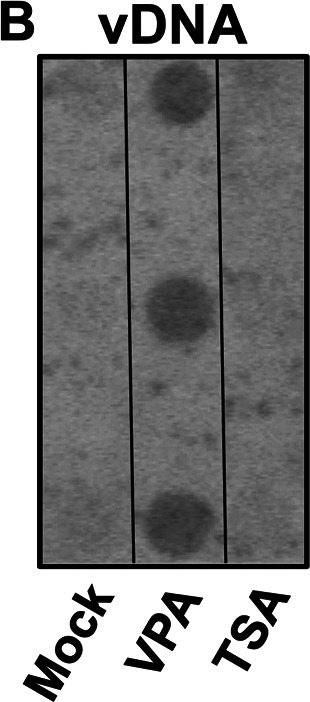
Figure 2B should appear as shown in this correction. The following statement should be added to the Fig. 2 legend: “Three regions of the blot image that demonstrate encapsidated vDNA produced by the indicated treatments were spliced together as indicated by the black outlines. The other regions of the blot did not pertain to this publication.”
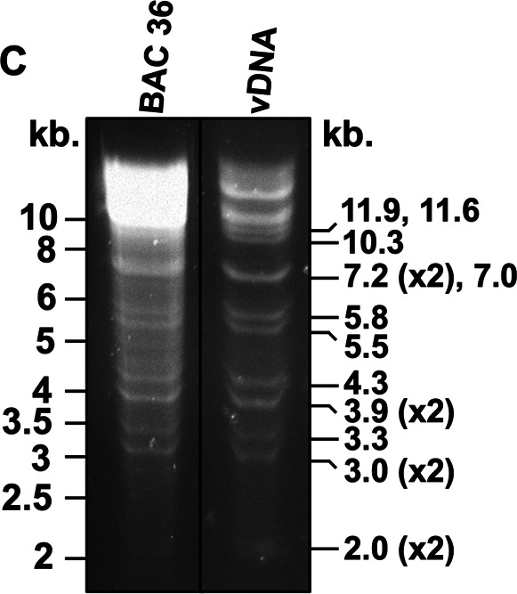
Figure 4C should appear as shown in this correction. The following statement should be added to the Fig. 4 legend: “Two regions of the gel image were spliced together as indicated by the black outlines. The other regions of the gel were blank and did not pertain to this publication.”
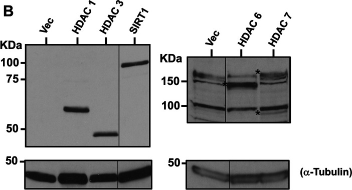
Figure 7B should appear as shown in this correction. The following statement should be added to the Fig. 7 legend: “Each panel was assembled from a single image from two independent gels/western blots. The left panel was assembled from one image of gel 1/western blot 1, and the right panel was assembled from one image from gel 2/blot 2. The gel in the right panel was loaded with 2× protein extract. Noncontiguous portions are indicated with black outlines.”
Fig 2.

Fig 4.

Fig 7.


