Abstract
RNA replicon particles derived from a vaccine strain of Venezuelan equine encephalitis virus (VEE) were used as a vector for expression of the major envelope proteins (GL and M) of equine arteritis virus (EAV), both individually and in heterodimer form (GL/M). Open reading frame 5 (ORF5) encodes the GL protein, which expresses the known neutralizing determinants of EAV (U. B. R. Balasuriya, J. F. Patton, P. V. Rossitto, P. J. Timoney, W. H. McCollum, and N. J. MacLachlan, Virology 232:114–128, 1997). ORF5 and ORF6 (which encodes the M protein) of EAV were cloned into two different VEE replicon vectors that contained either one or two 26S subgenomic mRNA promoters. These replicon RNAs were packaged into VEE replicon particles by VEE capsid protein and glycoproteins supplied in trans in cells that were coelectroporated with replicon and helper RNAs. The immunogenicity of individual replicon particle preparations (pVR21-GL, pVR21-M, and pVR100-GL/M) in BALB/c mice was determined. All mice developed antibodies against the recombinant proteins with which they were immunized, but only the mice inoculated with replicon particles expressing the GL/M heterodimer developed antibodies that neutralize EAV. The data further confirmed that authentic posttranslational modification and conformational maturation of the recombinant GL protein occur only in the presence of the M protein and that this interaction is necessary for induction of neutralizing antibodies.
The recently established order Nidovirales includes two families, the Arteriviridae (genus Arterivirus) and Coronavidae (genera Coronavirus and Torovirus [8]). Viruses from the different genera of the Nidovirales exhibit considerable differences in their genetic complexity and virion structure, but they are strikingly similar in their genome organization, replication strategy, and intracellular site of budding (reviewed in references 18 and 40). Equine arteritis virus (EAV) is the prototypic virus of the family Arteriviridae and the cause of equine viral arteritis (EVA), a sporadic respiratory and reproductive disease of horses (24, 40, 43).
The EAV genome is a positive-stranded RNA molecule of 12,704 nucleotides (nt), excluding the long 3′ polyadenylated tail (14, 40). The EAV genome includes two large open reading frames (ORFs)—1a and 1b—located at the 5′ end of the genome that encode the viral replicase. Seven other ORFs—2a, 2b, 3, 4, 5, 6, and 7—located at the 3′ end of the genome, are transcribed during replication and encode five structural proteins (E, GS, GL, M, and N) and two glycoproteins of unknown function (GP3 and GP4 [17, 40, 41]). ORF5 encodes the major envelope glycoprotein (GL), and ORF6 encodes an unglycosylated envelope protein (M). The GL protein may be single- or triple-membrane spanning, and it likely functions as both a receptor-binding and a membrane fusion protein (16, 17). The GL envelope protein expresses the known neutralization determinants of the virus, and we have identified four distinct neutralization sites in this protein (3, 4, 10, 15, 23). The M protein may be involved in virus budding and contains three membrane-spanning segments, with only 19 amino acids being exposed on the virion surface (16, 17, 40). The M and GL proteins form a disulfide-linked heterodimer in the virus particle (19). The M protein also forms covalently linked homodimers, but only the GL/M heterodimer is incorporated into virus particles.
Horses naturally infected with EAV or vaccinated with either live attenuated or inactivated whole-virus preparations are protected against clinical EAV (20, 21, 32). Neutralizing antibodies appear to prevent reinfection of horses with EAV. Horses immunized with portions of the GL protein expressed either in bacteria (residues 55 through 98) or as a synthetic oligopeptide (residues 75 through 97) developed antibodies that neutralize EAV (10). In contrast, we were unable to induce neutralizing antibodies in laboratory animals (mice, guinea pigs, and rabbits) immunized with EAV structural proteins (GL, M, and GL/M heterodimer) expressed in eukaryotic cells infected with recombinant baculo- and Sindbis viruses (1; U. B. R. Balasuriya, unpublished data).
Recombinant alphaviruses (Sindbis virus, Semliki Forest virus, and Venezuelan equine encephalitis virus [VEE]) derived from full-length cDNA clones recently have been developed as vectors for the expression of heterologous viral genes (37–39). In this study we have expressed the major EAV envelope proteins (GL and M) individually and in heterodimer (GL/M) form by using the VEE replicon vector system, and we have shown that the expression of both proteins as a heterodimer from EAV-VEE replicon particles (EAV-VRPs) is necessary for induction of neutralizing antibodies in inoculated mice.
MATERIALS AND METHODS
Cells and viruses.
Baby hamster kidney 21 (BHK-21 [ATCC CCL10]) cells were maintained in Eagle's minimal essential medium supplemented with 10% fetal bovine serum (Hyclone Laboratories, Inc.), 10% tryptose phosphate broth, and 1% each penicillin and streptomycin. Rabbit kidney 13 (RK-13 [ATCC CCL 37]) cells were maintained in Eagle's minimal essential medium supplemented with 10% calf serum (Hyclone Laboratories Inc.) and antibiotics. The Bucyrus strain of EAV was obtained from the American Type Culture Collection (ATCC VR-796). The virus was gradient purified on an 11 to 33% continuous CsCl gradient, as previously described (2). The purified virus was used as an antigen for Western immunoblotting and for immunization of mice.
Construction of recombinant plasmids.
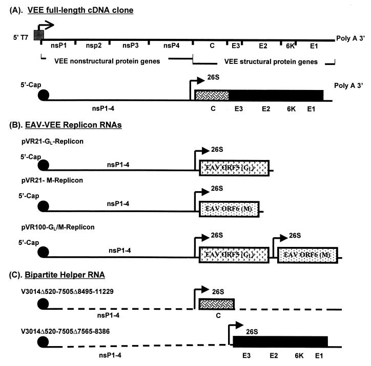
Three recombinant VEE replicon plasmids were constructed, two of which were designed to express the GL or M protein of EAV individually and a third which was designed to express the GL and M proteins together. The genes encoding GL (ORF5) and M (ORF6) from the ATCC strain of EAV were transferred from plasmids pEAV5A1–2 and pEAV6B1–1 (1, 30) into a VEE replicon plasmid designated pVR21, using a PCR-based cloning strategy as previously described (42). pVR21 was derived from the pVR2 replicon plasmid (analogous to the replicon plasmids described by Pushko et al. [37]) as follows. A PCR product was made using a forward primer spanning the unique MfeI (nt 7,621) restriction site and a reverse primer spanning the unique NotI (nt 7,752) restriction site that was designed to extend the poly(A) tract from 21 residues to 50 and insert a unique SapI site directly downstream of the poly(A) tract of the pVR2 plasmid. This amplicon was digested with MfeI and NotI and used to replace the homologous fragment in the pVR2 plasmid. A similar PCR strategy was used to replace the pVR2 multiple cloning site with a new polylinker containing four unique 8-base restriction sites and a unique ClaI site. The recombinant replicons containing the GL and M genes of EAV were designated pVR21-GL and pVR21-M, respectively (Fig. 1).
FIG. 1.
Schematic presentation of EAV-VEE replicon and helper RNAs. (A) VEE full-length infectious cDNA clone and in vitro-transcribed infectious RNA. The shaded box and arrow show the position of the T7 RNA polymerase promoter and direction of in vitro transcription, respectively. (B) EAV-VEE replicon RNAs. The GL, M, and GL/M replicon RNAs are similar to full-length RNA except that the ORF encoding the VEE structural proteins in the full-length RNA was replaced with ORF5, ORF6, or ORF5 and -6 of EAV, respectively. ORF5 and -6 were placed under the control of two different 26S promoters in the GL/M replicon. (C) Bipartite VEE helper RNAs. The bipartite helper system consists of two helper RNAs derived from the V3014Δ520-7505 monopartite helper (37). The dashed lines indicate deleted regions of the VEE genome. One RNA expresses the VEE capsid gene, and the second RNA expresses the envelope glycoprotein genes.
A replicon designed to express the GL and M proteins together was constructed in the genetic background of a VEE replicon plasmid designated pVR100. pVR100 was derived from a VEE cDNA clone that contains a second 26S mRNA promoter placed downstream of the structural genes (11). The Tth111I-SacII fragment of this cDNA clone, encompassing the structural gene region, was replaced by a synthetic DNA fragment containing unique PmeI, PacI, and AscI restriction sites. The resulting double-promoter replicon contains an upstream 26S promoter followed by a polylinker, a second copy of the 26S promoter, a unique ClaI site, and the 3′-untranslated region of the VEE genome. The cloning strategy for inserting the GL- and M-coding sequences into pVR100 was similar to that used to construct pVR21-GL and pVR21-M. This recombinant replicon was designated pVR100-GL/M (Fig. 1). The sequence of each recombinant replicon plasmid was confirmed by sequence analysis as previously described (26).
The bipartite helper system used for construction of replicon particles consists of two helper RNAs derived from the V3014Δ520-7505 monopartite helper (Fig. 1). The construction of the bipartite helper RNA system of individual capsid (C) and glycoprotein (GP) genes of VEE has been described in detail (37).
Production and titration of EAV-VRPs.
The recombinant plasmids were linearized by digestion with NotI at a unique restriction site downstream of the VEE cDNA sequence, and capped run-off transcripts were prepared in vitro with T7 RNA polymerase (Promega) as previously described (5). The in vitro-generated mixture of replicon RNA, capsid helper RNA, and GP helper RNA was cotransfected into BHK-21 cells by electroporation (37; Fig. 1) and incubated in 75-cm2 flasks at 37°C in 5% CO2 for 27 h. EAV-VRPs were partially purified, concentrated, and resuspended in phosphate-buffered saline (PBS [pH 7.4]) as previously described (12). BHK-21 cells were infected with 10-fold serial dilutions of EAV-VRPs. The cells were fixed in methanol and acetone and reacted with protein-specific antibodies. The infected cells were detected by avidin-biotin immunoperoxidase staining as previously described (1, 31), and EAV-VRP titers were determined as infectious units (IU) per milliliter.
Antibodies.
Monospecific rabbit antipeptide serum to the M protein and a neutralizing murine monoclonal antibody (MAb) to the GL protein (MAb 6D10) have been previously described (2, 4). An MAb that recognizes the β-COP protein (110 kDa; Sigma [36]) and a rabbit polyclonal antibody specific for calreticulin (60–63 kDa; StressGen Biotechnologies Corp. [44]) were used as markers for the Golgi complex and endoplasmic reticulum (ER), respectively, in immunofluorescence assays.
Immunofluorescence and confocal microscopy.
Cellular localization of expressed EAV proteins was determined by confocal immunofluorescence microscopy using antibodies specific for the GL and M proteins of EAV, the Golgi complex, and the ER. Briefly, subconfluent BHK-21 cells were infected with EAV-VRPs, fixed, and incubated with protein-specific antibodies as described above. Alexa Fluor 488-conjugated goat anti-mouse immunoglobulin G (IgG [0.6 μg/ml]) and Alexa Fluor 594-conjugated goat anti-rabbit IgG (0.6 μg/ml) were used as the secondary antibodies (Molecular Probes). The slides were mounted in Prolong Antifade Reagent (Molecular Probes) for microscopy. Confocal imaging was performed with the Bio-Rad MRC 1024 ES laser scanning confocal microscope with 488- and 568-nm krypton/argon laser excitation. The laser power, photomultiplier sensitivity, and number of averages were adjusted to generate images with optimal contrast.
Western immunoblotting and endoglycosidase treatment.
BHK-21 cell monolayers in six-well plates were infected with EAV-VRPs at a multiplicity of infection of ≥10 and incubated at 37°C for 15 h. The cells were washed twice in PBS and solubilized in solubilization buffer (20 mM Tris HCl [pH 7.6], 150 mM NaCl, 1% Nonidet P-40, 0.5% sodium deoxycholate, 0.1% sodium dodecyl sulfate [SDS] containing aprotinin, leupeptin, and pepstatin (1 μg/ml) and phenylmethylsulfonyl fluoride [17 μg/ml]). For endoglycosidase treatment, the cells were solubilized in endoglycosidase buffer (50 mM sodium phophate [pH 6.8], 20 mM EDTA, 0.15% SDS, 1% Nonidet P-40, 1% 2-mercaptoethanol containing aprotinin, leupeptin, and pepstatin [1 μg/ml] and phenylmethylsulfonyl fluoride [17 μg/ml]) and subjected to glyco F (Endoglycosidase F; Boehringer Mannheim Corp.) treatment. The solubilized or glyco F-treated proteins were mixed with an equal volume of Laemmli sample buffer containing 50 mM dithiothreitol (DTT) prior to SDS-polyacrylamide gel electrophoresis (PAGE) using a 12% resolving gel and a 4% stacking gel (Bio-Rad [2]). Proteins were then transferred to an Immobilon-P membrane (Millipore) for immunoblotting with MAb 6D10 and rabbit antipeptide serum and detected by chemiluminescence, as previously described (2, 4).
[35S]methionine labeling and immunoprecipitation.
BHK-21 cells that had been either infected with EAV-VRPs or mock infected were incubated with methionine- and cysteine-free medium containing 1% dialyzed fetal bovine serum (Gibco BRL) for 4 h at 6 h postinfection. Ten hours after infection, the cells were placed in medium containing 300 μCi of [35S]methionine (Express 35S protein labeling mix; NEN Life Science Products, Inc.) for 2 h. At 12 h postinfection, cells were washed once in ice-cold PBS and solubilized in 500 μl of solubilization buffer as described above. The cytoplasmic proteins were immunoprecipitated using protein G (Zymed) and either normal mouse serum, serum from mice inoculated with EAV-VRPs, MAb 6D10, or rabbit anti-M serum, as previously described (2). Selected samples were resuspended in 400 μl of endoglycosidase buffer and subjected to glyco F treatment as previously described (2, 4). Labeled cytoplasmic proteins and the immunoprecipitated proteins were resolved by SDS-PAGE as described above and visualized by fluorography with Amplify (Amersham Pharmacia Biotech).
Immunization of mice with EAV-VRPs and gradient-purified EAV.
The EAV-VRP preparations were diluted in PBS containing 1% calf serum, and groups of 5- to 6-week-old BALB/c mice (four per group) were inoculated subcutaneously in the rear footpads with 20 μl of one EAV-VRP preparation (6 × 10⁵ to 3.6 × 10⁶ IU [10 μl per footpad]). Mice were boosted at weeks 3 and 5 by footpad inoculation with the same EAV-VRP preparation. Mice were bled 3 weeks after the initial inoculation and 2 weeks after each booster immunization. A group of four 6-week-old BALB/c mice were inoculated intraperitoneally with 0.2 ml of the gradient-purified ATCC strain of EAV (1:2 emulsion of virus in complete Freund's adjuvant). Mice were boosted at weeks 4 and 7 with the same antigen (1:2 emulsion of virus in incomplete Fruend's adjuvant). The mice were bled 9 weeks after the initial inoculation.
Serum-neutralizing antibodies specific for EAV were detected by microneutralization assay using the EAV ATCC strain as the challenge virus in the presence of 5% guinea pig complement, as previously described (2, 4). Antibody titers were recorded as the reciprocal of the highest final dilution of serum that provided at least 50% protection of the RK-13 cell monolayer.
RESULTS
Characterization of recombinant EAV proteins expressed from EAV-VRPs.
The two major EAV envelope proteins (GL and M) were expressed individually as well as in heterodimer form (GL/M) using the VEE replicon system. ORF5 and -6 of EAV were cloned individually downstream of a 26S promoter in the pVR21 VEE replicon vector to express, respectively, the GL and M proteins (Fig. 1 [pVR21-GL and pVR21-M]). For dual expression, the same ORFs were cloned into the pVR100 double-promoter replicon vector that contained two 26S promoters (pVR100-GL/M). Expression of the EAV proteins was detected by immunoperoxidase staining of BHK-21 cells with GL and M protein-specific antibodies following transfection with in vitro-transcribed replicon RNAs (data not shown). Each EAV-VRP was titrated by enumeration of antigen-positive cells by immunoperoxidase staining of infected BHK-21 cells. The respective titers of pVR21-GL-VRP, pVR21-M-VRP, and pVR100-GL/M-VRP were 3 × 10⁷, 3 × 10⁸, and 1.8 × 10⁷ IU/ml.
The [35S]methionine-radiolabeled lysates of cells infected with each EAV-VRP were analyzed by SDS-PAGE to assess the level of protein expression (Fig. 2A), as staining with Coomassie brilliant blue G-250 dye did not identify unique protein bands in any of the lysates. The expression and authenticity of the recombinant GL and M proteins were further confirmed by Western immunoblotting (Fig. 2B) and immunoprecipitation (Fig. 3) assays. The GL protein-specific MAb 6D10 strongly reacted with distinct 25- and 30-kDa proteins in immunoblots of lysates of pVR21-GL-infected BHK-21 cells. The same MAb recognized a smear reflecting a 30- to 44-kDa heterogeneously glycosylated GL protein in the pVR100-GL/M-infected BHK-21 cell lysate, as well as in gradient-purified EAV. Deglycosylation of cell lysates and gradient-purified EAV prior to Western immunoblotting reduced the 30- and 30- to 44-kDa GL protein to a single band of 25 kDa. The 25-kDa protein corresponds to the unglycosylated precursor of the GL protein, and the 30- to 44-kDa protein smear represents variable posttranslational N-linked glycosylation (addition of N-acetyllactosamine) of this precursor protein. The GL protein was more heterogeneously glycosylated when it was expressed as a heterodimer with the M protein than when expressed by itself (Fig. 2B).
FIG. 2.
(A) SDS-PAGE of [35S]methionine-labeled cell lysate of individual EAV-VRP-infected BHK-21 cells (identified as GL, M, and GL/M). (B) Immunoblotting of recombinant proteins and EAV with various GL and M protein-specific antibodies. Antigens in each lane are indicated above the lanes. Lysate of uninfected BHK-21 cells was used as the negative control antigen (CT). Antibody (IgG) used in each lane is indicated above the antigens. The proteins were either treated with glyco F (+) or mock treated (−), as indicated below the lanes. Proteins were also analyzed by SDS-PAGE under reducing (with 50 mM DTT [+]) and nonreducing (without 50 mM DTT [−]) conditions. The proteins and their Mrs are indicated on either side of each panel.
FIG. 3.
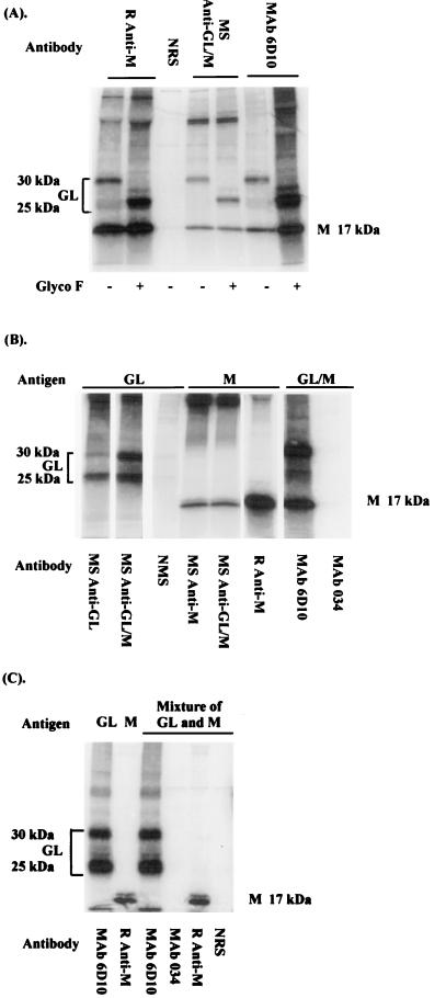
Radiolabeling and immunoprecipitation of EAV envelope proteins expressed from EAV-VRPs in BHK-21 cells by using protein-specific antibodies. The proteins and their Mrs are indicated on either side of each panel. (A) Lysate of pVR100-GL/M-infected cells was immunoprecipitated with antibodies specific for the M protein (rabbit anti-M [R anti-M]) or the GL protein (MAb 6D10) or with sera from mice inoculated with the GL/M heterodimer (mouse anti-GL/M [MS anti-GL/M]) or negative rabbit serum (NRS) and subsequently treated (+) or mock (−) treated with glyco F. (B) Lysates of pVR21-GL-, pVR21-M-, and pVR100-GL/M-infected cells were immunoprecipitated with protein-specific antibodies (R anti-M and MAb 6D10) or sera from mice inoculated with individual EAV-VRPs (e.g., MS anti-GL indicates serum from a mouse immunized with the pVR21-GL VRP) or negative control antibodies (normal mouse serum [NMS] or MAb specific for bluetongue virus [MAb 034]). The antigen (cell lysate) in each lane is identified above the lane, and the antibody used for immunoprecipitation is indicated below the lane. (C) Mixtures of lysates of pVR21-GL- and pVR21-M-infected cells were immunoprecipitated with protein-specific antibodies (R anti-M and MAb 6D10) and negative control antibodies (NRS and MAb 034).
The rabbit anti-peptide serum specific for the M protein strongly reacted with a 17-kDa protein in immunoblots of pVR21-M and pVR100-GL/M VRP-infected cell lysates, as well as gradient-purified EAV. Under nonreducing conditions, the anti-M serum recognized the 17-kDa M protein monomer as well as a 30-kDa homodimer by both immunoblotting (Fig. 2) and immunoprecipitation assays using the pVR21-M-infected cell lysate (data not shown). These data confirm that the M protein forms a covalently linked homodimer in replicon-infected cells, as well as in BHK-21 cells infected with EAV (19).
The MAbs that were specific for GL protein (6D10) and rabbit anti-M protein serum also were used to immunoprecipitate EAV proteins in lysates of radiolabeled EAV-VRP-infected cells in the absence of DTT. The MAb 6D10 and the anti-M serum each precipitated both the GL and M proteins from the radiolabeled lysate of pVR100-GL/M-infected cells (Fig. 3A). The coprecipitation of the GL and M proteins using monospecific antibodies to either protein in the absence of DTT indicates that these two proteins form a covalently linked heterodimer in pVR100-GL/M-infected cells, analogous to their association in the EAV particle (19). MAb 6D10 precipitated only 25- and 30-kDa proteins, consistent with the GL protein from the pVR21-GL-infected lysate, and the rabbit anti-M serum precipitated a 17-kDa protein, consistent with the M protein from the pVR21-M-infected cell lysate (Fig. 3B). Furthermore, mixing of radiolabeled lysates that included individually expressed GL and M proteins did not result in heterodimer formation, as determined by immunoprecipitation with MAb 6D10 and rabbit anti-M serum (Fig. 3C).
Localization of GL and M proteins in EAV-VRP-infected BHK-21 cells.
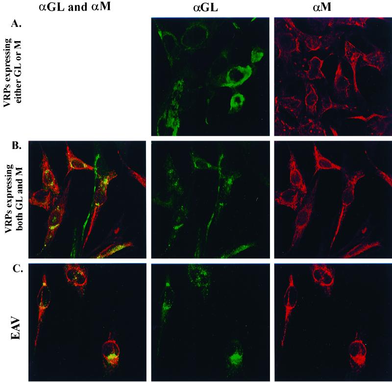
The intracellular localization of the EAV proteins (GL, M, and GL/M heterodimer) expressed in BHK-21 cells infected with the various EAV-VRPs was determined by confocal immunofluorescence microscopy (Fig. 4). The localization of these proteins was confirmed by immunofluorescent staining with organelle-specific antisera (data not shown). Cells expressing only the M protein exhibited perinuclear staining as well as a more diffuse reticular pattern consistent with the distribution of the ER. Similarly, cells expressing only the GL protein had a reticular pattern of staining consistent with ER localization. The localization of the M protein did not alter in cells expressing the GL/M heterodimer (pVR100-GL/M), whereas the GL protein consistently colocalized with the M protein in a polar perinuclear location in these cells as did the Golgi-specific β-COP protein. The colocalization of the GL and M proteins in the Golgi complex was identical to that in the EAV-infected BHK-21 cells (Fig. 4). Thus the localization of the GL protein to the Golgi complex was dependent on the presence of the M protein in the same cell.
FIG. 4.
Intracellular localization of GL and M proteins in BHK-21 cells infected with EAV-VRPs or EAV (15 h postinoculation). (A) Cells infected with pVR21-GL or pVR21-M are labeled with anti-GL (MAb 6D10)- and rabbit anti-M-purified IgG. The GL protein is stained green with Alexa Flour 488, and the M protein is stained red with Alexa Flour 594 conjugates. (B) Double labeling of pVR100-GL/M-infected cells with anti-GL and anti-M purified IgG. The yellow staining of overlays of both GL and M labels (merged images) indicates that the two proteins colocalize in the Golgi complex when they are present in the same cell and that the GL protein is concentrated in the Golgi complex. (C) The distribution of the GL and M proteins in EAV-infected cells is similar to that in the pVR100-GL/M-infected cells.
GL/M heterodimer induced neutralizing antibodies in mice.
The immunogenicity of individual EAV-VRPs was determined by immunizing BALB/c mice. All mice were inoculated three times (the primary inoculation and two boosts) with each EAV-VRP. Mice were bled 2 to 3 weeks after each immunization and evaluated for serum antibodies using virus neutralization, immunoprecipitation, and Western immunoblotting assays. Mice immunized with pVR21-GL or pVR21-M VRPs did not develop neutralizing antibodies to EAV. In contrast mice immunized with pVR100-GL/M, which expresses both the GL and M proteins, developed high titers (1,024 or 2,048) of neutralizing antibody to EAV (Table 1). The neutralizing-antibody titers in these mice were comparable to or higher than those in mice repeatedly immunized with whole EAV (Table 1).
TABLE 1.
Induction of neutralizing antibodies in mice with EAV-VRPs and whole EAV
| Immunogen | Immunization schedule (weeks) | Dose (IUa or TCID50b) | Weeks after primary immunization | Neutralizing antibody titersc of mouse no.:
|
|||
|---|---|---|---|---|---|---|---|
| 1 | 2 | 3 | 4 | ||||
| pVR21-GL VRP | Primary (0) | 6 × 10⁵ | 3 | 0 | 0 | 0 | 0 |
| First boost (3) | 5 | 0 | 0 | 0 | 0 | ||
| Second boost (7) | 9 | 0 | 0 | 0 | 0 | ||
| pVR21-M VRP | Primary (0) | 6 × 10⁶ | 3 | 0 | 0 | 0 | 0 |
| First boost (3) | 5 | 0 | 0 | 0 | 0 | ||
| Second boost (7) | 9 | 0 | 0 | 0 | 0 | ||
| pVR100-GL/M VRP | Primary (0) | 3.6 × 10⁶ | 3 | 32 | 32 | 64 | 64 |
| First boost (3) | 5 | 512 | 512 | 512 | 512 | ||
| Second boost (7) | 9 | 2,048 | 1,024 | 2,048 | 2,048 | ||
| EAV ATCC strain | Primary (0) | 2 × 10⁴b | N/Ad | NDe | ND | ND | ND |
| First boost (4) | N/A | ND | ND | ND | ND | ||
| Second boost (7) | 9 | 1,024 | 1,024 | 1,024 | 512 | ||
Infectious units as determined by immunoperoxidase staining of infected BHK-21 cells.
50% tissue culture infectious doses.
Neutralization titers are expressed as the inverse of the antibody dilution providing 50% protection of RK-13 cell monolayers against 200 50% tissue culture infective doses of virus.
N/A, not applicable.
ND, not determined.
Whereas all of the mice inoculated with the pVR100-GL/M VRPs developed neutralizing antibodies to the GL protein, serum from only one mouse weakly recognized the M protein in a Western immunoblotting assay. Sera from mice immunized with the EAV-VRPs that individually express the GL and M proteins contained antibodies to the respective immunizing proteins as determined by immunoprecipitation assays, confirming that the mice were immunized against each protein. The data clearly indicate that each EAV-VEE VRP successfully immunized mice against the appropriate expressed recombinant EAV protein but that coexpression of a GL/M heterodimer is essential for the induction of detectable neutralizing antibodies to EAV.
DISCUSSION
The VEE-based replicon system has previously been used to express several heterologous viral genes, including the influenza virus hemagglutinin (HA), the matrix/capsid (MA/CA) domain of human immunodeficiency virus type 1 (HIV-1), the MA/CA domain and gp160 of simian immunodeficiency virus, and the glycoprotein subunit of the Marburg virus (6, 7, 9, 12, 27, 37). Rodents and monkeys immunized with VRPs expressing these proteins were protected against subsequent challenge with the pathogen from which the heterologus genes were derived. Our objective was to use the VEE replicon vaccine vector system to express the two major envelope proteins (GL and M) of EAV and to determine the immunogenicity of these proteins in mice. Expression of the GL and M proteins as a heterodimer is clearly necessary for induction of neutralizing antibodies in mice, as mice immunized with replicons that individually express the GL and M proteins developed antibodies that immunoprecipitated each respective protein but did not neutralize the virus. Neutralizing antibody titers in mice immunized with the recombinant GL/M heterodimer were considerably higher than those in the serum of horses that were naturally infected with EAV (range, 4 to 512), as well as those repeatedly vaccinated with a modified live virus vaccine (≤512) (25, 33).
Inability of the expressed GL protein to induce neutralizing antibodies in mice was surprising, since the known neutralizing determinants of EAV all are located in the N-terminal hydrophilic ectodomain of this protein and all published EAV-neutralizing MAbs are specific for this region (3, 4, 10, 15, 23). Furthermore, horses and mice immunized with synthetic peptides (amino acid residues 75 through 97 or 93 through 112) or a bacterial fusion protein (amino acids 55 through 98) of the GL envelope protein developed EAV-neutralizing antibodies (10). We have previously demonstrated that linear epitopes in neutralization site D (amino acids 99 through 106) in the GL protein interact with amino acids in three other sites (A, B, and C) within the amino-terminal ectodomain of the protein (3). The fact that neutralizing antibodies were induced only in mice immunized with the GL/M heterodimer indicates that the presence of the M protein is critical to the expression of neutralization epitopes on the recombinant GL protein.
The GL and M proteins associate as a disulfide-linked heterodimer and form cuplike structures on the virion surface (40). Similarly, the GL and M proteins form a heterodimer after expression from the dual construct (pVR100-GL/M), as determined by immunoprecipitation with either GL or M protein-specific antibodies. The distribution of the GL protein in EAV-VRP-infected BHK-21 cells was markedly different in cells expressing GL alone (pVR21-GL infected) and in those expressing the GL/M heterodimer (pVR100-GL/M), as shown by confocal immunofluorescence microscopy. The GL protein accumulated in the Golgi complex only in association with the M protein, indicating that heterodimer formation is required for transport of the GL protein from the ER to the Golgi complex. The β3 and β4 galactosyltransferase enzymes are localized to the trans Golgi cisternae; thus, the heterogeneous glycosylation of the GL protein included in the heterodimer but not the recombinant GL protein alone further confirmed its movement from the ER to the Golgi when associated with the M protein (29). Our data indicate that the GL protein is a specific marker of the Golgi complex in EAV-infected cells only when it is associated with the M protein (41).
The M proteins of arteri- and coronaviruses are predicted to be functionally homologous and necessary for localizing viral envelope proteins to the site of virus budding in the intermediate compartment between the ER and the Golgi complex (41). The M protein associates with the major envelope glycoprotein (S protein of coronaviruses and GL protein of EAV) immediately after its synthesis and transport to the intermediate compartment (19, 35, 45). The coronavirus S protein is transported to the plasma membrane when expressed alone, whereas it is retained in the Golgi complex when expressed with the M protein (13, 28, 34, 35, 46). Similarly, the EAV GL protein accumulates in the ER when expressed alone and localizes to the Golgi complex only when it is coexpressed with the M protein. Assembly of viral-membrane protein oligomers usually takes place prior to their transport to the Golgi complex (minireviewed elsewhere [22 and references therein]), and, in the absence of the M protein, free sulfhydryl groups in the GL protein likely form aberrant intrachain disulfide bonds leading to misfolding and the formation of large protein aggregates. These aggregates would be relatively immobile in the ER due to their large size or to their association with fixed proteins of the rough ER (22). Thus, the M protein likely acts as an essential scaffold on which the GL protein folds to form the epitopes necessary to induce neutralizing antibodies in mice.
In summary, we have shown that the VEE-based replicon vector can express the GL and M proteins of EAV in authentic form. Our study unequivocally demonstrates that the recombinant GL and M proteins must be expressed together as a heterodimer to induce EAV-neutralizing antibodies in mice. We have also demonstrated that proper posttranslational modification and folding of the GL protein occur only in the presence of the M protein. Studies are now in progress to evaluate the immunogenicity of these recombinant proteins in horses and to determine their ability to protect against the infection with virulent strains of EAV.
ACKNOWLEDGMENTS
We thank Martha Collier for excellent technical support with construction of EAV-VRPs and Christopher D. DeMaula for help with immunization of mice.
These studies are supported by USDA National Research Initiative competitive grant 97-35204-4736 and the Center for Equine Health at University of California, Davis, with funds provided by the Harriet E. Pfleger Foundation, the Bernard and Gloria Salick endowment, the Oak Tree Racing Association, the State of California parimutuel fund, and contributions by private donors.
REFERENCES
- 1.Balasuriya U B R. Molecular characterization of neutralization determinants and phylogenetic analysis of open reading frame 5 (ORF5) of equine arteritis virus. Ph.D. dissertation. University of California, Davis; 1996. [Google Scholar]
- 2.Balasuriya U B R, MacLachlan N J, de Vries A A F, Rossitto P V, Rottier P J M. Identification of a neutralization site in the major envelope glycoprotein (GL) of equine arteritis virus. Virology. 1995;207:518–527. doi: 10.1006/viro.1995.1112. [DOI] [PubMed] [Google Scholar]
- 3.Balasuriya U B R, Patton J F, Rossitto P V, Timoney P J, McCollum W H, MacLachlan N J. Neutralization determinants of laboratory strains and field isolates of equine arteritis virus: identification of four neutralization sites in the amino-terminal ectodomain of the GL envelope glycoprotein. Virology. 1997;232:114–128. doi: 10.1006/viro.1997.8551. [DOI] [PubMed] [Google Scholar]
- 4.Balasuriya U B R, Rossitto P V, DeMaula C D, MacLachlan N J. A 29K envelope glycoprotein of equine arteritis virus expresses neutralization determinants recognized by murine monoclonal antibodies. J Gen Virol. 1993;74:2525–2529. doi: 10.1099/0022-1317-74-11-2525. [DOI] [PubMed] [Google Scholar]
- 5.Balasuriya U B R, Snijder E J, van Dinten L C, Heidner H W, Wilson W D, Hedges J F, Hullinger P J, MacLachlan N J. Equine arteritis virus derived from an infectious cDNA clone is attenuated and genetically stable in infected stallions. Virology. 1999;260:201–208. doi: 10.1006/viro.1999.9817. [DOI] [PubMed] [Google Scholar]
- 6.Caley I J, Betts M R, Irlbeck D M, Davis N L, Swanstrom R, Frelinger J A, Johnston R E. Humoral, mucosal, and cellular immunity in response to a human immunodeficiency virus type 1 immunogen expressed by a Venezuelan equine encephalitis virus vaccine vector. J Virol. 1997;71:3031–3038. doi: 10.1128/jvi.71.4.3031-3038.1997. [DOI] [PMC free article] [PubMed] [Google Scholar]
- 7.Caley I J, Davis N L, Swanstrom R, Frelinger J A, Johnston R E. Venezuelan equine encephalitis virus vectors expressing HIV-1 proteins: vector design strategies for improved vaccine efficacy. Vaccine. 1999;17:3124–3135. doi: 10.1016/s0264-410x(99)00142-5. [DOI] [PubMed] [Google Scholar]
- 8.Cavanagh D. Nidovirales: a new order comprising Coronaviridae and Arteriviridae. Arch Virol. 1997;142:629–633. [PubMed] [Google Scholar]
- 9.Charles P C, Brown K W, Davis N L, Hart M K, Johnston R E. Mucosal immunity induced by parental immunization with a live attenuated Venezuelan equine encephalitis virus vaccine candidate. Virology. 1997;228:153–160. doi: 10.1006/viro.1996.8381. [DOI] [PubMed] [Google Scholar]
- 10.Chirnside E D, de Vries A A F, Mumford J A, Rottier P J M. Equine arteritis virus-neutralizing antibody in the horse is induced by a determinant on the large envelope glycoprotein GL. J Gen Virol. 1995;76:1989–1998. doi: 10.1099/0022-1317-76-8-1989. [DOI] [PubMed] [Google Scholar]
- 11.Davis N L, Brown K W, Johnston R E. A viral vaccine vector that expresses foreign genes in lymph nodes and protects against mucosal challenge. J Virol. 1996;70:3781–3787. doi: 10.1128/jvi.70.6.3781-3787.1996. [DOI] [PMC free article] [PubMed] [Google Scholar]
- 12.Davis N L, Caley I J, Brown K W, Betts M R, Irlbeck D M, McGrath K M, Connel M J, Montefiori D C, Frelinger J A, Swanstrom R, Johnson P R, Johnston R E. Vaccination of macaques against pathogenic simian immunodeficiency virus with Venezuelan equine encephalitis virus replicon particles. J Virol. 2000;74:371–378. doi: 10.1128/jvi.74.1.371-378.2000. [DOI] [PMC free article] [PubMed] [Google Scholar]
- 13.Delmas B, Laude H. Assembly of coronavirus spike protein into trimers and its role in epitope expression. J Virol. 1990;64:5367–5375. doi: 10.1128/jvi.64.11.5367-5375.1990. [DOI] [PMC free article] [PubMed] [Google Scholar]
- 14.den Boon J A, Snijder E J, Chirnside E D, de Vries A A F, Horzinek M C, Spaan W J M. Equine arteritis virus is not a togavirus but belongs to the coronaviruslike superfamily. J Virol. 1991;65:2910–2920. doi: 10.1128/jvi.65.6.2910-2920.1991. [DOI] [PMC free article] [PubMed] [Google Scholar]
- 15.Deregt D, de Vries A A F, Raamsman M J B, Elmgren L D, Rottier P J M. Monoclonal antibodies to equine arteritis virus proteins identify the GL protein as a target for virus neutralization. J Gen Virol. 1994;75:2439–2444. doi: 10.1099/0022-1317-75-9-2439. [DOI] [PubMed] [Google Scholar]
- 16.de Vries A A F. The molecular biology of equine arteritis virus. Ph.D. dissertation. Utrecht, The Netherlands: Utrecht University; 1994. [Google Scholar]
- 17.de Vries A A F, Chirnside E D, Horzinek M C, Rottier P J M. Structural proteins of equine arteritis virus. J Virol. 1992;66:6294–6303. doi: 10.1128/jvi.66.11.6294-6303.1992. [DOI] [PMC free article] [PubMed] [Google Scholar]
- 18.de Vries A A F, Horzinek M C, Rottier P J M, de Groot R J. The genome organization of the Nidovirales: similarities and differences between arteri-, toro- and coronaviruses. Semin Virol. 1997;8:33–47. doi: 10.1006/smvy.1997.0104. [DOI] [PMC free article] [PubMed] [Google Scholar]
- 19.de Vries A A F, Post S M, Raamsman M J B, Horzinek M C, Rottier P J M. The two major envelope proteins of equine arteritis virus associate into disulfide-linked heterodimers. J Virol. 1995;69:4668–4674. doi: 10.1128/jvi.69.8.4668-4674.1995. [DOI] [PMC free article] [PubMed] [Google Scholar]
- 20.Doll E R, Bryans J T, McCollum W H, Crowe M E W. Isolation of a filterable agent causing arteritis of horses and abortion by mares: its differentiation from the equine abortion (influenza) virus. Cornell Vet. 1957;47:3–41. [PubMed] [Google Scholar]
- 21.Doll E R, Bryans J T, Wilson J C, McCollum W H. Immunization against equine viral arteritis using modified live virus propagated in cell cultures of rabbit kidney. Cornell Vet. 1968;48:497–524. [PubMed] [Google Scholar]
- 22.Doms R W, Lamb R A, Rose J K, Helenius A. Folding and assembly of viral membrane proteins. Virology. 1993;193:545–562. doi: 10.1006/viro.1993.1164. [DOI] [PubMed] [Google Scholar]
- 23.Glaser A L, de Vries A A F, Dubovi E J. Comparison of equine arteritis virus isolates using neutralizing monoclonal antibodies and identification of sequence changes in GL associated with neutralization resistance. J Gen Virol. 1995;76:2223–2233. doi: 10.1099/0022-1317-76-9-2223. [DOI] [PubMed] [Google Scholar]
- 24.Glaser A L, Rottier P J M, Horzinek M C, Colenbrander B. Equine arteritis virus: a review of clinical features and management aspects. Vet Q. 1996;18:95–99. doi: 10.1080/01652176.1996.9694625. [DOI] [PubMed] [Google Scholar]
- 25.Hedges J F, Balasuriya U B R, Ahmad S, Timoney P J, McCollum W H, Yilma T D, MacLachlan N J. Detection of antibodies to equine arteritis virus by enzyme linked immunosorbent assays utilizing GL, M and N proteins expressed from recombinant baculoviruses. J Virol Methods. 1998;76:127–137. doi: 10.1016/s0166-0934(98)00131-1. [DOI] [PubMed] [Google Scholar]
- 26.Hedges J F, Balasuriya U B R, Timoney P J, McCollum W H, MacLachlan N J. Genetic variation in open reading frame 2 of field isolates and laboratory strains of equine arteritis virus. Virus Res. 1996;42:41–52. doi: 10.1016/0168-1702(96)01294-4. [DOI] [PMC free article] [PubMed] [Google Scholar]
- 27.Hevey M, Negley D, Pushko P, Smith J, Schmaljohn A. Marburg virus vaccines based upon alphavirus replicons protects guinea pigs and nonhuman primates. Virology. 1998;251:28–37. doi: 10.1006/viro.1998.9367. [DOI] [PubMed] [Google Scholar]
- 28.Klumperman J, Locker J K, Meijer A, Horzinek M C, Geuze H J, Rottier P J M. Coronavirus M proteins accumulate in the Golgi complex beyond the site of virion budding. J Virol. 1994;68:6523–6534. doi: 10.1128/jvi.68.10.6523-6534.1994. [DOI] [PMC free article] [PubMed] [Google Scholar]
- 29.Kornfeld R, Kornfeld S. Assembly of asparagine-linked oligosaccharides. Annu Rev Biochem. 1985;54:631–664. doi: 10.1146/annurev.bi.54.070185.003215. [DOI] [PubMed] [Google Scholar]
- 30.MacLachlan N J, Balasuriya U B R, Hedges J F, Schweidler T M, McCollum W H, Timoney P J, Hullinger P J, Patton J F. Serologic response of horses to the structural proteins of equine arteritis virus. J Vet Diagn Investig. 1998;10:229–236. doi: 10.1177/104063879801000302. [DOI] [PubMed] [Google Scholar]
- 31.MacLachlan N J, Balasuriya U B R, Rossitto P V, Hullinger P J, Patton J F, Wilson W D. Fatal experimental equine arteritis virus infection of a pregnant mare: immunohistochemical staining of viral antigens. J Vet Diagn Investig. 1996;8:367–374. doi: 10.1177/104063879600800316. [DOI] [PubMed] [Google Scholar]
- 32.McCollum W H. Responses of horses vaccinated with avirulent modified-live equine arteritis virus propagated in the E. Derm (NBL-6) cell line to nasal inoculation with virulent virus. Am J Vet Res. 1986;47:1931–1934. [PubMed] [Google Scholar]
- 33.McCollum W H, Timoney P J, Roberts A W, Willard J E, Carswell G D. Response of vaccinated and non-vaccinated mares to artificial insemination with semen from stallions persistently infected with equine arteritis virus. In: Powell D G, editor. Proceedings of the 5th International Conference on Equine Infectious Diseases. Lexington, Ky: The University Press of Kentucky; 1988. pp. 13–18. [Google Scholar]
- 34.Nguyen V-P, Hohdatsu T. Protein interactions during coronavirus assembly. J Virol. 1997;71:9278–9284. doi: 10.1128/jvi.71.12.9278-9284.1997. [DOI] [PMC free article] [PubMed] [Google Scholar]
- 35.Opstelten D E, De Groote P, Horzinek M C, Vennema H, Rottier P J M. Disulfide bonds in folding and transport of mouse hepatitis coronavirus glycoproteins. J Virol. 1993;67:7394–7401. doi: 10.1128/jvi.67.12.7394-7401.1993. [DOI] [PMC free article] [PubMed] [Google Scholar]
- 36.Pepperkok R, Scheel J, Horstmann H, Hauri H P, Griffiths G, Kreis T E. Beta-COP is essential for biosynthetic membrane transport from the endoplasmic reticulum to the Golgi complex in vivo. Cell. 1993;74:71–82. doi: 10.1016/0092-8674(93)90295-2. [DOI] [PubMed] [Google Scholar]
- 37.Pushko P, Ludwig G V, Davis N L, Johnston R E, Smith J F. Replicon-helper systems from attenuated Venezuelan equine encephalitis virus: expression of heterologous genes in vitro and immunization against heterologous pathogens in vivo. Virology. 1997;239:389–401. doi: 10.1006/viro.1997.8878. [DOI] [PubMed] [Google Scholar]
- 38.Pushko P, Smith J F, Geisbert J, Negley D, Schmaljohn A, Jahrling P B, Smith J F. Venezuelan equine encephalitis viral replicon vector: immunogenicity studies with ebola NP and GP genes in guinea pigs. In: Brown F, Burton D, Doherty P, Mekalanos J, Norrby E, editors. Vaccines 97. Cold Spring Harbor, N.Y: Cold Spring Harbor Laboratory Press; 1997. pp. 253–258. [Google Scholar]
- 39.Schlesinger S. Alphavirus expression systems—promises and problems. ASM News. 1999;65:688–695. [Google Scholar]
- 40.Snijder E J, Meulenberg J J M. The molecular biology of arteriviruses. J Gen Virol. 1998;79:961–979. doi: 10.1099/0022-1317-79-5-961. [DOI] [PubMed] [Google Scholar]
- 41.Snijder E J, van Tol H, Pedersen K W, Raamsman M J B, de Vries A A F. Identification of a novel structural protein of arteriviruses. J Virol. 1999;73:6335–6345. doi: 10.1128/jvi.73.8.6335-6345.1999. [DOI] [PMC free article] [PubMed] [Google Scholar]
- 42.Sullivan K F. Enlightening mitosis: construction and expression of green fluorescent fusion proteins. Methods Cell Biol. 1999;61:113–135. doi: 10.1016/s0091-679x(08)61977-9. [DOI] [PubMed] [Google Scholar]
- 43.Timoney P J, McCollum W H. Equine viral arteritis. Vet Clin N Am Eq Pract. 1993;9:295–309. doi: 10.1016/S0749-0739(17)30397-8. [DOI] [PMC free article] [PubMed] [Google Scholar]
- 44.Vaux D, Tooze J, Fuller S. Identification by anti-idiotype antibodies of an intracellular membrane protein that recognizes a mammalian endoplasmic reticulum retention signal. Nature. 1990;345:495–502. doi: 10.1038/345495a0. [DOI] [PubMed] [Google Scholar]
- 45.Vennema H, Heijnen A, Zijderveld M C, Horzinek M C, Spaan W J M. Intracellular transport of recombinant coronavirus spike proteins: implications for virus assembly. J Virol. 1990;64:339–346. doi: 10.1128/jvi.64.1.339-346.1990. [DOI] [PMC free article] [PubMed] [Google Scholar]
- 46.Vennema H, Rottier P J M, Horzinek M C, Godeke G L, Spaan W J M. Biosynthesis and function of the coronavirus spike protein. In: Cavanagh D, Brown T D K, editors. Coronaviruses and their diseases. New York, N.Y: Plenum Press; 1990. pp. 9–19. [DOI] [PubMed] [Google Scholar]


