PLOS ONE logoLink to PLOS ONE
. 2024 May 16;19(5):e0303213. doi: 10.1371/journal.pone.0303213

Retinoic acid alleviates the reduction of Akt and Bad phosphorylation and regulates Bcl-2 family protein interactions in animal models of ischemic stroke

Ju-Bin Kang 1, Phil-Ok Koh 1,*
Editor: Ahmed E Abdel Moneim2
PMCID: PMC11098415  PMID: 38753710

Abstract

Ischemic stroke causes a lack of oxygen and glucose supply to brain, eventually leads to severe neurological disorders. Retinoic acid is a major metabolic product of vitamin A and has various biological effects. The PI3K-Akt signaling pathway is an important survival pathway in brain. Phosphorylated Akt is important in regulating survival and apoptosis. We examined whether retinoic acid has neuroprotective effects in stroke model by regulating Akt and its downstream protein, Bad. Moreover, we investigated the relationship between retinoic acid and Bcl-2 family protein interactions. Animals were intraperitoneally administered vehicle or retinoic acid (5 mg/kg) for four days before surgery and ischemic stroke was induced by middle cerebral artery occlusion (MCAO) surgery. Neurobehavioral tests were performed 24 h after MCAO and cerebral cortical tissues were collected. Cresyl violet staining and TUNEL histochemistry were performed, Western blot and immunoprecipitation analysis were performed to elucidate the expression of various proteins. Retinoic acid reduced neurological deficits and histopathological changes, decreased the number of TUNEL-positive cells, and alleviated reduction of phospho-PDK1, phospho-Akt, and phospho-Bad expression caused by MCAO damage. Immunoprecipitation analysis showed that MCAO damage reduced the interaction between phospho-Bad and 14-3-3, which was attenuated by retinoic acid. Furthermore, retinoic acid mitigated the increase in Bcl-2/Bad and Bcl-xL/Bad binding levels and the reduction in Bcl-2/Bax and Bcl-xL/Bax binding levels caused by MCAO damage. Retinoic acid alleviated MCAO-induced increase of caspase-3 and cleaved caspase-3 expression. We demonstrate that retinoic acid prevented apoptosis against cerebral ischemia through phosphorylation of Akt and Bad, maintenance of phospho-Bad and 14-3-3 binding, and regulation of Bcl-2 family protein interactions. 

Introduction

Stroke is a serious disease with high mortality and morbidity and is a major cause of human death [1]. The ischemic stroke is the main type of stroke, which is caused by blocked blood vessels and is also called cerebral infarction. Ischemic stroke blocks blood flow to the brain, resulting in a lack of oxygen and glucose supply [2]. It causes harmful changes in biochemical processes such as oxidative stress generation, ATP production reduction, neuroinflammatory response, and excitatory toxicity, which eventually cause irreversible brain damage [3, 4].

Retinoic acid is a representative bioactive derivative of vitamin A and performs a variety of biological functions including antioxidant, anti-inflammatory, and anti-apoptotic functions [57]. Retinoic acid is associated with brain development such as neuronal differentiation and axonal growth [8, 9]. It performs neuroprotective effects by regulating neuroinflammatory and neurodegenerative mechanisms [10]. Retinoic acid reduces cerebral infarction volume in focal cerebral ischemia and protects neurons [11]. In addition, retinoic acid has the advantage of easily passing the blood-brain barrier (BBB) and alleviates BBB disruption following ischemic stroke. Furthermore, low circulation level of retinoic acid is associated with increased risk of death. Thus, retinoic acid is considered to play an important role in neurological functions in the adult brain as well as in development of the central nervous system.

Akt, also known as protein kinase B, is a serine/threonine-specific protein kinase that plays an important role in regulating survival and cell death [12]. Akt is involved in the regulation of various processes, including glucose metabolism, cell proliferation, and cell death [13, 14]. Various growth and survival factors activate the phosphatidyl-inositol-3-kinase (PI3K)/Akt signaling pathway. Akt is phosphorylated directly at the threonine 308 site by phosphoinositide-dependent protein kinase 1 (PDK1) [15]. Akt inhibits apoptosis and promotes cell survival through phosphorylation of proapoptotic proteins including Bad, forkhead transcription factors (FKHR), and glycogen synthase kinase-3β (GSK-3β) [16, 17]. Among these proteins, Bad is accepted as a representative pro-apoptotic Bcl-2 family proteins. Bad promotes apoptotic cell death by binding with anti-apoptotic proteins such as Bcl-2 and Bcl-xL. However, phosphorylation of Bad dissociates Bad from the Bcl-2/Bad or Bad/Bcl-xL complex, prevents Bad interaction with Bcl-2 or Bcl-xL, and activates anti-apoptotic functions of Bcl-2 and Bcl-xL to inhibit apoptosis [18]. Phosphorylated Bad prevents pro-apoptotic processing of Bad by binding to the chaperone protein 14-3-3 [1921]. However, in the absence of growth or survival factors, Bad interacts with Bcl-2 or Bcl-xL and dissociates Bax from the Bcl-2/Bax and Bcl-xL/Bax complexes [22]. Activated Bax releases cytochrome c from mitochondria to the cytosol and continuously activates the caspase cascade, which results in apoptotic cell death [23]. The PI3K-Akt-Bad signaling pathway is important for cell survival, and phosphorylation of Bad is critical for cell survival.

Ischemic stroke induces cell excitotoxicity, mitochondrial dysfunction, blood-brain barrier damage, neuroinflammation, and apoptotic processes. It is also known that the mechanism of causing ischemic stroke is very complicated. Moreover, the signaling pathway of ischemic stroke is very diverse and complex. The signal pathways involved in stroke are PI3K/Akt signaling pathway, phosphatase and tensin homolog signaling pathway, death-associated protein kinase 1 signaling pathway, neuronal nitric oxide synthase signaling pathways, hypoxia-inducible factor signaling pathway, nuclear factor E2-related factor 2 signaling pathway, casein kinase 2 signaling pathway, mTOR-related signaling pathways, and p53-mediated apoptotic pathway [2432]. As mentioned above, stroke is associated with various signaling pathways, but it is difficult to discuss all pathways in this study. Among these various signaling pathways, we focused on the PI3K/Akt signal pathway, which is a representative survival pathway. In previous studies, retinoic acid activated the Akt signaling pathway and promoted cell survival in various tissues [5, 33, 34]. It has also been shown to enhance neural differentiation by upregulating DAX1 levels through the PI3K-Akt pathway [35]. Ischemic stroke is a complex neurological disorder in which signaling pathways are disrupted [36]. PI3K-Akt pathway is a representative signaling pathway involved in ischemic stroke [36]. Moreover, recent study has focused on the potential therapeutic significance through PI3K-Akt pathway activation in ischemic stroke [37]. However, data on the neuroprotective effects of retinoic acid on the PI3K-Akt signaling pathway in cerebral ischemia are limited. It has not been previously reported whether retinoic acid regulates the expression of phospho-Bad and the interaction between phospho-Bad and 14-3-3 in a stroke animal model. Therefore, this study was designed to investigate the neuroprotective effects of retinoic acid on cerebral ischemia and the regulation of phospho-Akt and phospho-Bad by retinoic acid. In addition, we examined whether retinoic acid inhibits apoptosis and protects brain tissues from cerebral ischemia by controlling the binding of phospho-Bad with 14-3-3 and binding of Bcl-2 and Bcl-xL to Bad or Bax.

Materials and methods

Experimental animal preparation

Male Sprague Dawley (n = 40, 210–220 g) rats were obtained from Samtako Co. (Animal Breeding Center, Osan, Korea). Male animals were used to eliminate potential confounding variables associated with sex hormones. All experimental procedures were conducted by following approved guidelines of the Institutional Animal Care and Use Committee of Gyeongsang National University (Approval number: GNU-190218-R0008). Animals were housed with controlled temperature condition with 25°C and lighting condition with 12 h light and 12 h dark cycle. They were randomly divided into four groups as follows: vehicle + sham, retinoic acid + sham, vehicle + middle cerebral artery occlusion (MCAO), and retinoic acid + MCAO. Retinoic acid (5 mg/kg, Sigma Aldrich, St. Louis, MO, USA) was dissolved in solvent agent (polyethylene glycol, 0.9% NaCl, and ethanol; 70%/20%/10% by volume) and injected via intraperitoneal cavity four days before surgery [38]. Animals were treated with 5 mg/kg of retinoic acid, and the dose and duration of treatment of retinoic acid were determined by the previously described method [38]. We previously confirmed the neuroprotective effect of retinoic acid at this dose and duration of retinoic acid administration [39]. Vehicle group animals were injected only solvent solutions.

Middle cerebral artery occlusion surgery

MCAO surgery was performed to induce cerebral ischemia in the following method [40]. Animals were anesthetized by intraperitoneal injection with Zoletil (50 mg/kg, Virbac, Carros, France) 30 min after retinoic acid injection and kept in a heating pad in a supine position to maintain body temperature. Right common carotid artery (CCA) was exposed through the midline cervical incision and separated from adjacent tissues. External carotid artery (ECA) and internal carotid artery (ICA) were exposed. CCA was temporarily ligated using microvascular clamps, the laryngeal artery and cranial thyroid artery were resected, and the ECA was amputated. A 4/0 monofilament with heat-rounded tip was inserted into the cut ECA, continuously moved to the ICA until resistance was felt to block the origin of the middle cerebral artery, and ligated with ECA. Microvascular clips were removed and incised skin was sutured. Middle cerebral artery was occluded for 24 h [38, 39, 41]. Sham animals were operated the same procedure except for insertion of nylon filament. Neurobehavioral tests including neurological deficits scoring test, corner test, and grip strength test are commonly used in rodent models of stroke [4244]. Animals were assessed on neurobehavioral tests 24 h after surgery. After anesthesia with Zoletil (Virbac), animals were quickly decapitated and sacrificed for experimentation. We tried to minimize pain to the animals and brain tissues were collected for further study.

Neurological deficits scoring test

Neurobehavioral disability was examined with neurological deficits scoring test [42]. Neurological deficits were scored according to the five-point scale: no recognizable neurological deficits (0); lack of spontaneous motor activity or flexion of the contralateral forelimb (1); instinctive circling to the contralateral side (2); falling to the contralateral side or lack of spontaneous motor activity (3); dysfunctional spontaneous activity (4). Three researchers simultaneously observed behavioral changes and scored each to reduce deviations. The obtained scores were averaged and presented as the final result.

Corner test

Corner test was performed to test the sensorimotor abnormalities according to following method [43]. Animals were placed in the corner which was made up with two board pieces (30 × 20 × 1 cm). The edges of the board were connected to the open end and the angle was 30°. When animals reached the corner, both sides of vibrissae were touched and they turned back to the open end. Animals were trained for seven days before MCAO surgery and the ratio of turning left or right side was similar. Ten times were tried and results were expressed as the number of turns. We provided a corner test interval of 1 min for the accuracy of the experiment.

Grip strength test

Grip strength of the left and right forelimbs were examined using a grip strength meter (Jeung Do Bio & Plant Co., Ltd., Seoul, Korea) and tested according to previously described method [44]. When animal grasped the bar, the force gauge was reset to 0 kg and animal’s tail was slowly pulled back. Maximum force tension was recorded. The test was performed with only one forelimb at a time. For example, when examining right forelimb, the other left forelimb was wrapped with masking tape. Each animal was tested five times and average was used as grip strength. The interval of the grip strength test was 5 min.

Cresyl violet staining

Brain tissues from bregma levels +2.0 mm to -2.0 mm were fixed with 4% paraformaldehyde solution and washed with tap water overnight to remove paraformaldehyde. They were dehydrated with the ethyl alcohol series from 70% to 100% and washed with xylene. They were infiltrated with paraplast® (Leica, Wetzlar, Germany) under vacuum and embedded in paraffin embedding center (Leica). Paraffin tissues were cut to 4 μm thicknesses and paraffin sections were mounted on slide glasses. They were dried on slide warmer (Thermo Fisher Scientific, Waltham, MA, USA) and deparaffinized with xylene. The tissues were hydrated by gradually progressing from 100% ethyl alcohol to 70% ethyl alcohol for 1 min each, and washed with water. Sections were stained with 1% cresyl violet solution (Sigma Aldrich) for 10 min and washed with distilled water. Stained sections were dehydrated by gradually progressing from 70% ethyl alcohol to 100% ethyl alcohol for 1 min each. They were cleared with xylene and mounted with a permount solution (Thermo Fisher Scientific). For histopathological observation, the level of brain slices was selected as bregma 1.20 mm. The level of bregma was determined based on previously reported [45]. They were visualized and photographed by Olympus light microscope (Olympus, Tokyo, Japan). Brain section images were analyzed by Image J software (National Institutes of Health, Bethesda, MD, USA). Intact area was stained with deep violet color, whereas infarct area was un-stained or stained with light violet color. Infarct volume was calculated according to a percentage value (%) by following formula: infarction area/whole section area × 100. Region of cerebral cortex was randomly selected. The number of damaged neurons was counted in each area using light microscope (Olympus) and analyzed by Image J software (National Institutes of Health) by observer-blinded manner. The value of damaged cells was determined as a percentage of the number of damaged cells to the number of total cells.

Terminal deoxynucleotidyl transferase dUTP nick-end labeling (TUNEL) assay

TUNEL assay was performed to detect apoptotic cells. ApopTag® peroxidase in situ apoptosis detection kit (Merck, Darmstadt, Germany) was used. Deparaffinized and hydrated sections were rinsed with phosphate buffered saline (PBS), pretreated with proteinase K (20 μg/mL, Thermo Fisher Scientific) for 5 min, quenched in 3% hydrogen peroxide in PBS for 5 min, and washed with PBS. They were incubated with equilibration buffer for 1 h at 4°C and reacted with working strength terminal deoxynucleotidyl transferase enzyme for 90 min at 37°C in humidified chamber. Reacted slides were applied with stop/wash buffer for 10 min and washed with PBS. Washed slides were incubated with anti-digoxigenin conjugate for 1 h at room temperature in humidified chamber and washed with PBS. They were reacted with peroxidase substrate 3,3’-diamino benzidine tetrahydrochloride (DAB, Sigma Aldrich), washed with PBS, and counterstained with Harris’ hematoxylin solution (Sigma Aldrich). They were dehydrated with ethyl alcohol series from 70% to 100%, cleared with xylene, and coverslipped with a permount solution (Thermo Fisher Scientific). Coverslipped sections were observed and photographed by Olympus light microscope (Olympus). Five areas of the cerebral cortex region were selected and TUNEL-positive cells were counted in each area. The result value of TUNEL assay was shown as a percentage of the number of TUNEL-positive cells to the number of total cells.

Western blot analysis

Right cerebral cortices were isolated from brain tissues and stored at -70°C until Western blot analysis was performed. Tissues were homogenized with lysis buffer [1% Triton X-100, 1 mM EDTA in PBS (pH 7.4)] with phenylmethanesulfonylfluoride (PMSF, Sigma Aldrich). They were kept on ice for 1 h and centrifuged at 15,000 g for 1 h at 4°C. Supernatants were collected and protein concentrations were measured using bicinchoninic acid protein assay kit (Pierce, Rockford, IL, USA). Protein samples (30 μg) were loaded on 10% sodium dodecyl sulfate polyacrylamide gel, electrophoresed at 10 mA for 30 min, and constantly electrophoresed at 20 mA for 90 min. Electrophoresed gel was transferred to polyvinylidene fluoride membrane (PVDF, Sigma Aldrich) using semi-dry blotting system (ATTO Co., Tokyo, Japan) at 25 V for 25 min. Transferred membrane was blocked with 5% skim milk solution with Tris-buffered saline containing 0.1% Tween-20 (TBST) and washed three times with TBST for 10 min. They were incubated with following primary antibodies; anti-phospho-PDK1 (Ser 241), anti-phospho-Akt (Thr 308), anti-phospho-Bad (Ser 136), anti-Akt (1: 1,000 dilution, Cell Signaling Technology, Danvers, MA, USA), anti-Bad, anti-14-3-3, and anti-β-actin (1:1,000 dilution, Santa Cruz Biotechnology, Dallas, TX, USA) for overnight at 4°C. Membrane was washed three times with TBST for 10 min and incubated with horseradish peroxide-conjugated anti-rabbit IgG or anti-mouse IgG (1: 5,000, Cell Signaling Technology) for 2 h at room temperature. They were washed three times with TBST for 10 min and incubated with enhanced chemiluminescent reagents (GE Healthcare, Chicago, IL, USA) to detect immunoreactive protein bands. They were exposed to Fuji medical X-ray film (Fuji Film, Tokyo, Japan) to visualize the bands. The intensity value of the protein bands were analyzed by Image J software (National Institutes of Health) and presented as a ratio of specific protein intensity to β-actin intensity.

Immunohistochemistry

Paraffin sections were deparaffinized with xylene, hydrated with ethyl alcohol series from 70% to 100%, and washed with tap water. Deparaffinized sections were incubated in 0.01 M sodium citrate buffer (pH 6.0) and were heated by microwave oven for antigen retrieval process. Sections were washed with PBS, reacted in 3% hydrogen peroxide for 20 min, and washed with PBS. They were blocked with 5% normal goat serum for 1 h at room temperature to block the non-specific binding, and were reacted with anti-phospho-Akt or anti-phospho-Bad (1:100, Cell Signaling Technology) for overnight at 4°C. They washed with PBS and incubated with biotin-conjugated secondary antibody for 2 h at room temperature. Sections were washed with PBS, incubated in avidin-biotin-peroxidase complex (Vector Laboratories Inc, Burlingame, CA, USA), and washed with PBS. They were colored with DAB (Sigma Aldrich), washed with tap water, and counterstained with Harris’ hematoxylin solution (Sigma Aldrich). They were dehydrated with ethyl alcohol series from 100% to 70%, cleared with xylene, and mounted with permount solution (Thermo Fisher Scientific). We observed stained sections using Olympus light microscope (Olympus). Five fields of the cerebral cortex region were randomly selected and the number of cells was counted in each region. The result value of reaction levels were expressed as a percentage of the number of phospho-Akt-positive cells or phospho-Bad-positive cells to the number of total cells.

Immunoprecipitation assay

Immunoprecipitation was performed to examine the interaction of phospho-Bad and 14-3-3 interaction and Bcl-2 family proteins interaction. Protein samples (200 μg) were precleared with protein A/G agarose beads (Santa Cruz Biotechnology) to eliminate nonspecific binding proteins. They were mixed with following antibodies: anti-14-3-3, anti-Bad, and anti-Bax antibody (Santa Cruz Biotechnology) and incubated for overnight at 4°C with mild shaking. Complexes were precipitated with protein A/G agarose beads for 2 h at 4°C, washed with radioimmunoprecipitation assay buffer (Sigma Aldrich) with PMSF, and centrifuged at 10,000 g for 1 min. Supernatants were removed and remnant were boiled with sample buffer. They were loaded on 10% sodium dodecyl sulfate polyacrylamide gel, electrophoresed, and transferred to PVDF membrane. Membrane were rinsed with TBST and reacted with following antibodies: anti-phospho-Bad (1:1,000, diluted with TBST, Cell Signaling Technology), anti-Bcl-2, and anti-Bcl-xL antibody (1:1,000, diluted with TBST, Santa Cruz Biotechnology). They were washed with TBST and continuously performed as above described method in Western blot analysis.

Statistical analysis

All results are presented as the mean ± standard error of means (S.E.M.). The results of each group were compared by two-way analysis of variance (ANOVA) followed by post-hoc Scheffe’s test. A value of p < 0.05 was considered statistically significant. # p < 0.05, * p < 0.01.

Results

Neuroprotective effects of retinoic acid in MCAO animals

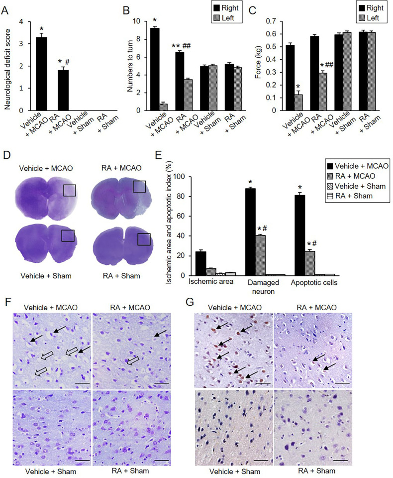
We found that MCAO caused severe neurological behavioral deficits such as instinctive circulation and lack of movement, and retinoic acid treatment alleviated the changes. However, in sham-operated animals, neurobehavioral disorders did not occur regardless of vehicle or retinoic acid treatment. Neurological deficit scores were 3.30 ± 0.15 in vehicle + MCAO animals and 1.80 ± 0.13 in retinoic acid + MCAO animals (Fig 1A). The corner test results showed a preference of response by stimuli. The number of right turns was 9.20 ± 0.20 and 6.50 ± 0.17 in vehicle + MCAO and retinoic acid + MCAO animals, respectively (Fig 1B). The number of left turns was 0.80 ± 0.20 in vehicle + MCAO animals and 3.50 ± 0.17 in retinoic acid + MCAO animals. However, sham-operated animals responded almost identically to the right or left stimuli. Grip strength of the contralateral forelimb was significantly decreased in MCAO animals compared with sham animals. Retinoic acid administration improved MCAO-induced grip loss. In the left forelimb, grip strength was 0.13 ± 0.02 kg in vehicle + MCAO animals and 0.29 ± 0.02 kg in retinoic acid + MCAO animals (Fig 1C). Grip strength of the right forelimb was 0.51 ± 0.02 kg and 0.58 ± 0.02 kg in vehicle + MCAO and retinoic acid + MCAO animals, respectively. In cresyl violet-stained brain tissues, we visually observed the pale areas of the right cerebral cortex of vehicle + MCAO animals, which was the ipsilateral side of MCAO damage (Fig 1D). However, retinoic acid treatment alleviated the area of pale lesions compared with vehicle + MCAO animals. Intact regions were completely stained dark purple, and the ischemic regions were not stained or stained light purple. Extensive ischemic areas were observed in vehicle + MCAO animals, retinoic acid treatment decreased the ischemic region (Fig 1D). Ischemic areas were 24.28 ± 2.19% and 7.24 ± 0.63% in vehicle + MCAO and retinoic acid + MCAO animals, respectively (Fig 1E). The microscopic observation results showed the histopathological changes caused by MCAO damage (Fig 1F). Sham-operated animals had normal neurons that included a pyramidal shape with large and round nuclei. We observed serious histopathological changes including pyknotic nuclei, cytoplasmic vacuolation, and shrunken dendrites in vehicle + MCAO animals. These changes were mitigated in retinoic acid + MCAO animals. We counted the number of damaged cells with abnormal forms and observed the increase in the number of damaged cells in vehicle + MCAO animals. These increases were alleviated in retinoic acid + MCAO animals. The number of damaged cells was 87.74 ± 2.36% in vehicle + MCAO and 40.68 ± 1.34% in retinoic acid + MCAO animals (Fig 1E). TUNEL assay was performed to confirm MCAO-induced apoptosis and TUNEL-positive cells were observed in the cerebral cortex of MCAO animals. The number of TUNEL-positive cells was significantly increased in the vehicle + MCAO animals, retinoic acid treatment attenuated this increase (Fig 1G). The number of TUNEL-positive cells was 81.43 ± 3.15% and 24.60 ± 2.39% in vehicle + MCAO and retinoic acid + MCAO animals, respectively (Fig 1E).

Fig 1. Retinoic acid improves neurobehavioral disorders caused by MCAO damage.

Fig 1

Neurological deficits scoring test (A), corner test (B), grip strength test (C), gross photographs (D), microscopic photographs of cresyl violet staining (F), and TUNEL staining (G) in vehicle + middle cerebral artery occlusion (MCAO), retinoic acid (RA) + MCAO, vehicle + sham, and RA + sham animals. Retinoic acid improves neurological behavior deficits (A-C) and histopathological changes (D, F, G) in ischemic brain injury. Intact area were stained dark purple and ischemic area were not stained or stained light purple (D). Retinoic acid alleviated the increase in ischemic area due to MCAO damage. F represents microscopic photos of the square in photograph D. Arrows indicate damaged neurons with pyknotic nuclei, cytoplasmic vacuolation, and shrunken dendrites (F). Open arrows indicate the TUNEL-positive cells (G). Retinoic acid attenuated the increase in the number of damaged neurons and the number of TUNEL-positive cells caused by MCAO (E and G). Data (neurobehavioral test, n = 10; histopathological test, n = 5) are shown as the mean ± S.E.M. *p < 0.001, **p < 0.01 vs. vehicle + sham animals, #p < 0.001, ##p < 0.01 vs. vehicle + MCAO animals. Scale bar = 100 μm.

Modulation of phospho-Akt and phospho-Bad expression by retinoic acid in MCAO animals

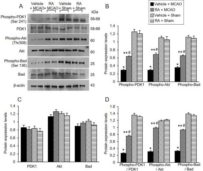
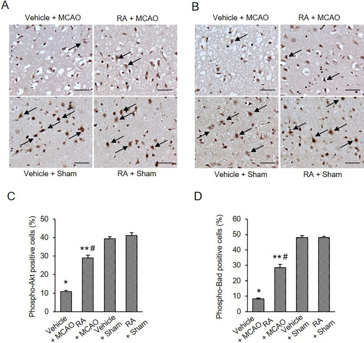
The expression of PDK1, phospho-PDK1, Akt, phospho-Akt, Bad, and phospho-Bad was investigated using Western blot analysis. Phospho-PDK1, phospho-Akt, and phospho-Bad expression was decreased in vehicle animals with MCAO damage, retinoic acid treatment alleviated these decreases (Fig 2A). Phospho-PDK1 levels were 0.29 ± 0.02 in the vehicle + MCAO animals and 0.63 ± 0.03 in the retinoic acid + MCAO animals (Fig 2B). Phospho-Akt levels were 0.29 ± 0.02 and 0.68 ± 0.04 in vehicle + MCAO and retinoic acid + MCAO animals, respectively. Furthermore, phospho-Bad levels were 0.36 ± 0.02 and 0.65 ± 0.02 in vehicle + MCAO and retinoic acid + MCAO animals, respectively (Fig 2B). However, there was no significant difference in PDK1 and Akt expression between vehicle- and retinoic acid-treated animals regardless of MCAO damage. The results of immunohistochemical analysis confirmed the change of phospho-Akt and phospho-Bad expression in all groups (Fig 3). Phospho-Akt and phospho-Bad expression was decreased in vehicle + MCAO animals, retinoic acid treatment mitigated this decrease (Fig 3A and 3B). Positive cells were observed at similar levels between vehicle + sham and retinoic acid + sham animals. The number of phospho-Akt-positive cells was 10.72 ± 0.65% and 28.94 ± 1.58% in vehicle + MCAO and retinoic acid + MCAO animals, respectively (Fig 3C). The number of phospho-Bad-positive cells was 8.27 ± 0.38% in vehicle + MCAO and 28.54 ± 2.17% in retinoic acid + MCAO animals (Fig 3D).

Fig 2. Retinoic acid alleviates decreases of phospho-Akt and phospho-Bad expression caused by MCAO damage.

Fig 2

Western blot analysis of phospho-PDK1, PDK1, phospho-Akt, Akt, phospho-Bad, and Bad in the cerebral cortex from vehicle + middle cerebral artery occlusion (MCAO), retinoic acid (RA) + MCAO, vehicle + sham, and RA + sham animals. Phospho-PDK1, phospho-Akt, phospho-Bad expressions were decreased in vehicle + MCAO animals, retinoic acid treatment alleviated these decreases. Each lane represents an individual experimental animal. Densitometric analysis is represented as a ratio, proteins intensity to β-actin intensity. Molecular weight (kDa) are depicted at right. Data (n = 5) are represented as mean ± S.E.M. *p < 0.001, **p < 0.01 vs. vehicle + sham animals, #p < 0.01 vs. vehicle + MCAO animals.

Fig 3. Retinoic acid attenuates reductions of phospho-Akt and phospho-Bad expression due to MCAO damage.

Fig 3

Immunohistochemical staining of phospho-Akt (A and C) and phospho-Bad (B and D) in cerebral cortex from vehicle + middle cerebral artery occlusion (MCAO), retinoic acid (RA) + MCAO, vehicle + sham, and RA + sham animals. The positive cells of phospho-Akt and phospho-Bad were stained dark brown. Retinoic acid alleviated the decrease of phospho-Akt and phospho-Bad expression due to MCAO damage. The value of positive cells was expressed as a percentage of the number of positive cells to the number of total cells. Arrows indicate positive cells. Data (n = 5) are represented as mean ± S.E.M. *p < 0.001, **p < 0.01 vs. vehicle + sham animals, #p < 0.01 vs. vehicle + MCAO animals. Scale bar = 100 μm.

Regulation of phospho-Bad and 14-3-3 interaction by retinoic acid in MCAO animals

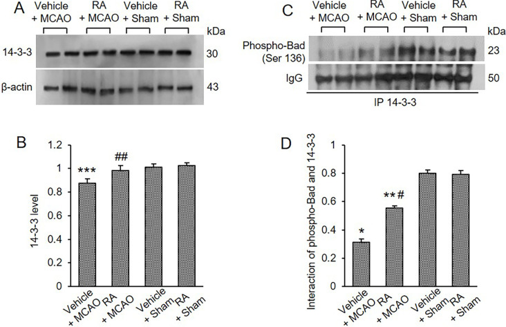
We examined the expression of 14-3-3 in the cerebral cortex of all groups. MCAO damage slightly reduced 14-3-3 expression, and retinoic acid treatment attenuated this reduction (Fig 4A). The 14-3-3 levels were 0.87 ± 0.03 and 0.97 ± 0.04 in vehicle + MCAO and retinoic acid + MCAO animals, respectively (Fig 4B). Immunoprecipitation was performed to determine the interaction between phospho-Bad and 14-3-3 (Fig 4C). Binding levels of these proteins were reduced in vehicle-treated animals with MCAO damage compared with sham animals. These reductions were alleviated in retinoic acid-treated animals with MCAO. Interaction levels were 0.32 ± 0.02 in vehicle + MCAO animals and 0.56 ± 0.02 in retinoic acid + MCAO animals (Fig 4D).

Fig 4. Retinoic acid alleviates decrease of phospho-Bad and 14-3-3 interaction caused by MCAO damage.

Fig 4

Western blot analysis of 14-3-3 (A and B) and immunoprecipitation analysis of phospho-Bad and 14-3-3 binding (C and D) in the cerebral cortex from vehicle + middle cerebral artery occlusion (MCAO), retinoic acid (RA) + MCAO, vehicle + sham, and RA + sham animals. Retinoic acid attenuated the decrease of phospho-Bad and 14-3-3 interaction caused by MCAO damage. Each lane represents an individual experimental animal. Densitometric analysis is represented as a ratio, proteins intensity to β-actin (B) or IgG (D) intensity. Molecular weights (kDa) are depicted at right. Data (n = 5) are represented as mean ± S.E.M. *p < 0.001, **p < 0.01, ***p < 0.05 vs. vehicle + sham animals, #p < 0.01, ##p < 0.05 vs. vehicle + MCAO animals.

Regulation of Bcl-2 family protein interactions by retinoic acid in MCAO animals

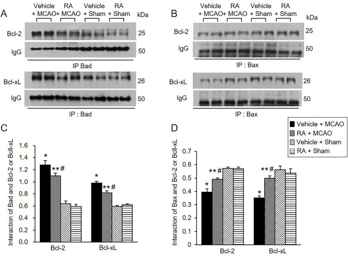
We also observed Bcl-2/Bad and Bcl-xL/Bad binding levels. MCAO damage increased Bcl-2/Bad and Bcl-xL/Bad binding, and retinoic acid treatment attenuated these increases (Fig 5A). Bcl-2/Bad binding levels were 1.27 ± 0.06 in vehicle + MCAO animals and 1.10 ± 0.04 in retinoic acid + MCAO animals (Fig 5C). Furthermore, Bcl-xL/Bad binding levels were 0.97 ± 0.03 and 0.82 ± 0.04 in vehicle + MCAO and retinoic acid + MCAO animals, respectively (Fig 5C). In addition, Bcl-2/Bax and Bcl-xL/Bax binding levels were evaluated and found to be decreased in vehicle-treated animals with MCAO. These decreases were attenuated in retinoic acid-treated animals with MCAO (Fig 5B). Bcl-2/Bax binding levels were 0.39 ± 0.02 in vehicle + MCAO animals and 0.49 ± 0.01 in retinoic acid + MCAO animals (Fig 5D). Bcl-xL/Bax binding levels were 0.35 ± 0.02 and 0.50 ± 0.02 in vehicle + MCAO and retinoic acid + MCAO animals, respectively (Fig 5D).

Fig 5. Retinoic acid regulates Bcl-2 family protein interactions in MCAO animals.

Fig 5

Immunoprecipitation analysis of Bcl-2 and Bcl-xL to Bad (A and C) or Bax (B and D) in the cerebral cortex from vehicle + middle cerebral artery occlusion (MCAO), retinoic acid (RA) + MCAO, vehicle + sham, and RA + sham animals. Retinoic acid alleviated the increase of Bcl-2/Bad and Bcl-xL/Bad binding and the decrease of Bcl/Bax and Bcl-xL and Bcl-xL/Bax caused by MCAO damage. Each lane represents an individual experimental animal. Densitometric analysis is represented as a ratio of proteins intensity to IgG intensity. Molecular weights (kDa) are depicted at right. Data (n = 5) are represented as mean ± S.E.M. *p < 0.001, **p < 0.01 vs. vehicle + sham animals, #p < 0.01 vs. vehicle + MCAO animals.

Modulation of caspase-3 expression by retinoic acid in MCAO animals

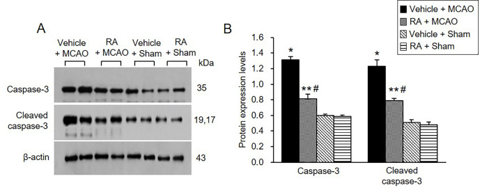
We also confirmed the expression of caspase-3 protein, a representative marker of apoptosis. Retinoic acid alleviated the increase of caspase-3 and cleaved caspase-3 expression caused by MCAO (Fig 6A). Caspase-3 levels were 1.31 ± 0.03 in vehicle + MCAO animals and 0.81 ± 0.06 in retinoic acid + MCAO animals (Fig 6B). Cleaved-caspase-3 levels were 1.23 ± 0.07 and 0.79 ± 0.03 in vehicle + MCAO and retinoic acid + MCAO animals, respectively (Fig 6B).

Fig 6. Retinoic acid attenuates increases of caspase-3 and cleaved caspase-3 expression caused by MCAO damage.

Fig 6

Western blot analysis of caspase-3 and cleaved caspase-3 in the cerebral cortex from vehicle + middle cerebral artery occlusion (MCAO), retinoic acid (RA) + MCAO, vehicle + sham, and RA + sham animals. Retinoic acid alleviated the increase of these proteins due to MCAO. Each lane represents an individual experimental animal. Densitometric analysis is represented as a ratio of proteins intensity to β-actin intensity. Molecular weights (kDa) are depicted at right. Data (n = 5) are represented as mean ± S.E.M. *p < 0.001, **p < 0.01 vs. vehicle + sham animals, #p < 0.01 vs. vehicle + MCAO animals.

Discussion

This study supported the neuroprotective effects of retinoic acid through various neurological behavior tests including neurological deficits scoring test, corner test, and grip strength test. Surgical MCAO was performed in the right brain of rats. A reduction of grip strength was observed in the left forelimb. These results showed neurological behavioral deficits in the contralateral side of the damaged brain. Retinoic acid mitigated the neurological disorder caused by MCAO and exerted neuroprotective effects. Furthermore, the morphological changes were confirmed in cresyl violet-stained tissues. Retinoic acid treatment decreased infarct areas caused by MCAO damage and reduced the number of damaged cells. We recently reported that retinoic acid alleviates the increases in brain edema and infarction volume due to MCAO damage [41]. The present study also confirmed that retinoic acid attenuates the histopathological changes and increase in the number of TUNEL-positive cells in the cerebral cortex with MCAO. Stroke is a complex disease involving a variety of mechanisms. The PI3K-Akt pathway is a representative pathway for neuronal survival and protection. PI3K-Akt is a key mediator of cerebral ischemic stroke [37]. The present study focused on the neuroprotective effects of retinoic acid by modulating the PI3K-Akt signaling pathway. This study additionally elucidated that retinoic acid performs neuroprotective functions through activation of the PI3K-Akt-Bad signaling pathway and regulation of Bcl-2 family protein interactions in a stroke animal model.

The PI3K-Akt signaling pathway regulates the survival and apoptosis of neurons [46, 47]. Activated Akt phosphorylates Bad and prevents the apoptotic function of Bad, leading to survival of neurons [48, 49]. However, in hypoxia and ischemia states, inactivated Akt dephosphorylates Bad and induces apoptotic cell death [50]. We previously showed that the PI3K/Akt signaling pathway was associated with neuroprotective mechanisms of various substances in ischemic stroke models [5153]. We also reported that these substances exert neuroprotective effects by regulating the expression of Akt downstream targets such as Bad, FKHR, and GSK-3β [54]. Therefore, we postulate that activation of Akt plays an important role in protecting neurons from stroke damage. Furthermore, retinoic acid activates the Akt pathway and protects cells against proteasome inhibition-associated cell death [33]. In the present study, retinoic acid alleviated the decrease in phospho-Akt as well as the reduction in phospho-PDK1 and phospho-Bad expression due to MCAO damage. PDK1 and Bad are upstream and downstream target proteins of Akt, respectively. PDK1 and Akt expression was maintained constant regardless of MCAO damage. Thus, the results provide evidence that phosphorylation of these proteins is important for performing neuroprotective effects against ischemic damage rather than changes in the total protein expression levels. In addition, immunohistochemical staining confirmed the changes in phospho-Akt and phospho-Bad expression were confirmed based on. The results were consistent with the results of Western blot analysis. MCAO decreased the number of positive cells in phospho-Akt and phospho-Bad, and retinoic acid attenuated this decrease. Bad is a representative Akt downstream protein and a proapoptotic protein. Previous studies have demonstrated the efficacy of retinoic acid in various diseases [55, 56]. Especially in the brain, retinoic acid has neuroprotective effects in neurodegenerative diseases such as amyotrophic lateral sclerosis, Parkinson’s and ischemic stroke. In this study, we focused on the specific protective mechanism of retinoic acid on cerebral ischemia [5759]. The PI3K-Akt pathway has shown therapeutic and preventive effects in various neurological disorders including stroke, Alzheimer’s disease, and Parkinson’s disease [60, 61]. Therefore, it is hypothesized that the regulation of the PI3K-Akt pathway by retinoic acid treatment may also be involved in the prevention and treatment of stroke. We previously showed a decrease in phospho-Bad due to MCAO damage and further described the mitigation of phospho-Bad decrease caused by retinoic acid. The changes in phospho-Bad expression due to retinoic acid treatment in MCAO indicate the importance of phospho-Bad in neuroprotective effects of retinoic acid. The maintenance of phospho-Akt and phospho-Bad plays a crucial role in cell survival. Thus, the results show that the PI3K/Akt signaling pathway contributes to the neuroprotective mechanism of retinoic acid in cerebral ischemia.

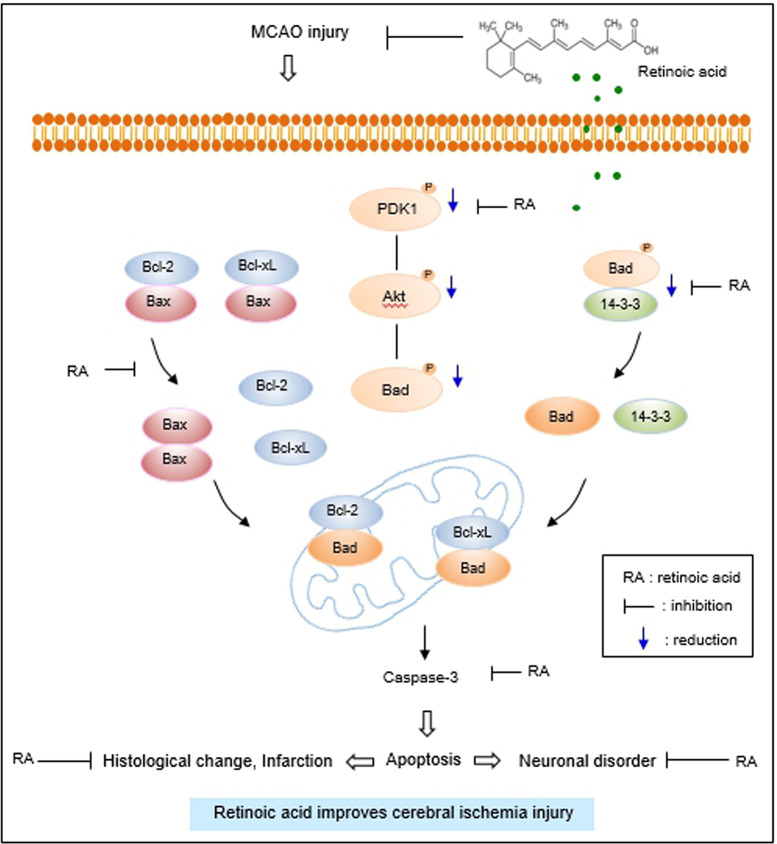
The interaction between phospho-Bad and 14-3-3 is important for cell survival. The binding of these proteins inhibits interaction with Bad and Bcl-xL or Bcl-2, induces Bcl-2/Bax or Bcl-xL/Bax complexes, inactivates pro-apoptotic function of Bax, and continuously prevents caspase cascade activation and apoptosis [20, 21, 24, 62]. The results of immunoprecipitation analysis showed a decrease in phospho-Bad and 14-3-3 binding in cerebral cortex with MCAO damage, which was attenuated in the presence of retinoic acid. The findings demonstrate that retinoic acid regulates phospho-Bad and 14-3-3 binding in rats with MCAO damage. Thus, the results provide evidence that retinoic acid regulates phospho-Bad and 14-3-3 binding and preserves neurons from ischemic damage. The binding of Bcl-2 and Bcl-xL to Bad or Bax in the cerebral cortex of MCAO animals was investigated. Bcl-2/Bad and Bcl-xL/Bad binding increased in MCAO-damaged rats, and retinoic acid prevented this increase. However, Bcl-2/Bax and Bcl-xL/Bax binding in rats with MCAO damage was reduced, and retinoic acid attenuated this decrease. Phosphorylated Bad prevented Bcl-2/Bad and Bcl-xL/Bad binding and inhibited the pro-apoptotic function of Bad. In addition, phosphorylated Bad induced Bcl-2/Bax and Bcl-xL/Bax binding and prevented pro-apoptotic activity of Bax. Our findings showed that retinoic acid alleviates the increase of Bcl-2/Bad and Bcl-xL/Bad binding and the decrease of Bcl-2/Bax and Bcl-xL/Bax binding due to MCAO damage. Retinoic acid regulates the phosphorylation of Bad and continuously modulates the interaction of Bcl-2 family proteins, eventually inhibiting apoptosis. We previously reported a decrease in Bcl-2 and an increase in Bax in MCAO damage, and retinoic acid attenuated those changes [41]. Furthermore, retinoic acid alleviated the increase in Bcl-2/Bax ratio caused by MCAO damage. The results show that retinoic acid exerts neuroprotective effects by regulating Bcl-2 family protein expression. In addition, we observed change in caspase-3 and cleaved caspase-3 expression, indicators of apoptosis. Retinoic acid significantly attenuated increases of caspase-3 and cleaved caspase-3 expression caused by MCAO. Our previous study showed that retinoic acid alleviates the increase of caspase-9, cleaved caspase-9, PARP, and cleaved-PARP in MCAO animals [41]. These results provide additional information confirming the current findings that retinoic acid prevents apoptosis from cerebral ischemic damage and protects brain tissue. Retinoic acid mitigated the decrease in phospho-Bad expression. It also alleviated the increase in Bcl-2/Bad and Bcl-xL/Bad binding and the decrease of Bcl-2/Bax and Bcl-xL/Bax in MCAO animals. The results indicate that retinoic acid inhibits the pro-apoptotic function of Bad and Bax. Finally, we confirmed that retinoic acid prevents the increase in caspase-3 and cleaved caspase-3 activity, thereby inhibiting apoptosis and protecting neurons from ischemic damage. The neuroprotective effect of retinoic acid on MCAO damage is due to Akt signaling pathway regulation and Bcl-2 family protein regulation (Fig 7). Retinoic acid exerts neuroprotective effects through various protective mechanisms during brain injury. However, it is difficult to discuss all protective mechanism of retinoic acid in this study. We focused on the regulatory mechanism of Akt and its downstream target Bad by retinoic acid in cerebral ischemia. We clearly showed that retinoic acid is involved in neuroprotection through the regulation of Akt and Bad in ischemic brain injury. We think that various protective mechanisms of retinoic acid including upstream and downstream target of Akt should be researched in the future. We also need to find more factors that are regulated by retinoic acid in the ischemic stroke. Further studies should be performed that provide therapeutic effects of retinoic acid in stoke model.

Fig 7. The neuroprotective mechanism of retinoic acid against MCAO damage.

Fig 7

Conclusions

Taken together, our results demonstrated that retinoic acid treatment induces the phosphorylation of Akt and Bad, regulates the interaction with Bcl-2 family proteins, and modulates the expression of caspase-3 in stroke animal models (S4S11 Files). Retinoic acid inhibits the process of apoptosis, promotes neuronal survival, and ultimately exerts neuroprotective effects in ischemic stroke (S1S3 Files). In conclusion, retinoic acid provides neuroprotective effects through mitigation of Akt and Bad phosphorylation and modulation of Bcl-2 family protein interactions in ischemic stroke.

Supporting information

S1 File. Gross photographs of cresyl violet staining.

This is full images of Fig 1D.

(PDF)

pone.0303213.s001.pdf (183.1KB, pdf)
S2 File. Microscopic photographs of cresyl violet staining.

Full images of Fig 1F.

(PDF)

pone.0303213.s002.pdf (450.1KB, pdf)
S3 File. TUNEL staining.

Full images of Fig 1G.

(PDF)

pone.0303213.s003.pdf (480KB, pdf)
S4 File. Western blot analysis of phospho-PDK1, PDK1, phospho-Akt, Akt, phospho-Bad, and Bad in the cerebral cortex.

Full images of Fig 2A.

(PDF)

pone.0303213.s004.pdf (184.7KB, pdf)
S5 File. Immunohistochemical staining of phospho-Akt.

Full images of Fig 3A.

(PDF)

pone.0303213.s005.pdf (567.3KB, pdf)
S6 File. Immunohistochemical staining of phospho-Bad.

Full images of Fig 3B.

(PDF)

pone.0303213.s006.pdf (567.7KB, pdf)
S7 File. Western blot analysis of 14-3-3.

Full images of Fig 4A.

(PDF)

pone.0303213.s007.pdf (129.3KB, pdf)
S8 File. Immunoprecipitation analysis of phospho-Bad and 14-3-3 binding.

Full images of Fig 4C.

(PDF)

pone.0303213.s008.pdf (97.4KB, pdf)
S9 File. Immunoprecipitation analysis of Bcl-2 and Bcl-xL to Bad.

Full images of Fig 5A.

(PDF)

pone.0303213.s009.pdf (135.3KB, pdf)
S10 File. Immunoprecipitation analysis of Bcl-2 and Bcl-xL to Bax.

Full images of Fig 5B.

(PDF)

pone.0303213.s010.pdf (144.3KB, pdf)
S11 File. Western blot analysis of caspase-3 and cleaved caspase-3.

Full images of Fig 6A.

(PDF)

pone.0303213.s011.pdf (120.6KB, pdf)

Data Availability

All relevant data are within the paper and its Supporting Information files.

Funding Statement

This research was supported by the National Research Foundation of Korea (NRF) grant funded by the Korea government [MEST][NRF-2021R1F1A105878711] and (MSIT) [RS-2023-00248145]. The funders had no role in study design, data collection and analysis, decision to publish, or preparation of the manuscript

References

  • 1.Kuriakose D, Xiao Z. Pathophysiology and treatment of stroke: present status and future perspectives. Int J Mol Sci. 2020;21: 7609. doi: 10.3390/ijms21207609 [DOI] [PMC free article] [PubMed] [Google Scholar]
  • 2.He Q, Ma Y, Liu J, Zhang D, Ren J, Zhao R, et al. Biological functions and regulatory mechanisms of hypoxia-inducible factor-1α in ischemic stroke. Front Immunol. 2021;12: 801985. [DOI] [PMC free article] [PubMed] [Google Scholar]
  • 3.Schädlich IS, Winzer R, Stabernack J, Tolosa E, Magnus T, Rissiek B. The role of the ATP-adenosine axis in ischemic stroke. Semin Immunopathol. 2023;45: 347–365. doi: 10.1007/s00281-023-00987-3 [DOI] [PMC free article] [PubMed] [Google Scholar]
  • 4.Simats A, Liesz A. Systemic inflammation after stroke: implications for post-stroke comorbidities. EMBO Mol Med. 2022;14: e16269. doi: 10.15252/emmm.202216269 [DOI] [PMC free article] [PubMed] [Google Scholar]
  • 5.García-Regalado A, Vargas M, García-Carrancá A, Aréchaga-Ocampo E, González-De la Rosa CH. Activation of Akt pathway by transcription-independent mechanisms of retinoic acid promotes survival and invasion in lung cancer cells. Mol Cancer. 2013;12: 44. doi: 10.1186/1476-4598-12-44 [DOI] [PMC free article] [PubMed] [Google Scholar]
  • 6.Dolin HH, Franco JH, Chen X, Pan ZK. Retinoic acid-induced regulation of inflammatory pathways is a potential sepsis treatment. Infect Immun. 2023;91: e0045722. doi: 10.1128/iai.00457-22 [DOI] [PMC free article] [PubMed] [Google Scholar]
  • 7.Keino H, Watanabe T, Sato Y, Okada AA. Anti-inflammatory effect of retinoic acid on experimental autoimmune uveoretinitis. Br J Ophthalmol. 2010;94: 802–807. doi: 10.1136/bjo.2009.171314 [DOI] [PubMed] [Google Scholar]
  • 8.Tan BT, Wang L, Li S, Long ZY, Wu YM, Liu Y. Retinoic acid induced the differentiation of neural stem cells from embryonic spinal cord into functional neurons in vitro. Int J Clin Exp Pathol. 2015;8: 8129–8135. [PMC free article] [PubMed] [Google Scholar]
  • 9.Lane MA. Bailey SJ. Role of retinoid signaling in the adult brain. Prog Neurobiol. 2005;75: 275–293. [DOI] [PubMed] [Google Scholar]
  • 10.Das BC, Dasgupta S, Ray SK. Potential therapeutic roles of retinoids for prevention of neuroinflammation and neurodegeneration in Alzheimer’s disease. Neural Regen Res. 2019;4: 1880–1892. doi: 10.4103/1673-5374.259604 [DOI] [PMC free article] [PubMed] [Google Scholar]
  • 11.Jiang W, Guo M, Gong M, Chen L, Bi Y, Zhang Y, et al. Vitamin A bio-modulates apoptosis via the mitochondrial pathway after hypoxic-ischemic brain damage. Mol Brain. 2018;11: 14. doi: 10.1186/s13041-018-0360-0 [DOI] [PMC free article] [PubMed] [Google Scholar]
  • 12.Ecker V, Stumpf M, Brandmeier L, Neumayer T, Pfeuffer L, Engleitner T, et al. Targeted PI3K/AKT-hyperactivation induces cell death in chronic lymphocytic leukemia. Nat Commun. 2021;12: 3526. doi: 10.1038/s41467-021-23752-2 [DOI] [PMC free article] [PubMed] [Google Scholar]
  • 13.Huang X, Liu G, Guo J, Su Z. The PI3K/AKT pathway in obesity and type 2 diabetes. Int J Biol Sci. 2018;14: 1483–1496. doi: 10.7150/ijbs.27173 [DOI] [PMC free article] [PubMed] [Google Scholar]
  • 14.Miao R, Fang X, Wei J, Wu H, Wang X, Tian J. Akt: a potential drug target for metabolic syndrome. Front Physiol. 2022;13: 822333. doi: 10.3389/fphys.2022.822333 [DOI] [PMC free article] [PubMed] [Google Scholar]
  • 15.Levina A, Fleming KD, Burke JE, Leonard TA. Activation of the essential kinase PDK1 by phosphoinositide-driven trans-autophosphorylation. Nat Commun. 2022;13: 1874. doi: 10.1038/s41467-022-29368-4 [DOI] [PMC free article] [PubMed] [Google Scholar]
  • 16.Brunet A, Bonni A, Zigmond MJ, Lin MZ, Juo P, Hu LS, et al. Akt promotes cell survival by phosphorylating and inhibiting a Forkhead transcription factor. Cell. 1999;96: 857–868. doi: 10.1016/s0092-8674(00)80595-4 [DOI] [PubMed] [Google Scholar]
  • 17.Endo H, Nito C, Kamada H, Nishi T, Chan PH. Activation of the Akt/GSK3beta signaling pathway mediates survival of vulnerable hippocampal neurons after transient global cerebral ischemia in rats. J Cereb Blood Flow Metab. 2006;26: 1479–1489. doi: 10.1038/sj.jcbfm.9600303 [DOI] [PubMed] [Google Scholar]
  • 18.Bae J, Hsu SY, Leo CP, Zell K, Hsueh AJ. Underphosphorylated BAD interacts with diverse antiapoptotic Bcl-2 family proteins to regulate apoptosis. Apoptosis. 2001;6: 319–330. doi: 10.1023/a:1011319901057 [DOI] [PubMed] [Google Scholar]
  • 19.Datta SR, Katsov A, Hu L, Petros A, Fesik SW, Yaffe MB, et al. 14-3-3 proteins and survival kinases cooperate to inactivate BAD by BH3 domain phosphorylation. Molecular. Cell. 2000;6: 41–51. [PubMed] [Google Scholar]
  • 20.Downward J. How BAD phosphorylation is good for survival. Nat Cell Biol. 1999;1: 33–35. [DOI] [PubMed] [Google Scholar]
  • 21.Masters SC, Fu H. 14-3-3 proteins mediate an essential anti-apoptotic signal. J Biol Chem. 2001;276: 45193–45200. doi: 10.1074/jbc.M105971200 [DOI] [PubMed] [Google Scholar]
  • 22.Garenne D, Renault TT, Manon S. Bax mitochondrial relocation is linked to its phosphorylation and its interaction with Bcl-xL. Microb Cell. 2016;3: 597–605. doi: 10.15698/mic2016.12.547 [DOI] [PMC free article] [PubMed] [Google Scholar]
  • 23.Garrido C, Galluzzi L, Brunet M, Puig PE, Didelot C, Kroemer G. Mechanisms of cytochrome c release from mitochondria. Cell Death Differ. 2006;13: 1423–33. doi: 10.1038/sj.cdd.4401950 [DOI] [PubMed] [Google Scholar]
  • 24.Masters SC, Yang H, Datta SR, Greenberg ME, Fu H. 14-3-3 inhibits Bad-induced cell death through interaction with serine-136. Mol Pharmacol. 2001;60: 1325–1331. doi: 10.1124/mol.60.6.1325 [DOI] [PubMed] [Google Scholar]
  • 25.Jo H, Mondal S, Tan D, Nagata E, Takizawa S, Sharma AK, et al. Small molecule-induced cytosolic activation of protein kinase Akt rescues ischemia-elicited neuronal death. Proc Natl Acad Sci USA. 2012;109: 10581–10586. doi: 10.1073/pnas.1202810109 [DOI] [PMC free article] [PubMed] [Google Scholar]
  • 26.Zhang S, Taghibiglou C, Girling K, Dong Z, Lin SZ, Lee W, et al. Critical role of increased PTEN nuclear translocation in excitotoxic and ischemic neuronal injuries. J Neurosci. 2013;33: 7997–8008. doi: 10.1523/JNEUROSCI.5661-12.2013 [DOI] [PMC free article] [PubMed] [Google Scholar]
  • 27.Shamloo M, Soriano L, Wieloch T, Nikolich K, Urfer R, Oksenberg D. Death-associated protein kinase is activated by dephosphorylation in response to cerebral ischemia. J Biol Chem. 2005;280: 42290–4229. doi: 10.1074/jbc.M505804200 [DOI] [PubMed] [Google Scholar]
  • 28.Guo S, Miyake M, Liu KJ, Shi H. Specific inhibition of hypoxia inducible factor 1 exaggerates cell injury induced by in vitro ischemia through deteriorating cellular redox environment. J Neurochem. 209;108: 1309–1321. doi: 10.1111/j.1471-4159.2009.05877.x [DOI] [PMC free article] [PubMed] [Google Scholar]
  • 29.Dinkova-Kostova AT, Abramov AY. The emerging role of Nrf2 in mitochondrial function. Free Radic Biol Med. 2015;88: 179–188. doi: 10.1016/j.freeradbiomed.2015.04.036 [DOI] [PMC free article] [PubMed] [Google Scholar]
  • 30.Bastian C, Quinn J, Tripathi A, Aquila D, McCray A, Dutta R, et al. CK2 inhibition confers functional protection to young and aging axons against ischemia by differentially regulating the CDK5 and AKT signaling pathways. Neurobiol Dis. 2019;126: 47–61. doi: 10.1016/j.nbd.2018.05.011 [DOI] [PMC free article] [PubMed] [Google Scholar]
  • 31.Perez-Alvarez MJ, Villa Gonzalez M, Benito-Cuesta I, Wandosell FG. Role of mTORC1 controlling proteostasis after brain ischemia. Front Neurosci. 2018;12: 60. doi: 10.3389/fnins.2018.00060 [DOI] [PMC free article] [PubMed] [Google Scholar]
  • 32.Leker RR, Aharonowiz M, Greig NH, Ovadia H. The role of p53-induced apoptosis in cerebral ischemia: effects of the p53 inhibitor pifithrin alpha. Exp Neurol. 2004;187: 478–486. doi: 10.1016/j.expneurol.2004.01.030 [DOI] [PubMed] [Google Scholar]
  • 33.Cheng B, Martinez AA, Morado J, Scofield V, Roberts JL, Maffi SK. Retinoic acid protects against proteasome inhibition associated cell death in SH-SY5Y cells via the AKT pathway. Neurochem Int. 2013;62: 31–42. doi: 10.1016/j.neuint.2012.10.014 [DOI] [PubMed] [Google Scholar]
  • 34.Zhang S, Chen X, Hu Y, Wu J, Cao Q, Chen S, et al. All-trans retinoic acid modulates Wnt3A-induced osteogenic differentiation of mesenchymal stem cells via activating the PI3K/AKT/GSK3β signalling pathway. Mol Cell Endocrino. 2016;1422: 243–253. [DOI] [PubMed] [Google Scholar]
  • 35.Nagl F, Schönhofer K, Seidler B, Mages J, Allescher HD, Schmid RM, et al. Retinoic acid-induced nNOS expression depends on a novel PI3K/Akt/DAX1 pathway in human TGW-nu-I neuroblastoma cells. Am J Physiol Cell Physiol. 2009;297: C1146–56. doi: 10.1152/ajpcell.00034.2009 [DOI] [PubMed] [Google Scholar]
  • 36.Wang MM, Zhang M, Feng YS, Xing Y, Tan ZX, Li WB, et al. Electroacupuncture inhibits neuronal autophagy and apoptosis via the PI3K/AKT pathway following ischemic stroke. Front Cell Neurosci. 2020;15: 14:134. doi: 10.3389/fncel.2020.00134 [DOI] [PMC free article] [PubMed] [Google Scholar]
  • 37.Gu C, Zhang Q, Li Y, Li R, Feng J, Chen W, et al. The PI3K/AKT pathway-the potential key mechanisms of traditional chinese medicine for stroke. Front Med. 2022;31: 9:900809. doi: 10.3389/fmed.2022.900809 [DOI] [PMC free article] [PubMed] [Google Scholar]
  • 38.Kong L, Wang Y, Wang XJ, Wang XT, Zhao Y, Wang LM, et al. Retinoic acid ameliorates blood-brain barrier disruption following ischemic stroke in rats. Pharmacol Res. 2015;99: 125–136. 293. doi: 10.1016/j.phrs.2015.05.014 [DOI] [PubMed] [Google Scholar]
  • 39.Kang JB, Park DJ, Shah MA, Koh PO. Retinoic acid exerts neuroprotective effects against focal cerebral ischemia by preventing apoptotic cell death. Neurosci Lett. 2021;757: 135979 doi: 10.1016/j.neulet.2021.135979 [DOI] [PubMed] [Google Scholar]
  • 40.Longa EZ, Weinstein PR, Carlson S, Cummins R. Reversible middle cerebral artery occlusion without craniectomy in rats. Stroke. 1989;20: 84–91. doi: 10.1161/01.str.20.1.84 [DOI] [PubMed] [Google Scholar]
  • 41.Kang JB, Shah MA, Park DJ, Koh PO. Retinoic acid regulates the ubiquitin-proteasome system in a middle cerebral artery occlusion animal model. Lab Anim Res. 2022;38: 13. doi: 10.1186/s42826-022-00123-6 [DOI] [PMC free article] [PubMed] [Google Scholar]
  • 42.Ruan J, Yao Y. Behavioral tests in rodent models of stroke. Brain Hemorrhages. 2020;1: 171–184. doi: 10.1016/j.hest.2020.09.001 [DOI] [PMC free article] [PubMed] [Google Scholar]
  • 43.Hattori K, Lee H, Hurn PD, Crain BJ, Traystman RJ, DeVries AC. Cognitive deficits after focal cerebral ischemia in mice. Stroke. 2000;31: 1939–1944. doi: 10.1161/01.str.31.8.1939 [DOI] [PubMed] [Google Scholar]
  • 44.Takeshita H, Yamamoto K, Nozato S, Inagaki T, Tsuchimochi H, Shirai M, et al. Modified forelimb grip strength test detects aging-associated physiological decline in skeletal muscle function in male mice. Sci Rep. 2017;7: 42323. doi: 10.1038/srep42323 [DOI] [PMC free article] [PubMed] [Google Scholar]
  • 45.Paxinos G, Watson C, The rat brain in stereotaxic coordinates, seventh ed., Press Academic, Diego San, 2005. [Google Scholar]
  • 46.Gong Z, Lao D, Wu Y, Li T, Lv S, Mo X, et al. Inhibiting PI3K/Akt-signaling pathway improves neurobehavior changes in anti-NMDAR encephalitis mice by ameliorating blood-brain barrier disruption and neuronal damage. Cell Mol Neurobiol. 2023;43: 3623–3637. doi: 10.1007/s10571-023-01371-3 [DOI] [PMC free article] [PubMed] [Google Scholar]
  • 47.Zhang L, Zhang ZG, Liu XS, Hozeska-Solgot A, Chopp M. The PI3K/Akt pathway mediates the neuroprotective effect of atorvastatin in extending thrombolytic therapy after embolic stroke in the rat. Arterioscler Thromb Vasc Biol. 2007;27: 2470–2475. doi: 10.1161/ATVBAHA.107.150748 [DOI] [PubMed] [Google Scholar]
  • 48.Ahn JY. Neuroprotection signaling of nuclear akt in neuronal cells. Exp Neurobiol. 2004;23: 200–206. [DOI] [PMC free article] [PubMed] [Google Scholar]
  • 49.Heras-Sandoval D, Pérez-Rojas JM, Hernández-Damián J, Pedraza-Chaverri J. The role of PI3K/AKT/mTOR pathway in the modulation of autophagy and the clearance of protein aggregates in neurodegeneration. Cell Signal. 2014;26: 2694–2701. doi: 10.1016/j.cellsig.2014.08.019 [DOI] [PubMed] [Google Scholar]
  • 50.Zhao H, Sapolsky RM, Steinberg GK. Phosphoinositide-3-kinase/akt survival signal pathways are implicated in neuronal survival after stroke. Mol Neurobiol. 2006;34: 249–270. doi: 10.1385/MN:34:3:249 [DOI] [PubMed] [Google Scholar]
  • 51.Cho JH, Sung JH, Cho EH, Won CK, Lee HJ, Kim MO, et al. Gingko biloba Extract (EGb 761) prevents ischemic brain injury by activation of the Akt signaling pathway. Am J Chin Med. 2009;37: 547–555. doi: 10.1142/S0192415X09007041 [DOI] [PubMed] [Google Scholar]
  • 52.Koh PO. Melatonin prevents hepatic injury-induced decrease in Akt downstream targets phosphorylations. J Pineal Res. 2011;51: 214–219. doi: 10.1111/j.1600-079X.2011.00879.x [DOI] [PubMed] [Google Scholar]
  • 53.Koh PO. Ferulic acid prevents the cerebral ischemic injury-induced decrease of Akt and Bad phosphorylation. Neurosci Lett. 2012;507: 156–160. doi: 10.1016/j.neulet.2011.12.012 [DOI] [PubMed] [Google Scholar]
  • 54.Koh PO, Cho JH, Won CK, Lee HJ, Sung JH, Kim MO. Estradiol attenuates the focal cerebral ischemic injury through mTOR/p70S6 kinase signaling pathway. Neurosci Lett. 2008;436: 62–66. doi: 10.1016/j.neulet.2008.02.061 [DOI] [PubMed] [Google Scholar]
  • 55.Caricasulo MA, Zanetti A, Terao M, Garattini E, Paroni G. Cellular and micro-environmental responses influencing the antitumor activity of all-trans retinoic acid in breast cancer. Cell Commun Signal. 2024;22: 127. doi: 10.1186/s12964-024-01492-2 [DOI] [PMC free article] [PubMed] [Google Scholar]
  • 56.Al-Marsoummi S, Mehus AA, Garrett SH, Sens DA, Somji S. The Effect of Retinoic Acid on Arsenite-Transformed Malignant UROtsa Bladder Cancer Cells: In Vitro Model of Basal Muscle-Invasive Bladder Cancer. Cancers (Basel). 2024;16: 1178. doi: 10.3390/cancers16061178 [DOI] [PMC free article] [PubMed] [Google Scholar]
  • 57.Zhu Y, Liu Y, Yang F, Chen W, Jiang J, He P, et al. All-trans retinoic acid exerts neuroprotective effects in amyotrophic lateral sclerosis-like Tg (SOD1*G93A)1Gur mice. Mol Neurobiol. 2020;57(8):3603–3615. doi: 10.1007/s12035-020-01973-8 [DOI] [PubMed] [Google Scholar]
  • 58.Esteves M, Cristóvão AC, Saraiva T, Rocha SM, Baltazar G, Ferreira L, et al. Retinoic acid-loaded polymeric nanoparticles induce neuroprotection in a mouse model for Parkinson’s disease. Front Aging Neurosci. 2015;7:20. doi: 10.3389/fnagi.2015.00020 [DOI] [PMC free article] [PubMed] [Google Scholar]
  • 59.Cai W, Wang J, Hu M, Chen X, Lu Z, Bellanti JA, et al. All trans-retinoic acid protects against acute ischemic stroke by modulating neutrophil functions through STAT1 signaling. J Neuroinflammation. 2019;16(1):175. doi: 10.1186/s12974-019-1557-6 [DOI] [PMC free article] [PubMed] [Google Scholar]
  • 60.Long HZ, Cheng Y, Zhou ZW, Luo HY, Wen DD, Gao LC. PI3K/AKT signal pathway: A target of natural products in the prevention and treatment of Alzheimer’s disease and Parkinson’s disease. Front Pharmacol. 2021;12: 648636. doi: 10.3389/fphar.2021.648636 [DOI] [PMC free article] [PubMed] [Google Scholar]
  • 61.Goyal A, Agrawal A, Verma A, Dubey N. The PI3K-AKT pathway: A plausible therapeutic target in Parkinson’s disease. Exp Mol Pathol. 2023;129: 104846. doi: 10.1016/j.yexmp.2022.104846 [DOI] [PubMed] [Google Scholar]
  • 62.Nomura M, Shimizu S, Sugiyama T, Narita M, Ito T, Matsuda H, et al. 14-3-3 interacts directly with and negatively regulates pro-apoptotic Bax. J Biol Chem. 2003;278: 2058–2065. doi: 10.1074/jbc.M207880200 [DOI] [PubMed] [Google Scholar]

Decision Letter 0

Ahmed E Abdel Moneim

28 Nov 2023

PONE-D-23-19266Retinoic acid alleviates the reduction of Akt and Bad phosphorylation and regulates Bcl-2 family protein interactions in animal models of ischemic strokePLOS ONE

Dear Dr. Koh,

Thank you for submitting your manuscript to PLOS ONE. After careful consideration, we feel that it has merit but does not fully meet PLOS ONE’s publication criteria as it currently stands. Therefore, we invite you to submit a revised version of the manuscript that addresses the points raised during the review process.

Please submit your revised manuscript by Jan 12 2024 11:59PM. If you will need more time than this to complete your revisions, please reply to this message or contact the journal office at plosone@plos.org. When you're ready to submit your revision, log on to https://www.editorialmanager.com/pone/ and select the 'Submissions Needing Revision' folder to locate your manuscript file.

Please include the following items when submitting your revised manuscript:

  • A rebuttal letter that responds to each point raised by the academic editor and reviewer(s). You should upload this letter as a separate file labeled 'Response to Reviewers'.

  • A marked-up copy of your manuscript that highlights changes made to the original version. You should upload this as a separate file labeled 'Revised Manuscript with Track Changes'.

  • An unmarked version of your revised paper without tracked changes. You should upload this as a separate file labeled 'Manuscript'.

If you would like to make changes to your financial disclosure, please include your updated statement in your cover letter. Guidelines for resubmitting your figure files are available below the reviewer comments at the end of this letter.

If applicable, we recommend that you deposit your laboratory protocols in protocols.io to enhance the reproducibility of your results. Protocols.io assigns your protocol its own identifier (DOI) so that it can be cited independently in the future. For instructions see: https://journals.plos.org/plosone/s/submission-guidelines#loc-laboratory-protocols. Additionally, PLOS ONE offers an option for publishing peer-reviewed Lab Protocol articles, which describe protocols hosted on protocols.io. Read more information on sharing protocols at https://plos.org/protocols?utm_medium=editorial-email&utm_source=authorletters&utm_campaign=protocols.

We look forward to receiving your revised manuscript.

Kind regards,

Ahmed E. Abdel Moneim

Academic Editor

PLOS ONE

Journal Requirements:

When submitting your revision, we need you to address these additional requirements.

1. Please ensure that your manuscript meets PLOS ONE's style requirements, including those for file naming. The PLOS ONE style templates can be found at 

https://journals.plos.org/plosone/s/file?id=wjVg/PLOSOne_formatting_sample_main_body.pdf and 

https://journals.plos.org/plosone/s/file?id=ba62/PLOSOne_formatting_sample_title_authors_affiliations.pdf

2. To comply with PLOS ONE submissions requirements, in your Methods section, please provide additional information regarding the experiments involving animals and ensure you have included details on (1) methods of sacrifice, (2) methods of anesthesia and/or analgesia, and (3) efforts to alleviate suffering.

3. Thank you for stating the following financial disclosure: 

"The funders had no role in study design, data collection and analysis, decision to publish, or preparation of the manuscript"

At this time, please address the following queries:

a) Please clarify the sources of funding (financial or material support) for your study. List the grants or organizations that supported your study, including funding received from your institution. 

b) State what role the funders took in the study. If the funders had no role in your study, please state: “The funders had no role in study design, data collection and analysis, decision to publish, or preparation of the manuscript.”

c) If any authors received a salary from any of your funders, please state which authors and which funders.

d) If you did not receive any funding for this study, please state: “The authors received no specific funding for this work.”

Please include your amended statements within your cover letter; we will change the online submission form on your behalf.

4. In your Data Availability statement, you have not specified where the minimal data set underlying the results described in your manuscript can be found. PLOS defines a study's minimal data set as the underlying data used to reach the conclusions drawn in the manuscript and any additional data required to replicate the reported study findings in their entirety. All PLOS journals require that the minimal data set be made fully available. For more information about our data policy, please see http://journals.plos.org/plosone/s/data-availability.

Upon re-submitting your revised manuscript, please upload your study’s minimal underlying data set as either Supporting Information files or to a stable, public repository and include the relevant URLs, DOIs, or accession numbers within your revised cover letter. For a list of acceptable repositories, please see http://journals.plos.org/plosone/s/data-availability#loc-recommended-repositories. Any potentially identifying patient information must be fully anonymized.

Important: If there are ethical or legal restrictions to sharing your data publicly, please explain these restrictions in detail. Please see our guidelines for more information on what we consider unacceptable restrictions to publicly sharing data: http://journals.plos.org/plosone/s/data-availability#loc-unacceptable-data-access-restrictions. Note that it is not acceptable for the authors to be the sole named individuals responsible for ensuring data access.

We will update your Data Availability statement to reflect the information you provide in your cover letter.

5. PLOS ONE now requires that authors provide the original uncropped and unadjusted images underlying all blot or gel results reported in a submission’s figures or Supporting Information files. This policy and the journal’s other requirements for blot/gel reporting and figure preparation are described in detail at https://journals.plos.org/plosone/s/figures#loc-blot-and-gel-reporting-requirements and https://journals.plos.org/plosone/s/figures#loc-preparing-figures-from-image-files. When you submit your revised manuscript, please ensure that your figures adhere fully to these guidelines and provide the original underlying images for all blot or gel data reported in your submission. See the following link for instructions on providing the original image data: https://journals.plos.org/plosone/s/figures#loc-original-images-for-blots-and-gels. 


In your cover letter, please note whether your blot/gel image data are in Supporting Information or posted at a public data repository, provide the repository URL if relevant, and provide specific details as to which raw blot/gel images, if any, are not available. Email us at plosone@plos.org if you have any questions.

[Note: HTML markup is below. Please do not edit.]

Reviewers' comments:

Reviewer's Responses to Questions

Comments to the Author

1. Is the manuscript technically sound, and do the data support the conclusions?

The manuscript must describe a technically sound piece of scientific research with data that supports the conclusions. Experiments must have been conducted rigorously, with appropriate controls, replication, and sample sizes. The conclusions must be drawn appropriately based on the data presented.

Reviewer #1: Partly

Reviewer #2: Yes

Reviewer #3: Yes

**********

2. Has the statistical analysis been performed appropriately and rigorously?

Reviewer #1: Yes

Reviewer #2: Yes

Reviewer #3: Yes

**********

3. Have the authors made all data underlying the findings in their manuscript fully available?

The PLOS Data policy requires authors to make all data underlying the findings described in their manuscript fully available without restriction, with rare exception (please refer to the Data Availability Statement in the manuscript PDF file). The data should be provided as part of the manuscript or its supporting information, or deposited to a public repository. For example, in addition to summary statistics, the data points behind means, medians and variance measures should be available. If there are restrictions on publicly sharing data—e.g. participant privacy or use of data from a third party—those must be specified.

Reviewer #1: Yes

Reviewer #2: Yes

Reviewer #3: Yes

**********

4. Is the manuscript presented in an intelligible fashion and written in standard English?

PLOS ONE does not copyedit accepted manuscripts, so the language in submitted articles must be clear, correct, and unambiguous. Any typographical or grammatical errors should be corrected at revision, so please note any specific errors here.

Reviewer #1: Yes

Reviewer #2: Yes

Reviewer #3: Yes

**********

5. Review Comments to the Author

Please use the space provided to explain your answers to the questions above. You may also include additional comments for the author, including concerns about dual publication, research ethics, or publication ethics. (Please upload your review as an attachment if it exceeds 20,000 characters)

Reviewer #1: Major Revisions

The authors showed that retinoic acid pretreatment can alleviate stroke induced by middle cerebral artery occlusion. They showed this by neurological scoring and other tests with histopathological changes and western blot analysis for protein expressions.

1. The author fails to show a direct mechanism in which retinoic acid is exerting the effect observed.

2. The signaling pathway observed is not the only major pathway involved in stroke, other pathways have been implicated as well. Moreso, retinoic acid not only activates Akt/PI3K pathway alone. The changes in other stroke pathways in relation to retinoic acid should also be investigated.

Minor revisions

1. Duration of the occlusion not mentioned.

2. Interval between the surgery and testing if 24 hours is short.

3. How did the authors avoid bias in their scoring?

4. The number of training sessions/day and interval of testing for corner test is missing.

5. Similarly, the interval of gripping test is missing.

6. Sampling of brain sections for histopathological analysis was not adequately mentioned. Were the sections selected randomly or a systemic approach?

7. There are some typos and grammar that need to be revised.

Other

Abstract

Line 30 – ‘and cerebral cortical tissues’ hanging statement.

Line 31 – analysis wrongly spelled.

Line 38 – ‘it alleviated………” need revision.

Line 39 – tautology

Introduction

Line 48-49 – need revision.

Methods

Line 111 – “Animals were performed neuronal behavioral tests” animals were assessed on neurobehavioral tests.

Line 123 – cm3 remove 3.

Line 137 – overnight wash with tap water – are you sure about this?

Duration of dehydrating and rehydrating of slides

Line 166 – “cleared” not “cleaned”.

Line 175 – “in ice” replace with “on ice”.

Line 287 – remove “continuously”.

Discussion

Line 316 – remove “strongly”.

Reviewer #2: In this research, the author provided the data about the neuroprotective effects of retinoic acid through various neurological behavior tests including neurological deficits scoring test, corner test, and grip strength test. And explained its mechanism that retinoic acid prevented apoptosis against cerebral ischemia through phosphorylation of Akt and Bad, maintenance of phospho-Bad and 14-3-3 binding, and regulation of Bcl-2

family protein interactions. Most of the results seem likely to be valid as experimental observations.

Minor points:

Please detect the phosphorylation status of brain tissue after MCAO injury.

Reviewer #3: 1) the references cited in the past three years are not sufficient and need to be updated. 2) The statistical significance in the annotations is not consistent with that in the figure and needs to be unified. 3) When exploring the protective mechanism of Retinoic acid, only the phenomenon has been observed in this study. Although the results obtained were encouraging, there was no validation through the reverse way. Therefore, the results of this study are not so reliable and powerful. Therefore, it is recommended not to use words such as “demonstrated” when describing the results of this study.

**********

6. PLOS authors have the option to publish the peer review history of their article (what does this mean?). If published, this will include your full peer review and any attached files.

If you choose “no”, your identity will remain anonymous but your review may still be made public.

Do you want your identity to be public for this peer review? For information about this choice, including consent withdrawal, please see our Privacy Policy.

Reviewer #1: No

Reviewer #2: No

Reviewer #3: No

**********

[NOTE: If reviewer comments were submitted as an attachment file, they will be attached to this email and accessible via the submission site. Please log into your account, locate the manuscript record, and check for the action link "View Attachments". If this link does not appear, there are no attachment files.]

While revising your submission, please upload your figure files to the Preflight Analysis and Conversion Engine (PACE) digital diagnostic tool, https://pacev2.apexcovantage.com/. PACE helps ensure that figures meet PLOS requirements. To use PACE, you must first register as a user. Registration is free. Then, login and navigate to the UPLOAD tab, where you will find detailed instructions on how to use the tool. If you encounter any issues or have any questions when using PACE, please email PLOS at figures@plos.org. Please note that Supporting Information files do not need this step.

PLoS One. 2024 May 16;19(5):e0303213. doi: 10.1371/journal.pone.0303213.r002

Author response to Decision Letter 0


28 Dec 2023

Reviewer #1: Major Revisions

The authors showed that retinoic acid pretreatment can alleviate stroke induced by middle cerebral artery occlusion. They showed this by neurological scoring and other tests with histopathological changes and western blot analysis for protein expressions.

1. The author fails to show a direct mechanism in which retinoic acid is exerting the effect observed.

Response:

We appreciate for your kind comments. We tried to answer your questions very carefully.

Retinoic acid has several properties such as anti-oxidation (Ahlemeyer., 2001), anti-inflammation (Oliveira et al., 2018), anti-cancer (Hunsu et at., 2021). Especially in the brain, retinoic acid has neuroprotective effects in neurodegenerative diseases such as amyotrophic lateral sclerosis (Zhu et al., 2020), Parkinson's disease (Esteves et al., 2015) and ischemic stroke (Cai et al., 2019). In this study, we focused on the specific protective mechanism of retinoic acid on cerebral ischemia. The signal pathways involved in stroke is very diverse and complex. The regulatory pathways of retinoic acid in ischemic strokes are also diverse and complex. Among these multiple pathways, we focused on the PI3K/Akt pathway, which is a signal transduction pathway that promotes survival and growth in response to extracellular signals. Akt pathway regulates glucose metabolism, protein synthesis, mitochondrial metabolism, lipid metabolism, angiogenesis, autophagy, proliferation, and cell growth (Avan et al., 2016). It is a representative pathway related to cell survival. The regulation of Akt pathway by retinoic acid in stroke has not been reported much. Akt inhibits apoptosis through phosphorylation of proapoptotic proteins such as Bad and forkhead transcription factor (FKHR). However, dephosphorylated Bad forms heterodimers with Bcl-2 and Bcl-xL to inactivate them, allowing apoptosis. We showed that retinoic acid treatment alleviates MCAO-induced decreases in phospho-PDK1, phospho-Akt and phospho-Bad expression. Retinoic acid attenuated decrease in interaction between phospho-Bad and 14-3-3 caused by MCAO damage. Furthermore, retinoic acid mitigated the increase in Bcl-2/Bad and Bcl-xL/Bad binding levels and the reduction in Bcl-2/Bax and Bcl-xL/Bax binding levels caused by MCAO damage. Retinoic acid alleviated MCAO-induced increase of caspase-3 and cleaved caspase-3 expression. We demonstrate that retinoic acid prevented apoptosis against cerebral ischemia through phosphorylation of Akt and Bad, maintenance of phospho-Bad and 14-3-3 binding, and regulation of Bcl-2 family protein interactions. Retinoic acid exerts neuroprotective effects through various protective mechanisms during brain injury. However, it is difficult to discuss all protective mechanism of retinoic acid in this study. Among these various mechanisms, we focused on the PI3K/Akt signal pathway, which is a representative survival pathway. Previous studies demonstrated that retinoic acid activated the Akt signaling pathway and promoted cell survival in various tissues. However, data on the neuroprotective effects of retinoic acid on the PI3K-Akt signaling pathway in cerebral ischemia are limited. It has not been previously reported whether retinoic acid regulates the expression of phospho-Bad and the interaction between phospho-Bad and 14-3-3 in a stroke animal model. Therefore, we focused on the regulatory mechanism of Akt and its downstream target Bad by retinoic acid in cerebral ischemia. We regret that this study did not show various neuroprotective mechanism of retinoic acid in stroke animal model. However, we clearly showed that retinoic acid is involved in neuroprotection through the regulation of Akt and its downstream targets Bad in ischemic brain injury. We suggest that various protective mechanisms of retinoic acid should be researched. Thus, we plan to study other mechanisms in future research with reference to your kind comments.

References

Ahlemeyer B, Bauerbach E, Plath M, Steuber M, Heers C, Tegtmeier F, et al. Retinoic acid reduces apoptosis and oxidative stress by preservation of SOD protein level. Free Radic Biol Med. 2001;30(10):1067-77.

Oliveira LM, Teixeira FME, Sato MN. Impact of retinoic acid on immune cells and inflammatory diseases. Mediators Inflamm. 2018;2018:3067126.

Hunsu VO, Facey COB, Fields JZ, Boman BM. Retinoids as chemo-preventive and molecular-targeted anti-cancer therapies. International Journal of Molecular Sciences. 2021; 22(14):7731.

Zhu Y, Liu Y, Yang F, Chen W, Jiang J, He P, et al. All-trans retinoic acid exerts neuroprotective effects in amyotrophic lateral sclerosis-like Tg (SOD1*G93A)1Gur mice. Mol Neurobiol. 2020;57(8):3603-3615.

Esteves M, Cristóvão AC, Saraiva T, Rocha SM, Baltazar G, Ferreira L, et al. Retinoic acid-loaded polymeric nanoparticles induce neuroprotection in a mouse model for Parkinson's disease. Front Aging Neurosci. 2015;7:20.

Cai W, Wang J, Hu M, Chen X, Lu Z, Bellanti JA, et al. All trans-retinoic acid protects against acute ischemic stroke by modulating neutrophil functions through STAT1 signaling. J Neuroinflammation. 2019;16(1):175.

Avan A, Narayan R, Giovannetti E, Peters GJ. Role of Akt signaling in resistance to DNA-targeted therapy. World J Clin Oncol. 2016;7(5):352-369.

We added a sentence to the discussion section to make it easier to understand.

Retinoic acid exerts neuroprotective effects through various protective mechanisms during brain injury. However, it is difficult to discuss all protective mechanism of retinoic acid in this study. We focused on the regulatory mechanism of Akt and its downstream target Bad by retinoic acid in cerebral ischemia. We clearly showed that retinoic acid is involved in neuroprotection through the regulation of Akt and Bad in ischemic brain injury. We suggest that various protective mechanisms of retinoic acid should be researched.

2. The signaling pathway observed is not the only major pathway involved in stroke, other pathways have been implicated as well. Moreso, retinoic acid not only activates Akt/PI3K pathway alone. The changes in other stroke pathways in relation to retinoic acid should also be investigated.

Response:

We appreciate for your kind comments

Ischemic stroke is caused primarily by an interruption of cerebral blood flow, causes severe nerve damage, and is one of the leading causes of death and disability worldwide. It induces cell excitotoxicity, mitochondrial dysfunction, neuroinflammation, blood-brain barrier damage, and apoptotic processes. It is also known that the mechanism of causing ischemic stroke is very complicated. Moreover, the signaling pathway of ischemic stroke is very diverse and complex. The typical signal pathway involved in stroke is as follows; phosphatidylinositol 3-kinase (PI3K)-Akt signaling pathway, phosphatase and tensin homolog (PTEN) signaling pathway, death-associated protein kinase 1 (DAPK1) signaling pathway, postsynaptic density protein-95 (PSD95)/neuronal nitric oxide synthase (nNOS) signaling pathways, hypoxia-inducible factor (HIF) signaling pathway, nuclear factor E2-related factor 2 (Nrf2) signaling pathway, casein kinase 2 (CK2) signaling pathway, mTOR-related signaling pathways, and p53-mediated apoptotic pathway (Jo et al., 2012, Zhang et al., 2013, Shamloo et al., 2005, Guo et al., 2009, Dinkova-Kostova and Abramov, 2015, Bastian et al., 2019, Perez-Alvarez et al., 2018, Leker et al., 2004). As mentioned above, stroke is associated with various signaling pathways, but it is difficult to discuss all pathways in this study. Among these various signaling pathways, we focused on the PI3K/Akt signal pathway, which is a representative survival pathway. We investigated the change of Akt and its downstream target Bad by retinoic acid in cerebral ischemia.

Reference

Jo H, Mondal S, Tan D, Nagata E, Takizawa S, Sharma AK, et al. Small molecule-induced cytosolic activation of protein kinase Akt rescues ischemia-elicited neuronal death. Proc Natl Acad Sci USA. 2012;109: 10581-10586.

Zhang S, Taghibiglou C, Girling K, Dong Z, Lin SZ, Lee W, et al. Critical role of increased PTEN nuclear translocation in excitotoxic and ischemic neuronal injuries. J Neurosci. 2013;33: 7997-8008.

Shamloo M, Soriano L, Wieloch T, Nikolich K, Urfer R, Oksenberg D. Death-associated protein kinase is activated by dephosphorylation in response to cerebral ischemia. J Biol Chem. 2005;280: 42290-4229.

Guo S, Miyake M, Liu KJ, Shi H. Specific inhibition of hypoxia inducible factor 1 exaggerates cell injury induced by in vitro ischemia through deteriorating cellular redox environment. J Neurochem. 209;108: 1309-1321.

Dinkova-Kostova AT, Abramov AY. The emerging role of Nrf2 in mitochondrial function. Free Radic Biol Med. 2015;88: 179-188.

Bastian C, Quinn J, Tripathi A, Aquila D, McCray A, Dutta R, et al. CK2 inhibition confers functional protection to young and aging axons against ischemia by differentially regulating the CDK5 and AKT signaling pathways. Neurobiol Dis. 2019;126: 47-61.

Perez-Alvarez MJ, Villa Gonzalez M, Benito-Cuesta I, Wandosell FG. Role of mTORC1 controlling proteostasis after brain ischemia. Front Neurosci. 2018;12: 60.

Leker RR, Aharonowiz M, Greig NH, Ovadia H. The role of p53-induced apoptosis in cerebral ischemia: effects of the p53 inhibitor pifithrin alpha. Exp Neurol. 2004;187: 478-486.

In addition, retinoic acid plays a variety of roles including anti-oxidation, anti-inflammation, and anti-cancer., Retinoic acid inhibited cell death through regulation of apoptosis-related proteins (Kang et al., 2021). It regulates calcium concentration through the regulation of calcium binding proteins and inhibits apoptosis in response to brain ischemic injury (Kang et al., 2023). It also modulated MAP kinase pathway and caspase cascade. In previous studies, retinoic acid activated the Akt signaling pathway and promoted cell survival in various tissues. However, data on the neuroprotective effects of retinoic acid on the PI3K-Akt signaling pathway in cerebral ischemia are limited. It has not been previously reported whether retinoic acid regulates the expression of phospho-Bad and the interaction between phospho-Bad and 14-3-3 in a stroke animal model. Therefore, this study was designed to investigate the neuroprotective effects of retinoic acid on cerebral ischemia and the regulation of phospho-Akt and phospho-Bad by retinoic acid. In addition, we examined whether retinoic acid inhibits apoptosis and protects brain tissues from cerebral ischemia by controlling the binding of phospho-Bad with 14-3-3 and binding of Bcl-2 and Bcl-xL to Bad or Bax. Therefore, we focused on the regulatory mechanism of AKT and its downstream target Bad by retinoic acid in cerebral ischemia.

We added sentences to introduction section to make it easier to understand.

Ischemic stroke induces cell excitotoxicity, mitochondrial dysfunction, blood-brain barrier damage, neuroinflammation, and apoptotic processes. It is also known that the mechanism of causing ischemic stroke is very complicated. Moreover, the signaling pathway of ischemic stroke is very diverse and complex. The signal pathways involved in stroke are PI3K/Akt signaling pathway, phosphatase and tensin homolog signaling pathway, death-associated protein kinase 1 signaling pathway, neuronal nitric oxide synthase signaling pathways, hypoxia-inducible factor signaling pathway, nuclear factor E2-related factor 2 signaling pathway, casein kinase 2 signaling pathway, mTOR-related signaling pathways, and p53-mediated apoptotic pathway [23-30]. As mentioned above, stroke is associated with various signaling pathways, but it is difficult to discuss all pathways in this study. Among these various signaling pathways, we focused on the PI3K/Akt signal pathway, which is a representative survival pathway

We added references to reference list.

23. Jo H, Mondal S, Tan D, Nagata E, Takizawa S, Sharma AK, et al. Small molecule-induced cytosolic activation of protein kinase Akt rescues ischemia-elicited neuronal death. Proc Natl Acad Sci USA. 2012;109: 10581-10586.

24. Zhang S, Taghibiglou C, Girling K, Dong Z, Lin SZ, Lee W, et al. Critical role of increased PTEN nuclear translocation in excitotoxic and ischemic neuronal injuries. J Neurosci. 2013;33: 7997-8008.

25. Shamloo M, Soriano L, Wieloch T, Nikolich K, Urfer R, Oksenberg D. Death-associated protein kinase is activated by dephosphorylation in response to cerebral ischemia. J Biol Chem. 2005;280: 42290-4229.

26. Guo S, Miyake M, Liu KJ, Shi H. Specific inhibition of hypoxia inducible factor 1 exaggerates cell injury induced by in vitro ischemia through deteriorating cellular redox environment. J Neurochem. 209;108: 1309-1321.

27. Dinkova-Kostova AT, Abramov AY. The emerging role of Nrf2 in mitochondrial function. Free Radic Biol Med. 2015;88: 179-188.

28. Bastian C, Quinn J, Tripathi A, Aquila D, McCray A, Dutta R, et al. CK2 inhibition confers functional protection to young and aging axons against ischemia by differentially regulating the CDK5 and AKT signaling pathways. Neurobiol Dis. 2019;126: 47-61.

29. Perez-Alvarez MJ, Villa Gonzalez M, Benito-Cuesta I, Wandosell FG. Role of mTORC1 controlling proteostasis after brain ischemia. Front Neurosci. 2018;12: 60.

30. Leker RR, Aharonowiz M, Greig NH, Ovadia H. The role of p53-induced apoptosis in cerebral ischemia: effects of the p53 inhibitor pifithrin alpha. Exp Neurol. 2004;187: 478-486.

Minor revisions

1. Duration of the occlusion not mentioned.

Middle cerebral artery was occluded for 24 hours.

We mentioned the occlusion period in the following sentences.

Animals were assessed on neurobehavioral tests 24 h after surgery and brain tissues were collected for further study.”

We added the sentence to make it easier to understand.

Middle cerebral artery was occluded for 24 h.

2. Interval between the surgery and testing if 24 hours is short.

Response:

Thank you for your kind review. Animal models of MCAO were used to induce cerebral ischemia, and the middle cerebral artery was blocked for 24 hours. This surgical method has been used in previous studies. We have previously reported the damage of cerebral cortex due to MCA occlusion for 24 hours. We think that this experimental model is suitable for the study of the neuroprotective effects of retinoic acid. We have already reported several papers demonstrating the neuroprotective effects of retinoic acid using this experimental model (Kang et al., 2021; 2022a; 2022b; 2023a;2023b)

Kang JB, Park DJ, Shah MA, Koh PO. Retinoic acid exerts neuroprotective effects against focal cerebral ischemia by preventing apoptotic cell death. Neurosci Lett. 2021. 757:135979.

Kang JB, Koh PO. Identification of changed proteins by retinoic acid in cerebral ischemic damage: a proteomic study. J Vet Med Sci. 2022a. 84(9):1194-1204.

Kang JB, Shah MA, Park DJ, Koh PO. Retinoic acid regulates the ubiquitin-proteasome system in a middle cerebral artery occlusion animal model. Lab Anim Res. 2022b. 38(1):13.

Kang JB, Koh PO. Retinoic Acid Has Neuroprotective effects by modulating thioredoxin in ischemic brain damage and glutamate-exposed neurons. Neuroscience. 2023a. 521:166-181.

Kang JB, Park DJ, Koh PO. Retinoic acid prevents the neuronal damage through the regulation of parvalbumin in an ischemic stroke model. Neurochem Res. 2023b. 48(2):487-501.

We cited the references

Middle cerebral artery was occluded for 24 h [35, 36].

We added the references

35. Kang JB, Shah MA, Park DJ, Koh PO. Retinoic acid regulates the ubiquitin-proteasome system in a middle cerebral artery occlusion animal model. Lab Anim Res. 2022;38: 13.

36. Kang JB, Park DJ, Shah MA, Koh PO. Retinoic acid exerts neuroprotective effects against focal cerebral ischemia by preventing apoptotic cell death. Neurosci Lett. 2021;757: 135979

3. How did the authors avoid bias in their scoring?

Response:

Thank you for your kind and exact comments.

Neurobehavioral deficits were assessed by the neurological deficits scoring test (Hattori et al., 2000). Neurobehavioral deficits were scored on a five-point scale according to the observation of behavioral changes as follows: no recognizable neurological deficits (0); lack of spontaneous motor activity or flexion of the contralate

Attachment

Submitted filename: Response to reviewer.docx

pone.0303213.s012.docx (43.3KB, docx)

Decision Letter 1

Ahmed E Abdel Moneim

8 Jan 2024

PONE-D-23-19266R1Retinoic acid alleviates the reduction of Akt and Bad phosphorylation and regulates Bcl-2 family protein interactions in animal models of ischemic strokePLOS ONE

Dear Dr. Koh,

Thank you for submitting your manuscript to PLOS ONE. After careful consideration, we feel that it has merit but does not fully meet PLOS ONE’s publication criteria as it currently stands. Therefore, we invite you to submit a revised version of the manuscript that addresses the points raised during the review process.

Please submit your revised manuscript by Feb 22 2024 11:59PM. If you will need more time than this to complete your revisions, please reply to this message or contact the journal office at plosone@plos.org. When you're ready to submit your revision, log on to https://www.editorialmanager.com/pone/ and select the 'Submissions Needing Revision' folder to locate your manuscript file.

Please include the following items when submitting your revised manuscript:

  • A rebuttal letter that responds to each point raised by the academic editor and reviewer(s). You should upload this letter as a separate file labeled 'Response to Reviewers'.

  • A marked-up copy of your manuscript that highlights changes made to the original version. You should upload this as a separate file labeled 'Revised Manuscript with Track Changes'.

  • An unmarked version of your revised paper without tracked changes. You should upload this as a separate file labeled 'Manuscript'.

If you would like to make changes to your financial disclosure, please include your updated statement in your cover letter. Guidelines for resubmitting your figure files are available below the reviewer comments at the end of this letter.

If applicable, we recommend that you deposit your laboratory protocols in protocols.io to enhance the reproducibility of your results. Protocols.io assigns your protocol its own identifier (DOI) so that it can be cited independently in the future. For instructions see: https://journals.plos.org/plosone/s/submission-guidelines#loc-laboratory-protocols. Additionally, PLOS ONE offers an option for publishing peer-reviewed Lab Protocol articles, which describe protocols hosted on protocols.io. Read more information on sharing protocols at https://plos.org/protocols?utm_medium=editorial-email&utm_source=authorletters&utm_campaign=protocols.

We look forward to receiving your revised manuscript.

Kind regards,

Ahmed E. Abdel Moneim

Academic Editor

PLOS ONE

Journal Requirements:

Please review your reference list to ensure that it is complete and correct. If you have cited papers that have been retracted, please include the rationale for doing so in the manuscript text, or remove these references and replace them with relevant current references. Any changes to the reference list should be mentioned in the rebuttal letter that accompanies your revised manuscript. If you need to cite a retracted article, indicate the article’s retracted status in the References list and also include a citation and full reference for the retraction notice.

[Note: HTML markup is below. Please do not edit.]

Reviewers' comments:

Reviewer's Responses to Questions

Comments to the Author

1. If the authors have adequately addressed your comments raised in a previous round of review and you feel that this manuscript is now acceptable for publication, you may indicate that here to bypass the “Comments to the Author” section, enter your conflict of interest statement in the “Confidential to Editor” section, and submit your "Accept" recommendation.

Reviewer #1: All comments have been addressed

Reviewer #2: All comments have been addressed

**********

2. Is the manuscript technically sound, and do the data support the conclusions?

The manuscript must describe a technically sound piece of scientific research with data that supports the conclusions. Experiments must have been conducted rigorously, with appropriate controls, replication, and sample sizes. The conclusions must be drawn appropriately based on the data presented.

Reviewer #1: Yes

Reviewer #2: Yes

**********

3. Has the statistical analysis been performed appropriately and rigorously?

Reviewer #1: Yes

Reviewer #2: Yes

**********

4. Have the authors made all data underlying the findings in their manuscript fully available?

The PLOS Data policy requires authors to make all data underlying the findings described in their manuscript fully available without restriction, with rare exception (please refer to the Data Availability Statement in the manuscript PDF file). The data should be provided as part of the manuscript or its supporting information, or deposited to a public repository. For example, in addition to summary statistics, the data points behind means, medians and variance measures should be available. If there are restrictions on publicly sharing data—e.g. participant privacy or use of data from a third party—those must be specified.

Reviewer #1: Yes

Reviewer #2: Yes

**********

5. Is the manuscript presented in an intelligible fashion and written in standard English?

PLOS ONE does not copyedit accepted manuscripts, so the language in submitted articles must be clear, correct, and unambiguous. Any typographical or grammatical errors should be corrected at revision, so please note any specific errors here.

Reviewer #1: Yes

Reviewer #2: Yes

**********

6. Review Comments to the Author

Please use the space provided to explain your answers to the questions above. You may also include additional comments for the author, including concerns about dual publication, research ethics, or publication ethics. (Please upload your review as an attachment if it exceeds 20,000 characters)

Reviewer #1: 1. Middle cerebral artery occlusion surgery section in methodology is repeated.

2. How many brain slices were mounted on glass slides for Cresyl Violet and TUNEL staining? How are these slices selected? was it random or after specific successions?

3. Line 39 there is double period

Reviewer #2: The author has provided a detailed response to the comments, and I recommend acceptance of the manuscript.

**********

7. PLOS authors have the option to publish the peer review history of their article (what does this mean?). If published, this will include your full peer review and any attached files.

If you choose “no”, your identity will remain anonymous but your review may still be made public.

Do you want your identity to be public for this peer review? For information about this choice, including consent withdrawal, please see our Privacy Policy.

Reviewer #1: No

Reviewer #2: Yes: Yang Xu

**********

[NOTE: If reviewer comments were submitted as an attachment file, they will be attached to this email and accessible via the submission site. Please log into your account, locate the manuscript record, and check for the action link "View Attachments". If this link does not appear, there are no attachment files.]

While revising your submission, please upload your figure files to the Preflight Analysis and Conversion Engine (PACE) digital diagnostic tool, https://pacev2.apexcovantage.com/. PACE helps ensure that figures meet PLOS requirements. To use PACE, you must first register as a user. Registration is free. Then, login and navigate to the UPLOAD tab, where you will find detailed instructions on how to use the tool. If you encounter any issues or have any questions when using PACE, please email PLOS at figures@plos.org. Please note that Supporting Information files do not need this step.

PLoS One. 2024 May 16;19(5):e0303213. doi: 10.1371/journal.pone.0303213.r004

Author response to Decision Letter 1


9 Jan 2024

Reviewer #1: Major Revisions

The authors showed that retinoic acid pretreatment can alleviate stroke induced by middle cerebral artery occlusion. They showed this by neurological scoring and other tests with histopathological changes and western blot analysis for protein expressions.

1. The author fails to show a direct mechanism in which retinoic acid is exerting the effect observed.

Response:

We appreciate for your kind comments. We tried to answer your questions very carefully.

Retinoic acid has several properties such as anti-oxidation (Ahlemeyer., 2001), anti-inflammation (Oliveira et al., 2018), anti-cancer (Hunsu et at., 2021). Especially in the brain, retinoic acid has neuroprotective effects in neurodegenerative diseases such as amyotrophic lateral sclerosis (Zhu et al., 2020), Parkinson's disease (Esteves et al., 2015) and ischemic stroke (Cai et al., 2019). In this study, we focused on the specific protective mechanism of retinoic acid on cerebral ischemia. The signal pathways involved in stroke is very diverse and complex. The regulatory pathways of retinoic acid in ischemic strokes are also diverse and complex. Among these multiple pathways, we focused on the PI3K/Akt pathway, which is a signal transduction pathway that promotes survival and growth in response to extracellular signals. Akt pathway regulates glucose metabolism, protein synthesis, mitochondrial metabolism, lipid metabolism, angiogenesis, autophagy, proliferation, and cell growth (Avan et al., 2016). It is a representative pathway related to cell survival. The regulation of Akt pathway by retinoic acid in stroke has not been reported much. Akt inhibits apoptosis through phosphorylation of proapoptotic proteins such as Bad and forkhead transcription factor (FKHR). However, dephosphorylated Bad forms heterodimers with Bcl-2 and Bcl-xL to inactivate them, allowing apoptosis. We showed that retinoic acid treatment alleviates MCAO-induced decreases in phospho-PDK1, phospho-Akt and phospho-Bad expression. Retinoic acid attenuated decrease in interaction between phospho-Bad and 14-3-3 caused by MCAO damage. Furthermore, retinoic acid mitigated the increase in Bcl-2/Bad and Bcl-xL/Bad binding levels and the reduction in Bcl-2/Bax and Bcl-xL/Bax binding levels caused by MCAO damage. Retinoic acid alleviated MCAO-induced increase of caspase-3 and cleaved caspase-3 expression. We demonstrate that retinoic acid prevented apoptosis against cerebral ischemia through phosphorylation of Akt and Bad, maintenance of phospho-Bad and 14-3-3 binding, and regulation of Bcl-2 family protein interactions. Retinoic acid exerts neuroprotective effects through various protective mechanisms during brain injury. However, it is difficult to discuss all protective mechanism of retinoic acid in this study. Among these various mechanisms, we focused on the PI3K/Akt signal pathway, which is a representative survival pathway. Previous studies demonstrated that retinoic acid activated the Akt signaling pathway and promoted cell survival in various tissues. However, data on the neuroprotective effects of retinoic acid on the PI3K-Akt signaling pathway in cerebral ischemia are limited. It has not been previously reported whether retinoic acid regulates the expression of phospho-Bad and the interaction between phospho-Bad and 14-3-3 in a stroke animal model. Therefore, we focused on the regulatory mechanism of Akt and its downstream target Bad by retinoic acid in cerebral ischemia. We regret that this study did not show various neuroprotective mechanism of retinoic acid in stroke animal model. However, we clearly showed that retinoic acid is involved in neuroprotection through the regulation of Akt and its downstream targets Bad in ischemic brain injury. We suggest that various protective mechanisms of retinoic acid should be researched. Thus, we plan to study other mechanisms in future research with reference to your kind comments.

References

Ahlemeyer B, Bauerbach E, Plath M, Steuber M, Heers C, Tegtmeier F, et al. Retinoic acid reduces apoptosis and oxidative stress by preservation of SOD protein level. Free Radic Biol Med. 2001;30(10):1067-77.

Oliveira LM, Teixeira FME, Sato MN. Impact of retinoic acid on immune cells and inflammatory diseases. Mediators Inflamm. 2018;2018:3067126.

Hunsu VO, Facey COB, Fields JZ, Boman BM. Retinoids as chemo-preventive and molecular-targeted anti-cancer therapies. International Journal of Molecular Sciences. 2021; 22(14):7731.

Zhu Y, Liu Y, Yang F, Chen W, Jiang J, He P, et al. All-trans retinoic acid exerts neuroprotective effects in amyotrophic lateral sclerosis-like Tg (SOD1*G93A)1Gur mice. Mol Neurobiol. 2020;57(8):3603-3615.

Esteves M, Cristóvão AC, Saraiva T, Rocha SM, Baltazar G, Ferreira L, et al. Retinoic acid-loaded polymeric nanoparticles induce neuroprotection in a mouse model for Parkinson's disease. Front Aging Neurosci. 2015;7:20.

Cai W, Wang J, Hu M, Chen X, Lu Z, Bellanti JA, et al. All trans-retinoic acid protects against acute ischemic stroke by modulating neutrophil functions through STAT1 signaling. J Neuroinflammation. 2019;16(1):175.

Avan A, Narayan R, Giovannetti E, Peters GJ. Role of Akt signaling in resistance to DNA-targeted therapy. World J Clin Oncol. 2016;7(5):352-369.

We added a sentence to the discussion section to make it easier to understand.

Retinoic acid exerts neuroprotective effects through various protective mechanisms during brain injury. However, it is difficult to discuss all protective mechanism of retinoic acid in this study. We focused on the regulatory mechanism of Akt and its downstream target Bad by retinoic acid in cerebral ischemia. We clearly showed that retinoic acid is involved in neuroprotection through the regulation of Akt and Bad in ischemic brain injury. We suggest that various protective mechanisms of retinoic acid should be researched.

2. The signaling pathway observed is not the only major pathway involved in stroke, other pathways have been implicated as well. Moreso, retinoic acid not only activates Akt/PI3K pathway alone. The changes in other stroke pathways in relation to retinoic acid should also be investigated.

Response:

We appreciate for your kind comments

Ischemic stroke is caused primarily by an interruption of cerebral blood flow, causes severe nerve damage, and is one of the leading causes of death and disability worldwide. It induces cell excitotoxicity, mitochondrial dysfunction, neuroinflammation, blood-brain barrier damage, and apoptotic processes. It is also known that the mechanism of causing ischemic stroke is very complicated. Moreover, the signaling pathway of ischemic stroke is very diverse and complex. The typical signal pathway involved in stroke is as follows; phosphatidylinositol 3-kinase (PI3K)-Akt signaling pathway, phosphatase and tensin homolog (PTEN) signaling pathway, death-associated protein kinase 1 (DAPK1) signaling pathway, postsynaptic density protein-95 (PSD95)/neuronal nitric oxide synthase (nNOS) signaling pathways, hypoxia-inducible factor (HIF) signaling pathway, nuclear factor E2-related factor 2 (Nrf2) signaling pathway, casein kinase 2 (CK2) signaling pathway, mTOR-related signaling pathways, and p53-mediated apoptotic pathway (Jo et al., 2012, Zhang et al., 2013, Shamloo et al., 2005, Guo et al., 2009, Dinkova-Kostova and Abramov, 2015, Bastian et al., 2019, Perez-Alvarez et al., 2018, Leker et al., 2004). As mentioned above, stroke is associated with various signaling pathways, but it is difficult to discuss all pathways in this study. Among these various signaling pathways, we focused on the PI3K/Akt signal pathway, which is a representative survival pathway. We investigated the change of Akt and its downstream target Bad by retinoic acid in cerebral ischemia.

Reference

Jo H, Mondal S, Tan D, Nagata E, Takizawa S, Sharma AK, et al. Small molecule-induced cytosolic activation of protein kinase Akt rescues ischemia-elicited neuronal death. Proc Natl Acad Sci USA. 2012;109: 10581-10586.

Zhang S, Taghibiglou C, Girling K, Dong Z, Lin SZ, Lee W, et al. Critical role of increased PTEN nuclear translocation in excitotoxic and ischemic neuronal injuries. J Neurosci. 2013;33: 7997-8008.

Shamloo M, Soriano L, Wieloch T, Nikolich K, Urfer R, Oksenberg D. Death-associated protein kinase is activated by dephosphorylation in response to cerebral ischemia. J Biol Chem. 2005;280: 42290-4229.

Guo S, Miyake M, Liu KJ, Shi H. Specific inhibition of hypoxia inducible factor 1 exaggerates cell injury induced by in vitro ischemia through deteriorating cellular redox environment. J Neurochem. 209;108: 1309-1321.

Dinkova-Kostova AT, Abramov AY. The emerging role of Nrf2 in mitochondrial function. Free Radic Biol Med. 2015;88: 179-188.

Bastian C, Quinn J, Tripathi A, Aquila D, McCray A, Dutta R, et al. CK2 inhibition confers functional protection to young and aging axons against ischemia by differentially regulating the CDK5 and AKT signaling pathways. Neurobiol Dis. 2019;126: 47-61.

Perez-Alvarez MJ, Villa Gonzalez M, Benito-Cuesta I, Wandosell FG. Role of mTORC1 controlling proteostasis after brain ischemia. Front Neurosci. 2018;12: 60.

Leker RR, Aharonowiz M, Greig NH, Ovadia H. The role of p53-induced apoptosis in cerebral ischemia: effects of the p53 inhibitor pifithrin alpha. Exp Neurol. 2004;187: 478-486.

In addition, retinoic acid plays a variety of roles including anti-oxidation, anti-inflammation, and anti-cancer., Retinoic acid inhibited cell death through regulation of apoptosis-related proteins (Kang et al., 2021). It regulates calcium concentration through the regulation of calcium binding proteins and inhibits apoptosis in response to brain ischemic injury (Kang et al., 2023). It also modulated MAP kinase pathway and caspase cascade. In previous studies, retinoic acid activated the Akt signaling pathway and promoted cell survival in various tissues. However, data on the neuroprotective effects of retinoic acid on the PI3K-Akt signaling pathway in cerebral ischemia are limited. It has not been previously reported whether retinoic acid regulates the expression of phospho-Bad and the interaction between phospho-Bad and 14-3-3 in a stroke animal model. Therefore, this study was designed to investigate the neuroprotective effects of retinoic acid on cerebral ischemia and the regulation of phospho-Akt and phospho-Bad by retinoic acid. In addition, we examined whether retinoic acid inhibits apoptosis and protects brain tissues from cerebral ischemia by controlling the binding of phospho-Bad with 14-3-3 and binding of Bcl-2 and Bcl-xL to Bad or Bax. Therefore, we focused on the regulatory mechanism of AKT and its downstream target Bad by retinoic acid in cerebral ischemia.

We added sentences to introduction section to make it easier to understand.

Ischemic stroke induces cell excitotoxicity, mitochondrial dysfunction, blood-brain barrier damage, neuroinflammation, and apoptotic processes. It is also known that the mechanism of causing ischemic stroke is very complicated. Moreover, the signaling pathway of ischemic stroke is very diverse and complex. The signal pathways involved in stroke are PI3K/Akt signaling pathway, phosphatase and tensin homolog signaling pathway, death-associated protein kinase 1 signaling pathway, neuronal nitric oxide synthase signaling pathways, hypoxia-inducible factor signaling pathway, nuclear factor E2-related factor 2 signaling pathway, casein kinase 2 signaling pathway, mTOR-related signaling pathways, and p53-mediated apoptotic pathway [23-30]. As mentioned above, stroke is associated with various signaling pathways, but it is difficult to discuss all pathways in this study. Among these various signaling pathways, we focused on the PI3K/Akt signal pathway, which is a representative survival pathway

We added references to reference list.

23. Jo H, Mondal S, Tan D, Nagata E, Takizawa S, Sharma AK, et al. Small molecule-induced cytosolic activation of protein kinase Akt rescues ischemia-elicited neuronal death. Proc Natl Acad Sci USA. 2012;109: 10581-10586.

24. Zhang S, Taghibiglou C, Girling K, Dong Z, Lin SZ, Lee W, et al. Critical role of increased PTEN nuclear translocation in excitotoxic and ischemic neuronal injuries. J Neurosci. 2013;33: 7997-8008.

25. Shamloo M, Soriano L, Wieloch T, Nikolich K, Urfer R, Oksenberg D. Death-associated protein kinase is activated by dephosphorylation in response to cerebral ischemia. J Biol Chem. 2005;280: 42290-4229.

26. Guo S, Miyake M, Liu KJ, Shi H. Specific inhibition of hypoxia inducible factor 1 exaggerates cell injury induced by in vitro ischemia through deteriorating cellular redox environment. J Neurochem. 209;108: 1309-1321.

27. Dinkova-Kostova AT, Abramov AY. The emerging role of Nrf2 in mitochondrial function. Free Radic Biol Med. 2015;88: 179-188.

28. Bastian C, Quinn J, Tripathi A, Aquila D, McCray A, Dutta R, et al. CK2 inhibition confers functional protection to young and aging axons against ischemia by differentially regulating the CDK5 and AKT signaling pathways. Neurobiol Dis. 2019;126: 47-61.

29. Perez-Alvarez MJ, Villa Gonzalez M, Benito-Cuesta I, Wandosell FG. Role of mTORC1 controlling proteostasis after brain ischemia. Front Neurosci. 2018;12: 60.

30. Leker RR, Aharonowiz M, Greig NH, Ovadia H. The role of p53-induced apoptosis in cerebral ischemia: effects of the p53 inhibitor pifithrin alpha. Exp Neurol. 2004;187: 478-486.

Minor revisions

1. Duration of the occlusion not mentioned.

Middle cerebral artery was occluded for 24 hours.

We mentioned the occlusion period in the following sentences.

Animals were assessed on neurobehavioral tests 24 h after surgery and brain tissues were collected for further study.”

We added the sentence to make it easier to understand.

Middle cerebral artery was occluded for 24 h.

2. Interval between the surgery and testing if 24 hours is short.

Response:

Thank you for your kind review. Animal models of MCAO were used to induce cerebral ischemia, and the middle cerebral artery was blocked for 24 hours. This surgical method has been used in previous studies. We have previously reported the damage of cerebral cortex due to MCA occlusion for 24 hours. We think that this experimental model is suitable for the study of the neuroprotective effects of retinoic acid. We have already reported several papers demonstrating the neuroprotective effects of retinoic acid using this experimental model (Kang et al., 2021; 2022a; 2022b; 2023a;2023b)

Kang JB, Park DJ, Shah MA, Koh PO. Retinoic acid exerts neuroprotective effects against focal cerebral ischemia by preventing apoptotic cell death. Neurosci Lett. 2021. 757:135979.

Kang JB, Koh PO. Identification of changed proteins by retinoic acid in cerebral ischemic damage: a proteomic study. J Vet Med Sci. 2022a. 84(9):1194-1204.

Kang JB, Shah MA, Park DJ, Koh PO. Retinoic acid regulates the ubiquitin-proteasome system in a middle cerebral artery occlusion animal model. Lab Anim Res. 2022b. 38(1):13.

Kang JB, Koh PO. Retinoic Acid Has Neuroprotective effects by modulating thioredoxin in ischemic brain damage and glutamate-exposed neurons. Neuroscience. 2023a. 521:166-181.

Kang JB, Park DJ, Koh PO. Retinoic acid prevents the neuronal damage through the regulation of parvalbumin in an ischemic stroke model. Neurochem Res. 2023b. 48(2):487-501.

We cited the references

Middle cerebral artery was occluded for 24 h [35, 36].

We added the references

35. Kang JB, Shah MA, Park DJ, Koh PO. Retinoic acid regulates the ubiquitin-proteasome system in a middle cerebral artery occlusion animal model. Lab Anim Res. 2022;38: 13.

36. Kang JB, Park DJ, Shah MA, Koh PO. Retinoic acid exerts neuroprotective effects against focal cerebral ischemia by preventing apoptotic cell death. Neurosci Lett. 2021;757: 135979

3. How did the authors avoid bias in their scoring?

Response:

Thank you for your kind and exact comments.

Neurobehavioral deficits were assessed by the neurological deficits scoring test (Hattori et al., 2000). Neurobehavioral deficits were scored on a five-point scale according to the observation of behavioral changes as follows: no recognizable neurological deficits (0); lack of spontaneous motor activity or flexion of the contralate

Attachment

Submitted filename: Response to Reviewers.docx

pone.0303213.s013.docx (43.3KB, docx)

Decision Letter 2

Ahmed E Abdel Moneim

5 Mar 2024

PONE-D-23-19266R2Retinoic acid alleviates the reduction of Akt and Bad phosphorylation and regulates Bcl-2 family protein interactions in animal models of ischemic strokePLOS ONE

Dear Dr. Koh,

Thank you for submitting your manuscript to PLOS ONE. After careful consideration, we feel that it has merit but does not fully meet PLOS ONE’s publication criteria as it currently stands. Therefore, we invite you to submit a revised version of the manuscript that addresses the points raised during the review process.

Please submit your revised manuscript by Apr 19 2024 11:59PM. If you will need more time than this to complete your revisions, please reply to this message or contact the journal office at plosone@plos.org. When you're ready to submit your revision, log on to https://www.editorialmanager.com/pone/ and select the 'Submissions Needing Revision' folder to locate your manuscript file.

Please include the following items when submitting your revised manuscript:

  • A rebuttal letter that responds to each point raised by the academic editor and reviewer(s). You should upload this letter as a separate file labeled 'Response to Reviewers'.

  • A marked-up copy of your manuscript that highlights changes made to the original version. You should upload this as a separate file labeled 'Revised Manuscript with Track Changes'.

  • An unmarked version of your revised paper without tracked changes. You should upload this as a separate file labeled 'Manuscript'.

If you would like to make changes to your financial disclosure, please include your updated statement in your cover letter. Guidelines for resubmitting your figure files are available below the reviewer comments at the end of this letter.

If applicable, we recommend that you deposit your laboratory protocols in protocols.io to enhance the reproducibility of your results. Protocols.io assigns your protocol its own identifier (DOI) so that it can be cited independently in the future. For instructions see: https://journals.plos.org/plosone/s/submission-guidelines#loc-laboratory-protocols. Additionally, PLOS ONE offers an option for publishing peer-reviewed Lab Protocol articles, which describe protocols hosted on protocols.io. Read more information on sharing protocols at https://plos.org/protocols?utm_medium=editorial-email&utm_source=authorletters&utm_campaign=protocols.

We look forward to receiving your revised manuscript.

Kind regards,

Ahmed E. Abdel Moneim

Academic Editor

PLOS ONE

[Note: HTML markup is below. Please do not edit.]

Reviewers' comments:

Reviewer's Responses to Questions

Comments to the Author

1. If the authors have adequately addressed your comments raised in a previous round of review and you feel that this manuscript is now acceptable for publication, you may indicate that here to bypass the “Comments to the Author” section, enter your conflict of interest statement in the “Confidential to Editor” section, and submit your "Accept" recommendation.

Reviewer #1: (No Response)

Reviewer #4: (No Response)

Reviewer #5: (No Response)

Reviewer #6: (No Response)

**********

2. Is the manuscript technically sound, and do the data support the conclusions?

The manuscript must describe a technically sound piece of scientific research with data that supports the conclusions. Experiments must have been conducted rigorously, with appropriate controls, replication, and sample sizes. The conclusions must be drawn appropriately based on the data presented.

Reviewer #1: Yes

Reviewer #4: Partly

Reviewer #5: Partly

Reviewer #6: Yes

**********

3. Has the statistical analysis been performed appropriately and rigorously?

Reviewer #1: Yes

Reviewer #4: No

Reviewer #5: Yes

Reviewer #6: Yes

**********

4. Have the authors made all data underlying the findings in their manuscript fully available?

The PLOS Data policy requires authors to make all data underlying the findings described in their manuscript fully available without restriction, with rare exception (please refer to the Data Availability Statement in the manuscript PDF file). The data should be provided as part of the manuscript or its supporting information, or deposited to a public repository. For example, in addition to summary statistics, the data points behind means, medians and variance measures should be available. If there are restrictions on publicly sharing data—e.g. participant privacy or use of data from a third party—those must be specified.

Reviewer #1: Yes

Reviewer #4: No

Reviewer #5: Yes

Reviewer #6: Yes

**********

5. Is the manuscript presented in an intelligible fashion and written in standard English?

PLOS ONE does not copyedit accepted manuscripts, so the language in submitted articles must be clear, correct, and unambiguous. Any typographical or grammatical errors should be corrected at revision, so please note any specific errors here.

Reviewer #1: Yes

Reviewer #4: No

Reviewer #5: No

Reviewer #6: Yes

**********

6. Review Comments to the Author

Please use the space provided to explain your answers to the questions above. You may also include additional comments for the author, including concerns about dual publication, research ethics, or publication ethics. (Please upload your review as an attachment if it exceeds 20,000 characters)

Reviewer #1: Last comments were not addressed. I don't know if the editor did not get across to you my minor comments. The MCAO surgery protocol was written twice in the methodology section, there was 2 full stops after a sentence in line 39 and the method of election of brain slices used for CFV and TUNEL staining is still missing.

Reviewer #4: Dear Author,

Thanks for your manuscript submission.

1. Clarity and Organization:

- The overall structure of the manuscript needs improvement for clarity.

- The introduction should provide more context on retinoic acid, ischemic stroke, and the PI3K-Akt pathway.

- Consider providing a clearer rationale for choosing the specific doses and duration of retinoic acid administration.

2. Background and Literature Review:

- The introduction lacks a comprehensive review of relevant literature. It should cover existing knowledge on retinoic acid, its role in stroke, and the connection with the PI3K-Akt pathway.

- The significance of the PI3K-Akt pathway in stroke needs more emphasis, along with recent developments in the field.

3. Methodology:

- The methods section needs more details, especially regarding the experimental design, animal model characteristics, and surgical procedures.

- Specify the criteria for selecting the 5 mg/kg dose of retinoic acid and the rationale for the four-day treatment period.

- Provide information on the choice of neurobehavioral tests used and justify why these specific tests were selected.

4. Results:

- The results section lacks clarity in presenting the findings. Consider reorganizing and presenting the results in a more logical sequence.

- Include statistical analyses and significance levels for the observed changes.

- Clarify the timeline of events and measurements, especially in relation to the administration of retinoic acid and the induction of ischemic stroke.

5. Discussion:

- The discussion needs to be more thorough and should interpret the results in the context of existing literature.

- Provide a detailed explanation of how retinoic acid affects the PI3K-Akt pathway and its downstream proteins.

- Discuss potential limitations of the study and avenues for future research.

- Address the clinical relevance of the findings and potential implications for stroke therapy.

6. Figures and Tables:

- Ensure that figures and tables are clear and easy to interpret.

- Include proper labeling and legends for all figures and tables.

- Consider whether additional figures or tables could enhance the presentation of results.

7. Language and Style:

- Improve the overall writing style for clarity and precision.

- Check grammar and sentence structure throughout the manuscript.

- Eliminate unnecessary repetition and jargon that may hinder understanding for readers outside the specific field.

8. Conclusion:

- The conclusion should be strengthened, summarizing the key findings and their implications more clearly.

- Avoid introducing new information in the conclusion; focus on summarizing and emphasizing the main contributions of the study.

Reviewer #5: Before the final comments on this manuscript, the author needs to address the comments in a scientific manner.

Reviewer #6: The paper written by Ju-Bin Kang et al. found that retinoic acid can effectively relieve the brain nerve injury caused by ischemic stroke. Overall, the study looks very interesting, but there are still some questions that need to be addressed:

1.Why were male rats chosen? Is there gender difference for ischemic stroke?

2.Why was retinoic acid injected four days before surgery?

3.Besides PI3K-Akt signaling pathway, what other pathways are involved in ischemic stroke?

4.How to understand that the grip strength of the left and right forelimbs was examined, but only one forelimb is used in each test?

5.Are there any references for supporting the content in Lines 57-59?

6.There are some grammatical errors in this manuscript, and it is better to find a native speaker to correct it.

7.In the Reference, please check them carefully and modify them according to the requirements of the Journal.

**********

7. PLOS authors have the option to publish the peer review history of their article (what does this mean?). If published, this will include your full peer review and any attached files.

If you choose “no”, your identity will remain anonymous but your review may still be made public.

Do you want your identity to be public for this peer review? For information about this choice, including consent withdrawal, please see our Privacy Policy.

Reviewer #1: No

Reviewer #4: Yes: Sidharth Mehan

Reviewer #5: Yes: Sumit Kumar

Reviewer #6: No

**********

[NOTE: If reviewer comments were submitted as an attachment file, they will be attached to this email and accessible via the submission site. Please log into your account, locate the manuscript record, and check for the action link "View Attachments". If this link does not appear, there are no attachment files.]

While revising your submission, please upload your figure files to the Preflight Analysis and Conversion Engine (PACE) digital diagnostic tool, https://pacev2.apexcovantage.com/. PACE helps ensure that figures meet PLOS requirements. To use PACE, you must first register as a user. Registration is free. Then, login and navigate to the UPLOAD tab, where you will find detailed instructions on how to use the tool. If you encounter any issues or have any questions when using PACE, please email PLOS at figures@plos.org. Please note that Supporting Information files do not need this step.

Attachment

Submitted filename: Dear Author.docx

pone.0303213.s014.docx (13.9KB, docx)
PLoS One. 2024 May 16;19(5):e0303213. doi: 10.1371/journal.pone.0303213.r006

Author response to Decision Letter 2


5 Apr 2024

Reviewer #1:

Last comments were not addressed. I don't know if the editor did not get across to you my minor comments. The MCAO surgery protocol was written twice in the methodology section, there was 2 full stops after a sentence in line 39 and the method of election of brain slices used for CFV and TUNEL staining is still missing.

Response:

Thank you for your valuable comment and thought review for our manuscript. We removed the duplicated section. We used the brain range from bregma levels +2.0 mm to -2.0 mm for further research [Chen SH et al., 2002].

References

Chen SH, Fung PC, Cheung RT. Neuropeptide Y-Y1 receptor modulates nitric oxide level during stroke in the rat. Free Radic Biol Med. 2002;32: 776-784.

We inserted the sentence in materials and methods section.

Brain tissues from bregma levels +2.0 mm to -2.0 mm were fixed with 4% paraformaldehyde solution and washed with tap water overnight to remove paraformaldehyde. They were dehydrated with the ethyl alcohol series from 70% to 100% and washed with xylene. 

Reviewer #4:

Thanks for your manuscript submission.

1. Clarity and Organization:

- The overall structure of the manuscript needs improvement for clarity. The introduction should provide more context on retinoic acid, ischemic stroke, and the PI3K-Akt pathway.

Response:

Thank you for your valuable comment and thought review for our manuscript. Retinoic acid has been widely studied for its regulatory mechanism in various cellular processes, including tissue damage and recovery. Retinoic acid treatment modulates the expression of key molecules involved in apoptosis such as PI3K, Akt, Bad, and caspase-3 [Jiang et al., 2018]. Retinoic acid has also been shown to enhance neural differentiation by upregulating DAX1 levels through the PI3K-Akt pathway [Nagl et al., 2009]. Ischemic stroke is a complex neurological disorder in which signaling pathways are disrupted [Wang et al., 2020]. PI3K-Akt pathway is a representative signaling pathway involved in ischemic stroke [Wang et al., 2020]. Moreover, recent study has focused on the potential therapeutic significance through PI3K-Akt pathway activation in ischemic stroke [Gu et al., 2022].

References

Jiang W, Guo M, Gong M, Chen L, Bi Y, Zhang Y, Shi Y, Qu P, Liu Y, Chen J, Li T. Vitamin A bio-modulates apoptosis via the mitochondrial pathway after hypoxic-ischemic brain damage. Mol Brain. 2018;13: 11:14.

Nagl F, Schönhofer K, Seidler B, Mages J, Allescher HD, Schmid RM, Schneider G, Saur D. Retinoic acid-induced nNOS expression depends on a novel PI3K/Akt/DAX1 pathway in human TGW-nu-I neuroblastoma cells. Am J Physiol Cell Physiol. 2009;297: C1146-56.

Wang MM, Zhang M, Feng YS, Xing Y, Tan ZX, Li WB, Dong F, Zhang F. Electroacupuncture inhibits neuronal autophagy and apoptosis via the PI3K/AKT pathway following ischemic stroke. Front Cell Neurosci. 2020;15: 14:134.

Gu C, Zhang Q, Li Y, Li R, Feng J, Chen W, Ahmed W, Soufiany I, Huang S, Long J, Chen L. The PI3K/AKT pathway-the potential key mechanisms of traditional chinese medicine for stroke. Front Med (Lausanne). 2022;31: 9:900809.

We revised and inserted the sentences the introduction part .

In previous studies, retinoic acid activated the Akt signaling pathway and promoted cell survival in various tissues [5, 33, 34]. It has also been shown to enhance neural differentiation by upregulating DAX1 levels through the PI3K-Akt pathway [35]. Ischemic stroke is a complex neurological disorder in which signaling pathways are disrupted [36]. PI3K-Akt pathway is a representative signaling pathway involved in ischemic stroke [36]. Moreover, recent study has focused on the potential therapeutic significance through PI3K-Akt pathway activation in ischemic stroke [37].

We inserted the references in reference section.

35. Nagl F, Schönhofer K, Seidler B, Mages J, Allescher HD, Schmid RM, Schneider G, Saur D. Retinoic acid-induced nNOS expression depends on a novel PI3K/Akt/DAX1 pathway in human TGW-nu-I neuroblastoma cells. Am J Physiol Cell Physiol. 2009;297: C1146-56.

36. Wang MM, Zhang M, Feng YS, Xing Y, Tan ZX, Li WB, Dong F, Zhang F. Electroacupuncture inhibits neuronal autophagy and apoptosis via the PI3K/AKT pathway following ischemic stroke. Front Cell Neurosci. 2020;15: 14:134.

37. Gu C, Zhang Q, Li Y, Li R, Feng J, Chen W, Ahmed W, Soufiany I, Huang S, Long J, Chen L. The PI3K/AKT Pathway-The Potential Key Mechanisms of Traditional Chinese Medicine for Stroke. Front Med (Lausanne). 2022;9: 900809.

- Consider providing a clearer rationale for choosing the specific doses and duration of retinoic acid administration.

Response:

We really appreciate your careful review and kind comments. We decided the dose of retinoic acid (5 mg/kg) by referring to the previously reported method [Kong et al., 2015]. The experimental animals were treated with 0.2, 1, 5, 25 mg/kg of retinoic acid to determine the protective effect of retinoic acid. Retinoic acid attenuates blood-brain barrier (BBB) permeability and infarction caused by MCAO operation. They showed that BBB-specific genes are significantly increased after treatment with either 5 or 25 mg/kg of retinoic acid compared with the vehicle group. They reported a neuroprotective effect on at least 5 mg/kg of retinoic acid. Based on the previous study, the dose of retinoic acid was determined to be 5 mg/kg, and the neuroprotective effect of retinoic acid was confirmed at this dose [Kang et al., 2021].

Kong L, Wang Y, Wang XJ, Wang XT, Zhao Y, Wang LM, et al. Retinoic acid ameliorates blood-brain barrier disruption following ischemic stroke in rats. Pharmacol Res. 2015;99: 125-136.

Kang JB, Park DJ, Shah MA, Koh PO. Retinoic acid exerts neuroprotective effects against focal cerebral ischemia by preventing apoptotic cell death. Neurosci Lett. 2021;757:135979.

We inserted the sentence in materials and methods

Animals were treated with 5 mg/kg of retinoic acid, and the dose and duration of treatment of retinoic acid were determined by the previously described method [38]. We previously confirmed the neuroprotective effect of retinoic acid at this dose and duration of retinoic acid administration [39].

We revised the references in reference list

38. Kong L, Wang Y, Wang XJ, Wang XT, Zhao Y, Wang LM, et al. Retinoic acid ameliorates blood-brain barrier disruption following ischemic stroke in rats. Pharmacol Res. 2015;99: 125-136.

293.

39. Kang JB, Shah MA, Park DJ, Koh PO. Retinoic acid regulates the ubiquitin-proteasome system in a middle cerebral artery occlusion animal model. Lab Anim Res. 2022;38: 13.

2. Background and Literature Review:

- The introduction lacks a comprehensive review of relevant literature. It should cover existing knowledge on retinoic acid, its role in stroke, and the connection with the PI3K-Akt pathway. The significance of the PI3K-Akt pathway in stroke needs more emphasis, along with recent developments in the field.

Response:

We revised and inserted the sentences the introduction part .

In previous studies, retinoic acid activated the Akt signaling pathway and promoted cell survival in various tissues [5, 33, 34]. It has also been shown to enhance neural differentiation by upregulating DAX1 levels through the PI3K-Akt pathway [35]. Ischemic stroke is a complex neurological disorder in which signaling pathways are disrupted [36]. PI3K-Akt pathway is a representative signaling pathway involved in ischemic stroke [36]. Moreover, recent study has focused on the potential therapeutic significance through PI3K-Akt pathway activation in ischemic stroke [37].

We inserted the references in reference section.

35. Nagl F, Schönhofer K, Seidler B, Mages J, Allescher HD, Schmid RM, Schneider G, Saur D. Retinoic acid-induced nNOS expression depends on a novel PI3K/Akt/DAX1 pathway in human TGW-nu-I neuroblastoma cells. Am J Physiol Cell Physiol. 2009;297: C1146-56.

36. Wang MM, Zhang M, Feng YS, Xing Y, Tan ZX, Li WB, Dong F, Zhang F. Electroacupuncture inhibits neuronal autophagy and apoptosis via the PI3K/AKT pathway following ischemic stroke. Front Cell Neurosci. 2020;15: 14:134.

37. Gu C, Zhang Q, Li Y, Li R, Feng J, Chen W, Ahmed W, Soufiany I, Huang S, Long J, Chen L. The PI3K/AKT Pathway-The Potential Key Mechanisms of Traditional Chinese Medicine for Stroke. Front Med (Lausanne). 2022;9: 900809.

3. Methodology:

- The methods section needs more details, especially regarding the experimental design, animal model characteristics, and surgical procedures.

Response:

We appreciate your careful review and kind comments. As you recommended, we inserted more details about experimental animal design and procedure.

We revised and inserted the sentences in materials and methods section.

Experimental animal preparation

Male Sprague Dawley (n=40, 210-220 g) rats were obtained from Samtako Co. (Animal Breeding Center, Osan, Korea). Male animals were used to eliminate potential confounding variables associated with sex hormones. All experimental procedures were conducted by following approved guidelines of the Institutional Animal Care and Use Committee of Gyeongsang National University (Approval number: GNU-190218-R0008). Animals were housed with controlled temperature condition with 25℃ and lighting condition with 12 h light and 12 h dark cycle. They were randomly divided into four groups as follows: vehicle + sham, retinoic acid + sham, vehicle + middle cerebral artery occlusion (MCAO), and retinoic acid + MCAO. Retinoic acid (5 mg/kg, Sigma Aldrich, St. Louis, MO, USA) was dissolved in solvent agent (polyethylene glycol, 0.9% NaCl, and ethanol; 70%/20%/10% by volume) and injected via intraperitoneal cavity four days before surgery [38]. Animals were treated with 5 mg/kg of retinoic acid, and the dose and duration of treatment of retinoic acid were determined by the previously described method [38]. We previously confirmed the neuroprotective effect of retinoic acid at this dose and duration of retinoic acid administration [39]. Vehicle group animals were injected only solvent solutions.

Middle cerebral artery occlusion surgery

MCAO surgery was performed to induce cerebral ischemia in the following method [40]. Animals were anesthetized by intraperitoneal injection with Zoletil (50 mg/kg, Virbac, Carros, France) 30 min after retinoic acid injection and kept in a heating pad in a supine position to maintain body temperature. Right common carotid artery (CCA) was exposed through the midline cervical incision and separated from adjacent tissues. External carotid artery (ECA) and internal carotid artery (ICA) were exposed. CCA was temporarily ligated using microvascular clamps, the laryngeal artery and cranial thyroid artery were resected, and the ECA was amputated. A 4/0 monofilament with heat-rounded tip was inserted into the cut ECA, continuously moved to the ICA until resistance was felt to block the origin of the middle cerebral artery, and ligated with ECA. Microvascular clips were removed and incised skin was sutured. Middle cerebral artery was occluded for 24 h [38, 39, 41]. Sham animals were operated the same procedure except for insertion of nylon filament. Neurobehavioral tests including neurological deficits scoring test, corner test, and grip strength test are commonly used in rodent models of stroke [42-44]. Animals were assessed on neurobehavioral tests 24 h after surgery. After anesthesia with Zoletil (Virbac), animals were quickly decapitated and sacrificed for experimentation. We tried to minimize pain to the animals and brain tissues were collected for further study.

We revised the refences.

42. Hattori K, Lee H, Hurn PD, Crain BJ, Traystman RJ, DeVries AC. Cognitive deficits after focal cerebral ischemia in mice. Stroke. 2000;31: 1939-1944.

43. Ruan J, Yao Y. Behavioral tests in rodent models of stroke. Brain Hemorrhages. 2020;1: 171-184.

44. Takeshita H, Yamamoto K, Nozato S, Inagaki T, Tsuchimochi H, Shirai M, et al. Modified forelimb grip strength test detects aging-associated physiological decline in skeletal muscle function in male mice. Sci Rep. 2017;7: 42323.

- Specify the criteria for selecting the 5 mg/kg dose of retinoic acid and the rationale for the four-day treatment period.

Response:

We appreciate your careful review and kind comments. We decided the dose of retinoic acid (5 mg/kg) by referring to the previously reported method [Kong et al., 2015]. The experimental animals were treated with 0.2, 1, 5, 25 mg/kg of retinoic acid to determine the protective effect of retinoic acid. Retinoic acid attenuates blood-brain barrier (BBB) permeability and infarction caused by MCAO operation. They showed that BBB-specific genes are significantly increased after treatment with either 5 or 25 mg/kg of retinoic acid compared with the vehicle group. They reported a neuroprotective effect on at least 5 mg/kg of retinoic acid. They also continuously treated retinoic acid 4 days before MCAO surgery, so we tried their method by referring to Kong et al. Actually, 5 mg/kg of retinoic acid was continuously administered to the experimental animals for 4 days before MCAO surgery, and we clearly identified the neuroprotective effect of retinoic acid at this dose and duration of retinoic acids [Kang et al., 2021].

References

Kong L, Wang Y, Wang XJ, Wang XT, Zhao Y, Wang LM, Chen ZY Retinoic acid ameliorates blood-brain barrier disruption following ischemic stroke in rats. Pharmacol Res. 2015;99: 125-136.

Kang JB, Park DJ, Shah MA, Koh PO. Retinoic acid exerts neuroprotective effects against focal cerebral ischemia by preventing apoptotic cell death. Neurosci Lett. 2021;757:135979.

We modified and inserted the sentence in materials and methods

Animals were treated with 5 mg/kg of retinoic acid, and the dose and duration of treatment of retinoic acid were determined by the previously described method [38]. We previously confirmed the neuroprotective effect of retinoic acid at this dose and duration of retinoic acid administration [39].

- Provide information on the choice of neurobehavioral tests used and justify why these specific tests were selected.

Response:

Thank you for your kind review and comment. The neurological deficit score test, corner test, and grip strength test are motor/sensory motor and cognitive tests commonly used in rodent models of stroke [Ruan et al., 2020]. Neurological deficits scoring test is a combined assessment method that observes various neurological functions such as balance, coordination, reflexes, and sensory perception. The corner test observes a turning pattern towards the strong side during the corner turn. The grip strength test assesses motor function by measuring force. Behavioral tests were performed to confirm the behavioral deficits induced by MCAO and to investigate the effects of retinoic acid administration on behavioral improvement and neuroprotection. Neurological deficit score tests showed that retinoic acid-treated animals with MCAO damage scored lower than vehicle-treated animals with MCAO damage. In the corner test, retinoic acid-treated animals with MCAO damage showed a lower frequency of rotation in the same direction as the lesion, compared with vehicle-treated animal with MCAO damage. In the grip test, retinoic acid-treated animals with MCAO damage had a higher grip strength on the contralateral side compared to vehicle-treated animal with MCAO damage. These results confirmed that MCAO induces behavioral deficits and retinoic acid administration improves behavioral deficits. Therefore, we can demonstrate that retinoic acid has neuroprotective effects in focal cerebral ischemia.

References

Ruan J, Yao Y. Behavioral tests in rodent models of stroke. Brain Hemorrhages. 2020;1: 171-184.

We inserted the sentence in materials and methods.

Neurobehavioral tests including neurological deficits scoring test, corner test, and grip strength test are commonly used in rodent models of stroke [42-44]. Animals were assessed on neurobehavioral tests 24 h after surgery.

We inserted the reference in reference section

42. Ruan J, Yao Y. Behavioral tests in rodent models of stroke. Brain Hemorrhages. 2020;1: 171-184.

43. Hattori K, Lee H, Hurn PD, Crain BJ, Traystman RJ, DeVries AC. Cognitive deficits after focal cerebral ischemia in mice. Stroke. 2000

Attachment

Submitted filename: Response to reviewer.docx

pone.0303213.s015.docx (203.5KB, docx)

Decision Letter 3

Ahmed E Abdel Moneim

22 Apr 2024

Retinoic acid alleviates the reduction of Akt and Bad phosphorylation and regulates Bcl-2 family protein interactions in animal models of ischemic stroke

PONE-D-23-19266R3

Dear Dr. Koh,

We’re pleased to inform you that your manuscript has been judged scientifically suitable for publication and will be formally accepted for publication once it meets all outstanding technical requirements.

Within one week, you’ll receive an e-mail detailing the required amendments. When these have been addressed, you’ll receive a formal acceptance letter and your manuscript will be scheduled for publication.

An invoice will be generated when your article is formally accepted. Please note, if your institution has a publishing partnership with PLOS and your article meets the relevant criteria, all or part of your publication costs will be covered. Please make sure your user information is up-to-date by logging into Editorial Manager at Editorial Manager® and clicking the ‘Update My Information' link at the top of the page. If you have any questions relating to publication charges, please contact our Author Billing department directly at authorbilling@plos.org.

If your institution or institutions have a press office, please notify them about your upcoming paper to help maximize its impact. If they’ll be preparing press materials, please inform our press team as soon as possible -- no later than 48 hours after receiving the formal acceptance. Your manuscript will remain under strict press embargo until 2 pm Eastern Time on the date of publication. For more information, please contact onepress@plos.org.

Kind regards,

Ahmed E. Abdel Moneim

Academic Editor

PLOS ONE

Additional Editor Comments (optional):

Reviewers' comments:

Reviewer's Responses to Questions

Comments to the Author

1. If the authors have adequately addressed your comments raised in a previous round of review and you feel that this manuscript is now acceptable for publication, you may indicate that here to bypass the “Comments to the Author” section, enter your conflict of interest statement in the “Confidential to Editor” section, and submit your "Accept" recommendation.

Reviewer #1: All comments have been addressed

Reviewer #4: All comments have been addressed

Reviewer #6: (No Response)

**********

2. Is the manuscript technically sound, and do the data support the conclusions?

The manuscript must describe a technically sound piece of scientific research with data that supports the conclusions. Experiments must have been conducted rigorously, with appropriate controls, replication, and sample sizes. The conclusions must be drawn appropriately based on the data presented.

Reviewer #1: Yes

Reviewer #4: Partly

Reviewer #6: Yes

**********

3. Has the statistical analysis been performed appropriately and rigorously?

Reviewer #1: Yes

Reviewer #4: Yes

Reviewer #6: Yes

**********

4. Have the authors made all data underlying the findings in their manuscript fully available?

The PLOS Data policy requires authors to make all data underlying the findings described in their manuscript fully available without restriction, with rare exception (please refer to the Data Availability Statement in the manuscript PDF file). The data should be provided as part of the manuscript or its supporting information, or deposited to a public repository. For example, in addition to summary statistics, the data points behind means, medians and variance measures should be available. If there are restrictions on publicly sharing data—e.g. participant privacy or use of data from a third party—those must be specified.

Reviewer #1: Yes

Reviewer #4: Yes

Reviewer #6: Yes

**********

5. Is the manuscript presented in an intelligible fashion and written in standard English?

PLOS ONE does not copyedit accepted manuscripts, so the language in submitted articles must be clear, correct, and unambiguous. Any typographical or grammatical errors should be corrected at revision, so please note any specific errors here.

Reviewer #1: Yes

Reviewer #4: No

Reviewer #6: Yes

**********

6. Review Comments to the Author

Please use the space provided to explain your answers to the questions above. You may also include additional comments for the author, including concerns about dual publication, research ethics, or publication ethics. (Please upload your review as an attachment if it exceeds 20,000 characters)

Reviewer #1: (No Response)

Reviewer #4: Dear author,

After careful revision, manuscript revised successfully, and can be proceed further for publication.

Reviewer #6: (No Response)

**********

7. PLOS authors have the option to publish the peer review history of their article (what does this mean?). If published, this will include your full peer review and any attached files.

If you choose “no”, your identity will remain anonymous but your review may still be made public.

Do you want your identity to be public for this peer review? For information about this choice, including consent withdrawal, please see our Privacy Policy.

Reviewer #1: Yes: Azeez Ishola

Reviewer #4: No

Reviewer #6: No

**********

Acceptance letter

Ahmed E Abdel Moneim

4 May 2024

PONE-D-23-19266R3

PLOS ONE

Dear Dr. Koh,

I'm pleased to inform you that your manuscript has been deemed suitable for publication in PLOS ONE. Congratulations! Your manuscript is now being handed over to our production team.

At this stage, our production department will prepare your paper for publication. This includes ensuring the following:

* All references, tables, and figures are properly cited

* All relevant supporting information is included in the manuscript submission,

* There are no issues that prevent the paper from being properly typeset

If revisions are needed, the production department will contact you directly to resolve them. If no revisions are needed, you will receive an email when the publication date has been set. At this time, we do not offer pre-publication proofs to authors during production of the accepted work. Please keep in mind that we are working through a large volume of accepted articles, so please give us a few weeks to review your paper and let you know the next and final steps.

Lastly, if your institution or institutions have a press office, please let them know about your upcoming paper now to help maximize its impact. If they'll be preparing press materials, please inform our press team within the next 48 hours. Your manuscript will remain under strict press embargo until 2 pm Eastern Time on the date of publication. For more information, please contact onepress@plos.org.

If we can help with anything else, please email us at customercare@plos.org.

Thank you for submitting your work to PLOS ONE and supporting open access.

Kind regards,

PLOS ONE Editorial Office Staff

on behalf of

Dr. Ahmed E. Abdel Moneim

Academic Editor

PLOS ONE

Associated Data

    This section collects any data citations, data availability statements, or supplementary materials included in this article.

    Supplementary Materials

    S1 File. Gross photographs of cresyl violet staining.

    This is full images of Fig 1D.

    (PDF)

    pone.0303213.s001.pdf (183.1KB, pdf)
    S2 File. Microscopic photographs of cresyl violet staining.

    Full images of Fig 1F.

    (PDF)

    pone.0303213.s002.pdf (450.1KB, pdf)
    S3 File. TUNEL staining.

    Full images of Fig 1G.

    (PDF)

    pone.0303213.s003.pdf (480KB, pdf)
    S4 File. Western blot analysis of phospho-PDK1, PDK1, phospho-Akt, Akt, phospho-Bad, and Bad in the cerebral cortex.

    Full images of Fig 2A.

    (PDF)

    pone.0303213.s004.pdf (184.7KB, pdf)
    S5 File. Immunohistochemical staining of phospho-Akt.

    Full images of Fig 3A.

    (PDF)

    pone.0303213.s005.pdf (567.3KB, pdf)
    S6 File. Immunohistochemical staining of phospho-Bad.

    Full images of Fig 3B.

    (PDF)

    pone.0303213.s006.pdf (567.7KB, pdf)
    S7 File. Western blot analysis of 14-3-3.

    Full images of Fig 4A.

    (PDF)

    pone.0303213.s007.pdf (129.3KB, pdf)
    S8 File. Immunoprecipitation analysis of phospho-Bad and 14-3-3 binding.

    Full images of Fig 4C.

    (PDF)

    pone.0303213.s008.pdf (97.4KB, pdf)
    S9 File. Immunoprecipitation analysis of Bcl-2 and Bcl-xL to Bad.

    Full images of Fig 5A.

    (PDF)

    pone.0303213.s009.pdf (135.3KB, pdf)
    S10 File. Immunoprecipitation analysis of Bcl-2 and Bcl-xL to Bax.

    Full images of Fig 5B.

    (PDF)

    pone.0303213.s010.pdf (144.3KB, pdf)
    S11 File. Western blot analysis of caspase-3 and cleaved caspase-3.

    Full images of Fig 6A.

    (PDF)

    pone.0303213.s011.pdf (120.6KB, pdf)
    Attachment

    Submitted filename: Response to reviewer.docx

    pone.0303213.s012.docx (43.3KB, docx)
    Attachment

    Submitted filename: Response to Reviewers.docx

    pone.0303213.s013.docx (43.3KB, docx)
    Attachment

    Submitted filename: Dear Author.docx

    pone.0303213.s014.docx (13.9KB, docx)
    Attachment

    Submitted filename: Response to reviewer.docx

    pone.0303213.s015.docx (203.5KB, docx)

    Data Availability Statement

    All relevant data are within the paper and its Supporting Information files.


    Articles from PLOS ONE are provided here courtesy of PLOS

    RESOURCES