Abstract
Hemimelia is a congenital disease of complete or partial absence of one or more bones. The most important hypothesis is that radial agenesis is a consequence of neural crest injury. Treatment selection depends on the degree of the deformity and the reduction of limb function. This report describes a case of bilateral radial hemimelia and multiple malformations in a kitten aged 2 months treated conservatively with splint bandage, until bone maturity. The re-evaluation was performed 4 years later.
Case Report
Hemimelia is a dysostosis, a congenital bone dysmorphology that includes amelia, ectrodactyly, polydactyly and syndactyly.1–3 These malformations are present from birth and are characterised by an abnormal development of one bone or a part of a bone. 4 The term hemimelia indicates the complete or partial absence of one or more bones of the limbs. Hemimelia is terminal if all, or part, of the middle and distal bones of a limb are absent, or intercalary if all or part of the middle bones of a limb are absent.1,4 Each of these two main groups can be divided into longitudinal hemimelia, which indicates the absence of one or more bones along the pre-axial (medial) or postaxial (lateral) side of a limb, and traverse hemimelia, which refers to the complete absence of one or more bones across the limb’s width.1,4 Pre-axial longitudinal intercalary radial hemimelia is one of several congenital limb malformations that may be found in small animals.1,3,5 The first hypothesis is that radial agenesis is a consequence of neural crest injury and lack of apical ectodermal ridge-mesodermal interaction during limb outgrowth.3,6
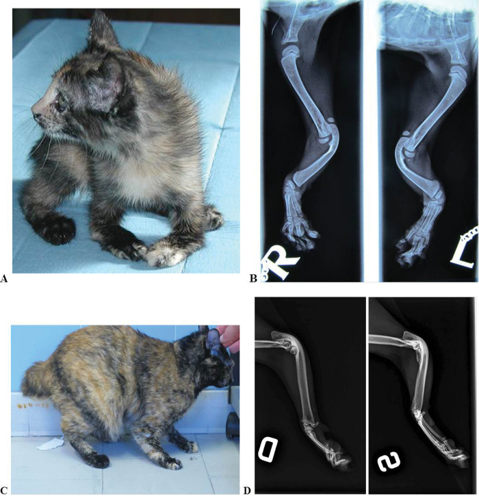
A 2-month-old female, domestic shorthair cat was presented to the Veterinary Teaching Hospital, Faculty of Veterinary Medicine, University of Bologna, for an orthopaedic examination. The malformation had been present since birth and on physical examination a marked varus deviation of both carpii was observed (Figure 1a). There was no pain, crepitation or evidence of fractures during physical examination of the deformed limbs, while the range of motion (ROM) of the carpal and elbow joints of both forelimbs was reduced: the extension of the right elbow was 90° and the flexion was 35° (ROM 55°), for the left elbow the extension was 85° and the flexion 35° (ROM 50°); the extension of both carpii was 180°, while the flexion of the right carpus was 30° (ROM 150°) and the flexion of left one was 85° (ROM 95°). The gait was abnormal and the cat walked with the entire forearm in contact with the floor. Radiographs of both forelimbs showed a suspected radial hypoplasia: small portions of the radii were present proximally on the left arm, but no sign of radii was found in the right forearm. Radiographs also showed bilateral malformations of the carpal joint, bilateral agenesis of the first digit and malformation of the tail, while the ulna was shorter, thicker and curved, and elbow joint subluxation was also observed (Figure 1b). This cat presented other malformations, such as 15 thoracic vertebrae, six lumbar vertebrae and 15 ribs, with hypoplasia of the first pair. The resulting diagnosis was bilateral pre-axial longitudinal intercalary radial hemimelia and multiple malformations.
Figure 1.
Kitten aged 2 months and mediolateral radiographs of both forelimbs; note the absence of the radius in the right forelimb and a small portion of the radius in the left forelimb (a, b). Cat aged 4 years and mediolateral radiographs; note the presence of small portion of the proximal radius in both forelimbs (c, d)
The treatment consisted of a Robert Jones splinted bandage type modified with two bilateral wooden tongue depressors of 2 mm thickness as support. The length of the splint was the same as the length of the forearm and the width was 8 mm. The aim was to reduce limb deformity to a level close to a normal position with a splinted bandage until bone maturity. During the first period the cat was not comfortable with the bandages, but it could walk in an acceptably normal position and got used to the new position. Each time the bandages did not fit adequately they were changed, usually every 1–2 weeks. The only complication caused by the bandage was the ischaemic necrosis of the right fifth finger, which was surgically removed.
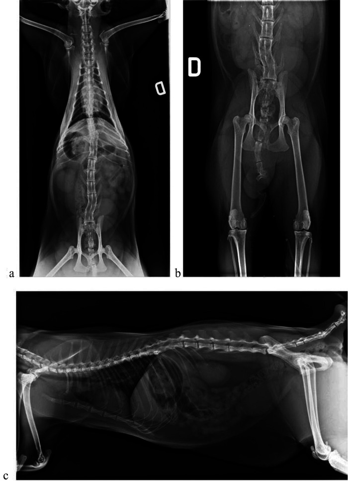
The cat was sent back to the referring veterinarian and he reported, after removing the bandages at 6 months, that there was still evidence of malformation in the forearms, but to a less severe degree, and that the gait of the cat had improved. After 4 years, the cat had an almost normal body conformation, except for the forelimbs where the malformation had improved but was still present; it could walk, jump and live normally (Figure 1c). At the clinical evaluation the cat showed a slight bilateral varus carpal deviation and an almost normal alignment of both elbows. The ROM of the right elbow had improved by 10°, with an extension of 110° and a flexion of 45°. Also, the left elbow had improved by 25°, with an extension of 115° and a flexion of 40°. The ROM of the carpii was the same as the first visit. Radiographical evaluation at 4 years clearly showed the presence of a small portion of proximal radii in both forelimbs, confirming the occurrence of radial hypoplasia (Figure 1d). Additionally, there were synostoses between the last three thoracic vertebrae (T13–T15) and the last three lumbar vertebrae (L5–L7), mineralisation of the vertebral disc at L1–L2, and malformation and fusion between the great ribs (Figure 2a–c); the malformations reported at 2 months of age were still present. Despite these malformations, the cat showed adequate limb function to permit a good quality of life.
Figure 2.
Cat aged 4 years. Ventrodorsal (a, b) and lateral (c) radiographs show the presence of multiple malformations in vertebrae, tail and ribs
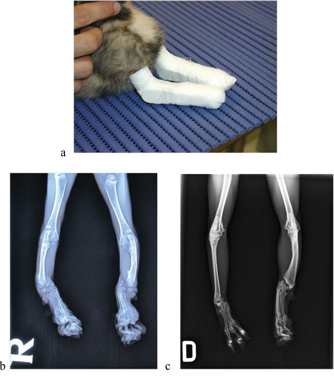
The pre-axial longitudinal intercalary radial hemimelia is the most common type of hemimelia in small animals.3,5,6 This condition is usually unilateral4,6–8 and is present in both dogs and cats.3,5,8,9 Moreover, dysostoses may affect either the forelimb or the hindlimb.1,2,10 Typical findings in animals affected by radial hemimelia include varus deformity of the ulnocarpal joint, absence of the radial carpal bone3,5,7,9 and first digit agenesis,5,7 as well as metacarpal and metatarsal synostosis,1,8 elbow joint subluxation3,6 and polydactyly. 3 The aetiology and pathogenesis of radial hemimelia is unknown.3,6 Some authors have suggested factors that may be involved, including intra-uterine compression, inflammation, maternal nutritional deficiencies, irradiation, teratogenic agents, vaccines and drugs such as thalidomide.1,4 There are more hypotheses for the pathogenesis of radial hemimelia, including genetic inheritance1,4,9,11 or vascular defects such as abnormal vasculogenesis or disruption of vessels that could result in radial agenesis. 6 However, the first hypothesis is that radial hemimelia is a consequence of neural crest injury, with a lack of apical ectodermal ridge-mesodermal interaction during limb outgrowth.1,9 It is possible that this pathology, like in humans, can be inherited. It has been suggested that radial aplasia or hypoplasia may be genetically linked to polydactyly.3,7 There are anecdotal reports from breeders that discuss how mating of cats with radial hemimelia can produce similarly affected kittens.5,7 Therefore, many authors recommend that animals affected by radial hemimelia should not be bred, as the offspring are likely to be affected by the deformation.3,5 Moreover, it has been suggested that radial hemimelia in Siamese and domestic shorthair cats may be a hereditary trait, 11 but there is no such evidence in dogs.1,4 Typical clinical signs are forelimb shortening, varus deformity of the ulnocarpal joint and elbow joint subluxation. 3 The range of motion of the elbow joint is reduced, walking is abnormal and the animal often carries the weight on the forelimb with the entire arm in contact with the ground. 7 Radial hemimelia is usually noticed soon after birth and, in this case, the ulnae are thickened, misshaped and curved. 4 Frequently, in these cases, complication factors, such as fractures, ankylosis of the radiocarpal joint, muscle contraction, muscle atrophy and arthrosis of the joint, can lead to a more difficult resolution.5,6 Radiographical documentation of the absence of hypoplastic radial structures is necessary to confirm the diagnosis. 1 In fact, in this case, radial agenesis or hypoplasia was suspected but it was necessary to confirm the diagnosis with radiographical examination. In this disease, it is very important to make an early diagnosis and treat to prevent flexural contracture, subsequent varus deformity, muscle atrophy and pathologic fracture. 3 If a limb deformity is reduced, we recommend stabilisation of the radiocarpal joint in a normal weight-bearing position with a fortified Robert Jones, such as in this case, until bone maturity in animals younger than 4–5 months.1,6 The treatment of radial hemimelia depends on whether the animal is affected unilaterally or bilaterally, and on the type of malformations. There are two types of treatment: conservative and surgical. Conservative treatment is indicated if limb function is acceptable and the deformity is minimal,1,6,7 while surgical treatment is indicated if limb function and the deformity are unacceptable. Surgical treatment, frequent in the dogs, may consist of ulnocarpal arthrodesis, 12 the Ilizarov method, 13 bone graft to fill the skeletal defect, 14 amputation or euthanasia if limb function is severely affected.4,5,7,15 To the authors’ knowledge, no previous conservative or surgical treatment in cat was proposed. In the case reported here, conservative treatment with splint bandage was used because the cat was young (2 months old) and the limb deformity could be reduced to a normal weight-bearing position (Figure 3a). The aim of the bandages was to prevent muscle contracture, in case it would worsen the angular deformity, and to force the kitten to stand on the hand instead of the elbow. The bandage could support the ulna function in bearing the weight without the radius and improve the physiological forelimb alignment. Treatment was therefore directed at improving the function of the limb, when possible, in addition to improving the cat’s quality of life. For the next 3 years, the veterinarian and owner told us that limb function and forelimb varus deformities had improved by about 10° in the right carpus (Figure 3b, c). Four years after the last examination, we re-examined the cat. Telephone contact with the owner and veterinarian, and orthopaedic examinations showed a cat with a good quality of life that had adapted well to its deformities and could walk and jump. In this case the conservative treatment gave good results; however, a limitation of this choice was that the cat had to adapt to wearing the splint bandages for long periods. Furthermore, the loss of the fifth finger can be considered a major complication, but it did not aggravate the cat’s condition.
Figure 3.
Kitten with bilateral splint bandage (a). Dorsoventral radiographs of kitten aged 2 months (b) and cat aged 4 years (c)
In this case the kitten was found and the mother is unknown. For this reason we can’t exclude that other kittens in the litter were affected by this disease. It would be interesting to investigate this disease with genetic testing in order to contribute to the understanding of the aetiology.
Bilateral radial hemimelia is a serious congenital pathology often associated with other malformations. Early diagnosis and appropriate treatment is necessary for a good prognosis.
Footnotes
Funding: The authors received no specific grant from any funding agency in the public, commercial or not-for-profit sectors for the preparation of this case report.
The authors do not have any potential conflicts of interest to declare.
Accepted: 15 March 2012
References
- 1. Towle HAM, Breur GJ. Dysostoses of the canine and feline appendicular skeleton. J Am Vet Med Assoc 2004; 225: 1685–1692. [DOI] [PubMed] [Google Scholar]
- 2. Barrad KR, Cornillie PK. Bilateral hindlimb adactyly in an adult cat. J Small Anim Pract 2008; 49: 252–253. [DOI] [PubMed] [Google Scholar]
- 3. Lockwood A, Montgomery R, McEwen V. Bilateral radial hemimelia, polydactyly and cardiomegaly in two cats. Vet Comp Orthop Traumatol 2009; 22: 511–513. [DOI] [PubMed] [Google Scholar]
- 4. Lewis RE, Van Sickle DC. Congenital hemimelia (agenesis) of the radius in a dog and cat. J Am Vet Med Assoc 1970; 156: 1892–1896. [PubMed] [Google Scholar]
- 5. O’Brien CR, Malik R, Nicoli RG. What is your diagnosis? J Feline Med Surg 2002; 4: 111–113. [DOI] [PMC free article] [PubMed] [Google Scholar]
- 6. Winterbotham EJ, Johnson KA, Francis DJ. Radial agenesis in a cat. J Small Anim Pract 1985; 26: 393–398. [Google Scholar]
- 7. Swalley J, Swalley M. Agenesis of the radius in a kitten. Feline Pract 1978; 8: 25–26. [Google Scholar]
- 8. Schultz VA, Watson AG. Lumbosacral transitional vertebra and thoracic limb malformations in a Chihuahua puppy. J Am Anim Hosp Assoc 1995; 31: 101–106. [DOI] [PubMed] [Google Scholar]
- 9. Vijayakumar G, Dharmaceelan S, Madhavan Unny N, Ezakial Napoleon R. Bilateral hemimelia in a dog. Indian Vet J 2007; 84: 1112–1113. [Google Scholar]
- 10. Arnbjerg J. Congenital partial hemimelia tibia in a kitten. Zentralbl Veterinarmed A 1979; 26: 73–77. [DOI] [PubMed] [Google Scholar]
- 11. Hoskins JD. Congenital defects of cats. Compend Contin Educ Pract Vet 1995; 17: 385–405. [Google Scholar]
- 12. McKee MW, Reynolds J. Ulnocarpal arthrodesis and limb lengthening for the management of radial agenesis in a dog. J Small Animal Pract 2007; 48: 591–595. [DOI] [PubMed] [Google Scholar]
- 13. Rahal SC, Volpi RS, Ciani RB, Vulcano LC. Use of the illizarov method of distraction osteogenesis for the treatment of radial hemimelia in a dog. J Am Vet Med Assoc 2005; 226: 65–68. [DOI] [PubMed] [Google Scholar]
- 14. Pedersen NC. Surgical correction of a congenital defect of the radius and ulna in a dog. J Am Vet Med Assoc 1968; 153: 1328–1331. [PubMed] [Google Scholar]
- 15. Ahalt BA, Bilbrey SA. What is your diagnosis? J Small Anim Pract 1997; 39: 539–571. [PubMed] [Google Scholar]


