Abstract
Purpose
Retinitis pigmentosa (RP) comprises a genetically and clinically heterogeneous group of inherited retinal degenerations, where 20–30% of patients exhibit extra-ocular manifestations (syndromic RP). Understanding the genetic profile of RP has important implications for disease prognosis and genetic counseling. This study aimed to characterize the genetic profile of syndromic RP in Portugal.
Methods
Multicenter, retrospective cohort study. Six Portuguese healthcare providers identified patients with a clinical diagnosis of syndromic RP and available genetic testing results. All patients had been previously subjected to a detailed ophthalmologic examination and clinically oriented genetic testing. Genetic variants were classified according to the American College of Medical Genetics and Genomics; only likely pathogenic or pathogenic variants were considered relevant for disease etiology.
Results
One hundred and twenty-two patients (53.3% males) from 100 families were included. Usher syndrome was the most frequent diagnosis (62.0%), followed by Bardet-Biedl (19.0%) and Senior-Løken syndromes (7.0%). Deleterious variants were identified in 86/100 families for a diagnostic yield of 86.0% (87.1% for Usher and 94.7% for Bardet-Biedl). A total of 81 genetic variants were identified in 25 different genes, 22 of which are novel. USH2A and MYO7A were responsible for most type II and type I Usher syndrome cases, respectively. BBS1 variants were the cause of Bardet-Biedl syndrome in 52.6% of families. Best-corrected visual acuity (BCVA) records were available at baseline and last visit for 99 patients (198 eyes), with a median follow-up of 62.0 months. The mean BCVA was 56.5 ETDRS letters at baseline (Snellen equivalent ~ 20/80), declining to 44.9 ETDRS letters (Snellen equivalent ~ 20/125) at the last available follow-up (p < 0.001).
Conclusion
This is the first multicenter study depicting the genetic profile of syndromic RP in Portugal, thus contributing toward a better understanding of this heterogeneous disease group. Usher and Bardet-Biedl syndromes were found to be the most common types of syndromic RP in this large Portuguese cohort. A high diagnostic yield was obtained, highlighting current genetic testing capabilities in providing a molecular diagnosis to most affected individuals. This has major implications in determining disease-related prognosis and providing targeted genetic counseling for syndromic RP patients in Portugal.
Supplementary information
The online version contains supplementary material available at 10.1007/s00417-023-06360-2.
Keywords: Inherited retinal diseases, Syndromic retinitis pigmentosa, Ophthalmic genetics, Genotype
Introduction
Retinitis pigmentosa (RP) comprises a genetically and clinically diverse group of inherited retinal degenerations (IRDs), primarily characterized by rod-cone degeneration. With an estimated prevalence of 1:4000 individuals, it is the most frequent form of IRD [1]. While most cases of RP are not associated with systemic abnormalities, 20–30% of patients exhibit extra-ocular disease and are referred to as syndromic RP [1–3]. Usher syndrome features sensorineural hearing loss (and in some forms vestibular impairment) in association with RP and is overall the most frequent form of syndromic RP [2–4], followed by Bardet-Biedl syndrome. In the latter, polydactyly, intellectual disability, and truncal obesity are among the most prevalent extra-ocular manifestations [2–4].
Genetic profiling of IRDs takes on an ever-growing significance for the affected individual, not only with regard to disease prognosis and genetic counseling but also for treatment prospects [5], which recently became a reality with the introduction of gene therapy for RPE65-associated retinal degeneration [6]. Even though therapies targeting the retinal phenotype of syndromic RP are not currently available, the genetic landscape of syndromic RP has been receiving increased interest worldwide, including a few European studies [7–10]. Although there are some similarities in genetic profiles, there is significant variation among regions and ethnic groups. This genetic diversity between populations may be partly explained by founder mutations [8, 11, 12], thus highlighting the importance of obtaining reference population-based data.
In Portugal, data on the genetic architecture of syndromic RP is currently scarce. By conducting a national, multicenter study, we aimed at characterizing the genetic landscape of syndromic RP in a large Portuguese cohort.
Methods
Study design
A nationwide, multicenter, retrospective cohort study was conducted in six Portuguese public healthcare providers (HCP): Centro Hospitalar e Universitário de Coimbra (CHUC), Instituto de Oftalmologia Dr. Gama Pinto (IOGP), Centro Hospitalar Universitário de Lisboa Norte (CHULN), Centro Hospitalar e Universitário de Santo António (CHUdSA), Centro Hospitalar de Entre o Douro e Vouga (CHEDV), and Hospital de Braga (HB). Patients with a clinical diagnosis of syndromic RP and available genetic testing results were retrieved from internal databases and the IRD-PT registry [12]. Every patient provided written informed consent prior to enrollment, and the study complied with the tenets of the Declaration of Helsinki for biomedical research. Of note, even though most of the data shown here has never been published, the study includes data that has been featured in previous publications [13–15].
Clinical/demographic features
Data regarding demographics (age, gender, district of residence), family history, presence of consanguinity, age of ophthalmologic symptom onset, presence of ocular and systemic comorbidities, best-corrected visual acuity (BCVA) at baseline, and last available follow-up was obtained from each patient clinical record. A clinical diagnosis was established based on history and compatible structural (multimodal retinal imaging) and functional (electrophysiology testing and visual field testing) retinal findings. However, such testing was not standardized among the different contributing HCPs.
Genetic testing
Peripheral blood samples were collected, and genomic DNA was isolated using a DNA extraction and purification kit based on the manufacturer’s protocol. A clinically oriented next-generation sequencing (NGS) approach was used, comprising whole-exome sequencing (WES) or WES-based NGS panels with copy number variation (CNV) screening, complemented by multiplex ligation-dependent probe amplification (MLPA), when necessary. Whenever possible, segregation analysis was performed on family members. Identified genetic variants were classified in compliance with the American College of Medical Genetics and Genomics (ACMG) standards and guidelines for the interpretation of sequence variants [16]. Only class IV (likely pathogenic) and class V (pathogenic) variants were deemed relevant to disease etiology. Variants were considered novel in the absence of previous reports featured in scientific publications. Genetic counseling provided by a medical geneticist was granted to all families.
Statistical analysis
Statistical analysis was performed using the software IBM SPSS Statistics version 26 (Armonk, New York, USA). Descriptive statistics were computed for all variables. A statistically significant result was defined as a p-value < 0.05.
Results
Clinical/demographic features
A total of 122 patients (100 different families) with a clinical diagnosis of syndromic RP and available genetic testing results were included (75 patients from CHUC, 26 from IOGP, 7 from CHULN, 7 from CHUdSA, 5 from CHEDV and 2 from HB). Most patients (53.3%) were males, and the mean age was 44.6 ± 15.1 years (range 11–79). Family history of the disease was present in 53.3%, while 36.1% of patients reported consanguinity. Age of ophthalmic disease onset, defined as the first instance of RP-attributable symptoms, along with the demographic characterization of the cohort, is presented in Table 1, while the cohort distribution per district of residence is presented in Fig. 1.
Table 1.
Demographic characterization of the cohort
| Number of families (number of patients) | 100 (122) | |||
| Male gender: n (%) | 65 (53.3%) | |||
| Age: mean ± SD (years) | 44.6 ± 15.1 | |||
| Family history, n (%) | 65 (53.3%) | |||
| Consanguinity, n (%) | 44 (36.1%) | |||
| Age of symptom onset, n (%) | Diagnosis | All patients | Usher syndrome | BBS |
| ≤ 5 years | 19 (15.6%) | 10 (13.5%) | 6 (24.0%) | |
| 6–10 years | 27 (22.1%) | 12 (16.2%) | 10 (40.0%) | |
| 11–20 years | 29 (23.8%) | 20 (27.0%) | 2 (8.0%) | |
| 21–30 years | 14 (11.5%) | 8 (10.8%) | 1 (4.0%) | |
| 31–50 years | 15 (12.3%) | 14 (18.9%) | 0 (0%) | |
| Unknown | 18 (14.8%) | 10 (13.5%) | 6 (24%) | |
Data presented per patient. Age of symptom onset is presented for all patients and the two most common diagnoses
BBS, Bardet-Biedl syndrome
Fig. 1.
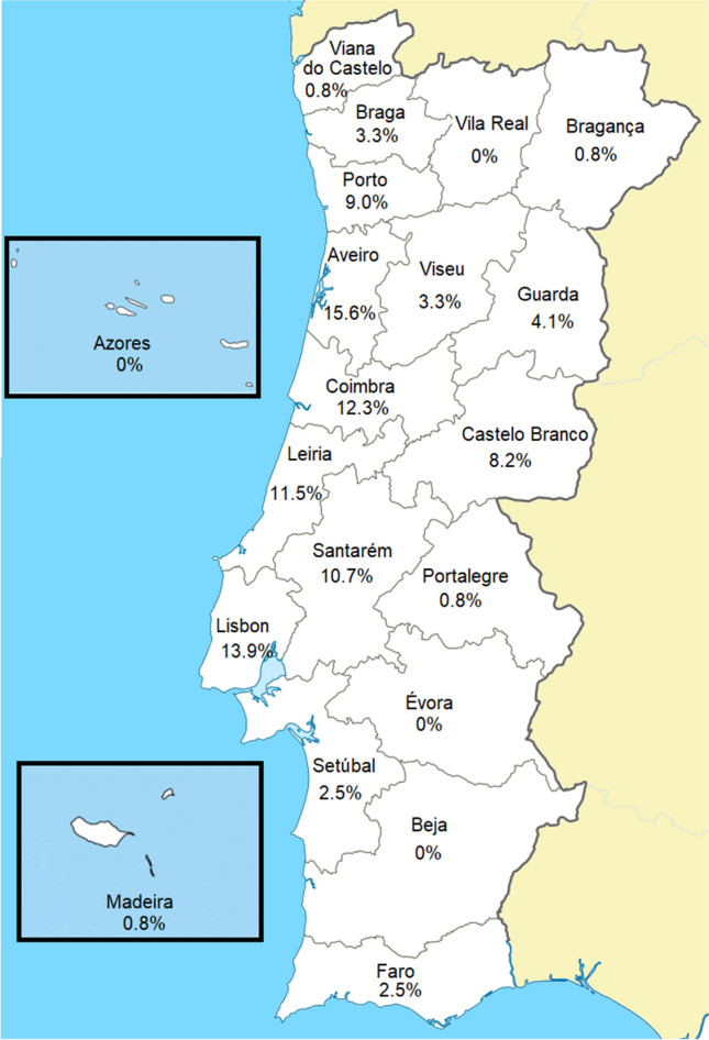
Cohort distribution by district of residence (data presented per patient)
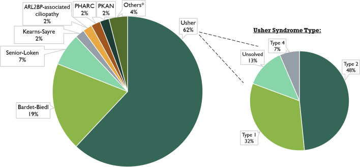
The most frequently encountered diagnosis was Usher syndrome, present in 62.0% of the families, followed by Bardet-Biedl (19.0%) and Senior-Løken (7.0%) syndromes. The remaining cases consisted of Kearns-Sayre syndrome (n = 2); ARL2BP-associated ciliopathy [14] (n = 2); polyneuropathy, hearing loss, ataxia, retinitis pigmentosa, and cataract (PHARC) (n = 2); pantothenate kinase-associated neurodegeneration (PKAN) (n = 2); bone marrow failure syndrome type 3 (n = 1); neuropathy, ataxia, retinitis pigmentosa (NARP) (n = 1); Jalili syndrome (n = 1), and a presumed mitochondrial DNA depletion syndrome (n = 1), as shown in Fig. 2. Regarding Usher syndrome, type II was the most frequent phenotype (48%), followed by type I (32%) and type IV (7%), with 13% of families remaining genetically unsolved.
Fig. 2.
Cohort diagnosis distribution (percentage per family). *Others include bone marrow failure syndrome type 3; neuropathy, ataxia, retinitis pigmentosa (NARP) syndrome; Jalili syndrome; and mitochondrial DNA depletion syndrome. PHARC: polyneuropathy, hearing loss, ataxia, retinitis pigmentosa, and cataract; PKAN: pantothenate kinase-associated neurodegeneration
Genetic findings
Disease-causing variants were identified in 86/100 families, hereby referred to as the solved cases, for a diagnostic yield of 86.0% (87.1% for Usher and 94.7% for Bardet-Biedl, the most common diagnoses). The most frequently implicated gene in cases of Usher syndrome was USH2A, containing disease-causing biallelic variants for 33.9% of families, followed by MYO7A in 24.2% of all families. For Bardet-Biedl syndrome, BBS1 was the most commonly mutated gene (52.6% of families), followed by BBS10 (21.1%). Further information on the diagnostic yield and all involved genes per diagnosis can be found in Table 2. All solved cases except for the mitochondrial DNA-dependent syndromes were associated with autosomal recessive inheritance. In such cases, a single disease-causing variant in homozygosity was identified in 65% of families (n = 54), while 35% (n = 29) harbored 2 different variants in compound heterozygosity. Please refer to Supplementary Table 1 for a detailed description.
Table 2.
Diagnostic yield and causative gene of syndromic RP (data presented per family)
| Diagnosis | Genetic testing result | Gene | N (%) | ||
|---|---|---|---|---|---|
| Solved | Unsolved | Total | |||
| Usher | 54 (87.1%) | 8 (12.9%) | 62 (100%) | ADGRV1 | 9 (14.5%) |
| ARSG | 4 (6.5%) | ||||
| CDH23 | 3 (4.8%) | ||||
| MYO7A | 15 (24.2%) | ||||
| PCDH15 | 1 (1.6%) | ||||
| USH1G | 1 (1.6%) | ||||
| USH2A | 21 (33.9%) | ||||
| Unsolved | 8 (12.9%) | ||||
| Bardet-Biedl | 18 (94.7%) | 1 (5.3%) | 19 (100%) | BBS1 | 10 (52.6%) |
| BBS2 | 1 (5.3%) | ||||
| BBS10 | 4 (21.1%) | ||||
| MKKS | 1 (5.3%) | ||||
| SDCCAG8 | 1 (5.3%) | ||||
| TTC8 | 1 (5.3%) | ||||
| Unsolved | 1 (5.3%) | ||||
| Senior- Løken | 5 (71.4%) | 2 (28.6%) | 7 (100%) | NPHP1 | 2 (28.6%) |
| SDCCAG8 | 1 (14.3%) | ||||
| TRAF3IP1 | 1 (14.3%) | ||||
| WDR19 | 1 (14.3%) | ||||
| Unsolved | 2 (28.6%) | ||||
| PKAN | 1 (50%) | 1 (50%) | 2 (100%) | PANK2 | 1 (50%) |
| Unsolved | 1 (50%) | ||||
| Kearns-Sayre | 2 (100%) | 0 (0%) | 2 (100%) | mtDNAa | 2 (100%) |
| ARL2BP-associated ciliopathy | 2 (100%) | 0 (0%) | 2 (100%) | ARL2BP | 2 (100%) |
| PHARC | 1 (50%) | 1 (50%) | 2 (100%) | ABHD12 | 1 (50%) |
| Unsolved | 1 (50%) | ||||
| Bone marrow failure syndrome 3 | 1 (100%) | 0 (0%) | 1 (100%) | DNAJC21 | 1 (100%) |
| Jalili | 1 (100%) | 0 (0%) | 1 (100%) | CNNM4 | 1 (100%) |
| NARP | 1 (100%) | 0 (0%) | 1 (100%) | MT-ATP6 | 1 (100%) |
| MDS | 0 (0%) | 1 (100%) | 1 (100%) | Unsolved | 1 (100%) |
| Total | 86 (86%) | 14 (14%) | 100 (100%) | 100 (100%) | |
PKAN, pantothenate kinase-associated neurodegeneration; NARP, neuropathy, ataxia, and retinitis pigmentosa; PHARC, polyneuropathy, hearing loss, ataxia, retinitis pigmentosa, and cataract; MDS, mitochondrial DNA depletion syndrome
aLarge deletion of mitochondrial DNA involving several genes
A total of 81 unique variants were identified in 25 different genes, 22 of which are novel and herein reported for the first time. The pathogenic variant c.920_923dup p.(His308Glnfs*16) was the most frequently encountered variant in USH2A-associated Usher syndrome (n = 5/5; families/patients), while c.397dup p.(His133Profs*7) was the most frequent variant for MYO7A-associated cases (n = 4/7; families/patients). For Bardet-Biedl syndrome, the BBS1 pathogenic variant c.1169 T > G p.(Met390Arg) was the most commonly identified causative variant (n = 9/10; families/patients). A detailed description of all identified genetic variants is available in Table 3.
Table 3.
Genetic data of identified variants
| dbSNP | Nucleotide change | Protein change | Variant type | Predicted effect | ACMG classification (applied criteriaA) | Count in cohort (n of patients/families) | First report |
|---|---|---|---|---|---|---|---|
| ABHD12 (NM_001042472.3) | |||||||
| c.728G > A | p.(Trp243*) | SNV | Nonsense | Likely pathogenic (PVS1, PM2) | 1 / 1 | This study | |
| ADGRV1 (NM_032119.4) | |||||||
| c.(17019 + 1_17020-1)_(17856 + 1_17857-1)dup | CNV | Exon 79–83 duplication | Likely pathogenic | 3 / 2 | This study | ||
| rs757696771 | c.17668_17669del | p.(Met5890Valfs*10) | Indel | Frameshift | Pathogenic (PVS1, PS4, PM2, PP5) | 3 / 3 | PMID: 21569298 |
| rs746618021 | c.2864C > A | p.(Ser955*) | SNV | Nonsense | Pathogenic (PVS1, PS4, PM2, PP5) | 1 / 1 | PMID: 22147658 |
| rs397517429 | c.2870dup | p.(Asn957Lysfs*10) | Indel | Frameshift | Pathogenic (PVS1, PM2, PP5) | 1 / 1 | This study |
| c.6515C > G | p.(Ser2172*) | SNV | Nonsense | Likely pathogenic (PVS1, PM2) | 1 / 1 | This study | |
| c.7336del | p.(Glu2446Asnfs*21) | Indel | Frameshift | Likely pathogenic (PVS1, PM2) | 1 / 1 | This study | |
| c.9484G > T | p.(Glu3162*) | SNV | Nonsense | Likely pathogenic (PVS1, PM2) | 2 / 1 | This study | |
| c.17669del | p.(Met5890Valfs*10) | Indel | Frameshift | Likely pathogenic (PVS1, PM2) | 5 / 4 | This study | |
| c.8832del | p.(Gly2945Valfs*2) | Indel | Frameshift | Likely pathogenic (PVS1, PM2) | 1 / 1 | This study | |
| ARL2BP (NM_012106.4) | |||||||
| rs199830550 | c.207 + 1G > A | p.? | SNV | Splicing | Pathogenic (PVS1, PM2, PM3, PP5) | 2 / 2 | PMID: 28041643 |
| ARSG (NM_001267727.2) | |||||||
| rs751461705 | c.1326del | p.(Ser443Alafs*12) | Indel | Frameshift | Pathogenic (PVS1, PS3, PM2, PM3, PP5) | 4 / 3 | PMID: 33300174 |
| rs141748845 | c.253 T > C | p.(Ser85Pro) | SNV | Missense | Pathogenic (PM2, PM3, PP3, PP5) | 1 / 1 | PMID: 33300174 |
| rs1244718647 | c.338G > A | p.(Gly113Asp) | SNV | Missense | Pathogenic (PM2, PM3 PP3, PP5) | 1 / 1 | PMID: 33300174 |
| BBS1 (NM_024649.5) | |||||||
| rs113624356 | c.1169 T > G | p.(Met390Arg) | SNV | Missense | Pathogenic (PS3, PM2, PM3, PP1, PP3, PP5) | 10 / 9 | PMID: 12118255 |
| rs1014835928 | c.1318C > T | p.(Arg440*) | SNV | Nonsense | Pathogenic (PVS1, PM2, PM3, PP5) | 1 / 1 | PMID: 12677556 |
| c.863 T > G | p.(Leu288Arg) | SNV | Missense | Likely pathogenic (PM2, PM3, PP3, PP4) | 1 / 1 | This study | |
| rs1490351829 | c.118del | p.(Cys40Alafs*2) | Indel | Frameshift | Pathogenic (PVS1, PM2, PM3, PP5) | 1 / 1 | PMID: 27032803 |
| rs121917777 | c.1645G > T | p.(Glu549*) | SNV | Nonsense | Pathogenic (PVS1, PM2, PM3, PP5) | 3 / 2 | PMID: 12118255 |
| c.17C > G | p.(Ser6*) | SNV | Nonsense | Likely pathogenic (PVS1, PM2) | 1 / 1 | This study | |
| BBS2 (NM_031885.5) | |||||||
| rs1368647604 | c.402del | p.(Ala136Argfs*65) | Indel | Frameshift | Pathogenic (PVS1, PM2, PM3, PP5) | 2 / 1 | PMID: 15770229 |
| rs121908178 | c.943C > T | p.(Arg315Trp) | SNV | Missense | Likely pathogenic (PS3, PM1, PM2, PM3, PM5, PP3, PP5) | 2 / 1 | PMID: 11567139 |
| BBS10 (NM_024685.4) | |||||||
| rs1057517031 | c.1542del | p.(Asp515Ilefs*9) | Indel | Frameshift | Pathogenic (PVS1, PM2, PP5) | 3 / 1 | PMID: 16582908 |
| rs549625604 | c.271dup | p.(Cys91Leufs*15) | Indel | Frameshift | Pathogenic (PVS1, PM2, PM3, PP5) | 1 / 1 | PMID: 10874630 |
| rs148374859 | c.273C > G | p.(Cys91Trp) | SNV? | Missense | Pathogenic (PS3, PM2, PM3, PP5) | 2 / 2 | PMID: 16582908 |
| c.1677del | p.(Tyr559*) | Indel | Frameshift | Pathogenic (PVS1, PM2, PM3, PP5) | 1 / 1 | PMID: 16582908 | |
| CDH23 (NM_022124.6) | |||||||
| rs1385831846 | c.3579 + 2 T > C | p.? | SNV | Splicing | Pathogenic (PVS1, PS4, PM2, PP5) | 1 / 1 | PMID: 11138009 |
| rs1306728898 | c.6319C > T | p.(Arg2107*) | SNV | Nonsense | Pathogenic (PVS1, PM2, PP5) | 1 / 1 | PMID: 11090341 |
| rs111033247 | c.6049 + 1G > A | p.? | SNV | Splicing | Pathogenic (PVS1, PS4, PM2, PP5) | 1 / 1 | PMID: 8894709 |
| c.753 + 2 T > A | p.? | SNV | Splicing | Likely Pathogenic (PVS1, PM2) | 1 / 1 | This study | |
| CNNM4 (NM_020184.4) | |||||||
| rs74552543 | c.971 T > C | p.(Leu324Pro) | SNV | Missense | Pathogenic (PM2, PM3, PP3, PP5) | 1 / 1 | PMID: 19200527 |
| DNAJC21 (NM_001012339.3) | |||||||
| c.805C > T | p.(Gln269*) | SNV | Nonsense | Likely pathogenic (PVS1, PM2) | 1 / 1 | This study | |
| MKKS (NM_170784.3) | |||||||
| rs768929313 | c.748G > A | p.(Gly250Arg) | SNV | Missense | Pathogenic (PM2, PM3, PM5, PP3, PP5) | 3 / 1 | PMID: 20142850 |
| MT-ATP6 | |||||||
| rs199476133 | m.8993 T > G | p.(Leu156Arg) | SNV | Missense | Pathogenic (PS2, PM3, PM5, PP3, PP5) | 2 / 1 | PMID: 2137962 |
| MYO7A (NM_000260.4) | |||||||
| c.1529 T > C | p.(Ile510Thr) | SNV | Missense | Likely pathogenic (PM1, PM2, PM3, PP3) | 1 / 1 | This study | |
| rs111033214 | c.3508G > A | p.(Glu1170Lys) | SNV | Missense | Pathogenic (PS4, PM2 PM5, PP3, PP5) | 5 / 3 | PMID: 10425080 |
| rs111033187 | c.397dup | p.(His133Profs*7) | Indel | Frameshift | Pathogenic (PVS1, PM2, PM3, PP5) | 7 / 4 | PMID: 21569298 |
| rs751769391 | c.4489G > C | p.(Gly1497Arg) | SNV | Missense | Pathogenic (PM2, PM5, PP3, PP5) | 3 / 3 | PMID: 27460420 |
| c.5510 T > A | p.(Leu1837His) | SNV | Missense | Pathogenic (PM2, PM5, PP3, PP5) | 4 / 4 | PMID: 36909829 | |
| c.5743-15_5746del | p.(Ala1915fs) | Indel | Frameshift | Likely pathogenic (PVS1, PM2) | 1 / 1 | This study | |
| rs1591514873 | c.6439-1G > A | p.? | SNV | Splicing | Pathogenic (PVS1, PM2, PP5) | 3 / 2 | PMID: 16199547 |
| rs111033285 | c.999 T > G | p.(Tyr333*) | SNV | Nonsense | Pathogenic (PVS1, PS4, PM2, PP5) | 1 / 1 | PMID: 8900236 |
| c.1929dup | p.(Pro644Alafs*67) | Indel | Frameshift | Pathogenic (PVS1, PM2, PP5) | 2 / 1 | PMID: 36909829 | |
| rs1173853484 | c.6026C > A | p.(Ala2009Asp) | SNV | Missense | Likely pathogenic (PP3, PP5, PM1, PM2, PM5) | 1 / 1 | PMID: 27460420 |
| NPHP1 (NM_001128178.3) | |||||||
| c.2065_2074del | p.(Thr689Leufs*37) | Indel | Frameshift | Likely pathogenic (PVS1, PM2) | 2 / 2 | This study | |
| NPHP4 (NM_015102.5) | |||||||
| rs370946873 | c.2956G > A | p.(Gly986Arg) | SNV | Missense | VUS (PM2, PP3) | 1 / 1 | PMID: 36909829 |
| NRL (NM_001354768.3) | |||||||
| rs774348345 | c.74G > A | p.(Arg25Gln) | SNV | Missense | VUS (PM2, PP2) | 1 / 1 | This study |
| PANK2 (NM_001386393.1) | |||||||
| rs779815683 | c.1268G > T | p.(Cys423Phe) | SNV | Missense | VUS (PM1, PM2, PP2, PP3) | 1 / 1 | This study |
| rs137852959 | c.1561G > A | p.(Gly521Arg) | SNV | Missense | Pathogenic (PS3, PM2, PM3, PP1, PP2, PP3, PP5) | 1 / 1 | PMID: 11479594 |
| rs754521581 | c.1070G > C | p.(Arg357Pro) | SNV | Missense | Likely pathogenic (PM1, PM2, PM3, PM5, PP2, PP5) | 1 / 1 | PMID: 28680084 |
| PCDH15 (NM_001384140.1) | |||||||
| c.(2220 + 1_2221-1)_(3122 + 1_3123-1)dup | CNV | Exon 19–23 duplication | Likely pathogenic | 1 / 1 | PMID: 20538994 | ||
| SDCCAG8 (NM_006642.5) | |||||||
| rs768207230 | c.397G > T | p.(Glu133*) | SNV | Nonsense | Pathogenic (PVS1, PM2, PM3, PP5) | 2 / 2 | This study |
| SLC7A14 (NM_020949.3) | |||||||
| rs116040996 | c.821C > T | p.(Thr274IIe) | SNV | Missense | VUS (PP3, PM2) | 1 / 1 | This study |
| TRAF3IP1 (NM_015650.4) | |||||||
| rs778376663 | c.916-4A > G | p.? | SNV | Splicing | Likely pathogenic (PP3, PM2, PM3) | 2 / 1 | PMID: 36909829 |
| TTC8 (NM_001288781.1) | |||||||
| c.647G > A | p.(Trp216*) | SNV | Missense | Likely pathogenic (PVS1, PM2, PP5) | 1 / 1 | This study | |
| c.(?_681-1)_(879 + 1_?)del | CNV | Exon 9–10 deletion | Likely pathogenic | 1 / 1 | |||
| USH1G (NM_173477.5) | |||||||
| c.183 T > A | p.(Cys61*) | SNV | Nonsense | Likely pathogenic (PVS1, PM2) | 1 / 1 | This study | |
| USH2A (NM_206933.4) | |||||||
| rs750228923 | c.1214del | p.(Asn405Ilefs*3) | Indel | Frameshift | Pathogenic (PVS1, PM2, PM3, PP5) | 1 / 1 | PMID:16098008 |
| c.12294 + 1559_14133 + 8144del | CNV | Exon 63–64 deletion | Likely pathogenic | 1 / 1 | PMID: 28041643 | ||
| rs998302546 | c.14134-3169A > G | p.? | SNV | Splicing | Likely pathogenic (PM2, PM3, PP5) | 2 / 1 | PMID: 29196752 |
| c.14423G > A | p.(Cys4808Tyr) | SNV | Missense | VUS (PM1, PM2, PM3) | 1 / 1 | PMID: 36909829 | |
| c.1879C > T | p.(Gln627*) | SNV | Nonsense | Likely pathogenic (PVS1, PM2) | 1 / 1 | This study | |
| rs111033334 | c.2209C > T | p.(Arg737*) | SNV | Nonsense | Pathogenic (PVS1, PM2, PM3, PP5) | 1 / 1 | PMID: 17296898 |
| rs80338902 | c.2276G > T | p.(Cys759Phe) | SNV | Missense | Pathogenic (PS4, PM1, PM2, PM3, PP1, PP3, PP4, PP5) | 1 / 1 | PMID: 1968399 |
| c.(7300 + 1_7301-1)_(9371 + 1_9372-1)del | CNV | Exon 38–47 deletion | Likely pathogenic | 3 / 2 | |||
| rs202175091 | c.10712C > T | p.(Thr3571Met) | SNV | Missense | Pathogenic (PM1, PM2, PM3, PM5, PP1, PP5) | 1 / 1 | PMID: 17085681 |
| rs527236139 | c.11156G > A | p.(Arg3719His) | SNV | Missense | Pathogenic (PP1, PP5, PM2, PM3) | 2 / 2 | PMID: 20507924 |
| rs397517994 | c.14911C > T | p.(Arg4971*) | SNV | Nonsense | Pathogenic (PVS1, PP5, PM2, PM3) | 2 / 1 | PMID: 10729113 |
| rs758660532 | c.15089C > A | p.(Ser5030*) | SNV | Nonsense | Pathogenic (PVS1, PP5, PM2, PM3) | 2 / 1 | PMID: 10729113 |
| rs80338903 | c.2299del | p.(Glu767Serfs*21) | Indel | Frameshift | Pathogenic (PVS1, PP1, PP5, PM2, PM3) | 2 / 2 | PMID: 9624053 |
| rs1052375050 | c.2302 T > C | p.(Cys768Arg) | SNV | Missense | Likely pathogenic (PP3, PM2) | 1 / 1 | PMID: 36909829 |
| rs759433119 | c.2809 + 1G > A | p.? | SNV | Splicing | Pathogenic (PVS1, PP5, PM2, PM3) | 2 / 1 | PMID: 10729113 |
| rs754374132 | c.5278del | p.(Asp1760Metfs*10) | Indel | Frameshift | Pathogenic (PVS1, PP5, PM2, PM3) | 1 / 1 | PMID: 10729113 |
| rs1571783742 | c.7932G > A | p.(Trp2644*) | SNV | Nonsense | Pathogenic (PVS1, PP5, PM2, PM3) | 2 / 2 | PMID: 10729113 |
| rs748465849 | c.907C > A | p.(Arg303Ser) | SNV | Missense | Pathogenic (PP5, PM2, PM3, PM5) | 3 / 3 | PMID: 14970843 |
| rs397518043 | c.920_923dup | p.(His308Glnfs*16) | Indel | Frameshift | Pathogenic (PVS1, PP5, PM2, PM3) | 5 / 5 | PMID: 18641288 |
| rs111033263 | c.9799 T > C | p.(Cys3267Arg) | SNV | Missense | Likely pathogenic (PP3, PP5,PM2, PM3, PM5) | 1 / 1 | PMID: 17085681 |
| c.9315del | p.(Val3106Trpfs*54) | Indel | Frameshift | Likely pathogenic (PVS1, PM2) | 1 / 1 | PMID: 36909829 | |
| rs150982499 | c.5039A > G | p.(Lys1680Arg) | SNV | Missense | VUS (PM1, PM2) | 1 / 1 | PMID: 28912962 |
| WDR19 (NM_025132.4) | |||||||
| rs1020915921 | c.2704-2A > C | p.? | SNV | Splicing | Pathogenic (PVS1, PP5, PM2) | 1 / 1 | PMID: 16199547 |
| rs387906980 | c.1649 T > C | p.(Leu550Ser) | SNV | Missense | Pathogenic (PP1, PP3, PP5, PM2, PM3) | 1 / 1 | PMID: 22019273 |
AEach ACMG pathogenicity criterion is weighted as very strong (PVS), strong (PS), moderate (PM), or supporting (PP)
dbSNP, single nucleotide polymorphism database; PMID, PubMed identifier; VUS, variant of uncertain significance
Ocular findings
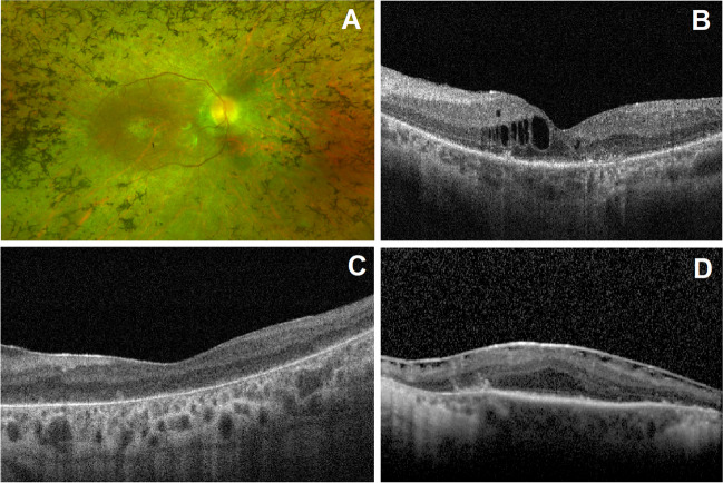
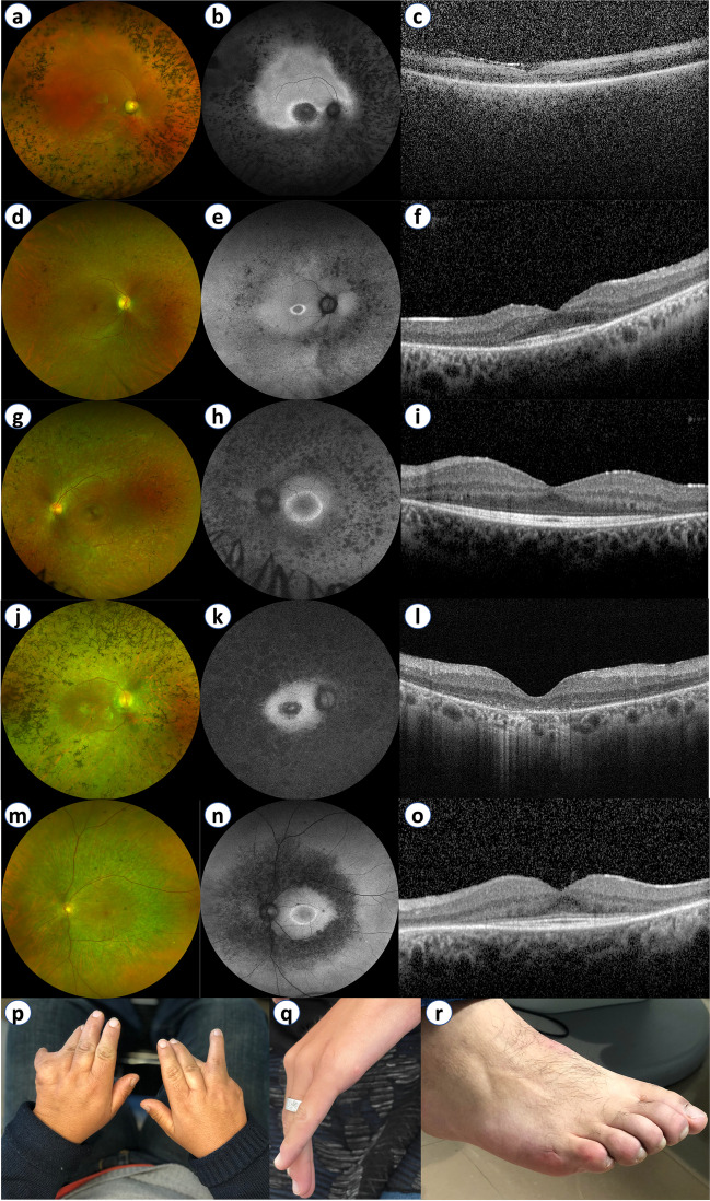
One hundred twenty-two patients were followed for a median period of 43 months. Best-corrected visual acuity (BCVA) records were available at both baseline and follow-up for 99 patients (198 eyes), followed for a median period of 62.0 months. The mean BCVA for this group was at baseline 56.5 Early Treatment Diabetic Retinopathy Study (ETDRS) letters (Snellen equivalent ~ 20/80), declining to 44.9 ETDRS letters (Snellen equivalent ~ 20/125) at the last available follow-up, a statistically significant change (p < 0.001). Ocular comorbidities were identified in 39.1% of all eyes, the most frequent being cystoid macular edema, present in 13.6% of eyes, followed by epiretinal membrane (9.9% of eyes) (Fig. 3). Figure 4 depicts the retinal phenotype of 5 patients from our cohort.
Fig. 3.
Ultra-widefield color fundus photography and spectral-domain optical coherence tomography (OCT) imaging of syndromic RP patients. (A) Classic fundus findings of retinitis pigmentosa: blood vessel attenuation and bone spicule hyperpigmentation in an Usher syndrome patient (macular atrophy is also present). (B) Cystoid macular edema present in USH2A-associated Usher syndrome. (C) OCT imaging displaying foveal atrophy of the outer retinal layers and RPE/Bruch’s membrane complex in BBS1-associated Bardet-Biedl syndrome. (D) Epiretinal membrane causing loss of foveal depression and presence of ectopic inner foveal layers in an Usher syndrome patient
Fig. 4.
(a–o) Ultra-widefield color fundus photography (UWF-CFP), ultra-widefield fundus autofluorescence (UWF-FAF), and spectral-domain optical coherence tomography (OCT) imaging of five syndromic RP patients: (a–c) BBS10-associated Bardet-Biedl syndrome; (d–f) SDCCAG8-associated Bardet-Biedl syndrome; (g–i) MYO7A-associated Usher syndrome; (j–l) USH2A-associated Usher syndrome; (m–o) ARSG-associated Usher syndrome. Bone spicule hyperpigmentation and patches of outer retinal atrophy seen on UWF-CFP (a, d, g, j, and m) directly correspond to hypoautofluorescent patches on UWF-FAF (b, e, h, k, and n). The parafoveal hyperautofluorescent ring (e, h, and n) directly correlates to the extent of outer retinal layer preservation in the corresponding OCT imaging (f, l, and o). Foveal atrophy of the outer retinal layers and RPE/Bruch’s membrane complex are typically found earlier in Bardet-Biedl syndrome (c) comparatively to Usher syndrome (i and o), where it is usually found in the latter stages of the disease. (p–r) Clinical photographs depicting congenital limb malformations in Bardet-Biedl syndrome: syndactyly in BBS10-associated Bardet-Biedl syndrome (p); residual hand appendage in BBS1-associated Bardet-Biedl syndrome (q); and patient with BBS1-associated Bardet-Biedl syndrome born with clinically evident polydactyly, subject to correcting surgery during childhood (r)
Discussion
Genetic profiling of IRDs is of major importance for patients and, through genetic counseling, for family members as well. Nevertheless, constraints in access to genetic testing may hinder the goal of obtaining a molecular diagnosis for every affected patient [17]. A paradigm shift is in progress, with a recent increase in the number of publications contributing to improve knowledge of the genetic landscape of IRDs in Portugal [13, 18–24]. One of such publications included a cohort of 230 Portuguese families with IRDs, but only 23 probands had syndromic RP [13]. In this nationwide, multicenter study including 122 patients from 100 families, we describe the genetic landscape of syndromic RP in Portugal.
Overall, disease-causing variants were identified in 86/100 families for a diagnostic yield of 86%. Even though this figure is much higher than what is usually obtained for non-syndromic forms of the disease [7, 25], it is in line with a previous study by Karali et al. [10], reporting genetic testing sensitivity upwards of 80% for syndromic IRDs.
Given the geographic proximity between Portugal and Spain, as well as the genetic similarities observed between its inhabitants [26], studies on the genetic landscape of syndromic RP in Spanish cohorts are a natural reference for comparison purposes, and thus, one could anticipate somewhat similar genetic findings for a Portuguese cohort. As expected, Usher (n = 62 families) and Bardet-Biedl (n = 19 families) syndromes were found to be the most frequent causes of syndromic RP in our cohort. USH2A and MYO7A variants were the major causes of Usher syndrome type II and type I, respectively. Similar findings were reported by Perea-Romero et al. [7] in their large Spanish cohort (n = 577 syndromic IRD families) and are observed as well in most studies from different populations [27–29]. Additionally, the BBS1 variant c.1169 T > G p.(Met380Arg) was the most frequently identified causative variant for Bardet-Biedl cases. This is in line with other Caucasian cohorts, where it was shown that ~ 80% of patients with BBS1-related disease carry this pathogenic variant [30, 31].
Even so, significant differences were found in the genetic architecture of Usher syndrome for the present cohort, as illustrated by the comparatively high prevalence of ADGRV1 variants, present in 14.5% of families, but found to be less common in Spanish [8] or North American [25] cohorts. Conversely, PCDH15 mutations were a prevalent cause of type 1 Usher syndrome, responsible for over 15% of such cases in both Spanish [7] and North American [25] cohorts, but were identified in just a single family in this study.
Eighty-one distinct genetic variants in 25 different genes were identified, 22 of which are novel. For USH2A-associated Usher syndrome, the most prevalent disease-causing variant was c.920_923dup p.(His308Glnfs*16), previously reported in multiple European cohorts [32–34]. The frameshift variant c.397dup p.(His133Profs*7), first reported by Bonnet et al. [35], was the most prevalent cause of MYO7A-associated Usher syndrome. The ADGRV1 gene contained the most novel variants (n = 7), all of which were disease-causing, i.e., ACMG class IV or V. The remaining novel variants were distributed across 13 different genes (Table 3).
We found that most patients (61.5%) experience a symptomatic onset of vision loss during the first 20 years of age, with Bardet-Biedl syndrome patients reporting the earliest visual symptom onset, i.e., within the first decade of life (Table 1). Although a direct comparison cannot be established, this appears to be before than most cases of non-syndromic RP, where a mean age of onset of 19.5 ± 12.6 years and 23.2 ± 16.6 years has been reported by Colombo et al. for autosomal dominant and autosomal recessive non-syndromic RP, respectively, in a large Italian cohort [36]. A mean loss of 11.6 ETDRS letters (p < 0.001) was observed over a follow-up period of 62.0 months, corresponding to an annual reduction in BCVA of 2.24 letters. A similar reduction (2.3 letters) was previously reported by Iftikhar et al. [37] in their cohort of non-syndromic RP patients, illustrating the slowly progressive nature of the disease. Cystoid macular edema was present in 13.6% of eyes. The previously reported prevalence for this comorbidity is widely variable, ranging from ~ 5% [38] to 50.9% [39] of eyes (in non-syndromic RP), and has been noticed not to differ significantly between syndromic or non-syndromic RP [20]. Regardless, ophthalmologists should be aware of the importance of screening patients for the presence of this potentially treatable condition [20, 39].
Our study presents some limitations. First, the absence of standardization in multimodal retinal imaging across different contributing HCPs may have led to differences in the reporting of comorbidities such as cystoid macular edema and epiretinal membrane, as patients were not required to have performed regular optical coherence tomography (OCT) imaging to be included in the cohort. Also, not all Portuguese regions were represented in this cohort, as there were 4 districts for which no patients were included (Fig. 1). Naturally, there is a selection bias toward patients who can visit the ophthalmology clinics of the contributing HCPs. Patients with severe comorbidities and those living in more remote areas may have difficulties accessing these specialized centers and may be underrepresented in this sample. Nevertheless, we were able to enroll a large number of syndromic RP patients from six different HCPs, providing genetic data from 100 families.
In conclusion, as ophthalmology takes a deep dive into precision medicine, nationwide efforts to improve knowledge of the genetic background of IRDs are of utmost importance. The present study illustrates the diverse genetic landscape and provides reference data for syndromic RP in Portugal. Twenty-two novel variants in syndromic RP-associated genes are herein reported for the first time, thus contributing to expand the mutational spectrum of syndromic RP.
Supplementary information
Below is the link to the electronic supplementary material.
Acknowledgements
We would like to acknowledge all patients and their families for participating in this study. We are also grateful to Mathieu Quinodoz for the bioinformatic analysis of the data regarding patients from IOGP.
Author contribution
All authors contributed to the study’s conception and design. Material preparation and data collection were performed by Telmo Cortinhal, Cristina Santos, Luísa Coutinho Santos, Karolina Kaminska, Sara Vaz-Pereira, Ana Marta, Vítor Miranda, and José Costa. Data analysis was performed by Telmo Cortinhal, Célia Azevedo Soares, and João Pedro Marques. The first draft of the manuscript was written by Telmo Cortinhal and João Pedro Marques. All authors substantially revised the manuscript and approved its final version.
Funding
Open access funding provided by FCT|FCCN (b-on). Part of this research was supported by EJPRD19-234 (Solve-RET) (to CR) regarding patients from Instituto Gama Pinto (IOGP). No other funding was received.
Declarations
Research involving human participants
The present study complied with the ethical standards of the Human Research Ethics Committee (HREC) of CHUC/Faculty of Medicine, University of Coimbra (Reference Number: CE 125/2019), and with the tenets of the 1964 Helsinki declaration for biomedical research and its later amendments.
Informed consent
Every patient included in the study provided written informed consent prior to enrollment.
Conflict of interest
The authors declare no competing interests.
Footnotes
Publisher's Note
Springer Nature remains neutral with regard to jurisdictional claims in published maps and institutional affiliations.
References
- 1.Verbakel SK, van Huet RAC, Boon CJF, et al. Non-syndromic retinitis pigmentosa. Prog Retin Eye Res. 2018;66:157–186. doi: 10.1016/j.preteyeres.2018.03.005. [DOI] [PubMed] [Google Scholar]
- 2.Ferrari S, Di Iorio E, Barbaro V, et al. Retinitis pigmentosa: genes and disease mechanisms. Curr Genomics. 2011;12:238–249. doi: 10.2174/138920211795860107. [DOI] [PMC free article] [PubMed] [Google Scholar]
- 3.Hartong DT, Berson EL, Dryja TP. Retinitis pigmentosa. Lancet Lond Engl. 2006;368:1795–1809. doi: 10.1016/S0140-6736(06)69740-7. [DOI] [PubMed] [Google Scholar]
- 4.Pierrottet CO, Zuntini M, Digiuni M, et al. Syndromic and non-syndromic forms of retinitis pigmentosa: a comprehensive Italian clinical and molecular study reveals new mutations. Genet Mol Res GMR. 2014;13:8815–8833. doi: 10.4238/2014.October.27.23. [DOI] [PubMed] [Google Scholar]
- 5.Thompson DA, Ali RR, Banin E, et al. Advancing therapeutic strategies for inherited retinal degeneration: recommendations from the Monaciano symposium. Invest Ophthalmol Vis Sci. 2015;56:918–931. doi: 10.1167/iovs.14-16049. [DOI] [PMC free article] [PubMed] [Google Scholar]
- 6.Maguire AM, Russell S, Wellman JA, et al. Efficacy, safety, and durability of voretigene neparvovec-rzyl in RPE65 mutation-associated inherited retinal dystrophy: results of phase 1 and 3 trials. Ophthalmology. 2019;126:1273–1285. doi: 10.1016/j.ophtha.2019.06.017. [DOI] [PubMed] [Google Scholar]
- 7.Perea-Romero I, Gordo G, Iancu IF, et al. Genetic landscape of 6089 inherited retinal dystrophies affected cases in Spain and their therapeutic and extended epidemiological implications. Sci Rep. 2021;11:1526. doi: 10.1038/s41598-021-81093-y. [DOI] [PMC free article] [PubMed] [Google Scholar]
- 8.Bertelsen M, Jensen H, Bregnhøj JF, Rosenberg T. Prevalence of generalized retinal dystrophy in Denmark. Ophthalmic Epidemiol. 2014;21:217–223. doi: 10.3109/09286586.2014.929710. [DOI] [PubMed] [Google Scholar]
- 9.Pontikos N, Arno G, Jurkute N, et al. Genetic basis of inherited retinal disease in a molecularly characterized cohort of more than 3000 families from the United Kingdom. Ophthalmology. 2020;127:1384–1394. doi: 10.1016/j.ophtha.2020.04.008. [DOI] [PMC free article] [PubMed] [Google Scholar]
- 10.Karali M, Testa F, Di Iorio V, et al. Genetic epidemiology of inherited retinal diseases in a large patient cohort followed at a single center in Italy. Sci Rep. 2022;12:20815. doi: 10.1038/s41598-022-24636-1. [DOI] [PMC free article] [PubMed] [Google Scholar]
- 11.Sharon D, Ben-Yosef T, Goldenberg-Cohen N, et al. A nationwide genetic analysis of inherited retinal diseases in Israel as assessed by the Israeli inherited retinal disease consortium (IIRDC) Hum Mutat. 2020;41:140–149. doi: 10.1002/humu.23903. [DOI] [PubMed] [Google Scholar]
- 12.Marques JP, Carvalho AL, Henriques J, et al. Design, development and deployment of a web-based interoperable registry for inherited retinal dystrophies in Portugal: the IRD-PT. Orphanet J Rare Dis. 2020;15:304. doi: 10.1186/s13023-020-01591-6. [DOI] [PMC free article] [PubMed] [Google Scholar]
- 13.Peter V, Kaminska K, Santos C, et al. The first genetic landscape of inherited retinal dystrophies in Portuguese patients identifies recurrent homozygous mutations as a frequent cause of pathogenesis. PNAS Nexus. 2023 doi: 10.1093/pnasnexus/pgad043. [DOI] [PMC free article] [PubMed] [Google Scholar]
- 14.Moye AR, Bedoni N, Cunningham JG, et al. Mutations in ARL2BP, a protein required for ciliary microtubule structure, cause syndromic male infertility in humans and mice. PLoS Genet. 2019;15:e1008315. doi: 10.1371/journal.pgen.1008315. [DOI] [PMC free article] [PubMed] [Google Scholar]
- 15.Peter VG, Quinodoz M, Sadio S, et al. New clinical and molecular evidence linking mutations in ARSG to Usher syndrome type IV. Hum Mutat. 2022;43:2326–2327. doi: 10.1002/humu.24496. [DOI] [PubMed] [Google Scholar]
- 16.Richards S, Aziz N, Bale S, et al. Standards and guidelines for the interpretation of sequence variants: a joint consensus recommendation of the American College of Medical Genetics and Genomics and the Association for Molecular Pathology. Genet Med Off J Am Coll Med Genet. 2015;17:405–424. doi: 10.1038/gim.2015.30. [DOI] [PMC free article] [PubMed] [Google Scholar]
- 17.Marques JP, Pires J, Costa J, et al. Inherited retinal degenerations in Portugal: addressing the unmet needs. Acta Med Port. 2021;34:332–334. doi: 10.20344/amp.15802. [DOI] [PubMed] [Google Scholar]
- 18.Marques JP, Porto FBO, Carvalho AL, et al. EYS-associated sector retinitis pigmentosa. Graefes Arch Clin Exp Ophthalmol Albrecht Von Graefes Arch Klin Exp Ophthalmol. 2022;260:1405–1413. doi: 10.1007/s00417-021-05411-w. [DOI] [PubMed] [Google Scholar]
- 19.Marques JP, Marta A, Geada S, et al. Clinical/demographic functional testing and multimodal imaging differences between genetically solved and unsolved retinitis pigmentosa. Ophthalmol J Int Ophtalmol Int J Ophthalmol Z Augenheilkd. 2022;245:134–143. doi: 10.1159/000520305. [DOI] [PubMed] [Google Scholar]
- 20.Marques JP, Neves E, Geada S, et al. Frequency of cystoid macular edema and vitreomacular interface disorders in genetically solved syndromic and non-syndromic retinitis pigmentosa. Graefes Arch Clin Exp Ophthalmol Albrecht Von Graefes Arch Klin Exp Ophthalmol. 2022;260:2859–2866. doi: 10.1007/s00417-022-05649-y. [DOI] [PubMed] [Google Scholar]
- 21.Marques JP, Pinheiro R, Carvalho AL, et al. Genetic spectrum, retinal phenotype, and peripapillary RNFL thickness in RPGR heterozygotes. Graefes Arch Clin Exp Ophthalmol Albrecht Von Graefes Arch Klin Exp Ophthalmol. 2022 doi: 10.1007/s00417-022-05809-0. [DOI] [PubMed] [Google Scholar]
- 22.Soares RM, Carvalho AL, Simão S, et al. EYS-associated retinal degeneration: natural history, genetic landscape and phenotypic spectrum. Ophthalmol Retina. 2023;S2468–6530(23):00054–64. doi: 10.1016/j.oret.2023.02.001. [DOI] [PubMed] [Google Scholar]
- 23.Geada S, Santos C, Vaz-Pereira S, et al. Molecular and multimodal retinal imaging findings in a multicentric Portuguese cohort of Stargardt disease. Rev Soc Port Oftalmol. 2022;46:15–24. doi: 10.48560/rspo.25968. [DOI] [Google Scholar]
- 24.Cortinhal T, Geada S, Neves E, et al. Genomic landscape and natural history of sector retinitis pigmentosa. Rev Soc Port Oftalmol. 2022;46:25–32. doi: 10.48560/rspo.25958. [DOI] [Google Scholar]
- 25.Stone EM, Andorf JL, Whitmore SS, et al. Clinically focused molecular investigation of 1000 consecutive families with inherited retinal disease. Ophthalmology. 2017;124:1314–1331. doi: 10.1016/j.ophtha.2017.04.008. [DOI] [PMC free article] [PubMed] [Google Scholar]
- 26.Bycroft C, Fernandez-Rozadilla C, Ruiz-Ponte C, et al. Patterns of genetic differentiation and the footprints of historical migrations in the Iberian Peninsula. Nat Commun. 2019;10:551. doi: 10.1038/s41467-018-08272-w. [DOI] [PMC free article] [PubMed] [Google Scholar]
- 27.Fuster-García C, García-Bohórquez B, Rodríguez-Muñoz A, et al. Usher syndrome: genetics of a human ciliopathy. Int J Mol Sci. 2021;22:6723. doi: 10.3390/ijms22136723. [DOI] [PMC free article] [PubMed] [Google Scholar]
- 28.Millán JM, Aller E, Jaijo T, et al. An update on the genetics of Usher syndrome. J Ophthalmol. 2011;2011:417217. doi: 10.1155/2011/417217. [DOI] [PMC free article] [PubMed] [Google Scholar]
- 29.Jouret G, Poirsier C, Spodenkiewicz M, et al. Genetics of usher syndrome: new insights from a meta-analysis. Otol Neurotol Off Publ Am Otol Soc Am Neurotol Soc Eur Acad Otol Neurotol. 2019;40:121–129. doi: 10.1097/MAO.0000000000002054. [DOI] [PubMed] [Google Scholar]
- 30.Cox KF, Kerr NC, Kedrov M, et al. Phenotypic expression of Bardet-Biedl syndrome in patients homozygous for the common M390R mutation in the BBS1 gene. Vision Res. 2012;75:77–87. doi: 10.1016/j.visres.2012.08.005. [DOI] [PubMed] [Google Scholar]
- 31.Niederlova V, Modrak M, Tsyklauri O, et al. Meta-analysis of genotype-phenotype associations in Bardet-Biedl syndrome uncovers differences among causative genes. Hum Mutat. 2019;40:2068–2087. doi: 10.1002/humu.23862. [DOI] [PubMed] [Google Scholar]
- 32.Pennings RJE, Te Brinke H, Weston MD, et al. USH2A mutation analysis in 70 Dutch families with Usher syndrome type II. Hum Mutat. 2004;24:185. doi: 10.1002/humu.9259. [DOI] [PubMed] [Google Scholar]
- 33.Dad S, Rendtorff ND, Tranebjærg L, et al. Usher syndrome in Denmark: mutation spectrum and some clinical observations. Mol Genet Genomic Med. 2016;4:527–539. doi: 10.1002/mgg3.228. [DOI] [PMC free article] [PubMed] [Google Scholar]
- 34.Leroy BP, Aragon-Martin JA, Weston MD, et al. Spectrum of mutations in USH2A in British patients with Usher syndrome type II. Exp Eye Res. 2001;72:503–509. doi: 10.1006/exer.2000.0978. [DOI] [PubMed] [Google Scholar]
- 35.Bonnet C, Grati M, Marlin S, et al. Complete exon sequencing of all known Usher syndrome genes greatly improves molecular diagnosis. Orphanet J Rare Dis. 2011;6:21. doi: 10.1186/1750-1172-6-21. [DOI] [PMC free article] [PubMed] [Google Scholar]
- 36.Colombo L, Maltese PE, Castori M, et al. Molecular epidemiology in 591 Italian probands with nonsyndromic retinitis pigmentosa and usher syndrome. Invest Ophthalmol Vis Sci. 2021;62:13. doi: 10.1167/iovs.62.2.13. [DOI] [PMC free article] [PubMed] [Google Scholar]
- 37.Iftikhar M, Usmani B, Sanyal A, et al. Progression of retinitis pigmentosa on multimodal imaging: The PREP-1 study. Clin Experiment Ophthalmol. 2019;47:605–613. doi: 10.1111/ceo.13458. [DOI] [PubMed] [Google Scholar]
- 38.Fragiotta S, Rossi T, Carnevale C, et al. Vitreo-macular interface disorders in retinitis pigmentosa. Graefes Arch Clin Exp Ophthalmol Albrecht Von Graefes Arch Klin Exp Ophthalmol. 2019;257:2137–2146. doi: 10.1007/s00417-019-04418-8. [DOI] [PubMed] [Google Scholar]
- 39.Liew G, Strong S, Bradley P, et al. Prevalence of cystoid macular oedema, epiretinal membrane and cataract in retinitis pigmentosa. Br J Ophthalmol. 2019;103:1163–1166. doi: 10.1136/bjophthalmol-2018-311964. [DOI] [PubMed] [Google Scholar]
Associated Data
This section collects any data citations, data availability statements, or supplementary materials included in this article.


