Abstract
Hepatocellular carcinoma (HCC) is a prominent contributor to cancer-related mortality worldwide. Early detection and diagnosis of liver cancer can significantly improve its prognosis and patient survival. Ultrasound technology, serving has undergone substantial advances as the primary method of HCC surveillance and has broadened its scope in recent years for effective management of HCC. This article is a comprehensive overview of ultrasound technology in the treatment of HCC, encompassing early detection, diagnosis, staging, treatment evaluation, and prognostic assessment. In addition, the authors summarized the application of contrast-enhanced ultrasound in the diagnosis of HCC and assessment of prognosis. Finally, the authors discussed further directions in this field by emphasizing overcoming existing obstacles and integrating cutting-edge technologies.
Keywords: Ultrasonography, Hepatocellular carcinoma, Contrast-enhanced ultrasound, Diagnosis, Staging, Prognosis, Surveillance
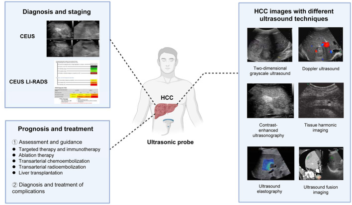
Graphical abstract
Introduction
Liver cancer is one of the most commonly diagnosed malignancies and ranks as the fourth leading cause of cancer-related mortality worldwide. Hepatocellular carcinoma (HCC) accounts for approximately 75–85% of primary liver cancers and is a high-incidence tumor that is sixth among all cancer types and has an upward trend in prevalence. From 1990 to 2015, there was a notable 75% rise in the global prevalence of liver cancer. Projections based on current trends indicate that the number of newly diagnosed liver cancer cases may increase by up to 62% in 2040. HCC is also characterized by a high fatality rate, with a morbidity and mortality ratio nearing unity, and its mortality rate is increasing, especially in the USA and Europe, where it has emerged as the fastest-growing contributor to cancer-related death.1 Increased understanding of the pathogenesis and development of HCC has led to increasingly diverse treatment. However, other than HCC diagnosed at an early stage, the prognosis remains unsatisfactory. Because it is often detected in an advanced stage, the therapeutic efficacy of HCC is limited, and the overall prognosis is poor. To improve long-term survival and prognosis, early detection and diagnosis are crucial for improving the potential of curative treatment, including surgical resection and liver transplantation.
Clinical practice guidelines widely recommend ultrasound technology as the primary method for screening and monitoring HCC due to its noninvasive, cost effective, and convenient nature. The recent development of contrast-enhanced ultrasonography has improved the diagnostic accuracy of this imaging technique and improved the characterization of HCC. Ultrasound is expected to provide more biological information on HCC, which will enable personalized treatment decisions in the era of precision medicine. This review summarizes the latest advancements in ultrasound and methods of monitoring HCC, its diagnosis, treatment, prognosis, and identifying common complications.
Ultrasound techniques applied to HCC
Two-dimensional gray-scale ultrasound
Two-dimensional (2D) gray-scale ultrasound is the primary imaging modality for HCC screening. It provides real-time visualization of the liver with insights into tumor location. It also facilitates prompt assessment of lesion characteristics, tumor borders, morphology, and the dynamic evaluation of echogenicity at the tumor margins, within the lesion itself and posterior to it. The formation of fibrous capsules and a nodule in nodule appearance are distinctive morphologic features of HCC and are of significant importance in early-stage diagnosis.2 Two-dimensional gray-scale ultrasound is widely used in clinical practice to screen HCC in healthy populations and is the foundation of HCC ultrasound diagnosis. It can provide a preliminary description of the morphology of HCC (Supplementary Fig. 1A/a). However, due to the limited information about HCC, it has not become a means for further diagnosis and adjunctive therapy.
Doppler ultrasound
Color Doppler ultrasound can visualize intricate details of blood flow both inside and surrounding HCC. This imaging modality encompasses various techniques such as color Doppler flow imaging, color Doppler energy, super microvascular imaging, and micro flow imaging. Color Doppler flow imaging is effective for visualizing blood vessels with a diameter of >1 mm and a flow velocity >3–5 cm/s, both within the tumor and at its periphery.3 Color Doppler energy is a well-established method of observing low-speed blood flow and small blood vessels by displaying an intravascular energy-based signal. Super microvascular imaging and micro flow imaging are recently developed Doppler microflow imaging modes that enable clear visualization of low-speed small blood flow without the need for contrast agents.4,5 There is currently a lack of research on the diagnostic and therapeutic applications of these two techniques in managing HCC, especially regarding accurate diagnosis and differential diagnosis. Their clinical value needs confirmation by large multicenter studies. Color Doppler ultrasound provides information about the blood flow within HCC tumors and in the surrounding tissue. This information helps clinicians understand tissue differentiation and the extent of vascular invasion of HCC. It not only provides important diagnostic value but also serves as a therapeutic basis for selecting subsequent treatments for HCC, especially for those with a rich or relatively poor blood supply. For example, color Doppler ultrasound can image vessels appropriate for transarterial chemoembolization (TACE) (Supplementary Fig. 1B/b). In clinical practice, color Doppler ultrasound has become an indispensable tool.
Contrast-enhanced ultrasonography
Contrast-enhanced ultrasonography is a revolutionary advance in the field of ultrasound imaging following the development of 2D gray-scale ultrasound and Doppler ultrasound. The technique is an effective means of evaluating focal liver lesions based on hemodynamic changes. Administration of contrast agents allows dynamic monitoring of changes in blood flow specifically for HCC. It has been extensively utilized across various stages of management, including preoperative diagnosis, guided tumor biopsy, intraoperative guidance during radiofrequency ablation procedures, and evaluation of early treatment response and complications, along with monitoring tumor recurrence. This technique is being refined for use in diagnosing and planning treatment for HCC. In addition, three-dimensional contrast-enhanced ultrasound (3D-CEUS) is an advance of 2D-CEUS. This technique enables visualization of tumor spatial vascularity and intratumoral perfusion by the reconstruction of 3D images (Supplementary Fig. 1C/c). CEUS is preferred by many clinicians and patients because it provides more accurate tumor information and is not affected by issues such as radiation from computed tomography (CT) contrast agents.
Ultrasonic contrast agents are materials that include microbubbles, and their characteristics depend on the type of gas and the enclosure composition. As the microvessel diameter is smaller than that of a red blood cell, it can move through the bloodstream after intravenous injection. We listed the commercially available clinical ultrasound contrast agents described by Chang6 and Frinking et al.7 in Supplementary Table 1. They include Levovist, a first-generation contrast agent comprising galactose-based gas-filled microbubbles containing palmitic acid (Schering AG, Berlin, Germany). There are three second-generation contrast agents, SonoVue (sulfur hexafluoride; Bracco, Milan, Italy), Definity (Perflutren lipid microspheres; Lantheus Medical Imaging, Billerica, MA, USA), and Sonazoid (perfluorobutane; GE Healthcare United Kingdom Ltd., Pollards Wood, UK). SonoVue and Definity are blood-pool contrast agents that circulate within blood vessels and Sonazoid specifically accumulates in the reticuloendothelial system to enable imaging of Kupffer cells. These advanced microbubbles consist of a gas core with low diffusion properties enclosed by a highly flexible and soft envelope. This design improves stability and persistence during diagnostic medical procedures.8,9
Tissue harmonic imaging
Tissue harmonic imaging (THI) detects the harmonics generated by sound waves as they propagate through tissue, with wave signals being meticulously received and intensified for imaging purposes. Currently, THI is widely used in diagnosing HCC.10 It can image the morphology and structure of the liver, liver cancers, outlines of portal vein tumor thrombi, internal echo, the relationship of the tumor with surrounding tissues, and enlarged lymph nodes in the hilar region. It improves the detection rate of subtle tissue lesions and HCC, while being especially valuable in distinguishing hyperplastic nodules from malignant nodules among patients with liver cirrhosis.11 Previous studies have examined the use of contrast agent-enhanced harmonic imaging to evaluate HCC diagnosis and treatment response. These studies revealed a distinct contrast-enhanced difference between tumors and the surrounding normal liver tissue.12 THI significantly improves the accuracy of liver cancer diagnosis but because of its principles and technical limitations, it is not currently the primary method of evaluating HCC in the clinical setting. However, with the integration and development of new technologies, such as CEUS, it has been gradually adopted for the diagnosis and treatment of HCC.
Ultrasound elastography
Ultrasound elastography is an imaging technique that uses ultrasound to detect the molecular and microstructural features of target tissues, providing valuable information on tissue elasticity and its distribution. Ultrasound elastography has two modes, strain elastography and shear wave elastography (SWE).13,14 SWE overcomes subjective factors that could impact strain elastography results, such as probe pressure and frequency. It has advantages for diagnosing abdominal organ disease, including liver fibrosis.13,15,16 SWE can effectively detect and assess the severity of liver fibrosis outside tumor lesions in patients with HCC (Supplementary Fig. 1D/d).
Acoustic radiation force impulse (ARFI) imaging is a key component of SWE. Virtual touch tissue quantification is an integral component of ARFI. It collects lateral shear wave data by sequential detection of pulsed waves and determines tissue stiffness by calculating velocity values. ARFI uses stable acoustic pulses to differentiate benign from malignant tumors while enabling elastic assessment of deep-seated tissues such as the liver, kidneys, and spleen.17 The advantages of ARFI include decreased inter-operator variability, high-level repeatability, tissue stiffness quantification, deep tissue penetration by focused ultrasound beams, and efficient contrast conversion.18
Ultrasound fusion imaging
Investigation of the clinical applications of ultrasound fusion imaging has expanded because of advancements in ultrasound technology and computing power.19–21 This innovative technique depends on an automated combination of ultrasound and radiological imaging that enables synchronous correlation of ultrasound images and one or more cross-sectional radiological images such as CT, magnetic resonance imaging (MRI) or positron emission tomography-CT. These correlated images are then reconstructed in a 3D format within their corresponding plane.22 Providing a simultaneous display of multiplanar reconstruction images on a single screen, allows physicians to quickly make diagnostic or procedural decisions with greater efficiency.23,24 This method integrates diverse ultrasound techniques, such as color Doppler ultrasound, elastography, and CEUS, for precise localization and characterization of lesions based on specific requirements.22 The procedure uses complex algorithms, restricted computing competence, and integration of multiple ultrasound and radiology techniques. The process relies on skillful operators, and has the potential of image offset during fusion. Moreover, challenges arise from changes in patient positioning that interfere with the accurate alignment of the ultrasound image with CT or MRI image. While this innovative technology holds broad application potential, further study is needed to standardize technological parameters across various scenarios. In recent years, ultrasound fusion imaging has been rapidly developing, and clinical use has begun advances that make it easier to integrate ultrasound and CT/MRI images (Supplementary Fig. 1E-F/e-f), and further technological innovations are underway. In the future, an important direction for ultrasound imaging will be the development of ultrasound fusion imaging technology.
Application of ultrasound technologies in different stages of diagnosis and treatment of HCC
Surveillance
Ultrasound is an essential technique for HCC surveillance. Abdominal ultrasonography is the preferred method for monitoring patients at high risk of HCC, including those with cirrhosis, and noncirrhotic patients with chronic hepatitis B virus (HBV) and/or hepatitis C virus (HCV) infection or high HBV-DNA levels. Noncirrhotic patients with a family history of HCC and those with nonalcoholic fatty liver disease should also receive regular ultrasound monitoring. A randomized controlled clinical trial including over 18,000 Chinese HCC patients found that ultrasound screening reduced the mortality risk by 37%.25 Moreover, long-term prospective follow-up of patients with compensated viral cirrhosis suggested that consistent adherence to the recommended 6-month screening interval resulted detection of more early HCC cases, which exhibited a survival benefit of the earlier treatment procedures.26 Two-dimensional gray-scale ultrasound imaging is a practical clinical application that can be used for early screening of HCC. This technique is both convenient to implement and capable of achieving the purpose of preliminary screening. Ultrasound has a sensitivity of 40–81% and a specificity of 80–100% for HCC monitoring.1 One reason for differences in the effectiveness of surveillance is that the surveillance populations had different high-risk factors for HCC. For patients with HBV, ultrasound detected HCC with a sensitivity of 84% for any stage and 63% for the early stage. However, for patients with cirrhosis, the combined sensitivity of ultrasound for early HCC was only 47% (95% CI: 33–61%). One reason is that cirrhosis with fibrous septa and regenerative nodules that appear as a coarse pattern on ultrasound may mask the existence of early HCC. Another important factor influencing the sensitivity is the size of the HCC lesion. Smaller lesions are often associated with a lower sensitivity of surveillance, with those <2 cm in diameter having a sensitivity of only 65%. Indeed, the above data are largely dependent on the expertise of the operator and the influence of several patient-level factors including obesity, alcoholic or nonalcoholic steatohepatitis cirrhosis, and Child-Pugh class B or C cirrhosis.27,28 These factors are the primary cause of the wide variation in ultrasound sensitivity observed in different clinics and patients.
To enhance the sensitivity, it is imperative to reinforce the professional and standardized training for personnel who perform HCC ultrasound monitoring. It is also effective to monitor HCC with sensitive biological indicators and ultrasound. A meta-analysis that compared the performance of ultrasound alone with ultrasound plus alpha-fetoprotein (AFP) found that the former was 63% (95% CI: 48–75%) and the latter was 45% (95% CI: 30–62%), showing that the combination of ultrasound and AFP improved HCC detection at an early stage. The diagnostic odds ratio, considering both sensitivity and specificity, was higher when combining the two tests than using ultrasound alone. The 2017 Asian-Pacific Association for the Study of the Liver guidelines recommend a combination of AFP level and ultrasound for routine monitoring of HCC.29 However, emerging data suggest that AFP has a limited diagnostic advantage (6–8%), and when combined with ultrasound, the increase in the percentage of detected cases includes false-positive results and increases surveillance costs.30 It remains to be seen whether AFP improves the sensitivity of ultrasound and needs more research and clinical experience.
Diagnosis and staging
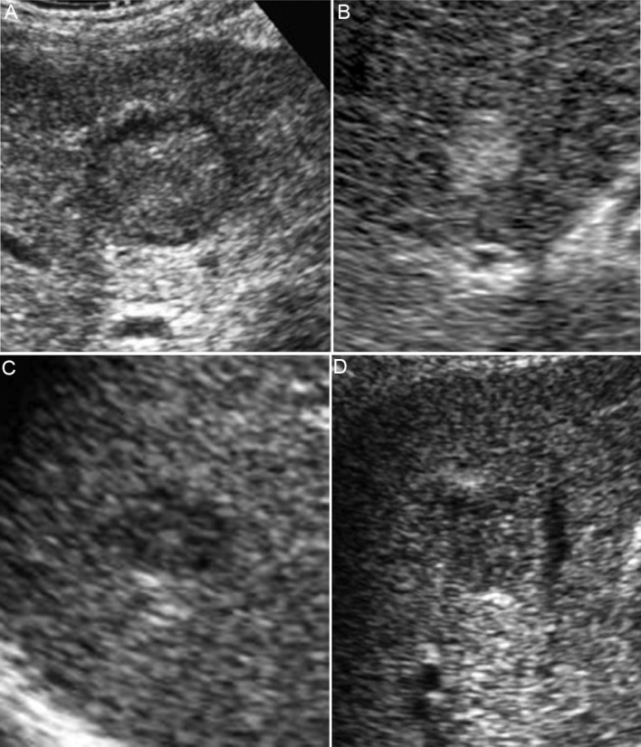
If liver nodules are detected, diagnosis and staging are necessary to guide subsequent treatment strategies. B-mode ultrasound serves as a reliable for imaging the macroscopic characteristics of HCC with distinct presentations varying by size on ultrasonography. The 2 cm threshold represents a significant differentiation between HCC sizes in ultrasound appearance. Nodules that are <2 cm often lack characterization, whereas those >2 cm usually have typical ultrasound features such as a mosaic pattern, nodular appearance in nodules, peripheral sound-through (halo sign), and silhouette.31 HCC can be classified into five types by its macroscopic characteristics: small nodular type with inconspicuous margin, simple nodular type, simple nodular type with the outgrowth of nodules, confluent multinodular type, and infiltrating type. These types are ordered in terms of malignancy increment. Small nodules are classified into two groups by Moribana et al (Fig. 1).32 depending on the presence of halos, type 1 (with halos) and type 2 (without halos). Type 2 is further divided into three subgroups. Type 2a is homogeneously hyperechoic, type 2b is hypoechoic with smooth margins, and type 2c is hypoechoic with irregular or indistinct margins. There is a gradual increase in malignant potential among the three subtypes.
Fig. 1. Classification of B-mode ultrasonographic images of small hepatocellular carcinoma.
Hepatocellular carcinoma nodules <3 cm were divided into two groups using B-mode ultrasonography, type 1 (A) with and type 2 without a halo. Type 2 was divided into three subgroups, type 2a, homogenous hyperechoic (B); type 2b, hypoechoic with a smooth margin (C); and type 2c, hypoechoic with an irregular or unclear margin (D).
In addition to CT and MRI, CEUS is a frequently utilized imaging modality for diagnosing liver cancer. All guidelines recommend the use of contrast-enhanced imaging when ultrasound has identified focal lesions larger than 1 cm. CEUS is a dynamic imaging technique that enables quantitative evaluation of microcirculation within tissues following the injection of a contrast agent. Rapid real-time acquisition of sequential images with contrast agent concentrations over time enables subsequent extraction and adjustment of perfusion parameters. Perfusion imaging provides important quantitative information regarding tumor vasculature and angiogenesis. CEUS thus enables the dynamic observation of nodules during the influx, dispersion, and absorption of contrast medium in a specific liver, which makes it more frequently used for high-risk HCC screening and assessment of suspected liver nodules. A recent review describes important ultrasound updates and CEUS techniques.33 CEUS increases diagnostic accuracy by improving sensitivity and specificity and facilitating a more professional and optimized diagnostic process. In contrast, CT scans involve radiation exposure and possible risk of allergy to the iodine contrast agent. Although without radiation, MRI is more expensive and requires a gadolinium contrast agent that can lead to nephrotoxicity in some patients. Furthermore, recent data indicated a long-term risk of gadolinium accumulation, with uncertain clinical implications.34
Diagnosis and staging of HCC by CEUS imaging
Significant hemodynamic changes take place during hepatocarcinogenesis. The blood supply to the nodule increases with the degree of malignant transformation. Specifically, the portal bundle in the nodule decreases while the number of unpaired arteries increases. Ultimately, HCC is primarily supplied by the hepatic arterial system by abnormal unpaired arteries. This leads to excessive enhancement in the hepatic arterial phase of the liver on the CEUS background accompanied by a distinguishing pattern during the portal venous phase and/or delayed phase. The blood supply in HCC is positively associated with its malignancy, the detection of which is a key reference index in the diagnosis and staging of HCC. Notably, CEUS is effective for showing the distribution of tumor vessels and its sensitivity is superior to that of enhanced CT.
Superparamagnetic iron oxide, like Sonazoid, can be phagocytosed by hepatic Kupffer cells,35 and. It can assist in estimating the histological grade of HCC by post-vascular phase ratio (post-contrast echo of the tumor lesion/post-contrast echo of adjacent liver). Sonazoid CEUS uses the contrast echogenicity of neoplastic lesions or non-neoplastic liver parenchyma as a quantitative parameter to evaluate HCC staging. The Kupffer phase ratio decreases with decreasing HCC differentiation. Under this condition, all moderately and poorly differentiated HCCs had a hypoechoic pattern characterized by perfusion defects.36 In contrast, the majority (69.2%) of well-differentiated HCCs had an isoechoic pattern, which was consistent with the histology of the HCC category.36
The American College of Radiology launched the Liver Imaging Reporting and Data System (LI-RADS) for CEUS in 2016.37 This system utilizes SonoVue (Bracco Imaging SpA, Milan, Italy) as a contrast agent to classify hepatic nodules based on their size, pattern of arterial phase enhancement, type of clearance, and duration between enhancement and clearance. The system categorizes nodules into five types (LR-1 to 5) that determine the possibility of HCC and the LR-M category that indicates probable malignancy without specificity (Supplementary Fig. 2). In 2017, CEUS incorporated a revised edition of LI-RADS to enhance diagnostic efficiency. Notably, the LI-RADS system proposed by the American College of Radiology provides a distinct algorithm with auxiliary imaging capabilities for ultrasound screening and surveillance of HCC, as well as for the diagnosis of non-HCC malignancy and large vessel invasion, which surpasses previously established diagnosis. Although supporting evidence is still limited, it has significant potential to enhance the detection and characterization of hepatic nodules.
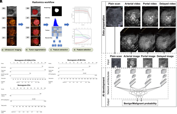
Vascular invasion is a common feature of HCC. Microvascular invasion (MVI) is usually detected by postoperative pathology, and when observed in pathological tissues it often indicates the progress of HCC. MVI is known to indicate the risk of recurrence and prognosis of HCC, even though its definition in HCC has yet to be standardized. Owing to the strong diagnostic performance of CEUS in HCC, an increasing number of studies have explored its potential in the diagnosis of MVI. A study developed a CEUS nomogram for predicting pre-operative MVI in HCC. The investigators compared it with a nomogram based on gadopentetate dimeglumine-enhanced MRI (commonly referred to as Gd-MRI) and verified that it was a reliable tool for predicting MVI in HCC with a predictive performance equal to that of Gd-MRI.38 An ultrasound-based radiomics score for pre-operative prediction of MVI in HCC developed in another study was found to be an independent predictor.39 Another study used CEUS-based radiomics and deep convolutional neural network models as noninvasive predictors of MVI status in HCC (Fig. 2).40 CEUS predicted poor prognosis of patients, which is of great significance for developing treatment strategies. In clinical practice, surgeons examine pathological tissue after liver cancer resection to detect MVI. This process relies on the examination and analysis of pathological tissue. However, if CEUS can successfully predict MVI and gain consensus among experts, it would be a significant innovation.
Fig. 2. Progress in predicting microvascular invasion in hepatocellular carcinoma using contrast-enhanced ultrasound.
(A) A flow chart of a radiomic model for predicting MVI status in HCC patients and the study of radiomic workflow. (B) The use of artificial intelligence to help radiologists identify malignant and focal liver lesions using contrast-enhanced ultrasound. (C) A nomogram of contrast-enhanced ultrasonography - liver imaging reporting model and clinical parameters predicting MVI in patients with liver cancer. AFP, alpha fetoprotein; AI, artificial intelligence; BM, B-mode; AP, artery phase; PVP, portal venous phase; DP, delay phase; HCC, hepatocellular carcinoma; MVI, microvascular invasion.
However, CEUS has some limitations. Firstly, inter-observer and intra-observer differences can always arise during the diagnosis of HCC using CEUS given the operator-dependent nature. Secondly, use of ultrasound is restricted in patients with severe obesity, air in the intestine, or heterogeneous cirrhosis. Thirdly, ultrasound findings are often insufficient in cases with deep, subdiaphragmatic, and multiple lesions. Finally, its ability to visualize the entire liver is also limited. Fortunately, implementing standardized protocols and using safe, stable contrast agents has strengthened the reproducibility and dependability of CEUS.
Ultrasound combined with other new techniques in the diagnosis of HCC
The primary dispute regarding CEUS is its limited universality in interpreting real-time imaging by different readers. The development of computer-aided diagnostic (CAD) systems and artificial intelligence (AI)-assisted identification has been proved to have remarkable capabilities to address this challenging issue. CAD has the potential to augment the diagnostic effectiveness of clinical physicians, and computer analysis results can provide clinicians with secondary reference opinions, which enhance the consistency of image interpretation by healthcare providers. A recent study has proven the potential of CEUS with CAD systems to identify poorly differentiated liver cancers including HCC. The results offer substantial promise for hepatologists to improve the diagnostic ability to distinguish among three levels of tissue differentiation in HCC.41 The technique may also assist in the noninvasive assessment of the malignancy level of liver cancer in the context of personalized medicine.
Deep learning models can learn predictive features directly from raw image pixels, eliminating the need for subjective feature engineering required in traditional machine learning. They have a higher tolerance for variation and noise in data. Some studies reported that AI strategies based on CEUS resulted in better performance of the healthcare provider and decreased inter-observer variability when distinguishing HCC from other tumor-related or benign conditions. In one study, Hu et al.42 developed an AI system based on CEUS to differentiate between benign and malignant liver focal lesions. The diagnostic performance of the AI system was superior to that of radiologists. A study by Liu F et al.43 used radiomics modeling to classify the progression-free survival (PFS) of different treatment groups with two radiomic features, radiofrequency ablation (RFA) and surgical resection (SR), that enabled personalized 2-year PFS predictions, which indicated that radiomics based on CEUS can optimize curative treatment of patients with early-stage HCC.
Evaluation of treatment effect
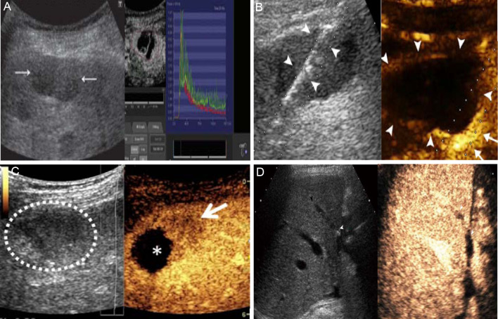
Early-stage HCC is primarily recommended for curative surgical resection. However, even with this treatment, HCC has a recurrence rate of 12% after 5 years. Unfortunately, the majority of patients with HCC are initially diagnosed at intermediate or advanced stages, which deprives them of the opportunity of curative resection. Recent noteworthy advances in the systemic treatment of HCC, local administration of TACE, hepatic arterial infusion chemotherapy, selective internal radiation therapy, stereotactic body radiation therapy, and ablation techniques including RFA and microwave ablation (MWA). Systemic drug treatment includes molecular targeted therapies and immunotherapy. In addition to diagnostic and surveillance functions, CEUS has been used to evaluate locoregional treatments such as RFA and TACE in patients with HCC (Fig. 3). CEUS can assess the efficacy of these interventions by identifying alterations of tumor vascularization and detecting residual or recurrent tumors.
Fig. 3. Assessment of treatment efficacy and adjunct therapy in hepatocellular carcinoma patients based on ultrasound.
(A) Ultrasound monitoring images of patients with HCC (arrow) 3 months after sorafenib treatment. (B) Ultrasound-guided surgical procedure of patients with recurrent HCC (arrowhead). (C) Ultrasound images of nodules of patients with HCC undergoing transcatheter arterial chemoembolization. Circles are the preoperative lesion, and asterisk is the postoperative lesion. (D) Results of ultrasound monitoring 1 year after liver transplantation, showed good liver growth. HCC, hepatocellular carcinoma.
Assessment of targeted therapy and immunotherapy
The insidious progression of HCC often leads to a delayed diagnosis at an advanced stage, necessitating the reliance on systemic treatments such as targeted therapy, chemotherapy, and immunotherapy. Recent studies of tumor molecular signaling pathways and microenvironments have highlighted targeted therapy as the pivotal point for the management of late-stage HCC. Sorafenib was approved in 2007 as the first drug for systemic treatment of HCC. This anti-angiogenic drug targets endothelial cells rather than tumor cells, thereby inhibiting the formation of tumor microvessels without causing significant clinical alterations in tumor size. The efficacy of sorafenib treatment is influenced by the incidence of severe adverse events. As it is vital to promptly assess the effectiveness of targeted drug therapy, CEUS might serve as a valuable early predictor of sorafenib treatment effectiveness in patients with advanced liver cancer. CEUS has been shown to accurately image tumor blood supply and perfusion, to reflect the influence of targeted drug therapy on the tumor microenvironment, helped to predict tumor response to drugs, to improve the accuracy of predicting PFS and overall survival.26,44 Previous studies consistently demonstrated that CEUS could predict the efficacy of drugs that target angiogenesis at an early stage, which is a reference important for the precise treatment of liver cancer and possibly prolonging life.44,45
Assessment of ablation therapy
Thermal ablation is the preferred nonsurgical treatment option for HCC patients with Barcelona Clinic Liver Cancer (BCLC) 0-A stage. Promising results have been achieved with techniques such as MWA and RFA.46 Within a week or month after RFA, CEUS had a sensitivity exceeding 80% and an almost perfect specificity (100%) for assessing the response to RFA. However, in certain instances, the immediate assessment of the effectiveness of RFA therapy during the interventional procedure is necessary for prompt retreatment. CEUS with its unique temporal and spatial resolution, as well as its portability, can effectively facilitate this process while significantly reducing incomplete ablation following initial treatment.47,48 Furthermore, there is evidence suggesting that the fusion of 3D-CEUS is a feasible and accurate tool for evaluating the immediate efficacy of thermal ablation in HCC patients. Recent studies have used a novel single-modal fusion imaging technique that employed 3D-CEUS to assess the ablation borders following MWA of HCC, achieving a success rate of >95% in image fusion. The performance. of 3D-CEUS fusion has been excellent in terms of time efficiency and a success rate consistent with contrast-enhanced CT fusion.49
Assessment of TACE
TACE is a minimally invasive treatment for patients with HCC at the BCLC B stage. It involves the administration of a combination of chemotherapy and embolic agents through a catheter into the blood vessels of the tumor, including the hepatic artery and its branches. It can also be used for BCLC A stage patients and for downgrading or palliative treatment of BCLC C stage patients.50 Enhanced CT and MRI evaluations are usually conducted 4 weeks after TACE to diminish arterial phase hyperenhancement caused by inflammation and congestion in and around the tumor following ablation, and eliminating the iodized oil artifact observed in CT scans. This delay in retreatment, normally lasting 6–8 weeks, impacts patients who have undergone incomplete ablation. CEUS can identify treated lesions in less than 4 weeks. After successful TACE treatment of liver masses, CEUS images show specific characteristics such as well-defined margins, absence of internal blood flow throughout the enhancement stage, and lack of peripheral nodular enhancement. Unlike CT and MRI, CEUS is rarely affected by iodized oil deposition artifacts, and can differentiate viable tumors from postoperative inflammation. Relevant studies have demonstrated that CEUS is able to detect viable tumors 4–6 weeks earlier than CT and MRI for assessment of TACE treatment response. This result has significant importance for the disease management of HCC patients.
Assessment of transarterial radioembolization
Transarterial radiation embolization is the recommended treatment for HCC patients with stage B BCLC. The Y-90 isotope generates pure beta radiation as it decays to stable Y-90 with a half-life of 64 h, effectively eliminating cancerous cells and achieving the treatment objective.51,52 It has been observed that CEUS is able to detect changes in tumor blood perfusion as early as 1 week after treatment. Furthermore, early alterations in tumor vasculature and hemodynamics have a strong association with long-term CT or MRI findings 3–4 months after treatment. The aforementioned advantages of temporal resolution, cost effectiveness, and accessibility position CEUS as an ideal method for monitoring the liver cancer response to transarterial radiation embolization.
Liver transplantation
Although the indications for CEUS in in-situ liver transplantation have not been fully validated by large multicenter studies, its use is progressively increasing in specialized centers. The purpose of preoperative imaging for liver transplantation is to select appropriate candidates for in-situ liver transplantation by excluding contraindications such as HCC or extrahepatic malignancies, while also evaluating the patency of the hepatic vascular and biliary systems alongside anatomical variations.53 CEUS is a potential diagnostic tool for assessing focal liver lesions and portal vein thrombosis prior to surgery. Prompt identification and management of various complications that may occur following in-situ liver transplantation are crucial to ensure graft longevity and optimal functionality, particularly when they manifest shortly after surgery. CEUS has a key role in the surveillance of postoperative complications, but its use is currently limited to specific instances identified by CT or MRI, where it is an adjunct diagnostic tool for problem-solving purposes. Its primary objective is to function as a reliable and integrated initial screening procedure to avoid potential complications.54,55
Summary
In general, ultrasonography is a valuable imaging technique for diagnosis, surveillance, and management of liver tumors and provides reliable information for clinicians and radiologists. Compared with other imaging modalities, its noninvasiveness, absence of radiation exposure, ease-of-use, cost effectiveness, minimal renal burden, and real-time imaging make it an appealing option for assessing liver lesions in clinical practice, particularly in patients with liver cirrhosis or undergoing locoregional treatment.
Previous reviews focused on the diagnostic use of ultrasound in HCC or its assistive role in treatment. They did not link the ultrasound techniques or provide a unified explanation of the selection and uses of different ultrasound techniques throughout the entire diagnostic and treatment process of HCC. This review comprehensively covers the use of ultrasound for screening, diagnosis, staging, monitoring, adjunctive therapy, and prognosis of HCC. The advantages and disadvantages of different ultrasound stages and techniques are discussed following the sequence of patient evaluation from diagnosis to treatment, along with corresponding adjunctive measures or alternative technologies to help clinicians choose among. available ultrasound techniques. We also review the latest ultrasound advances described in HCC studies, including imagingomics, AI, and others. These technological developments enable a more precise and efficient application of ultrasound in HCC. For example, imagingomics analyzes big data and pattern recognition to extract valuable information from a large number of ultrasound images, assisting physicians in more accurately diagnosing and staging HCC. AI, on the other hand, enhances the interpretation and diagnostic accuracy of ultrasound images by machine learning and deep learning algorithms. It also increases the speed of image analysis, providing more reliable evidence for clinical decision-making. These latest advancements continuously increase the benefit of ultrasound technology in the management of HCC, making it an indispensable tool.
However, there are several limitations of ultrasound in the diagnosis and treatment of HCC. Firstly, accurate interpretation of the results relies heavily on the expertise and experience of the examiner. Secondly, lack of standardization across centers and modalities may be attributed to insufficient high-quality studies. Thirdly, advantages of ultrasound relative to other noninvasive imaging techniques are not complete as far as detecting complications such as thrombi in patients with HCC. Furthermore, there is a dearth of novel contrast agents, contrast-specific imaging modalities, and analytical methods. Therefore, it is imperative to acknowledge that CEUS can often only serve in clinical practice as an adjunctive approach alongside other noninvasive techniques. Moreover, it is advisable to carry out the procedure under the guidance of experienced operators to ensure the reproducibility of data. Future investigations should focus on improving image quality for perfusion imaging, and exploring standardized protocols for forthcoming studies.
In this context, future research on the application of ultrasound in HCC might proceed by (1) incorporating international large-scale settings with cohorts from diverse backgrounds to obtain more comprehensive data on ultrasound imaging, especially CEUS in HCC management, (2) comparing and analyzing various ultrasound imaging methods, optimizing and integrating various imaging models to provide accurate biological information for the diagnosis and treatment of HCC, (3) actively promoting collaborative research by industry, universities, and scientific institutions, along with enhancing the development of new contrast agents and contrast-specific imaging methods, and (4) giving full play to the advantages of an AI algorithm, improve the quality and interpretability of images, and enhance the ability of diagnosis and evaluation of liver tumors.
Supporting information
(A, a) A 59-year-old woman with HBV with nodules on ultrasound monitoring, continuous arterial CEUS showing progressive centripetal filling. (B, b) A 50-year-old woman with hepatitis C virus (HCV)cirrhosis with hypoechoic nodules >3 cm and early arterial CEUS showing isovascular distribution. (C, c) A 64-year-old man with alcoholic cirrhosis with an exogenous hypoechoic nodule >2 cm with isointensification at the arterial peak. (D, d) Ultrasound monitoring of a 27-year-old man with occult cirrhosis. (F, f) B-mode ultrasound of a 61-year-old man with alcoholic cirrhosis and hypoechoic 2-cm lesions of in the right liver. CEUS showed high arterial stage enhancement of nodules. (G, g) Sagittal B-mode ultrasound of a 59-year-old man with HCV cirrhosis and new ascites and a focal, large, echogenic liver mass with high enhancement of both portal vein thrombosis and mass in the arterial phase.
(A, a) The accuracy of two-dimensional ultrasound in measuring the size of HCC lesions is not significantly different from that measured on ultrasonography images. (B, b) Color Doppler measurements of hepatic arterial beat index and resistance index abnormalities can predict HCC recurrence after surgery and show abnormal portal vein internal diameters. (C, c) Dynamic 3D-CEUS images showed an area of enhancement at the edge of an intrahepatic lesion in an HCC patient after radiofrequency ablation that matched the lesion shown on MRI, suggesting recurrence of the tumor. (D, d) Shear wave elastography can well detect and evaluate the severity of liver fibrosis, and the results are more objective and reproducible. (E, e) MR-US fusion imaging. Biopsy of very small liver nodule, hypointense on the late hepatobiliary phase of MR, with a final diagnosis of a metastatic lesion. (F, f) CT-US fusion imaging. Treatment of very small hypervascular nodular recurrence of HCC adjacent to a previously ablated area.
Abbreviations
- AFP
Alpha-fetoprotein
- AI
Artificial Intelligence
- ARFI
Acoustic radiation force impulse imaging
- CEUS
Contrast-enhanced ultrasound
- HCC
Hepatocellular carcinoma
- LI-RADS
Liver Imaging Reporting and Data System
- MWA
Microwave ablation
- RFA
Radiofrequency ablation
- SWE
Shear wave elastography
- TACE
Transarterial chemoembolization
- THI
Tissue harmonic imaging
- US
Ultrasound
References
- 1.Yang JD, Hainaut P, Gores GJ, Amadou A, Plymoth A, Roberts LR. A global view of hepatocellular carcinoma: trends, risk, prevention and management. Nat Rev Gastroenterol Hepatol. 2019;16(10):589–604. doi: 10.1038/s41575-019-0186-y. [DOI] [PMC free article] [PubMed] [Google Scholar]
- 2.Kim WK, Meyer A, Möllmann H, Rolf A, Möllmann S, Blumenstein J, et al. Cyclic changes in area- and perimeter-derived effective dimensions of the aortic annulus measured with multislice computed tomography and comparison with metric intraoperative sizing. Clin Res Cardiol. 2016;105(7):622–629. doi: 10.1007/s00392-016-0971-3. [DOI] [PubMed] [Google Scholar]
- 3.Han H, Ji Z, Ding H, Zhang W, Zhang R, Wang W. Assessment of blood flow in the hepatic tumors using non-contrast micro flow imaging: Initial experience. Clin Hemorheol Microcirc. 2019;73(2):307–316. doi: 10.3233/CH-180532. [DOI] [PubMed] [Google Scholar]
- 4.Aghabaglou F, Ainechi A, Abramson H, Curry E, Kaovasia TP, Kamal S, et al. Ultrasound monitoring of microcirculation: An original study from the laboratory bench to the clinic. Microcirculation. 2022;29(6-7):e12770. doi: 10.1111/micc.12770. [DOI] [PMC free article] [PubMed] [Google Scholar]
- 5.Mao Y, Mu J, Zhao J, Yang F, Zhao L. The comparative study of color doppler flow imaging, superb microvascular imaging, contrast-enhanced ultrasound micro flow imaging in blood flow analysis of solid renal mass. Cancer Imaging. 2022;22(1):21. doi: 10.1186/s40644-022-00458-2. [DOI] [PMC free article] [PubMed] [Google Scholar]
- 6.Chang EH. An Introduction to Contrast-Enhanced Ultrasound for Nephrologists. Nephron. 2018;138(3):176–185. doi: 10.1159/000484635. [DOI] [PMC free article] [PubMed] [Google Scholar]
- 7.Frinking P, Segers T, Luan Y, Tranquart F. Three Decades of Ultrasound Contrast Agents: A Review of the Past, Present and Future Improvements. Ultrasound Med Biol. 2020;46(4):892–908. doi: 10.1016/j.ultrasmedbio.2019.12.008. [DOI] [PubMed] [Google Scholar]
- 8.Arita J, Takahashi M, Hata S, Shindoh J, Beck Y, Sugawara Y, et al. Usefulness of contrast-enhanced intraoperative ultrasound using Sonazoid in patients with hepatocellular carcinoma. Ann Surg. 2011;254(6):992–999. doi: 10.1097/SLA.0b013e31822518be. [DOI] [PubMed] [Google Scholar]
- 9.Kang HJ, Lee JM, Yoon JH, Lee K, Kim H, Han JK. Contrast-enhanced US with Sulfur Hexafluoride and Perfluorobutane for the Diagnosis of Hepatocellular Carcinoma in Individuals with High Risk. Radiology. 2020;297(1):108–116. doi: 10.1148/radiol.2020200115. [DOI] [PubMed] [Google Scholar]
- 10.Sodhi KS, Sidhu R, Gulati M, Saxena A, Suri S, Chawla Y. Role of tissue harmonic imaging in focal hepatic lesions: comparison with conventional sonography. J Gastroenterol Hepatol. 2005;20(10):1488–1493. doi: 10.1111/j.1440-1746.2005.03780.x. [DOI] [PubMed] [Google Scholar]
- 11.Lencioni R, Cioni D, Bartolozzi C. Tissue harmonic and contrast-specific imaging: back to gray scale in ultrasound. Eur Radiol. 2002;12(1):151–165. doi: 10.1007/s003300101022. [DOI] [PubMed] [Google Scholar]
- 12.Kono M, Minami Y, Iwanishi M, Minami T, Chishina H, Arizumi T, et al. Contrast-Enhanced Tissue Harmonic Imaging versus Phase Inversion Harmonic Sonographic Imaging for the Delineation of Hepatocellular Carcinomas. Oncology. 2017;92(Suppl 1):29–34. doi: 10.1159/000451014. [DOI] [PubMed] [Google Scholar]
- 13.Xie LT, Yan CH, Zhao QY, He MN, Jiang TA. Quantitative and noninvasive assessment of chronic liver diseases using two-dimensional shear wave elastography. World J Gastroenterol. 2018;24(9):957–970. doi: 10.3748/wjg.v24.i9.957. [DOI] [PMC free article] [PubMed] [Google Scholar]
- 14.Shiina T, Nightingale KR, Palmeri ML, Hall TJ, Bamber JC, Barr RG, et al. WFUMB guidelines and recommendations for clinical use of ultrasound elastography: Part 1: basic principles and terminology. Ultrasound Med Biol. 2015;41(5):1126–1147. doi: 10.1016/j.ultrasmedbio.2015.03.009. [DOI] [PubMed] [Google Scholar]
- 15.Tsai E, Lee TP. Diagnosis and Evaluation of Nonalcoholic Fatty Liver Disease/Nonalcoholic Steatohepatitis, Including Noninvasive Biomarkers and Transient Elastography. Clin Liver Dis. 2018;22(1):73–92. doi: 10.1016/j.cld.2017.08.004. [DOI] [PubMed] [Google Scholar]
- 16.Sporea I, Sirli RL. Hepatic elastography for the assessment of liver fibrosis—present and future. Ultraschall Med. 2012;33(6):550–558. doi: 10.1055/s-0032-1313011. [DOI] [PubMed] [Google Scholar]
- 17.Shen YN, Zheng ML, Guo CX, Bai XL, Pan Y, Yao WY, et al. The role of imaging in prediction of post-hepatectomy liver failure. Clin Imaging. 2018;52:137–145. doi: 10.1016/j.clinimag.2018.07.019. [DOI] [PubMed] [Google Scholar]
- 18.Idilman IS, Ozdeniz I, Karcaaltincaba M. Hepatic Steatosis: Etiology, Patterns, and Quantification. Semin Ultrasound CT MR. 2016;37(6):501–510. doi: 10.1053/j.sult.2016.08.003. [DOI] [PubMed] [Google Scholar]
- 19.Faletra FF, Pozzoli A, Agricola E, Guidotti A, Biasco L, Leo LA, et al. Echocardiographic-fluoroscopic fusion imaging for transcatheter mitral valve repair guidance. Eur Heart J Cardiovasc Imaging. 2018;19(7):715–726. doi: 10.1093/ehjci/jey067. [DOI] [PubMed] [Google Scholar]
- 20.Minami Y, Kudo M. Ultrasound fusion imaging of hepatocellular carcinoma: a review of current evidence. Dig Dis. 2014;32(6):690–695. doi: 10.1159/000368001. [DOI] [PubMed] [Google Scholar]
- 21.Zur G, Andraous M, Bercovich E, Litvin M, Ofer A, Gaitini D, et al. CT-Ultrasound Fusion for Abdominal Aortic Aneurysm Measurement. AJR Am J Roentgenol. 2020;214(2):472–476. doi: 10.2214/AJR.19.21670. [DOI] [PubMed] [Google Scholar]
- 22.European Society of Radiology (ESR) Abdominal applications of ultrasound fusion imaging technique: liver, kidney, and pancreas. Insights Imaging. 2019;10(1):6. doi: 10.1186/s13244-019-0692-z. [DOI] [PMC free article] [PubMed] [Google Scholar]
- 23.Tanaka H. Current role of ultrasound in the diagnosis of hepatocellular carcinoma. J Med Ultrason (2001) 2020;47(2):239–255. doi: 10.1007/s10396-020-01012-y. [DOI] [PMC free article] [PubMed] [Google Scholar]
- 24.Xu E, Li K, Long Y, Luo L, Zeng Q, Tan L, et al. Intra-Procedural CT/MR-Ultrasound Fusion Imaging Helps to Improve Outcomes of Thermal Ablation for Hepatocellular Carcinoma: Results in 502 Nodules. Ultraschall Med. 2021;42(2):e9–e19. doi: 10.1055/a-1021-1616. [DOI] [PubMed] [Google Scholar]
- 25.Singal AG, Tiro JA, Marrero JA, McCallister K, Mejias C, Adamson B, et al. Mailed Outreach Program Increases Ultrasound Screening of Patients With Cirrhosis for Hepatocellular Carcinoma. Gastroenterology. 2017;152(3):608–615.e4. doi: 10.1053/j.gastro.2016.10.042. [DOI] [PMC free article] [PubMed] [Google Scholar]
- 26.Zocco MA, Garcovich M, Lupascu A, Di Stasio E, Roccarina D, Annicchiarico BE, et al. Early prediction of response to sorafenib in patients with advanced hepatocellular carcinoma: the role of dynamic contrast enhanced ultrasound. J Hepatol. 2013;59(5):1014–1021. doi: 10.1016/j.jhep.2013.06.011. [DOI] [PubMed] [Google Scholar]
- 27.Faccia M, Garcovich M, Ainora ME, Riccardi L, Pompili M, Gasbarrini A, et al. Contrast-Enhanced Ultrasound for Monitoring Treatment Response in Different Stages of Hepatocellular Carcinoma. Cancers (Basel) 2022;14(3):481. doi: 10.3390/cancers14030481. [DOI] [PMC free article] [PubMed] [Google Scholar]
- 28.Fraquelli M, Nadarevic T, Colli A, Manzotti C, Giljaca V, Miletic D, et al. Contrast-enhanced ultrasound for the diagnosis of hepatocellular carcinoma in adults with chronic liver disease. Cochrane Database Syst Rev. 2022;9(9):CD013483. doi: 10.1002/14651858.CD013483.pub2. [DOI] [PMC free article] [PubMed] [Google Scholar]
- 29.Omata M, Cheng AL, Kokudo N, Kudo M, Lee JM, Jia J, et al. Asia-Pacific clinical practice guidelines on the management of hepatocellular carcinoma: a 2017 update. Hepatol Int. 2017;11(4):317–370. doi: 10.1007/s12072-017-9799-9. [DOI] [PMC free article] [PubMed] [Google Scholar]
- 30.Tzartzeva K, Obi J, Rich NE, Parikh ND, Marrero JA, Yopp A, et al. Surveillance Imaging and Alpha Fetoprotein for Early Detection of Hepatocellular Carcinoma in Patients With Cirrhosis: A Meta-analysis. Gastroenterology. 2018;154(6):1706–1718.e1. doi: 10.1053/j.gastro.2018.01.064. [DOI] [PMC free article] [PubMed] [Google Scholar]
- 31.Cheng MQ, Hu HT, Huang H, Pan JM, Xian MF, Huang Y, et al. Pathological considerations of CEUS LI-RADS: correlation with fibrosis stage and tumour histological grade. Eur Radiol. 2021;31(8):5680–5688. doi: 10.1007/s00330-020-07660-5. [DOI] [PubMed] [Google Scholar]
- 32.Moribata K, Tamai H, Shingaki N, Mori Y, Enomoto S, Shiraki T, et al. Assessment of malignant potential of small hypervascular hepatocellular carcinoma using B-mode ultrasonography. Hepatol Res. 2011;41(3):233–239. doi: 10.1111/j.1872-034X.2010.00763.x. [DOI] [PubMed] [Google Scholar]
- 33.Battaglia V, Cervelli R. Liver investigations: Updating on US technique and contrast-enhanced ultrasound (CEUS) Eur J Radiol. 2017;96:65–73. doi: 10.1016/j.ejrad.2017.08.029. [DOI] [PubMed] [Google Scholar]
- 34.Pasquini L, Napolitano A, Visconti E, Longo D, Romano A, Tomà P, et al. Gadolinium-Based Contrast Agent-Related Toxicities. CNS Drugs. 2018;32(3):229–240. doi: 10.1007/s40263-018-0500-1. [DOI] [PubMed] [Google Scholar]
- 35.Suzuki S, Iijima H, Moriyasu F, Sasaki S, Yanagisawa K, Miyahara T, et al. Differential diagnosis of hepatic nodules using delayed parenchymal phase imaging of levovist contrast ultrasound: comparative study with SPIO-MRI. Hepatol Res. 2004;29(2):122–126. doi: 10.1016/j.hepres.2004.02.010. [DOI] [PubMed] [Google Scholar]
- 36.Jeong WK, Kang HJ, Choi SH, Park MS, Yu MH, Kim B, et al. Diagnosing Hepatocellular Carcinoma Using Sonazoid Contrast-Enhanced Ultrasonography: 2023 Guidelines From the Korean Society of Radiology and the Korean Society of Abdominal Radiology. Korean J Radiol. 2023;24(6):482–497. doi: 10.3348/kjr.2023.0324. [DOI] [PMC free article] [PubMed] [Google Scholar]
- 37.Kono Y, Lyshchik A, Cosgrove D, Dietrich CF, Jang HJ, Kim TK, et al. Contrast Enhanced Ultrasound (CEUS) Liver Imaging Reporting and Data System (LI-RADS®): the official version by the American College of Radiology (ACR) Ultraschall Med. 2017;38(1):85–86. doi: 10.1055/s-0042-124369. [DOI] [PubMed] [Google Scholar]
- 38.Bo J, Xiang F, XiaoWei F, LianHua Z, ShiChun L, YuKun L. A Nomogram Based on Contrast-Enhanced Ultrasound to Predict the Microvascular Invasion in Hepatocellular Carcinoma. Ultrasound Med Biol. 2023;49(7):1561–1568. doi: 10.1016/j.ultrasmedbio.2023.02.020. [DOI] [PubMed] [Google Scholar]
- 39.Hu HT, Wang Z, Huang XW, Chen SL, Zheng X, Ruan SM, et al. Ultrasound-based radiomics score: a potential biomarker for the prediction of microvascular invasion in hepatocellular carcinoma. Eur Radiol. 2019;29(6):2890–2901. doi: 10.1007/s00330-018-5797-0. [DOI] [PubMed] [Google Scholar]
- 40.Zhang D, Wei Q, Wu GG, Zhang XY, Lu WW, Lv WZ, et al. Preoperative Prediction of Microvascular Invasion in Patients With Hepatocellular Carcinoma Based on Radiomics Nomogram Using Contrast-Enhanced Ultrasound. Front Oncol. 2021;11:709339. doi: 10.3389/fonc.2021.709339. [DOI] [PMC free article] [PubMed] [Google Scholar]
- 41.Căleanu CD, Sîrbu CL, Simion G. Deep Neural Architectures for Contrast Enhanced Ultrasound (CEUS) Focal Liver Lesions Automated Diagnosis. Sensors (Basel) 2021;21(12):4126. doi: 10.3390/s21124126. [DOI] [PMC free article] [PubMed] [Google Scholar]
- 42.Hu HT, Wang W, Chen LD, Ruan SM, Chen SL, Li X, et al. Artificial intelligence assists identifying malignant versus benign liver lesions using contrast-enhanced ultrasound. J Gastroenterol Hepatol. 2021;36(10):2875–2883. doi: 10.1111/jgh.15522. [DOI] [PMC free article] [PubMed] [Google Scholar]
- 43.Liu F, Liu D, Wang K, Xie X, Su L, Kuang M, et al. Deep Learning Radiomics Based on Contrast-Enhanced Ultrasound Might Optimize Curative Treatments for Very-Early or Early-Stage Hepatocellular Carcinoma Patients. Liver Cancer. 2020;9(4):397–413. doi: 10.1159/000505694. [DOI] [PMC free article] [PubMed] [Google Scholar]
- 44.Sugimoto K, Moriyasu F, Saito K, Rognin N, Kamiyama N, Furuichi Y, et al. Hepatocellular carcinoma treated with sorafenib: early detection of treatment response and major adverse events by contrast-enhanced US. Liver Int. 2013;33(4):605–615. doi: 10.1111/liv.12098. [DOI] [PubMed] [Google Scholar]
- 45.Watanabe R, Munemasa T, Matsumura M. Contrast-enhanced ultrasound with perflubutane in the assessment of anti-angiogenic effects: early prediction of the anticancer activity of bevacizumab in a mouse xenografted model. Ultrasound Med Biol. 2015;41(9):2497–505. doi: 10.1016/j.ultrasmedbio.2015.04.018. [DOI] [PubMed] [Google Scholar]
- 46.Head HW, Dodd GD., 3rd Thermal ablation for hepatocellular carcinoma. Gastroenterology. 2004;127(5 Suppl 1):S167–78. doi: 10.1053/j.gastro.2004.09.031. [DOI] [PubMed] [Google Scholar]
- 47.Zhao X, Wang W, Zhang S, Liu J, Zhang F, Ji C, et al. Improved outcome of percutaneous radiofrequency ablation in renal cell carcinoma: a retrospective study of intraoperative contrast-enhanced ultrasonography in 73 patients. Abdom Imaging. 2012;37(5):885–891. doi: 10.1007/s00261-011-9828-4. [DOI] [PubMed] [Google Scholar]
- 48.Mauri G, Porazzi E, Cova L, Restelli U, Tondolo T, Bonfanti M, et al. Intraprocedural contrast-enhanced ultrasound (CEUS) in liver percutaneous radiofrequency ablation: clinical impact and health technology assessment. Insights Imaging. 2014;5(2):209–216. doi: 10.1007/s13244-014-0315-7. [DOI] [PMC free article] [PubMed] [Google Scholar]
- 49.Ding J, Wang D, Zhou Y, Zhao L, Zhou H, Jing X, et al. A novel mono-modality fusion imaging method based on three-dimensional contrast-enhanced ultrasound for the evaluation of ablation margins after microwave ablation of hepatocellular carcinoma. J Gastrointest Oncol. 2021;12(1):184–195. doi: 10.21037/jgo-21-46. [DOI] [PMC free article] [PubMed] [Google Scholar]
- 50.Bruix J, Llovet JM. Prognostic prediction and treatment strategy in hepatocellular carcinoma. Hepatology. 2002;35(3):519–524. doi: 10.1053/jhep.2002.32089. [DOI] [PubMed] [Google Scholar]
- 51.European Association for the Study of the Liver EASL Clinical Practice Guidelines: Management of hepatocellular carcinoma. J Hepatol. 2018;69(1):182–236. doi: 10.1016/j.jhep.2018.03.019. [DOI] [PubMed] [Google Scholar]
- 52.Llovet JM, Villanueva A, Marrero JA, Schwartz M, Meyer T, Galle PR, et al. Trial Design and Endpoints in Hepatocellular Carcinoma: AASLD Consensus Conference. Hepatology. 2021;73(Suppl 1):158–191. doi: 10.1002/hep.31327. [DOI] [PubMed] [Google Scholar]
- 53.Girometti R, Pancot M, Como G, Zuiani C. Imaging of liver transplantation. Eur J Radiol. 2017;93:295–307. doi: 10.1016/j.ejrad.2017.05.014. [DOI] [PubMed] [Google Scholar]
- 54.Ma L, Lu Q, Luo Y. Vascular complications after adult living donor liver transplantation: Evaluation with ultrasonography. World J Gastroenterol. 2016;22(4):1617–1626. doi: 10.3748/wjg.v22.i4.1617. [DOI] [PMC free article] [PubMed] [Google Scholar]
- 55.Rübenthaler J, Paprottka KJ, Hameister E, Hoffmann K, Joiko N, Reiser M, et al. Diagnostic accuracy of contrast-enhanced ultrasound (CEUS) in monitoring vascular complications in patients after liver transplantation - diagnostic performance compared with histopathological results. Clin Hemorheol Microcirc. 2017;66(4):311–316. doi: 10.3233/CH-179105. [DOI] [PubMed] [Google Scholar]
Associated Data
This section collects any data citations, data availability statements, or supplementary materials included in this article.
Supplementary Materials
(A, a) A 59-year-old woman with HBV with nodules on ultrasound monitoring, continuous arterial CEUS showing progressive centripetal filling. (B, b) A 50-year-old woman with hepatitis C virus (HCV)cirrhosis with hypoechoic nodules >3 cm and early arterial CEUS showing isovascular distribution. (C, c) A 64-year-old man with alcoholic cirrhosis with an exogenous hypoechoic nodule >2 cm with isointensification at the arterial peak. (D, d) Ultrasound monitoring of a 27-year-old man with occult cirrhosis. (F, f) B-mode ultrasound of a 61-year-old man with alcoholic cirrhosis and hypoechoic 2-cm lesions of in the right liver. CEUS showed high arterial stage enhancement of nodules. (G, g) Sagittal B-mode ultrasound of a 59-year-old man with HCV cirrhosis and new ascites and a focal, large, echogenic liver mass with high enhancement of both portal vein thrombosis and mass in the arterial phase.
(A, a) The accuracy of two-dimensional ultrasound in measuring the size of HCC lesions is not significantly different from that measured on ultrasonography images. (B, b) Color Doppler measurements of hepatic arterial beat index and resistance index abnormalities can predict HCC recurrence after surgery and show abnormal portal vein internal diameters. (C, c) Dynamic 3D-CEUS images showed an area of enhancement at the edge of an intrahepatic lesion in an HCC patient after radiofrequency ablation that matched the lesion shown on MRI, suggesting recurrence of the tumor. (D, d) Shear wave elastography can well detect and evaluate the severity of liver fibrosis, and the results are more objective and reproducible. (E, e) MR-US fusion imaging. Biopsy of very small liver nodule, hypointense on the late hepatobiliary phase of MR, with a final diagnosis of a metastatic lesion. (F, f) CT-US fusion imaging. Treatment of very small hypervascular nodular recurrence of HCC adjacent to a previously ablated area.


