Abstract
Purpose
Lipid-lowering therapy is integral in acute ischemic stroke (AIS), yet the connection between lipid parameters and parenchymal hemorrhage (PH) after endovascular treatment (EVT) for AIS is not well-defined. This research aims to assess the association between various lipid parameters and the PH risk following EVT.
Patients and Methods
We examined a database of patients who underwent EVT for AIS between September 2021 and May 2023 retrospectively. Traditional and non-traditional lipid parameters were documented. PH was identified on dual energy computed tomography images within 48 h. We employed logistic regression analysis and restricted cubic splines to examine the association between various lipid parameters and the risk of PH. The predictive capacity of the lipid parameters for PH was evaluated by comparing the area under the curve.
Results
The study included 384 patients, 65 of whom (17.7%) developed PH. After adjusting for potential confounders, only triglyceride was associated with PH among the traditional lipid parameters, while all non-traditional lipid parameters were related to PH. Based on ROC curve, the ratio of remnant cholesterol to high-density lipoprotein cholesterol (RC/HDL-C) exhibited the highest predictive capability for PH. Furthermore, our analysis revealed a significant nonlinear correlation between triglyceride, non-high-density lipoprotein cholesterol, RC, RC/HDL-C and PH risk.
Conclusion
In assessing the risk of PH after EVT, non-traditional lipid parameters are often superior to traditional lipid parameters. It is recommended that routine evaluation of non-traditional lipid parameters could also be conducted in clinical practice as well.
Keywords: endovascular treatment, hemorrhagic transformation, serum lipids
Introduction
The results of the Global Burden of Disease study show that in 2019, stroke was the leading cause of death in China, with the incidence of stroke increasing year by year.1 The Burden of Stroke in China in 2020 further shows that in 2020, the prevalence, incidence, and mortality rate of stroke in China were as high as 2.6%, 505.2/100,000 person-years, and 343.4/100,000 person-years, respectively, with ischemic stroke accounting for 86.8% of all stroke events bringing a great burden to the Chinese people.2
A part of the reason for the high disability and mortality rates of ischemic stroke lies in its complications. Hemorrhagic transformation (HT) is considered as a complication of ischemic stroke with an occurrence rate of 10–40%.3 Parenchymal hemorrhage (PH) is an important type of HT in acute ischemic stroke, which is the main type responsible for worsening symptoms in patients.4
Variations in serum lipid levels are implicated in the initiation and progression of stroke, exerting a discernible influence on patient prognosis.5 Lipid-lowering therapy stands as a pivotal element in the management of acute ischemic stroke (AIS).6 However, high-dose statin therapy has been found to be associated with hemorrhagic stroke.7 Meanwhile, some evidence suggests that low serum lipid levels may lead to increased red blood cell permeability and vascular leakage.8,9 It remains unclear whether changes in serum lipid markers impact HT.
Over recent years, there has been a growing focus on an expanding array of non-traditional lipid parameters. Non-traditional lipid parameters are derived through calculations of traditional lipid parameters, and they are better able to reflect the interactions between serum lipids.10 The ratio of triglycerides to high-density lipoprotein cholesterol (TG/HDL-C), non-high-density lipoprotein cholesterol (non-HDL-C) have been reported to be associated with HT.11,12 These studies have indicated a correlation between serum lipid parameters and the HT following cerebral infarction. However, the existing research findings lack consistency, and researchers often only focus on a few individual traditional lipid parameters. For example, previous studies have overlooked remnant cholesterol(RC), a lipid parameter that has been shown to play an important role in the occurrence and development of AIS13 and associated with a higher risk of bleeding in patients with coronary heart disease.14 In addition, although patients with AIS undergoing endovascular treatment (EVT) have a higher risk of postoperative hemorrhagic transformation compared to those treated with thrombolysis alone, most previous studies have focused solely on thrombolysis patients.15,16
Thus, this study aims to explore the association between various lipid parameters and the occurrence of substantial PH in patients with AIS who have undergone EVT.
Materials and Methods
Study Population
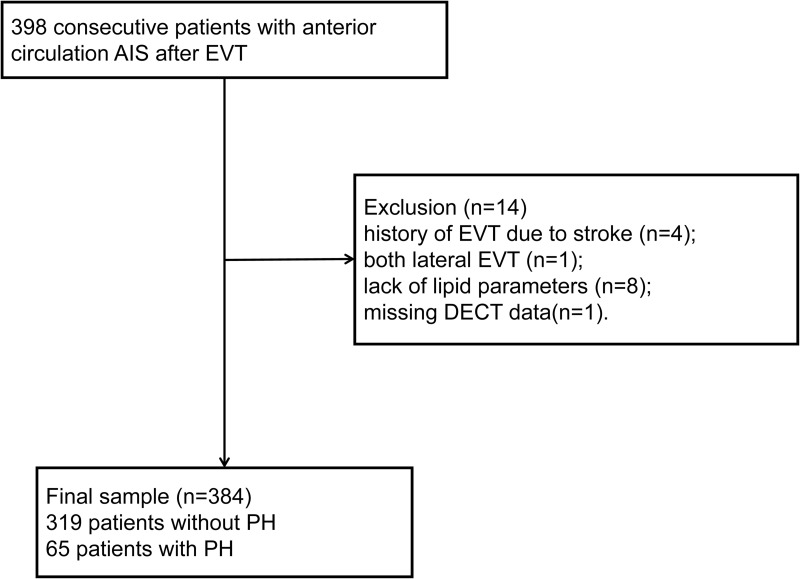
This research encompassed consecutive anterior circulation AIS patients who received EVT and successfully recanalized (eTICI ≥ 2b) at the Municipal Central Hospital of Lishui between September 1, 2021, and May 31, 2023, retrospectively. These patients include those who underwent direct thrombectomy and bridging therapy. Patients with EVT aged over 18 and who present within 24 h of ischemic stroke onset were included in the study according to the guideline in China.6 The exclusion criteria were as follows: (1) history of endovascular thrombectomy due to stroke; (2) a brain tumor; (3) both lateral endovascular thrombectomy; (4) lack of baseline lipid concentration data; (5) incomplete non-contrast computed tomography (NCCT) data at admission or dual energy computed tomography (DECT) data within 48 h. A flow chart was shown in Figure 1.
Figure 1.
Study patients flow chart.
Abbreviations: AIS, acute ischemic stroke; PH, parenchymal hemorrhage; EVT, endovascular treatment; DECT, dual energy computed tomography.
Data Collection
The data of participants were extracted from their respective medical records, which included age, gender, medical history (hypertension, diabetes, and atrial fibrillation), National Institutes of Health Stroke Scale (NIHSS) at admission, systolic blood pressure (SBP) upon admission, administration of intravenous thrombolysis (IVT), time from ischemic stroke onset to revascularization (OTR), fasting plasma glucose (FPG) levels, uric acid (UA) levels, serum creatinine (Scr) levels, cystatin C (CysC) levels, TG levels, total cholesterol (TC) levels, low density lipoprotein cholesterol (LDL-C) levels, and high-density lipoprotein cholesterol (HDL-C) levels. All the blood samples were collected in the next postoperative morning after admission and were tested for by a professional from the laboratory department of the Municipal Central Hospital of Lishui.
The calculation of non-traditional lipid parameters:
Non-HDL-C = TC – HDL-C;11
RC = TC – HDL-C – LDL-C;17
The ratio of TG to HDL-C = TG/HDL-C;12
The ratio of TC to HDL-C = TC/HDL-C;18
The ratio of RC to HDL-C = RC/HDL-C.19
It is worth noting that since the Non-HDL in this study is calculated by TC - HDL-C, TC can be expressed as HDL-C + Non-HDL. Thus, TC/HDL-C = (HDL-C + Non-HDL) / HDL-C, which can ultimately be represented as the ratio of Non-HDL to HDL-C + 1. Therefore, the statistical effect of TC/HDL-C and Non-HDL/HDL-C indicators is the same.20
All patients underwent preoperative NCCT examination, followed by postoperative DECT completion within 48 h. DECT has the ability to accurately differentiate between a hematoma and contrast agent. In accordance with the Heidelberg bleeding classification, PH constitutes a subtype of HT, encompassing parenchymal hematoma type 1 (PH1), characterized by hematoma within infarcted tissue occupying less than 30% without significant mass effect, and parenchymal hematoma type 2 (PH2), characterized by hematoma occupying 30% or more of the infarcted tissue, accompanied by evident mass effect.21 PH was independently identified by two researchers, with consultation from a third researcher in case of any discrepancies.
Statistical Analysis
Continuous variables were presented as means and standard deviation (SD) or median and interquartile range (IQR), while categorical variables were reported as frequencies and percentage (%). The differences in continuous variables were evaluated using Student’s t test or Mann–Whitney U-test. The chi-squared test was conducted to assess the differences in categorical variables. The correlation between lipid parameters were assessed using Spearman’s rank correlation analysis and visualized through heat maps. To evaluate the relationships between lipid parameters and PH, we categorized the lipid parameters into tertiles and employed logistic regression. For each lipid parameter, we developed four models. Covariates including age, gender, history of hypertension, atrial fibrillation, NIHSS at admission, FPG, UA, CysC, thrombolysis, and OTR time were included for variable adjustment. These covariates were chosen based on their clinical relevance to PH or their significant univariate relationship with PH (p < 0.1). Additionally, receiver operating characteristic (ROC) curve analysis was employed to evaluate the predictive ability of lipid parameters for PH. Additionally, we utilized restricted cubic splines (RCS) curves, employing three knots (at the 10th, 50th, and 90th percentiles), to visually analyze the relationships between lipid parameters and PH. Besides, subgroup analysis was used to evaluate the risk of PH in EVT alone group and combination-therapy (IVT + EVT) group, separately. The results were presented as odds ratios (OR) with accompanying 95% confidence intervals (CI). Statistical significance was determined at a two-sided p-value threshold of <0.05. All statistical analyses were conducted by R version 4.1.3.
Results
Three hundred and ninety-eight patients with AIS undergoing EVT were consecutively screened, and 384 patients were enrolled in this analysis (Figure 1). The reasons of exclusion are as follow: history of EVT due to stroke (n = 4); both lateral EVT (n = 1); lack of lipid parameters (n = 8); missing DECT data (n = 1). PH was observed in 65 (16.9%) patients. Table 1 presented comparisons of baseline characteristics between patients with and without PH. The age and gender composition of the two groups of patients did not differ significantly.
Table 1.
Baseline Characteristics
| Characteristic | Total(n=384) | Without PH(n=319) | With PH(n=65) | p-value |
|---|---|---|---|---|
| Age, median (IQR), years | 72(63–80) | 72(63–80) | 72(63–80) | 0.976 |
| Male, n (%) | 231(60.2) | 196(61.4) | 35(53.8) | 0.254 |
| Medical history, n (%) | ||||
| Hypertension, n (%) | 247(64.3) | 211(66.1) | 36(50.0) | 0.099 |
| Diabetes mellitus, n (%) | 92(24.0) | 79(24.8) | 13(18.1) | 0.412 |
| Atrial fibrillation, n (%) | 186(48.4) | 145(45.5) | 41(56.9) | 0.010 |
| NIHSS on admission, median (IQR) | 15(12–19) | 15(11–18) | 17.00(14–20) | <0.001 |
| SBP, (Mean ± SD), mmHg | 154±22 | 155±22 | 153±22 | 0.505 |
| FPG, median (IQR), mmol/L | 6.52(5.58–8.12) | 6.36(5.54–7.82) | 7.70(6.41–9.54) | <0.001 |
| Thrombolysis, n (%) | 148(38.5) | 122(38.2) | 25(34.7) | 0.988 |
| OTR time, median (IQR), hours | 9.1(6.8–12.6) | 9.2(6.8–12.8) | 8.5(7.2–11.0) | 0.439 |
| UA, median (IQR), μmol/L | 306.0(251.0–377.5) | 311.0(256.5–386.5) | 285.5(241.0–347.0) | 0.056 |
| Cr, median (IQR), μmol/L | 66.0(54.5–81.5) | 66.0(56.0–82.0) | 62.0(49.5–80.0) | 0.120 |
| Cys C, median (IQR), mg/L | 0.96(0.79–1.16) | 1.00(0.81–1.17) | 0.85(0.71–1.05) | 0.027 |
| Lipid profile | ||||
| TG, median (IQR), mmol/L | 0.86(0.62–1.23) | 0.88(0.66–1.25) | 0.73(0.53–1.12) | 0.009 |
| TC, median (IQR), mmol/L | 3.80(3.23–4.44) | 3.84(3.25–4.45) | 3.64(3.14–4.30) | 0.186 |
| HDL-C, median (IQR), mmol/L | 1.00(0.84–1.16) | 0.99(0.81–1.15) | 1.06(0.94–1.19) | 0.015 |
| LDL-C, median (IQR), mmol/L | 2.09(1.66–2.62) | 2.15(1.69–2.62) | 1.98(1.54–2.50) | 0.187 |
| Non-HDL-C, median (IQR), mmol/L | 2.76(2.28–3.44) | 2.80(2.30–3.44) | 2.50(2.02–3.02) | 0.021 |
| RC, median (IQR), mmol/L | 0.63(0.48–0.82) | 0.65(0.50–0.84) | 0.51(0.39–0.73) | 0.001 |
| TG/HDL-C, median (IQR) | 0.90(0.54–1.44) | 0.93(0.60–1.47) | 0.68(0.47–1.16) | 0.005 |
| TC/HDL-C, median (IQR) | 3.88(3.08–4.78) | 4.03(3.22–4.83) | 3.26(2.77–4.33) | 0.001 |
| LDL-C/HDL-C, median (IQR) | 2.15(1.56–2.89) | 2.23(1.66–2.91) | 1.81(1.32–2.59) | 0.009 |
| RC/HDL-C, median (IQR) | 0.63(0.44–0.92) | 0.65(0.48–0.97) | 0.53(0.34–0.76) | <0.001 |
Abbreviations: PH, parenchymal hematoma; IQR, interquartile range; SD, standard deviation; NIHSS, National Institutes of Health Stroke Scale; SBP, systolic blood pressure; FPG, fasting plasma glucose; OTR, onset to revascularization; UA, Uric Acid; Cr, creatinine; Cys C, cystatin C; TG, triglyceride; TC, total cholesterol; HDL-C, high density lipoprotein cholesterol; LDL-C, low density lipoprotein cholesterol; Non-HDL-C, non-high-density lipoprotein cholesterol; RC, remnant cholesterol; TG/HDL-C, the ratio of triglyceride to high density lipoprotein cholesterol; TC/HDL-C, the ratio of total cholesterol to high density lipoprotein cholesterol; LDL-C/HDL-C, the ratio of low density lipoprotein cholesterol to high density lipoprotein cholesterol; RC/HDL-C, the ratio of low density lipoprotein cholesterol to high density lipoprotein cholesterol remnant cholesterol.
Compared to the patients without PH after EVT, those with PH demonstrated higher NIHSS at admission, FPG level, proportions of history of hypertension and atrial fibrillation. However, no significant difference was observed concerning the history of diabetes. Regarding lipid profile, patients with PH exhibited lower median values of TG, non-HDL-C, RC, TG/HDL-C, TC/HDL-C, LDL-C/HDL-C, and RC/HDL-C, while also showing higher HDL-C levels. Despite these significant differences, the LDL-C and TC levels between the two groups did not show statistically significant differences. The correlation between different lipid parameters were illustrated in Figure S1.
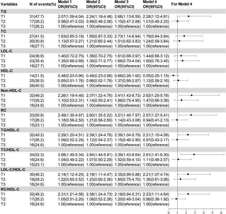
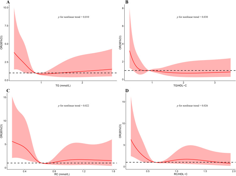
Model 1 for lipid parameters were unadjusted (Figure 2). In model 2, after adjusting for age and gender, patients in the lower TG, non-HDL-C, RC, TG/HDL-C, TC/HDL-C, LDL-C/HDL-C, RC/HDL-C tertiles showed increased risks of PH. When compared to the highest tertiles, the first tertiles of TG, TC, Non-HDL-C, RC, TG/HDL-C, TC/HDL-C demonstrated an elevated risk of PH (adjusted ORs 2.66 [95% CI 1.13–6.59], 2.73 [95% CI 1.14–6.94], 3.41[95% CI 1.43–8.73], 3.21[95% CI 1.40–7.97], 2.39[95% CI 1.04–5.79], and 3.39[95% CI 1.43–8.64], respectively) after adjusting for age, gender, history of hypertension, atrial fibrillation, NIHSS at admission, FPG, UA, CysC, thrombolysis, OTR time in model 3. After adjusting for history of atrial fibrillation and FPG in model 4, patients in the lower TG, Non-HDL-C, RC, TG/HDL-C, TC/HDL-C, LDL-C/HDL-C, RC/HDL-C tertiles exhibited increased risks of PH(2.28 [95% CI 1.12–4.81], 2.62 [95% CI 1.25–5.78], 2.57[95% CI 1.27–5.41], 2.21[95% CI 1.10–4.58], 2.91[95% CI 1.41–6.30], 2.21[95% CI 1.07–4.74], and 2.23[95% CI 1.11–4.64], respectively). Additionally, patients in the lower HDL-C tertiles were related to a decreased risk of PH in model 1 (0.48[95% CI 0.26–1.60]) and model 2 (0.49[95% CI 0.23–0.98]), while the association did not reach statistical significance in model 3 (0.66[95% CI 0.23–0.98]) and model 4 (0.55[95% CI 0.25–1.15]). Using RCS curves, a nonlinear trend was identified between TG, RC, TG/HDL-C, RC/HDL-C and PH after adjusting for variates based on model 4 (Figure 3). When setting the median RC level (0.63 mmol/L) as a reference, a substantial increased risk of PH was observed in patients with lower RC level. In subgroup analysis (Table S1), there was no significant difference in the risk of EVT group and IVT + EVT group regarding of the lipid parameters involved in this study.
Figure 2.
Association of tertiles of lipid parameters and risk of PH.
Notes: Model 2 was adjusted for age and gender; Model 3 was adjusted for age, gender, history of hypertension, atrial fibrillation, NIHSS on admission, fasting plasma glucose, uric acid, cystatin-C, thrombolysis, and time from stroke onset to revascularization; Model 4 adjusted for history of atrial fibrillation and fasting plasma glucose, which based on model 3. T1 of TG: ≤0.70 mmol/L; T2 of TG: >0.70–1.07 mmol/L; T3 of TG: >1.07 mmol/L; T1 of TC: ≤3.45 mmol/L; T2 of TC: >0.53–4.23 mmol/L; T3 of TC: >4.23 mmol/L; T1 of LDL-C: ≤1.82 mmol/L; T2 of LDL-C: >1.82–2.46 mmol/L; T3 of LDL-C: >2.46 mmol/L; T1 of HDL-C: ≤2.48 mmol/L; T2 of HDL-C: >2.48–3.16 mmol/L; T3 of HDL-C: >3.16mmol/L; T1 of Non-LDL-C: ≤0.89 mmol/L; T2 of Non-LDL-C: >0.89–1.09 mmol/L; T3 of Non-LDL-C: >1.09 mmol/L; T1 of RC: ≤0.53 mmol/L; T2 of RC: >0.53–0.75 mmol/L; T3 of RC: >0.75 mmol/L; T1 of TG/HDL-C: ≤0.66; T2 of TG/HDL-C: >0.66–1.19; T3 of TG/HDL-C: >1.19; T1 of TC/HDL-C: ≤3.30; T2 of TC/HDL-C: >3.30–4.44; T3 of TC/HDL-C: >4.44; T1 of LDL-C/HDL-C: ≤1.78; T2 of LDL-C/HDL-C: >1.78–2.64; T3 of LDL-C/HDL-C: >2.64; T1 of RC/HDL-C: ≤0.50; T2 of RC/HDL-C: >0.50–0.78; T3 of RC/HDL-C: >0.78.
Abbreviations: T1, first tertile; T2, second tertile; T3, third tertile; OR, odds ratios; CI, confidence interval; N, number; TG, triglyceride; TC, total cholesterol; HDL-C, high density lipoprotein cholesterol; LDL-C, low density lipoprotein cholesterol; Non-HDL-C, non-high-density lipoprotein cholesterol; RC, remnant cholesterol; TG/HDL-C, the ratio of triglyceride to high density lipoprotein cholesterol; TC/HDL-C, the ratio of total cholesterol to high density lipoprotein cholesterol; LDL-C/HDL-C, the ratio of low density lipoprotein cholesterol to high density lipoprotein cholesterol; RC/HDL-C, the ratio of low density lipoprotein cholesterol to high density lipoprotein cholesterol remnant cholesterol.
Figure 3.
Relationship of lipid parameters with PH risk.
Notes: Multiple spline regression analyses were used to analyze the association between TG (A), TG/HDL-C (B), RC (C), RC/HDL-C (D) and PH with three knots (at the 10th, 50th, 90th percentiles). The edges of the red area mark 95% confidence intervals. The red solid curve represents the ORs value. ORs and 95% confidence intervals were obtained through restricted cubic spline regression, with adjustments made for history of atrial fibrillation and glucose.
Abbreviations: OR, odds ratios; CI, confidence interval; TG, triglyceride; RC, remnant cholesterol; TG/HDL-C, the ratio of triglyceride to high density lipoprotein cholesterol; RC/HDL-C, the ratio of low density lipoprotein cholesterol to high density lipoprotein cholesterol remnant cholesterol.
In terms of predictive capacity, the ROC curves revealed that the TG, HDL-C, Non-HDL-C, RC, TG/HDL-C, TC/HDL-C, LDL-C/HDL-C, and RC/HDL-C exhibited predictive capability for PH, whereas TC and LDL-C showed no predictive ability for PH (Table 2). Among these lipid parameters, the largest AUC was observed for RC/HDL-C with a value of 0.643 (95% CI 0.564–0.723).
Table 2.
The AUC and Cut-off Value of Lipid Parameters for PH
| Variable | AUC | 95% CI | p-value | Cutoff value | Sensitivity | Specificity |
|---|---|---|---|---|---|---|
| TG | 0.603 | 0.521–0.685 | 0.011 | 0.80 | 0.600 | 0.614 |
| TC | 0.552 | 0.472–0.632 | 0.196 | 3.96 | 0.677 | 0.458 |
| LDL-C | 0.552 | 0.473–0.631 | 0.205 | 2.23 | 0.677 | 0.458 |
| HDL-C | 0.596 | 0.524–0.667 | 0.008 | 1.04 | 0.585 | 0.586 |
| Non-HDL-C | 0.591 | 0.510–0.672 | 0.028 | 2.84 | 0.723 | 0.492 |
| RC | 0.626 | 0.545–0.707 | 0.002 | 0.51 | 0.508 | 0.730 |
| TG/HDL-C | 0.596 | 0.524–0.667 | 0.009 | 1.04 | 0.585 | 0.586 |
| TC/HDL-C | 0.627 | 0.549–0.705 | 0.001 | 3.27 | 0.523 | 0.727 |
| LDL-C/HDL-C | 0.603 | 0.525–0.681 | 0.009 | 1.68 | 0.462 | 0.746 |
| RC/HDL-C | 0.643 | 0.564–0.723 | <0.001 | 0.42 | 0.463 | 0.837 |
Abbreviations: AUC, area under the curve; CI, confidence interval; TG, triglyceride; TC, total cholesterol; HDL-C, high density lipoprotein cholesterol; LDL-C, low density lipoprotein cholesterol; Non-HDL-C, non-high-density lipoprotein cholesterol; RC, remnant cholesterol; TG/HDL-C, the ratio of triglyceride to high density lipoprotein cholesterol; TC/HDL-C, the ratio of total cholesterol to high density lipoprotein cholesterol; LDL-C/HDL-C, the ratio of low density lipoprotein cholesterol to high density lipoprotein cholesterol; RC/HDL-C, the ratio of low density lipoprotein cholesterol to high density lipoprotein cholesterol remnant cholesterol.
Discussion
We have conducted the first investigation into the association between traditional and non-traditional lipid parameters and PH among AIS patients undergoing EVT. The primary results of our research were as follows: (1) Low TG, non-HDL-C, RC, TG/HDL-C, TC/HDL-C, LDL-C/HDL-C, and RC/HDL-C level showed significant related to the risk of PH during hospitalization; (2) RC/HDL-C exhibited the most robust predictive capacity for PH following EVT, while non-HDL-C demonstrated the highest sensitivity; (3) Additionally, a nonlinear relationship was found between TG, RC, TG/HDL-C, and RC/HDL-C with the PH risk.
Past researches have shown that the incidence rate of PH after thrombectomy varies between 5% and 25.7%.22,23 It was 16.9% in our study, which is consistent with previous research. The impact of pre-treatment IVT on the occurrence of PH lacks clarity. A post hoc analysis indicated that alteplase thrombolytic therapy did not elevate the risk of HT, sICH, or PH following EVT.24 In our research, we did not observed any significant statistical difference in the IVT rate between the PH group and the non-PH group.
Correlation Between Traditional Lipid Parameters and Hemorrhagic Transformation Following AIS
In this analysis, we revealed that among the traditional lipid parameters, only TG levels exhibited a significant association with an increased risk of PH in AIS patients undergoing EVT. A study published in 2019 supported the aforementioned finding, demonstrating an independent correlation between TG levels and the risk of HT in AIS patients.12 However, in other studies concentrating on AIS patients undergoing IVT, an independent association between TG levels and the development of HT was not observed.25–27 Previous studies also suggested that TC or HDL-C levels were not significantly associated with HT.25–27 Regarding LDL-C, it has been one of the early indicators of interest. Some studies did not identify a relationship between the LDL-C levels and HT,12,16,25,28 while others suggested that low levels of LDL-C might increase the risk of HT in AIS patients.11,26,29 It was important to note that these studies were not specifically designed for patients undergoing EVT, except for one study conducted by Ahn et al.29 In their research, encompassing 159 patients diagnosed with AIS who received EVT, the researches observed an independent association between LDL-C levels ≤50 mg/dL and delayed PH following thrombectomy. Another article published by Ahn et al indicated that LDL-C was not predictive of immediate post-thrombectomy PH, which was consistent with our own findings.30
Correlation Between Non-Traditional Lipid Parameters and Hemorrhagic Transformation Following AIS
Over recent years, the association between non-traditional lipid parameters and the prognosis of AIS has gradually attracted attention. There are also some controversies among studies of non-traditional lipid parameters and HT following AIS. In research analyzing some non-traditional indicators, it was found that lower TC/HDL-C, TG/HDL-C, and LDL/HDL-C (rather than non-HDL-C) levels were related to an higher risk of HT following IVT.31 While, also for thrombolytic patients, Chua Ming et al’s study28 indicated that non-HDL-C, instead of LDL-C, showed a significant correlation with SICH, and a U-shaped association was found between non-HDL-C and sICH. However, this U-shaped relationship was not evident in our study. Additionally, Yanan Wang et al’s11 research suggested that non-HDL-C has a similar role to LDL-C in predicting HT among AIS patients. As for TG/HDL-C, Qi-Wen Deng et al12 confirmed that a lower TG/HDL-C ratio was related to a higher incidence of HT among patients caused by LAA but not cardioembolism and small-vessel occlusion. Sadly, they did not involve the rest of non-traditional lipid parameters mentioned in this research. In summary, the research conclusions regarding the association between non-traditional lipid parameters and HT are currently not consistent. Most studies have only focused on one or a few non-traditional lipid parameters. Our study comprehensively incorporated the majority of non-traditional lipid parameters and pioneered the examination of the relationship between RC/HDL-C and PH.
Interestingly, among traditional parameters, TG is the only lipid parameter independently correlated to PH, and its predictive ability for PH is similar to that of other non-traditional indicators. This may be caused by a certain correlation between TG and RC. RC, also known as triglyceride-rich lipoprotein cholesterol (TRL-C), is a residue produced from the metabolism of triglyceride-rich lipoproteins (TGRLs), namely chylomicrons and very low-density lipoproteins. During lipolysis by lipoprotein lipase, these particles lose TG and generate residue rich in cholesterol esters.32 Under both fasting and non-fasting conditions, serum TG levels are strongly associated with TGRLs concentration.32 Thus, TG and RC may have similar predictive effects in some diseases.33 It is worth noting, however, that although TG is used as a marker for RC, its clinical significance is not entirely the same as that of RC.34 This may be due to the fact that most laboratories use enzymatic methods to test TG levels, which measure not only TG in lipoproteins as mentioned above, but also include the level of free glycerol. Therefore, we cannot directly substitute TG for RC.
Furthermore, it is worth noting that although the non-traditional lipid parameters we included all showed a correlation with PH in patients with AIS, it is still unclear which specific lipid parameter plays a major role in post- thrombectomy PH in AIS. Further confirmation is needed through large-sample prospective studies.
Potential Mechanisms Between Lipid Parameters and the Risk of Hemorrhagic Transformation Following AIS
The precise biological mechanism by through which lipid parameters increases the risk of HT remains incompletely understood. There are several possible speculations that could explain it: Firstly, lipid parameters may participate in maintaining the integrity of cerebral small blood vessels and the blood-brain barrier. Some researches have suggested that low serum lipid levels could lead to increased red blood cell permeability and leakage of vascular.8,9 In addition, after reperfusion therapy, serum lipid levels are also associated with an increased risk of brain edema, suggesting that serum lipids may affect the integrity of the blood-brain barrie;35 Secondly, serum lipid levels may also affect platelet aggregation, thereby impacting bleeding;36 Thirdly, the vascular ability to resist damage may depend on the coordination between different types of cholesterol. In our study, non-traditional lipid parameters (usually composite lipid indicators) were found to be associated with PH, rather than non-composite non-traditional parameters (except for TG). This suggests that it is not a specific lipid parameter but the appropriate balance between various lipid parameters that influences the outcomes.
Limitations
Several limitations exist within our research. Firstly, it encompasses a single-center retrospective study, which can only suggest a certain correlation between TG and non-traditional lipid parameters with the PH following EVT but cannot establish a causal relationship. Secondly, it should be noted that in our study, we only assessed lipid levels once in the next postoperative morning after admission and did not track the dynamic changes. Thus we were unable to establish whether fluctuations in lipid levels may have an impact on the occurrence of PH. Future research should include continuous monitoring of lipid levels to gain a better understanding of their potential influence on PH development. Lastly, due to the limitations of retrospective studies, we did not obtain complete data of the number of device passes during EVT. If the number of device passes >3 is considered one of the risk factors for HT after intravascular therapy in some studies.37 However, in other studies, the number of device passes >3, it is not considered relevant to HT.38
Conclusion
Low TG, non-HDL-C, RC, TG/HDL-C, TC/HDL-C, LDL-C/HDL-C, and RC/HDL-C levels are associated with PH after EVT in AIS patients. Incorporating routine assessment of non-traditional lipid levels is advised in clinical practice.
Acknowledgments
We acknowledge all the patients and their families.
Funding Statement
This study was supported by Medical and Health Science and Technology Program of Zhejiang Province (2021RC145) and Science and Technology Program of Lishui City (2020GYX16). The funding organization had no role in the study or in the preparation of this report.
Ethics Statement
This research was approved by the Ethics Committee of Lishui Municipal Central Hospital (ID:2023-748). Informed consent for the research was obtained from the patients or their legal representative. The guidelines outlined in the Declaration of Helsinki were followed.
Disclosure
All the authors report no conflicts of interest in this work.
References
- 1.Tu WJ, Wang LD.; Special Writing Group Of China Stroke Surveillance R. China stroke surveillance report 2021. Mil Med Res. 2023;10(1):33. doi: 10.1186/s40779-023-00463-x [DOI] [PMC free article] [PubMed] [Google Scholar]
- 2.Tu WJ, Zhao Z, Yin P, et al. estimated Burden of stroke in China in 2020. JAMA Network Open. 2023;6(3):e231455. doi: 10.1001/jamanetworkopen.2023.1455 [DOI] [PMC free article] [PubMed] [Google Scholar]
- 3.Spronk E, Sykes G, Falcione S, et al. Hemorrhagic transformation in ischemic stroke and the role of inflammation. Front Neurol. 2021;12:661955. doi: 10.3389/fneur.2021.661955 [DOI] [PMC free article] [PubMed] [Google Scholar]
- 4.Van Kranendonk KR, Treurniet KM, Boers AMM, et al. Hemorrhagic transformation is associated with poor functional outcome in patients with acute ischemic stroke due to a large vessel occlusion. J Neurointerv Surg. 2019;11(5):464–468. doi: 10.1136/neurintsurg-2018-014141 [DOI] [PubMed] [Google Scholar]
- 5.Hackam DG, Hegele RA. Cholesterol lowering and prevention of stroke. Stroke. 2019;50(2):537–541. doi: 10.1161/STROKEAHA.118.023167 [DOI] [PubMed] [Google Scholar]
- 6.Chinese Society Of Nurology, Chinses Stroke Society, Neurouvascular Intervention Group Of Chinese Of NeuroloGY. Chinese guideline for the endovascular treatment of acute ischemic stroke 2018. Chin J Neurol. 2018;51(9):683–691. [Google Scholar]
- 7.Lee M, Cheng CY, Wu YL, et al. Association between intensity of low-density lipoprotein cholesterol reduction with statin-based therapies and secondary stroke prevention: a meta-analysis of randomized clinical trials. JAMA Neurol. 2022;79(4):349–358. doi: 10.1001/jamaneurol.2021.5578 [DOI] [PMC free article] [PubMed] [Google Scholar]
- 8.Thrift A, Mcneil J, Donnan G, et al. Reduced frequency of high cholesterol levels among patients with intracerebral haemorrhage. J Clin Neurosci. 2002;9(4):376–380. doi: 10.1054/jocn.2002.1111 [DOI] [PubMed] [Google Scholar]
- 9.Wieberdink RG, Poels MM, Vernooij MW, et al. Serum lipid levels and the risk of intracerebral hemorrhage: the Rotterdam Study. Arterioscler Thromb Vasc Biol. 2011;31(12):2982–2989. doi: 10.1161/ATVBAHA.111.234948 [DOI] [PubMed] [Google Scholar]
- 10.Sheng G, Kuang M, Yang R, et al. Evaluation of the value of conventional and unconventional lipid parameters for predicting the risk of diabetes in a non-diabetic population. J Transl Med. 2022;20(1):266. doi: 10.1186/s12967-022-03470-z [DOI] [PMC free article] [PubMed] [Google Scholar]
- 11.Wang Y, Song Q, Cheng Y, et al. Association between non-high-density lipoprotein cholesterol and haemorrhagic transformation in patients with acute ischaemic stroke. BMC Neurol. 2020;20(1):47. doi: 10.1186/s12883-020-1615-9 [DOI] [PMC free article] [PubMed] [Google Scholar]
- 12.Deng QW, Liu YK, Zhang YQ, et al. Low triglyceride to high-density lipoprotein cholesterol ratio predicts hemorrhagic transformation in large atherosclerotic infarction of acute ischemic stroke. Aging. 2019;11(5):1589–1601. doi: 10.18632/aging.101859 [DOI] [PMC free article] [PubMed] [Google Scholar]
- 13.Yang XH, Zhang BL, Cheng Y, et al. Association of remnant cholesterol with risk of cardiovascular disease events, stroke, and mortality: a systemic review and meta-analysis [J]. Atherosclerosis. 2023;371:21–31. doi: 10.1016/j.atherosclerosis.2023.03.012 [DOI] [PubMed] [Google Scholar]
- 14.Li J, Li Y, Zhu P, et al. Remnant cholesterol but not LDL cholesterol is associated with 5-year bleeding following percutaneous coronary intervention. iScience. 2023;26(10):107666. doi: 10.1016/j.isci.2023.107666 [DOI] [PMC free article] [PubMed] [Google Scholar]
- 15.Bang OY, Saver JL, Liebeskind DS, et al. Cholesterol level and symptomatic hemorrhagic transformation after ischemic stroke thrombolysis [J]. Neurology. 2007;68(10):737–742. doi: 10.1212/01.wnl.0000252799.64165.d5 [DOI] [PubMed] [Google Scholar]
- 16.Hong CT, Chiu WT, Chi NF, et al. Low-density lipoprotein level on admission is not associated with postintravenous thrombolysis intracranial hemorrhage in patients with acute ischemic stroke [J]. J Investig Med. 2019;67(3):659–662. doi: 10.1136/jim-2018-000827 [DOI] [PubMed] [Google Scholar]
- 17.Nordestgaard BG, Varbo A. Triglycerides and cardiovascular disease [J]. Lancet. 2014;384(9943):626–635. doi: 10.1016/S0140-6736(14)61177-6 [DOI] [PubMed] [Google Scholar]
- 18.Chen L, Xu J, Sun H, et al. The total cholesterol to high-density lipoprotein cholesterol as a predictor of poor outcomes in a Chinese population with acute ischaemic stroke [J]. J Clin Lab Anal. 2017;31(6). doi: 10.1002/jcla.22139 [DOI] [PMC free article] [PubMed] [Google Scholar]
- 19.Yang WS, Li R, Shen YQ, et al. Importance of lipid ratios for predicting intracranial atherosclerotic stenosis [J]. Lipids Health Dis. 2020;19(1):160. doi: 10.1186/s12944-020-01336-1 [DOI] [PMC free article] [PubMed] [Google Scholar]
- 20.Hsia SH, Pan D, Berookim P, et al. A population-based, cross-sectional comparison of lipid-related indexes for symptoms of atherosclerotic disease [J]. Am J Cardiol. 2006;98(8):1047–1052. doi: 10.1016/j.amjcard.2006.05.024 [DOI] [PubMed] [Google Scholar]
- 21.Von Kummer R, Broderick JP, Campbell BC, et al. The Heidelberg bleeding classification: classification of bleeding events after ischemic stroke and reperfusion therapy [J]. Stroke. 2015;46(10):2981–2986. doi: 10.1161/STROKEAHA.115.010049 [DOI] [PubMed] [Google Scholar]
- 22.Xu C, Zhou Y, Zhang R, et al. Metallic hyperdensity sign on noncontrast ct immediately after mechanical thrombectomy predicts parenchymal hemorrhage in patients with acute large-artery occlusion [J]. AJNR Am J Neuroradiol. 2019;40(4):661–667. doi: 10.3174/ajnr.A6008 [DOI] [PMC free article] [PubMed] [Google Scholar]
- 23.Bracard S, Ducrocq X, Mas JL, et al. Mechanical thrombectomy after intravenous alteplase versus alteplase alone after stroke (THRACE): a randomised controlled trial [J]. Lancet Neurol. 2016;15(11):1138–1147. doi: 10.1016/S1474-4422(16)30177-6 [DOI] [PubMed] [Google Scholar]
- 24.Tian B, Tian X, Shi Z, et al. Clinical and imaging indicators of hemorrhagic transformation in acute ischemic stroke after endovascular thrombectomy [J]. Stroke. 2022;53(5):1674–1681. doi: 10.1161/STROKEAHA.121.035425 [DOI] [PubMed] [Google Scholar]
- 25.Zhang K, Luan J, Li C, et al. Nomogram to predict hemorrhagic transformation for acute ischemic stroke in Western China: a retrospective analysis [J]. BMC Neurol. 2022;22(1):156. doi: 10.1186/s12883-022-02678-2 [DOI] [PMC free article] [PubMed] [Google Scholar]
- 26.Yang C, Zhang J, Liu C, et al. Comparison of the risk factors of hemorrhagic transformation between large artery atherosclerosis stroke and cardioembolism after intravenous thrombolysis [J]. Clin Neurol Neurosurg. 2020;196:106032. doi: 10.1016/j.clineuro.2020.106032 [DOI] [PubMed] [Google Scholar]
- 27.Wang Y, Wei C, Song Q, et al. Reduction in the ratio of low-density lipoprotein cholesterol to high density lipoprotein cholesterol is associated with increased risks of hemorrhagic transformation in patients with acute ischemic stroke [J]. Curr Neurovasc Res. 2019;16(3):266–272. doi: 10.2174/1567202616666190619151914 [DOI] [PubMed] [Google Scholar]
- 28.Ming C, Toh EMS, Yap QV, et al. Impact of traditional and non-traditional lipid parameters on outcomes after intravenous thrombolysis in acute ischemic stroke [J]. J Clin Med. 2022;11(23):7148. doi: 10.3390/jcm11237148 [DOI] [PMC free article] [PubMed] [Google Scholar]
- 29.Ahn S, Roth SG, Jo J, et al. low levels of low-density lipoprotein cholesterol increase the risk of post-thrombectomy delayed parenchymal hematoma [J]. Neurointervention. 2023;18(3):172–181. doi: 10.5469/neuroint.2023.00269 [DOI] [PMC free article] [PubMed] [Google Scholar]
- 30.Ahn S, Mummareddy N, Roth SG, et al. The clinical utility of dual-energy CT in post-thrombectomy care: part 1, predictors and outcomes of subarachnoid and intraparenchymal hemorrhage [J]. J Stroke Cerebrovasc Dis. 2023;32(8):107217. doi: 10.1016/j.jstrokecerebrovasdis.2023.107217 [DOI] [PubMed] [Google Scholar]
- 31.Luo Y, Chen J, Yan XL, et al. Association of non-traditional lipid parameters with hemorrhagic transformation and clinical outcome after thrombolysis in ischemic stroke patients [J]. Curr Neurovasc Res. 2020;17(5):736–744. doi: 10.2174/1567202617999210101223129 [DOI] [PubMed] [Google Scholar]
- 32.Varbo A, Nordestgaard BG. Remnant lipoproteins [J]. Curr Opin Lipidol. 2017;28(4):300–307. doi: 10.1097/MOL.0000000000000429 [DOI] [PubMed] [Google Scholar]
- 33.Wadstrom BN, Pedersen KM, Wulff AB, et al. Elevated remnant cholesterol, plasma triglycerides, and cardiovascular and non-cardiovascular mortality [J]. Eur Heart J. 2023;44(16):1432–1445. doi: 10.1093/eurheartj/ehac822 [DOI] [PubMed] [Google Scholar]
- 34.Cheng Y, Zhang Q, Li H, et al. Remnant cholesterol, stronger than triglycerides, is associated with incident non-alcoholic fatty liver disease [J]. Front Endocrinol. 2023;14:1098078. doi: 10.3389/fendo.2023.1098078 [DOI] [PMC free article] [PubMed] [Google Scholar]
- 35.Escudero-Martinez I, Thoren M, Matusevicius M, et al. Association of cholesterol levels with hemorrhagic transformation and cerebral edema after reperfusion therapies [J]. Eur Stroke J. 2023;8(1):294–300. doi: 10.1177/23969873221148229 [DOI] [PMC free article] [PubMed] [Google Scholar]
- 36.Fessler MB, Rose K, Zhang Y, et al. Relationship between serum cholesterol and indices of erythrocytes and platelets in the US population [J]. J Lipid Res. 2013;54(11):3177–3188. doi: 10.1194/jlr.P037614 [DOI] [PMC free article] [PubMed] [Google Scholar]
- 37.Jiang L, Zhou L, Zhang H, et al. MRI predictors of intracranial hemorrhage in acute ischemic stroke after endovascular thrombectomy therapy [J]. Am J Transl Res. 2020;12(8):4532–4541. [PMC free article] [PubMed] [Google Scholar]
- 38.Hassan AE, Kotta H, Shariff U, et al. There is no association between the number of stent retriever passes and the incidence of hemorrhagic transformation for patients undergoing mechanical thrombectomy [J]. Front Neurol. 2019;10:818. doi: 10.3389/fneur.2019.00818 [DOI] [PMC free article] [PubMed] [Google Scholar]


