Abstract
A retrospective search was conducted to evaluate the diagnostic accuracy of the vertebral heart score (VHS) and other related radiographic indices in the detection of cardiac enlargement associated with different cardiac disorders in the cat. One hundred and five cats with a complete echocardiographic examination and radiographic examination of the thorax with at least two orthogonal views were enrolled. Eighty-three cats had different cardiac disorders, 72 with left-sided cardiac disorders (LSCD) and 11 with right-sided cardiac disorders; 22 cats were free of cardiovascular abnormalities. Measurements of VHS and cardiac long and short axes on lateral (L) and dorsoventral or ventrodorsal radiographs were obtained. Receiver operating characteristic curves were calculated to evaluate the diagnostic accuracy of each radiographic index in differentiating between cats with cardiac disorders or cats with LSCD and cats without cardiac abnormalities and, among cats with LSCD, between those with no or mild left atrial enlargement (LAE) or those with moderate-to-severe LAE and healthy cats. The L-VHS at the cut-off of 7.9 had high diagnostic accuracy in distinguishing cats with LSCD and moderate-to-severe LAE from healthy cats, but all the other radiographic indices were moderately accurate in distinguishing between cats with overall cardiac disorders or LSCD, either with no or mild LAE and moderate-to-severe LAE, and healthy cats. The considered radiographic indices were also moderately accurate in predicting different degrees of LAE in cats with LSCD. Radiographic indices are reasonably specific, but less sensitive predictors of cardiac enlargement in cats with heart disorders.
Introduction
The clinical diagnosis of feline cardiac disorders can be challenging as some cats with cardiac diseases may not have appreciable cardiac murmurs on physical examination and, similarly, cats with murmurs may not have structural cardiac disease.1–4 Thoracic radiography is a useful tool in the diagnostic work-up of patients affected by cardiac disorders. Radiographic diagnosis of cardiac disorders is based on the recognition of roentgen signs suggesting abnormal size and shape of the cardiac silhouette, abnormal size and shape of the pulmonary vessels, and the presence of congestive heart failure (ie, pulmonary oedema, pleural effusion, enlarged caudal vena cava and ascites).5–7 Determination of heart size is important in evaluating patients with suspected heart disease as an enlarged cardiac silhouette may be a reliable index of pathological cardiac changes.5–7
The vertebral heart score (VHS) is a method for objectively evaluating the dimensions of the cardiac silhouette in thoracic radiograms, first described by Buchanan and Bücheler in the dog. 8 Using this method, the cardiac long axis (CLA) and short axis (CSA) are measured on the lateral (L) and ventrodorsal (VD) or dorsoventral (DV) thoracic radiographic view, and are then compared with the length of the thoracic vertebrae. Normal VHS values have been established for cats,9,10 ferrets 11 and rabbits, 12 as well as for different canine breeds.13–17 In particular, an upper limit of ⩽8.1 is considered normal for L-VHS in cats.7,9 In the dog, this technique has been demonstrated to be particularly useful for evaluating cardiac enlargement associated with eccentric hypertrophy due to volume overload.18,19 In particular, the VHS has been applied to evaluate cardiac enlargement in different cardiovascular disorders, including chronic degenerative valvular disease, dilated cardiomyopathy, congenital cardiac diseases, heartworm disease and pericardial effusion.18,20–23 Furthermore, VHS has been used to discriminate between a cardiac and non-cardiac origin of cough in dogs with chronic degenerative valvular disease. 24 In feline medicine, the VHS has been employed to evaluate the cardiac silhouette in obese cats, 25 cats with heartworm disease 22 and cardiogenic pulmonary oedema, 26 as well as to differentiate congestive heart failure from other causes of dyspnoea. 4
To the best of our knowledge, systematic studies establishing the overall diagnostic value of VHS in differentiating cats with different cardiac disorders from cats free of cardiovascular disorders are lacking. Similarly, no study was carried out to establish the diagnostic accuracy of the VHS for the evaluation of cardiac enlargement in cats with cardiac disorders. Therefore, the aims of the study reported here were to evaluate the diagnostic accuracy of VHS and other correlated radiographic cardiac indices in differentiating cats with cardiac disorders from cats without cardiac disorders, and to evaluate the diagnostic accuracy of these radiographic indices in differentiating cats with different degrees of cardiac enlargement from healthy cats.
Materials and methods
Animals
Medical records of cats presented at the cardiology service of the Veterinary Teaching Hospital of the University of Padua and Bologna from January 2009 to June 2013 were retrospectively reviewed. Cats were included in the study if they had a radiographic examination of the thorax with at least two orthogonal views (ie, L and DV and/or VD thoracic view) and an echocardiographic examination within 24 h of thoracic radiographic examination. Cats with poor-quality thoracic radiographs were excluded from the study. In particular, exclusion criteria included incorrect positioning of the patient, radiographic features of pleural effusion and/or mediastinal mass obscuring the cardiac silhouette or vertebral anomalies reducing the accuracy of cardiac measurements. Transthoracic echocardiography was performed by the same experienced operator (CG and MC) at the two hospitals. Cats with echocardiographic evidence of pericardial effusion were also excluded from the study.
Cardiac diagnoses were determined on the basis of combined clinical, radiographic, echocardiographic and Doppler echocardiographic findings. During the same study period, 22 healthy cats (nine males, 13 females) weighing 4.9 ± 1.4 kg, without clinical or echocardiographic evidence of cardiac disorders, were recruited. Cats with cardiac disorders were subdivided into different groups according to the cardiac side prevalently affected into cats with left-sided cardiac disorders (LSCD) and cats with right-sided cardiac disorders (RSCD). In cats with LSCD, the ratio between the echocardiographic measurement of the left atrial diameter and the aortic root diameter (LA:Ao) was employed to categorise those without left atrial enlargement (LAE) (LA:Ao <1.5) and those with mild (LA:Ao ⩾1.51 and ⩽1.79), moderate (LA:Ao ⩾1.8 and ⩽1.99) and severe (LA:Ao >2) LAE. 27
Radiographic measurements
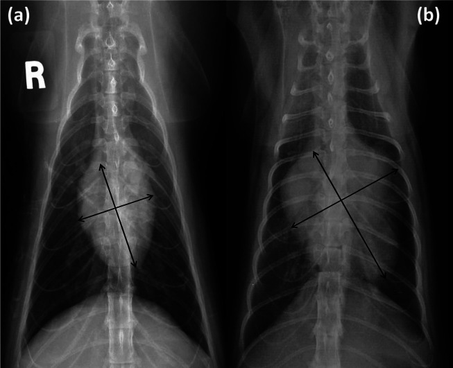
Blinded evaluations of right L and DV or VD radiographic views of the thorax were performed by the same experienced radiologist (AD), who was unaware of the echocardiographic findings. The cardiac dimensions were assessed by measuring the VHS, as described.9,10 Briefly, the CLA and CSA were measured on both thoracic views and then compared with the length of the vertebral bodies of thoracic vertebrae in the L view. In particular, in the right L view, the CLA was measured from the ventral border of the largest of the main stem bronchi seen in cross-section to the most distant point of the cardiac apex; the CSA was measured perpendicular to the measurement of the long axis at the point of maximum cardiac width. In the DV or VD view, the CLA was measured as the distance from midline of the cranial edge of the cardiac silhouette to the apex; the CSA was measured perpendicular to the measurement of the long axis at the point of maximum cardiac width. The method of measurement of VD-VHS is shown in Figure 1. The L-VHS and the DV/VD-VHS are the vertebral scale sum of the long and short axes obtained from right L and DV or VD radiographic views, respectively, each measured caudally from the cranial edge of the fourth thoracic vertebra. Additionally, a modified VHS system to evaluate the left atrium (LA-VHS) on L radiographic view was applied as previously described. 28 In brief, starting from the line of the CLA on the cardiac silhouette, a line perpendicular to this line was drawn approaching the caudal contour of the left atrial wall just dorsal to the border of the caudal vena cava.
Figure 1.
(a) Ventrodorsal (VD) thoracic radiograph of a cat without clinical and echocardiographic evidence of cardiovascular disorders showing measurement points for cardiac long axis (CLA) and short axis (CSA). The corresponding VD–vertebral heart score (VHS) is 7.8. (b) VD thoracic radiograph of a cat with echocardiographic evidence of mitral and tricuspid regurgitation showing measurement points for CLA and CSA. The corresponding VD–VHS is 11.2
Statistical analysis
The Shapiro–Wilk test was applied to test for normal distribution of the data. One-way analysis of variance was used to analyse normally distributed data, whereas Mann–Whitney and Kruskal–Wallis tests were used to analyse non-normally distributed data.
In normal cats and cats with LSCD, the association between the echocardiographic LA:Ao and each radiographic index (L-VHS, DV/VD-VHS, L-CLA, DV/VD-CLA, L-CSA, DV/VD-CSA and LA-VHS) was investigated using the Spearman’s rank correlation.
The ability of each radiographic index to distinguish between cats with cardiac disorders and healthy cats was evaluated by receiver operating characteristic (ROC) curve analysis. In particular, the sensitivity, specificity, positive likelihood ratio and negative likelihood ratio were calculated at various cut-off points. Furthermore, the same analysis was carried out to distinguish between cats with LSCD and healthy cats, between cats with LSCD and no or mild LAE and healthy cats, and between cats with LSCD and moderate-to-severe LAE and healthy cats. Finally, the ROC curve analysis was employed to determine the sensitivity and specificity of each radiographic index to discriminate between healthy cats and cats with LSCD and LAE at different values of the echocardiographic LA:Ao.
The value of the area under the curve (AUC) as a criterion of the accuracy of the tested indices was defined as low (0.5–0.7), moderate (0.7–0.9) and high (>0.9). 29
All analyses were performed with statistical software packages (SPSS 15.0, SAS v. 9.3 and MedCalc v. 12.4.0). P values of <0.05 were considered significant.
Results
The inclusion criteria were met by 83 cats of different breeds (54 males, 29 females), weighing 4.3 ± 1.7 kg, with various cardiac disorders.
Seventy-two cats (86.7%) were affected by LSCD and 11 (13.3%) cats were affected by RSCD. The most frequently diagnosed cardiac disorders included hypertrophic cardiomyopathy [42/83 (50.6%) cats], unclassified cardiomyopathy (9/83 [10.8%] cats), mitral regurgitation (8/83 [9.6%] cats), restrictive cardiomyopathy (6/83 [7.2%] cats), tricuspid regurgitation (5/83 [6.0%] cats) and dilated cardiomyopathy (4/83 [4.8%] cats). Of the 72 cats with LSCD, 24 and 13 cats (51.4%) had no or mild LAE, respectively, and six and 29 cats (48.6%) had moderate and severe LAE, respectively. The echocardiographic diagnoses in the 83 cats with cardiac disorders are summarised in Table 1.
Table 1.
Echocardiographic diagnosis and severity of left atrial enlargement (LAE) in 72 cats with left-sided cardiac disorders (LSCD) and 11 cats with right-sided cardiac disorders (RSCD)
| n | No LAE | Mild LAE |
Moderate LAE |
Severe LAE |
||
|---|---|---|---|---|---|---|
| LSCD | ||||||
| HCM | 42 | 18 | 7 | 2 | 15 | |
| UCM | 9 | 3 | 0 | 2 | 4 | |
| MR | 8 | 3 | 2 | 1 | 2 | |
| RCM | 6 | 0 | 0 | 0 | 6 | |
| DCM | 4 | 0 | 2 | 1 | 1 | |
| VSD | 2 | 0 | 1 | 0 | 1 | |
| SVMS + HCM | 1 | 0 | 1 | 0 | 0 | |
| Total | 72 | 24 | 13 | 6 | 29 | |
| RSCD | ||||||
| TR | 5 | 5 | ||||
| PS | 3 | 3 | ||||
| ARVC | 2 | 2 | ||||
| ToF | 1 | 1 | ||||
| Total | 11 | 11 | ||||
HCM = hypertrophic cardiomyopathy; UCM = unclassified cardiomyopathy; MR = mitral regurgitation; RCM = restrictive cardiomyopathy; DCM = dilated cardiomyopathy; VSD = ventricular septal defect; SVMS = supra-valvular mitral stenosis; TR = tricuspid regurgitation; PS = pulmonic stenosis; ARVC = arrhythmogenic right ventricular cardiomyopathy; ToF = tetralogy of Fallot
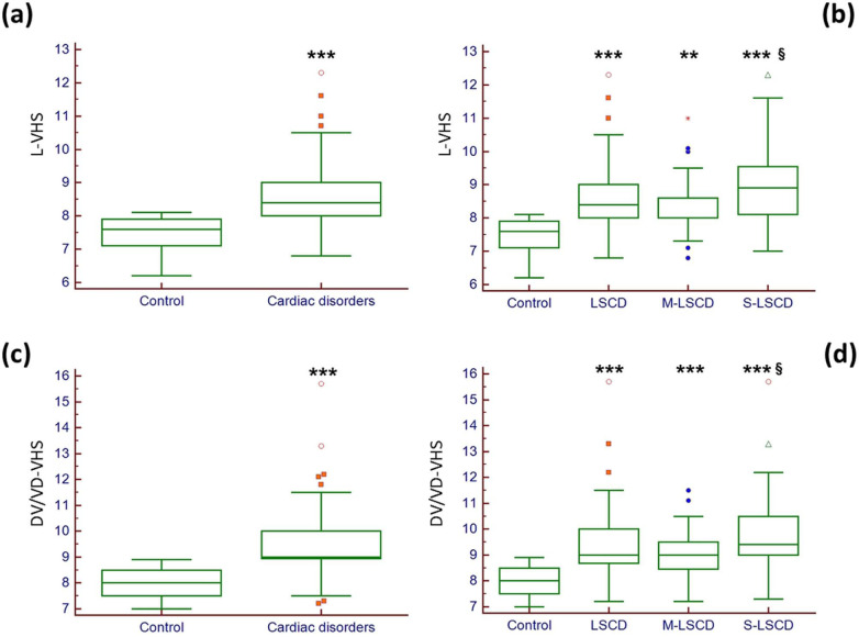
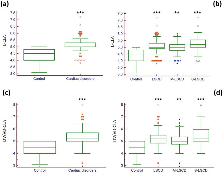
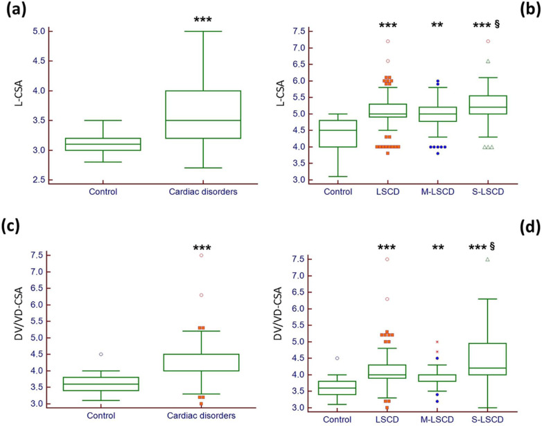
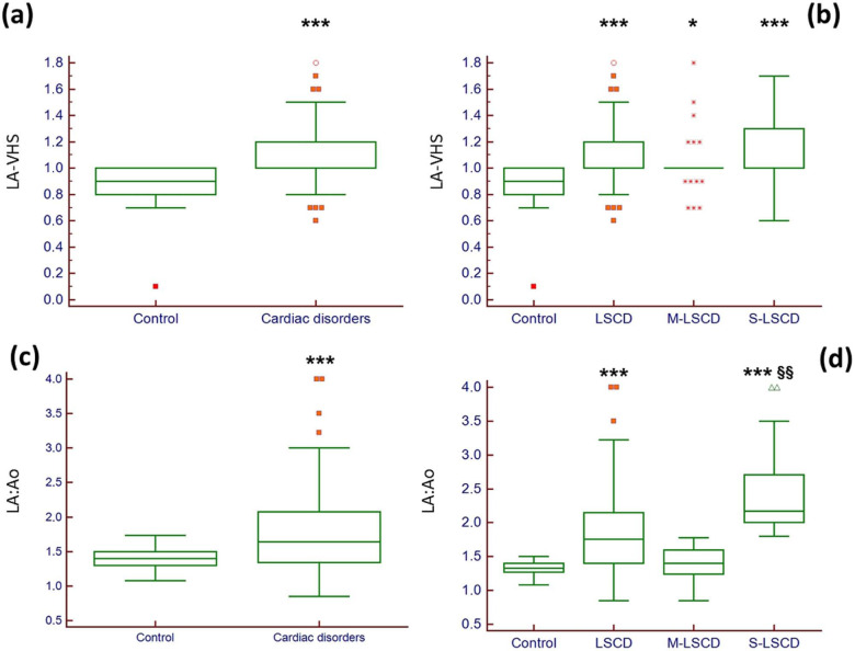
All cats had a right recumbent L radiographic view. Forty-eight cats with cardiac disorders and nine healthy cats had a DV radiographic view of the thorax available, while 35 cats with cardiac disorders and 13 healthy cats had a VD radiograph available. Radiographic indices for different groups of cats (control, overall cardiac disorders, LSCD, LSCD with no or mild LAE and LSCD with moderate-to-severe LAE) of the cats are summarised in Table 2 and Figures 2–5. All radiographic indices had significantly higher values in cats with cardiac disorders (P <0.001) and cats with LSCD (P <0.001) compared with those of control cats. The L-VHS, DV/VD-VHS, L-CSA and DV/VD-CSA had significantly higher values (P <0.05) in cats with LSCD and moderate-to-severe LAE compared with those of cats with LSCD and no or mild LAE. The echocardiographic LA:Ao was significantly higher in cats with cardiac disorders (P <0.001) and cats with LSCD (P <0.001) compared with that of control cats, and was significantly higher in cats with LSCD and moderate-to-severe LAE compared (P <0.001) with that of cats with LSCD and no or mild LAE (Table 2 and Figure 5).
Table 2.
Measured values for seven radiographic indices and the echocardiographic left atrium–aorta ratio (LA:Ao) in 22 healthy cats (control) and 83 cats with different cardiac disorders. Normally distributed and non-normally distributed data are listed as mean ± SD and median (range), respectively
| Control | Cardiac disorders |
|||||
|---|---|---|---|---|---|---|
| Overall | LSCD | M-LSCD | S-LSCD | |||
| n | 22 | 83 | 72 | 37 | 35 | |
| VHS | Lateral | 7.56 ± 0.54 | 8.65 ± 1.02 * | 8.62 ± 1.04 * | 8.29 ± 0.83 † | 8.95 ± 1.13 * § |
| DV or VD | 8.0 (7.0–8.9) | 9.0 (7.2–15.7) * | 9.0 (7.2–15.7) * | 9.0 (7.2–11.5) * | 9.4 (7.3–15.7) * § | |
| CLA | Lateral | 4.44 ± 0.46 | 5.09 ± 0.59 * | 5.08 ± 0.63 * | 4.92 ± 0.53 † | 5.24 ± 0.67 * |
| DV or VD | 4.53 ± 0.52 | 5.31 ± 0.80 * | 5.24 ± 0.79 * | 5.10 ± 0.68 † | 5.41 ± 0.86 * | |
| CSA | Lateral | 3.1 (2.8–3.5) | 3.5 (2.7–5.0) * | 3.5 (2.7–5.0) * | 3.3 (2.7–5.0) † | 3.7 (2.7–5.0) * § |
| DV or VD | 3.6 (3.1–4.5) | 4.0 (3.0–7.5) * | 4.0 (3.0–7.5) * | 4.0 (3.0–5.2) † | 4.2 (3.0–7.5) * § | |
| LA-VHS LA:Ao | 0.87 ± 0.21 1.33 (1.08–1.50) |
1.08 ± 0.22
*
1.73 (0.85–4.00) * |
1.08 ± 0.23
*
1.76 (0.85–4.00) * |
1.03 ± 0.20
‡
1.4 (0.85–1.78) |
1.14 ± 0.25
*
2.2 (1.8–4.0) * ¶ |
|
P <0.001 compared with control cats
P <0.01 compared with control cats
P <0.05 compared with control cats
P <0.05 compared with cats with M-LSCD
P <0.001 compared with cats with M-LSCD
LSCD = left-sided cardiac disorders; M-LSCD = cats with LSCD with no or mild left atrial enlargement; S-LSCD = cats with LSCD and moderate-to-severe left atrial enlargement; VHS = vertebral heart score; CLA = cardiac long axis; CSA = cardiac short axis; LA-VHS = left atrium VHS; DV = dorsoventral; VD = ventrodorsal
Figure 2.
Box and whiskers plots of (a) the lateral vertebral heart score (L-VHS) in healthy cats (control) and in cats with overall cardiac disorders; (b) L-VHS in healthy cats, cats with left-sided cardiac disorders (LSCD), cats with LSCD and no or mild left atrial enlargement (M-LSCD), and cats with LSCD and moderate-to-severe left atrial enlargement (S-LSCD); (c) dorsoventral or ventrodorsal vertebral heart score (DV/VD-VHS) in healthy cats (control) and in cats with overall cardiac disorders; (d) DV/VD-VHS in healthy cats, cats with LSCD, cats with M-LSCD and cats with S-LSCD.
**P <0.01 compared with control cats
***P <0.001 compared with control cats
§P <0.05 compared with cats with M-LSCD
Figure 3.
Box and whiskers plots of (a) the lateral cardiac long axis (L-CLA) in healthy cats (control) and in cats with overall cardiac disorders; (b) L-CLA in healthy cats, cats with left-sided cardiac disorders (LSCD), cats with LSCD and no or mild left atrial enlargement (M-LSCD), and cats with LSCD and moderate-to-severe left atrial enlargement (S-LSCD); (c) dorsoventral or ventrodorsal cardiac long axis (DV/VD-CLA) in healthy cats (control) and in cats with overall cardiac disorders; (d) DV/VD-CLA in healthy cats, cats with LSCD, cats with M-LSCD and cats with S-LSCD.
**P <0.01 compared with control cats
***P <0.001 compared with control cats
Figure 4.
Box and whiskers plots of (a) the lateral cardiac short axis (L-CSA) in healthy cats (control) and in cats with overall cardiac disorders; (b) L-CSA in healthy cats, cats with left-sided cardiac disorders (LSCD), cats with LSCD and no or mild left atrial enlargement (M-LSCD), and cats with LSCD and moderate-to-severe left atrial enlargement (S-LSCD); (c) dorsoventral or ventrdorsal cardiac short axis (DV/VD-CSA) in healthy cats (control) and in cats with overall cardiac disorders; (d) DV/VD-CSA in healthy cats, cats with LSCD, cats with M-LSCD and cats with S-LSCD.
**P <0.01 compared with control cats
***P <0.001 compared with control cats
§P <0.05 compared with cats with M-LSCD
Figure 5.
Box and whiskers plots of (a) a modified radiographic measurement of the left atrium (LA-VHS) in healthy cats (control) and in cats with overall cardiac disorders; (b) LA-VHS in healthy cats, cats with left-sided cardiac disorders (LSCD), cats with LSCD and no or mild left atrial enlargement (M-LSCD), and cats with LSCD and moderate-to-severe left atrial enlargement (S-LSCD); (c) ratio of the echocardiographic measurement of the left atrial diameter to aortic root diameter (LA:Ao) in healthy cats (control) and in cats with overall cardiac disorders; (d) LA:Ao in healthy cats, cats with LSCD, cats with M-LSCD and cats with S-LSCD.
*P <0.05 compared with control cats
***P <0.001 compared with control cats
§§P <0.001 compared with cats with M-LSCD
The echocardiographic LA:Ao measured in control cats and cats with LSCD had a significant (P <0.001), but weak and positive, correlation with all radiographic indices (Table 3) with higher correlation coefficients for L-VHS (0.334) and L-CSA (0.308).
Table 3.
Bivariate analysis of radiographic variables with the echocardiographic left atrium to aortic root diameter ratio in 22 healthy cats and 73 cats with left-sided cardiac disorders
| Variable | Correlation coefficient | P |
|---|---|---|
| L-VHS | 0.334 | <0.001 |
| DV/VD-VHS | 0.199 | <0.001 |
| L-CLA | 0.255 | <0.001 |
| DV/VD-CLA | 0.110 | <0.001 |
| L-CSA | 0.308 | <0.001 |
| DV/VD-CSA | 0.258 | <0.001 |
| LA-VHS | 0.262 | <0.001 |
L-VHS = lateral vertebral heart score; DV/VD–VHS = dorsoventral or ventrodorsal VHS; L-CLA = lateral cardiac long axis; DV/VD-CLA = dorso-ventral or ventrodorsal CLA; L-CSA = lateral cardiac short axis; DV/VD-CSA = dorsoventral or ventrodorsal CSA; LA-VHS = left atrium VHS
ROC curve analyses for each of the radiographic indices used to distinguish cats with overall cardiac disorders and cats with LSCD from healthy cats are summarised in Tables 4 and 5, respectively. In both populations of cats with cardiac disorders, all radiographic indices were moderately accurate in discriminating cats with cardiac disorders from healthy cats. Higher accuracy (AUC ± SE) was found for the L-VHS (0.879 ± 0.036 and 0.866 ± 0.039 for cats with overall cardiac disorders and cats with LSCD, respectively) and the DV/VD-VHS–VHS (0.874 ± 0.034 and 0.857 ± 0.039 for cats with overall cardiac disorders and cats with LSCD, respectively). Cut-offs of >7.9 and >8.9 for L-VHS and DV/VD-VHS, respectively, had the best equilibrium between sensitivity and specificity and were the most accurate radiographic indices for distinguishing cats with either overall cardiac disorders (sensitivity 0.86 and 0.75, respectively; specificity 0.82 and 1.00, respectively) and cats with LSCD (sensitivity 0.85 and 0.72, respectively; specificity 0.82 and 1.00, respectively) from healthy cats.
Table 4.
Diagnostic accuracy of seven radiographic indices in distinguishing healthy cats from cats with cardiac disorders
| Index | AUC ± SE | 95% CI | Cut-off | Sensitivity | Specificity | PLR | NLR |
|---|---|---|---|---|---|---|---|
| L-VHS | 0.879 ± 0.036 | 0.800–0.935 | >6.5 | 1 | 0.14 | 1.16 | 0 |
| >7.9* | 0.86 | 0.82 | 4.75 | 0.17 | |||
| >8.2 | 0.56 | 1 | NA | 0.44 | |||
| DV/VD-VHS | 0.874 ± 0.034 | 0.792–0.933 | >7 | 1 | 0.05 | 1.05 | 0 |
| >8.9* | 0.75 | 1 | NA | 0.25 | |||
| L-CLA | 0.867 ± 0.035 | 0.786–0.926 | >3.7 | 1 | 0.14 | 1.16 | 0 |
| >4.8* | 0.78 | 0.95 | 17.17 | 0.23 | |||
| >5 | 0.43 | 1 | NA | 0.57 | |||
| DV/VD-CLA | 0.839 ± 0.040 | 0.752–0.905 | >3.1 | 1 | 0.05 | 1.05 | 0 |
| >5* | 0.55 | 1 | NA | 0.45 | |||
| L-CSA | 0.830 ± 0.040 | 0.744–0.897 | ⩾2.7 | 1 | 0 | 1 | NA |
| >3.3* | 0.65 | 0.91 | 7.20 | 0.38 | |||
| >3.5 | 0.49 | 1 | NA | 0.51 | |||
| DV/VD-CSA | 0.836 ± 0.047 | 0.747–0.903 | ⩾3 | 1 | 0 | 1 | NA |
| >3.9* | 0.76 | 0.82 | 4.20 | 0.29 | |||
| >4.5 | 0.20 | 1 | NA | 0.80 | |||
| LA-VHS | 0.782 ± 0.047 | 0.690–0.857 | >0.1 | 1 | 0.05 | 1.05 | 0 |
| >0.9* | 0.85 | 0.59 | 2.08 | 0.25 | |||
| >1 | 0.28 | 1 | NA | 0.72 |
AUC = area under the curve; CI = confidence interval; PLR = positive likelihood ratio; NLR = negative likelihood ratio; L-VHS = lateral vertebral heart score; DV/VD-VHS = dorsoventral or ventrodorsal VHS; L-CLA = lateral cardiac long axis; DV/VD-CLA = dorsoventral or ventrodorsal CLA; L-CSA = lateral cardiac short axis; DV/VD-CSA = dorsoventral or ventrodorsal CSA; LA-VHS = left atrium VHS; NA = not applicable
Values with the best equilibrium between sensitivity and specificity
Table 5.
Diagnostic accuracy of seven radiographic indices in distinguishing healthy cats from cats with left-sided cardiac disorders
| Index | AUC ± SE | 95% CI | Cut-off | Sensitivity | Specificity | PLR | NLR |
|---|---|---|---|---|---|---|---|
| L-VHS | 0.866 ± 0.039 | 0.779–0.928 | >6.5 | 1 | 0.14 | 1.16 | 0 |
| >7.9* | 0.85 | 0.82 | 4.65 | 0.19 | |||
| >8.2 | 0.52 | 1 | NA | 0.48 | |||
| DV/VD-VHS | 0.857 ± 0.039 | 0.767–0.922 | >7 | 1 | 0.05 | 1.05 | 0 |
| >8.9* | 0.72 | 1 | NA | 0.28 | |||
| L-CLA | 0.850 ± 0.038 | 0.762–0.916 | >3.7 | 1 | 0.14 | 1.16 | 0 |
| >4.8* | 0.75 | 0.95 | 16.50 | 0.26 | |||
| >5 | 0.43 | 1 | NA | 0.57 | |||
| DV/VD-CLA | 0.821 ± 0.044 | 0.725–0.894 | >3.1 | 1 | 0.05 | 1.05 | 0 |
| >5* | 0.51 | 1 | NA | 0.49 | |||
| L-CSA | 0.812 ± 0.044 | 0.717–0.885 | ⩾2.7 | 1 | 0 | 1 | NA |
| >3.3* | 0.62 | 0.91 | 6.82 | 0.42 | |||
| >3.5 | 0.46 | 1 | NA | 0.54 | |||
| DV/VD-CSA | 0.821 ± 0.050 | 0.726–0.894 | ⩾3 | 1 | 0 | 1 | NA |
| >3.9* | 0.73 | 0.82 | 4.02 | 0.33 | |||
| >4.5 | 0.18 | 1 | NA | 0.82 | |||
| LA-VHS | 0.774 ± 0.048 | 0.675–0.855 | >0.1 | 1 | 0.05 | 1.05 | 0 |
| >0.9* | 0.83 | 0.59 | 2.03 | 0.29 | |||
| >1 | 0.3 | 1 | NA | 0.70 |
AUC = area under the curve; CI = confidence interval; PLR = positive likelihood ratio; NLR = negative likelihood ratio; L-VHS = lateral vertebral heart score; DV/VD-VHS = dorsoventral or ventrodorsal VHS; L-CLA = lateral cardiac long axis; DV/VD-CLA = dorsoventral or ventrodorsal CLA; L-CSA = lateral cardiac short axis; DV/VD-CSA = dorsoventral or ventrodorsal CSA; LA-VHS = left atrium VHS; NA = not applicable
Values with the best equilibrium between sensitivity and specificity
When considering the two subpopulations of cats with LSCD (ie, those with no or mild LAE and those with moderate-to-severe LAE [Tables 6 and 7, respectively]) only the L-VHS was highly accurate in distinguishing cats with moderate-to-severe LAE from healthy cats (AUC ± SE = 0.910 ± 0.040). All the other indices were only moderately accurate in distinguishing cats with LSCD and different degrees of LAE and healthy cats, with higher values of AUC ± SE for DV/VD–VHS (0.878 ± 0.050), L-CLA (0.891 ± 0.044) and L-CSA (0.896 ± 0.043) in distinguishing cats with moderate-to-severe LAE and healthy cats. Cut-offs of >7.9, >8.9, >4.8 and >3.4 for L-VHS, DV/VD-VHS, L-CLA and L-CSA, respectively, had the best equilibrium between sensitivity (0.91, 0.81, 0.83 and 0.74, respectively) and specificity (0.82, 1.00, 0.95 and 0.95, respectively), and these were the most accurate radiographic indices for distinguishing cats with LSCD and moderate-to-severe LAE from healthy cats.
Table 6.
Diagnostic accuracy of seven radiographic indices in distinguishing healthy cats from cats with left-sided cardiac disorders and no or mild left atrial enlargement
| Index | AUC ± SE | 95% CI | Cut-off | Sensitivity | Specificity | PLR | NLR |
|---|---|---|---|---|---|---|---|
| L-VHS | 0.823 ± 0.056 | 0.700–0.911 | >6.5 | 1 | 0.14 | 1.16 | 0 |
| >7.9* | 0.78 | 0.82 | 4.28 | 0.27 | |||
| >8.2 | 0.33 | 1 | NA | 0.67 | |||
| DV/VD-VHS | 0.840 ± 0.051 | 0.720–0.923 | >7 | 1 | 0.05 | 1.05 | 0 |
| >8.8* | 0.69 | 0.95 | 15.28 | 0.32 | |||
| >8.9 | 0.64 | 1 | NA | 0.36 | |||
| L-CLA | 0.812 ± 0.056 | 0.689–0.902 | >3.7 | 1 | 0.14 | 1.16 | 0 |
| >4.8* | 0.68 | 0.95 | 14.86 | 0.34 | |||
| >5 | 0.32 | 1 | NA | 0.68 | |||
| DV/VD-CLA | 0.800 ± 0.056 | 0.674–0.893 | >3.1 | 1 | 0.05 | 1.05 | 0 |
| >5* | 0.47 | 1 | NA | 0.53 | |||
| L-CSA | 0.730 ± 0.064 | 0.597–0.838 | >2.8 | 1 | 0.05 | 1.05 | 0 |
| >3.3* | 0.47 | 0.91 | 5.19 | 0.58 | |||
| >3.5 | 0.28 | 1 | NA | 0.72 | |||
| DV/VD-CSA | 0.795 ± 0.063 | 0.668–0.890 | >3.1 | 1 | 0.09 | 1.10 | 0 |
| >3.7* | 0.83 | 0.68 | 2.62 | 0.24 | |||
| >4.5 | 0.06 | 1 | NA | 0.94 | |||
| LA-VHS | 0.736 ± 0.061 | 0.604–0.843 | >0.1 | 1 | 0.05 | 1.05 | 0 |
| >0.9* | 0.81 | 0.59 | 1.97 | 0.33 | |||
| >1 | 0.17 | 1 | NA | 0.83 |
AUC = area under the curve; CI = confidence interval; PLR = positive likelihood ratio; NLR = negative likelihood ratio; L-VHS = lateral vertebral heart score; DV/VD-VHS = dorsoventral or ventrodorsal VHS; L-CLA = lateral cardiac long axis; DV/VD-CLA = dorsoventral or ventrodorsal CLA; L-CSA = lateral cardiac short axis; DV/VD-CSA = dorsoventral or ventrodorsal CSA; LA-VHS = left atrium VHS; NA = not applicable
Values with the best equilibrium between sensitivity and specificity
Table 7.
Diagnostic accuracy of seven radiographic indices in distinguishing healthy cats from cats with left-sided cardiac disorders and moderate or severe left atrial enlargement
| Index | AUC ± SE | 95% CI | Cut-off | Sensitivity | Specificity | PLR | NLR |
|---|---|---|---|---|---|---|---|
| L-VHS | 0.910 ± 0.040 | 0.804–0.969 | >6.5 | 1 | 0.14 | 1.16 | 0 |
| >7.9* | 0.91 | 0.82 | 5.03 | 0.10 | |||
| >8.2 | 0.71 | 1 | NA | 0.29 | |||
| DV/VD-VHS | 0.878 ± 0.050 | 0.758–0.951 | >7.2 | 1 | 0.14 | 1.16 | 0 |
| >8.9* | 0.81 | 1 | NA | 0.19 | |||
| L-CLA | 0.891 ± 0.044 | 0.780–0.958 | >3.7 | 1 | 0.14 | 1.16 | 0 |
| >4.8* | 0.83 | 0.95 | 18.23 | 0.18 | |||
| >5 | 0.54 | 1 | NA | 0.46 | |||
| DV/VD-CLA | 0.845 ± 0.050 | 0.719–0.929 | >3.9 | 1 | 0.14 | 1.16 | 0 |
| >5* | 0.55 | 1 | NA | 0.45 | |||
| L-CSA | 0.896 ± 0.043 | 0.787–0.961 | ⩾2.7 | 1 | 0 | 1 | NA |
| >3.4* | 0.74 | 0.95 | 16.34 | 0.27 | |||
| >3.5 | 0.66 | 1 | NA | 0.34 | |||
| DV/VD-CSA | 0.852 ± 0.055 | 0.727–0.934 | ⩾3 | 1 | 0 | 1 | NA |
| >3.9* | 0.84 | 0.82 | 4.61 | 0.20 | |||
| >4.5 | 0.32 | 1 | NA | 0.68 | |||
| LA-VHS | 0.814 ± 0.052 | 0.688–0.906 | >0.1 | 1 | 0.05 | 1.05 | 0 |
| >0.9* | 0.85 | 0.59 | 2.08 | 0.25 | |||
| >1 | 0.44 | 1 | NA | 0.56 |
AUC = area under the curve; CI = confidence interval; PLR = positive likelihood ratio; NLR = negative likelihood ratio; L-VHS = lateral vertebral heart score; DV/VD-VHS = dorsoventral or ventrodorsal VHS; L-CLA = lateral cardiac long axis; DV/VD-CLA = dorsoventral or ventrodorsal CLA; L-CSA = lateral cardiac short axis; DV/VD-CSA = dorsoventral or ventrodorsal CSA; LA-VHS = left atrium VHS; NA = not applicable
Values with the best equilibrium between sensitivity and specificity
The results of ROC curve analyses for each of the radiographic indices to identify different degrees of LAE (ie, mild, moderate and severe LAE) in cats with LSCD are summarised in Tables 8, 9 and 10, respectively. All the considered radiographic indices were moderately accurate in identifying LAE in cats with LSCD, with progressively higher values of AUC ± SE going from cats with mild LAE to cats with severe LAE. In particular, higher values of AUC ± SE to predict severe LAE were found for L-VHS (0.885 ± 0.046) and L-CLA (0.890 ± 0.049). Cut-offs of >8.2 and >3.5, for L-VHS and L-CSA, respectively, had the best equilibrium between sensitivity (0.78 for both) and specificity (0.92 for both), and these were the most accurate radiographic indices for identifying severe LAE in cats with LSCD.
Table 8.
Diagnostic accuracy of seven radiographic indices in predicting mild left atrial enlargement in healthy cats and cats with left-sided cardiac disorders
| Index | AUC ± SE | 95% CI | Cut-off | Sensitivity | Specificity | PLR | NLR |
|---|---|---|---|---|---|---|---|
| L-VHS | 0.776 ± 0.048 | 0.678–0.856 | >6.2 | 1 | 0.03 | 1.03 | 0 |
| >8.2* | 0.63 | 0.92 | 8.19 | 0.40 | |||
| >9 | 0.28 | 1 | NA | 0.72 | |||
| DV/VD-VHS | 0.706 ± 0.055 | 0.600–0.798 | >7 | 1 | 0.03 | 1.03 | 0 |
| >9.6* | 0.37 | 1 | NA | 0.63 | |||
| L-CLA | 0.740 ± 0.050 | 0.639–0.825 | >3.1 | 1 | 0.03 | 1.03 | 0 |
| >4.8* | 0.75 | 0.64 | 2.08 | 0.40 | |||
| >5.3 | 0.29 | 1 | NA | 0.71 | |||
| DV/VD-CLA | 0.668 ± 0.056 | 0.560–0.764 | >3.1 | 1 | 0.03 | 1.03 | 0 |
| >5.3* | 0.41 | 0.92 | 5.22 | 0.64 | |||
| >5.5 | 0.29 | 1 | NA | 0.71 | |||
| L-CSA | 0.733 ± 0.053 | 0.631–0.820 | ⩾2.7 | 1 | 0 | 1 | NA |
| >3.6* | 0.52 | 0.97 | 20.22 | 0.49 | |||
| >4 | 0.13 | 1 | NA | 0.87 | |||
| DV/VD-CSA | 0.717 ± 0.054 | 0.612–0.808 | ⩾3 | 1 | 0 | 1 | NA |
| >3.9* | 0.76 | 0.63 | 2.08 | 0.37 | |||
| >4.7 | 0.20 | 1 | NA | 0.80 | |||
| LA-VHS | 0.686 ± 0.050 | 0.581–0.779 | >0.1 | 1 | 0.03 | 1.03 | 0 |
| >1* | 0.38 | 0.97 | 14.72 | 0.64 | |||
| >1.2 | 0.28 | 1 | NA | 0.72 |
AUC = area under the curve; CI = confidence interval; PLR = positive likelihood ratio; NLR = negative likelihood ratio; L-VHS = lateral vertebral heart score; DV/VD-VHS = dorsoventral or ventrodorsal VHS; L-CLA = lateral cardiac long axis; DV/VD-CLA = dorsoventral or ventrodorsal CLA; L-CSA = lateral cardiac short axis; DV/VD-CSA = dorsoventral or ventrodorsal CSA; LA-VHS = left atrium VHS; NA = not applicable
Values with the best equilibrium between sensitivity and specificity
Table 9.
Diagnostic accuracy of seven radiographic indices in predicting moderate left atrial enlargement in healthy cats and cats with left-sided cardiac disorders
| Index | AUC ± SE | 95% CI | Cut-off | Sensitivity | Specificity | PLR | NLR |
|---|---|---|---|---|---|---|---|
| L-VHS | 0.848 ± 0.048 | 0.746–0.921 | >6.8 | 1 | 0.05 | 1.05 | 0 |
| >8.2* | 0.71 | 0.92 | 9.29 | 0.31 | |||
| >9 | 0.31 | 1 | NA | 0.69 | |||
| DV/VD-VHS | 0.784 ± 0.060 | 0.668–0.874 | >7.2 | 1 | 0.05 | 1.06 | 0 |
| >9* | 0.68 | 0.84 | 4.29 | 0.38 | |||
| >9.6 | 0.45 | 1 | NA | 0.55 | |||
| L-CLA | 0.790 ± 0.053 | 0.680–0.876 | >3.8 | 1 | 0.05 | 1.05 | 0 |
| >4.8* | 0.83 | 0.64 | 2.31 | 0.27 | |||
| >5.3 | 0.34 | 1 | NA | 0.66 | |||
| DV/VD-CLA | 0.726 ± 0.061 | 0.606–0.827 | >3.9 | 1 | 0.05 | 1.06 | 0 |
| >5.3* | 0.48 | 0.92 | 6.13 | 0.56 | |||
| >5.5 | 0.32 | 1 | NA | 0.68 | |||
| L-CSA | 0.836 ± 0.051 | 0.732–0.912 | ⩾2.7 | 1 | 0 | 1 | NA |
| >3.4* | 0.74 | 0.85 | 4.83 | 0.30 | |||
| >4 | 0.11 | 1 | NA | 0.89 | |||
| DV/VD-CSA | 0.781 ± 0.058 | 0.665–0.871 | ⩾3 | 1 | 0 | 1 | NA |
| >3.9* | 0.84 | 0.63 | 2.28 | 0.26 | |||
| >4.7 | 0.29 | 1 | NA | 0.71 | |||
| LA-VHS | 0.738 ± 0.054 | 0.622–0.834 | >0.1 | 1 | 0.03 | 1.03 | 0 |
| >1* | 0.44 | 0.97 | 17.21 | 0.57 | |||
| >1.2 | 0.35 | 1 | NA | 0.65 |
AUC = area under the curve; CI = confidence interval; PLR = positive likelihood ratio; NLR = negative likelihood ratio; L-VHS = lateral vertebral heart score; DV/VD-VHS = dorsoventral or ventrodorsal VHS; L-CLA = lateral cardiac long axis; DV/VD-CLA = dorsoventral or ventrodorsal CLA; L-CSA = lateral cardiac short axis; DV/VD-CSA = dorsoventral or ventrodorsal CSA; LA-VHS = left atrium VHS; NA = not applicable
Values with the best equilibrium between sensitivity and specificity
Table 10.
Diagnostic accuracy of seven radiographic indices in predicting severe left atrial enlargement in healthy cats and cats with left-sided cardiac disorders
| Index | AUC ± SE | 95% CI | Cut-off | Sensitivity | Specificity | PLR | NLR |
|---|---|---|---|---|---|---|---|
| L-VHS | 0.885 ± 0.046 | 0.778–0.952 | >7.3 | 1 | 0.21 | 1.26 | 0 |
| >8.2* | 0.78 | 0.92 | 10.17 | 0.24 | |||
| >9 | 0.26 | 1 | NA | 0.74 | |||
| DV/VD-VHS | 0.819 ± 0.063 | 0.699–0.907 | >7.2 | 1 | 0.05 | 1.06 | 0 |
| >9* | 0.68 | 0.84 | 4.32 | 0.38 | |||
| >9.6 | 0.50 | 1 | NA | 0.50 | |||
| L-CLA | 0.829 ± 0.057 | 0.712–0.913 | >3.8 | 1 | 0.05 | 1.05 | 0 |
| >4.8* | 0.91 | 0.64 | 2.54 | 0.14 | |||
| >5.3 | 0.35 | 1 | NA | 0.65 | |||
| DV/VD-CLA | 0.734 ± 0.070 | 0.605–0.840 | >3.9 | 1 | 0.05 | 1.06 | 0 |
| >5.3* | 0.50 | 0.92 | 6.33 | 0.54 | |||
| >5.5 | 0.32 | 1 | NA | 0.68 | |||
| L-CSA | 0.890 ± 0.049 | 0.784–0.955 | >2.9 | 1 | 0.05 | 1.05 | 0 |
| >3.5* | 0.78 | 0.92 | 10.17 | 0.24 | |||
| >4 | 0.04 | 1 | NA | 0.96 | |||
| DV/VD-CSA | 0.807 ± 0.065 | 0.685–0.898 | ⩾3 | 1 | 0 | 1 | NA |
| >3.9* | 0.86 | 0.63 | 2.34 | 0.22 | |||
| >4.7 | 0.32 | 1 | NA | 0.68 | |||
| LA-VHS | 0.809 ± 0.049 | 0.688–0.898 | >0.8 | 1 | 0.26 | 1.34 | 0 |
| >1* | 0.50 | 0.97 | 19.50 | 0.51 | |||
| >1.2 | 0.36 | 1 | NA | 0.64 |
AUC = area under the curve; CI = confidence interval; PLR = positive likelihood ratio; NLR = negative likelihood ratio; L-VHS = lateral vertebral heart score; DV/VD-VHS = dorsoventral or ventrodorsal VHS; L-CLA = lateral cardiac long axis; DV/VD-CLA = dorsoventral or ventrodorsal CLA; L-CSA = lateral cardiac short axis; DV/VD-CSA = dorsoventral or ventrodorsal CSA; LA-VHS = left atrium VHS; NA = not applicable
Values with the best equilibrium between sensitivity and specificity
Discussion
The type and prevalence of cardiac disorders observed in cats in the present study reflect the overall prevalence of cardiac disorders diagnosed in cats, with congenital cardiac disorders and RSCD being less prevalent than acquired cardiac disorders and LSCD, respectively.30–32 Furthermore, cardiomyopathies, particularly hypertrophic cardiomyopathy, and atrio-ventricular valve regurgitation were the most frequently observed cardiac disorders, similar to previous reports.30–32 Cats with severe cardiac disorders (ie, cats with decompensated cardiac diseases associated with pleural and/or pericardial effusion) were excluded from the study.
Results of measurements of the VHS, CLA and CSA in L and DV or VD view in 22 cats without cardiac abnormalities in the present study were similar to those previously observed in studies carried out on larger numbers of cats.9,10 In our control cats, the absence of even subtle cardiovascular abnormalities was based on physical and echocardiographic examination, while previous studies reporting values of VHS and related radiographic indices in normal cats were carried out without the use of cardiac ultrasonographic examination.9,10 The absence of remarkable differences in the VHS and other related radiographic indices in healthy cats among different study populations and feline breeds confirms that cats have relatively uniform thoracic shapes compared with dogs, in which different normal ranges of VHS have been proposed for different canine breeds.13–17 Conversely, our measurements of LA-VHS in healthy cats (0.87 ± 0.05) were different from those previously reported (median 1.10, range 0.75–1.30) in cats with echocardiographically measured normal left atria. 28 The observed discrepancy is likely related to the difficulty in clearly identifying the actual margin of the LA due to overlying soft tissue structures in feline thoracic radiographs. 28 Indeed, this radiographic index was constantly the least accurate in all the successive analyses in distinguishing healthy cats from cats with cardiac disorders and in predicting LAE in cats with LSCD.
All the radiographic indices measured in this study were significantly higher in the overall population of cats with cardiac disorders than those of control cats. Furthermore, all the radiographic indices were significantly higher in cats with LSCD, either those with no or mild LAE or those with moderate-to-severe LAE, compared with control cats. The echocardiographic LA:Ao was significantly higher in cats with LSCD and moderate-to-severe LAE than that of cats with LSCD and no or mild LAE, but only the L-VHS, DV/VD-VHS, L-CSA and DV/VD-VHS were significantly higher in the former compared with the latter group of cats. In healthy cats and cats with LSCD the LA:Ao was significantly, but weakly and positively, correlated with all radiographic indices. Because of the relatively small sample size and the heterogeneous pattern of cardiac remodelling observed in cats with RSCD (four cats with cardiac disorders leading to concentric cardiac hypertrophy and seven cats with cardiac disorders leading to eccentric cardiac hypertrophy), no further analyses were carried out for this group of cats.
Considering the radiographic indices most commonly employed in the clinical setting, namely L-VHS and DV/VD-VHS, clinically normal cats usually have a L-VHS of ⩽8.1 and a DV/VD-VHS <9.0. 9 Values of L-VHS (8.65 ± 1.02) and DV/VD-VHS (median 9.0, range 7.2–15.7) in our cats with cardiac disorders and values of L-VHS (8.62 ± 1.04) and DV/VD-VHS (median 9.0, range 7.2–15.7) in our cats with LSCD can be compared with those previously observed in cats with cardiogenic pulmonary oedema (L-VHS range 8.3–10.8) 26 and heartworm disease (7.96 ± 0.13 and 8.64 ± 0.17 for L-VHS and DV/VD-VHS, respectively). 22 Furthermore, in cats presenting to an emergency service for dyspnoea, none of the cats with a L-VHS ⩽8 had primary cardiac disease, while all cats with a L-VHS >9.3 had cardiac disease. 4 Our results confirm that feline cardiac diseases are associated with increased dimensions of the overall cardiac silhouette, as well as of its long and short axes.
The accuracy of all radiographic indices employed in the present study in distinguishing between cats with overall cardiac disorders or cats with LSCD and cats without cardiac disorders was only moderate, with higher values for L-VHS and DV/VD-VHS. In cats with LSCD, the L-VHS, L-CLA and L-CSA had high or ‘nearly high’ accuracy in distinguishing between cats with moderate-to-severe LAE and control cats with an AUC ± SE of 0.910 ± 0.040, 0.891 ± 0.044 and 0.896 ± 0.043, respectively. In particular, a cut-off of >7.9 for L-VHS had a sensitivity of 0.91 and specificity of 0.82 in differentiating cats with LSCD and moderate-to-severe LAE and cats without cardiac disorders. The accuracy of all the other considered radiographic indices at distinguishing cats with LSCD and no or mild LAE from cats without cardiac disorders was only moderate. At the cut-offs of >8.2 and >8.9 for L-VHS and DV/VD-VHS, respectively, the specificity in distinguishing cats with overall cardiac disorders and cats with LSCD, either those with no or mild LAE and those with moderate-to-severe LAE, and healthy cats was 1.00 for both indices, but the sensitivity ranged from 0.33 to 0.71 and from 0.64 to 0.81 for L-VHS and DV/VD-VHS, respectively. The successive analyses, conducted in healthy cats and cats with LSCD, to predict different degree of LAE evidenced a moderate accuracy for all radiographic indices, with values of AUC ± SE ‘nearly high’ only for L-VHS (0.885 ± 0.046) and L-CSA (0.890 ± 0.049) in predicting severe LAE (LA:Ao >2). At the cut-off of >8.2 for L-VHS, the specificity for predicting any degree of LAE was 0.92, but the sensitivity ranged from 0.63 to 0.78 in cats with mild and severe LAE, respectively. These findings suggest that thoracic radiography and measuring the dimensions of the cardiac silhouette using the VHS system can be only moderately useful in differentiating cats with cardiac disorders from those without cardiac disorders and in predicting LAE in cats with LSCD. The prevalence and type of cardiac disorders investigated are important factors influencing the diagnostic accuracy of this radiographic method of measurement. Most feline cardiac disorders are cardiomyopathies usually associated with no or mild left ventricular chamber enlargement. Ventricular wall hypertrophy or left ventricular dysfunction can be evidenced using echocardiography, but are often underestimated using survey thoracic radiography, making this latter technique less accurate in recognising a cardiac disease in cats. In canine chronic mitral valve disease, the VHS system is useful in recognising and following up during time left-sided cardiac dilation and remodelling because of the enlarged cardiac shape associated with chamber volume overload.20,21 These different pathophysiological mechanisms can explain the different accuracy of thoracic radiography in evaluating the cardiac dimension of the most common cardiac disorders between dogs and cats. In cats with LSCD the VHS likely estimates the size of the LA that can become severely enlarged in some subjects. Indeed, the accuracy of the L-VHS in distinguishing between cats with LSCD and moderate-to-severe LAE and normal cats was high at the cut-off of 7.9. An accuracy ‘nearly high’ was also found for L-CSA, which also reflects the left atrial dimension. However, all radiographic indices were only moderately accurate in predicting any degree of LAE in cats with LSCD, as previously demonstrated in cats with cardiomyopathy using either qualitative evaluation of thoracic radiographs and the quantitative index LA-VHS. 28 These findings are likely correlated to the actual location of the LA and auricle of cats that overlaps the overall cardiac silhouette differently from dogs.
This study has some limitations that need highlighting. First, because we used a retrospective study design, adherence to a consistent standardised protocol for obtaining the thoracic radiographs was not possible. All cats in the present study had a right L thoracic view available, while 57 cats had a DV and 48 cats had a VD thoracic view available. In the dog, the calculated VHS can vary according to the type of recumbency (ie, left vs right L view and DV vs VD view), 33 but no significant difference has been previously evidenced in values of VHS of normal cats obtained in DV and VD views.9,10 Radiographic images of the thorax in DV and VD views were combined for successful statistical analysis. The combined results of DV-VHS and VD-VHS have also been reported in a study carried out in cats with heartworm disease. 20 Second, values of the VHS calculated in cats could be influenced by the observer’s experience and choice of landmarks for measurements.4,34 Thus, evaluation of inter- and intra-observer variability is advisable. An overall level of agreement of 83% between one observer, with 10 years of experience, and another, with 1 year of experience, evaluating thoracic radiographs and measuring the VHS in cats with cardiac and non-cardiac causes of respiratory distress was recently reported. 4 The same experienced observer made all the VHS measurements in cats in the present study using published methods of measurement.9,10 Finally, the evaluation of LAE is influenced by the diagnostic method employed either using thoracic radiography and trans-thoracic echocardiography. We used the echocardiographic LA:Ao, which is the most commonly employed method in predicting left atrial enlargement in dogs with cardiac diseases. 35 In cats with cardiac disorders, only left auricle dilation may occur without significant increase of the left atrial short axis, making the LA:Ao suboptimal in predicting left atrial dimensions. We decided to use this method of measurement as it makes easier to create different categories of left atrial dilation; insufficient data are available regarding LA area measurement in cats with heart disease. 27
Conclusions
This study confirms that the cardiac silhouettes in cats with cardiac disorders are larger than those of healthy cat as objectively identified using the VHS and other quantitative radiographic indices. However, the diagnostic accuracy of these indices in discriminating between cats with cardiac disorders and healthy cats, and in predicting LAE in cats with LSCD is only moderate.
Acknowledgments
We appreciate the assistance of Ms Lorenza Guerriero for her help in the statistical analyses of data.
Footnotes
The authors do not have any potential conflicts of interest to declare.
Funding: This research received no specific grant from any funding agency in the public, commercial or not-for-profit sectors.
Accepted: 7 January 2014
References
- 1. Paige C, Abbott JA, Elvinger F. Prevalence of cardiomyopathy in apparently healthy cats. J Am Vet Med Assoc 2009; 234: 1398–1403. [DOI] [PubMed] [Google Scholar]
- 2. Wagner T, Luis Fuentes V, Payne JR, et al. Comparison of auscultatory and echocardiographic findings in healthy adult cats. J Vet Cardiol 2010; 12: 171–182. [DOI] [PubMed] [Google Scholar]
- 3. Nakamura RK, Rishniw M, King MK, et al. Prevalence of echocardiographic evidence of cardiac disease in apparently healthy cats with murmurs. J Feline Med Surg 2011; 13: 266–271. [DOI] [PMC free article] [PubMed] [Google Scholar]
- 4. Sleeper MM, Roland R, Drobatz KJ. Use of the vertebral heart scale for differentiation of cardiac and noncardiac causes of respiratory distress in cats: 67 cases (2002–2003). J Am Vet Med Assoc 2013; 242: 366–371. [DOI] [PubMed] [Google Scholar]
- 5. Lamb CR, Boswood A. Role of survey radiography in diagnosing canine cardiac disease. Compend Contin Educ Pract Vet 2002; 24: 316–326. [Google Scholar]
- 6. Bahr RJ. Heart and pulmonary vessels. In: Thrall DE. (ed). Textbook of veterinary diagnostic radiology. 5th ed. St Louis, MO: Saunders Elsevier, 2007, pp 568–590. [Google Scholar]
- 7. Côté E, MacDonald KA, Meurs KM, et al. Radiology. In: Côté E, MacDonald KA, Meurs KM, et al. (eds). Feline cardiology. Chichester: Wiley Blackwell, 2011, pp 85–100. [Google Scholar]
- 8. Buchanan JW, Bücheler J. Vertebral scale system to measure canine heart size in radiographs. J Am Vet Med Assoc 1995; 206: 194–199. [PubMed] [Google Scholar]
- 9. Lister AL, Buchanan JW. Vertebral scale system to measure heart size in radiographs of cats. J Am Vet Med Assoc 2000; 216: 210–214. [DOI] [PubMed] [Google Scholar]
- 10. Ghadiri A, Avizeh R, Rasekh A, et al. Radiographic measurement of vertebral heart size in healthy stray cats. J Feline Med Surg 2008; 10: 61–65. [DOI] [PMC free article] [PubMed] [Google Scholar]
- 11. Stepien RL, Benson KG, Forrest LJ. Radiographic measurement of cardiac size in normal ferrets. Vet Radiol Ultrasound 1999; 40: 606–610. [DOI] [PubMed] [Google Scholar]
- 12. Onuma M, Ono S, Ishida T, et al. Radiographic measurements of cardiac size in 27 rabbits. J Vet Med Sci 2010; 72: 529–531. [DOI] [PubMed] [Google Scholar]
- 13. Lamb CR, Wikeley H, Boswood A, et al. Use of breed-specific ranges for the vertebral heart scale as an aid to the radiographic diagnosis of cardiac disease in dogs. Vet Rec 2001; 148: 707–711. [DOI] [PubMed] [Google Scholar]
- 14. Bavegems V, Van Caelenberg A, Duchateau L, et al. Vertebral heart size ranges specific for whippets. Vet Radiol Ultrasound 2005; 46: 400–403. [DOI] [PubMed] [Google Scholar]
- 15. Marin LM, Brown J, McBrien C, et al. Vertebral heart size in retired racing Greyhounds. Vet Radiol Ultrasound 2007; 48: 332–334. [DOI] [PubMed] [Google Scholar]
- 16. Kraetschmer S, Ludwig K, Meneses F, et al. Vertebral heart scale in the beagle dog. J Small Anim Pract 2008; 49: 240–243. [DOI] [PubMed] [Google Scholar]
- 17. Jepsen–Grant K, Pollard RE, Johnson LR. Vertebral heart scores in eight dog breeds. Vet Radiol Ultrasound 2013; 54: 3–8. [DOI] [PubMed] [Google Scholar]
- 18. Lamb CR, Tyler M, Boswood A, et al. Assessment of the value of the vertebral heart scale in the radiographic diagnosis of cardiac disease in dogs. Vet Rec 2000; 146: 687–690. [DOI] [PubMed] [Google Scholar]
- 19. Nakayama H, Nakayama T, Hamlin R. Correlation of cardiac enlargement as assessed by vertebral heart size and echocardiographic and electrocardiographic findings in dogs with evolving cardiomegaly due to rapid ventricular pacing. J Vet Intern Med 2001; 15: 217–221. [DOI] [PubMed] [Google Scholar]
- 20. Lord P, Hansson K, Kvart C, et al. Rate of change of heart size before congestive heart failure in dogs with mitral regurgitation. J Small Anim Pract 2010; 51: 210–218. [DOI] [PubMed] [Google Scholar]
- 21. Lord P, Hansson K, Carnabuci C, et al. Radiographic heart size and its rate of increase for onset of congestive heart failure in Cavalier King Charles spaniels with mitral valve regurgitation. J Vet Intern Med 2011; 25: 1312–1319. [DOI] [PubMed] [Google Scholar]
- 22. Litster A, Atkins C, Atwell R, et al. Radiographic cardiac size in cats and dogs with heartworm disease compared with reference values using the vertebral heart scale method: 53 cases. J Vet Cardiol 2005; 7: 33–40. [DOI] [PubMed] [Google Scholar]
- 23. Guglielmini C, Diana A, Santarelli G, et al. Accuracy of radiographic vertebral heart score and sphericity index in the detection of pericardial effusion in dogs. J Am Vet Med Assoc 2012; 241: 1048–1055. [DOI] [PubMed] [Google Scholar]
- 24. Guglielmini C, Diana A, Pietra M, et al. Use of the vertebral heart score in coughing dogs with chronic degenerative mitral valve disease. J Vet Med Sci 2009; 71: 9–13. [DOI] [PubMed] [Google Scholar]
- 25. Litster AL, Buchanan JW. Radiographic and echocardiographic measurement of the heart in obese cats. Vet Radiol Ultrasound 2000; 41: 320–325. [DOI] [PubMed] [Google Scholar]
- 26. Benigni L, Morgan N, Lamb C. Radiographic appearance of cardiogenic pulmonary oedema in 23 cats. J Small Anim Pract 2009; 50: 9–14. [DOI] [PubMed] [Google Scholar]
- 27. Côté E, MacDonald KA, Meurs KM, et al. Echocardiography. In: Côté E, MacDonald KA, Meurs KM, et al. (eds). Feline cardiology. Ames: Wiley–Blackwell, 2011, pp 51–67. [Google Scholar]
- 28. Schober KE, Maerz I, Ludewig E, et al. Diagnostic accuracy of electrocardiography and thoracic radiography in the assessment of left atrial size in cats: comparison with transthoracic 2-dimensional echocardiography. J Vet Intern Med 2007; 21: 709–718. [DOI] [PubMed] [Google Scholar]
- 29. Gardner IA, Greiner M. Receiver-operating characteristics curves and likelihood ratios: improvements over traditional methods for the evaluation and application of veterinary clinical pathology tests. Vet Clin Pathol 2006; 35: 8–17. [DOI] [PubMed] [Google Scholar]
- 30. Buchanan J. Prevalence of cardiovascular disorders. In: Fox PR, Sisson D, Moïse NS. (eds). Textbook of canine and feline cardiology. 2nd ed. Philadelphia: WB Saunders, 1999, pp 457–470. [Google Scholar]
- 31. Riesen SC, Kovacevic A, Lombard CW. Prevalence of heart disease in symptomatic cats: an overview from 1998 to 2005. Schweiz Arch Tierheilkd 2005; 149: 65–71. [DOI] [PubMed] [Google Scholar]
- 32. MacDonald KA. Congenital heart diseases of puppies and kittens. Vet Clin North Am Small Anim Pract 2006; 36: 503–531. [DOI] [PubMed] [Google Scholar]
- 33. Greco A, Meomartino L, Raiano V, et al. Effect of left vs. right recumbency on the vertebral heart score in normal dogs. Vet Radiol Ultrasound 2008; 49: 454–455. [DOI] [PubMed] [Google Scholar]
- 34. Hansson K, Häggström J, Kvart C, et al. Interobserver variability of vertebral heart size measurements in dogs with normal and enlarged hearts. Vet Radiol Ultrasound 2005; 46: 122–130. [DOI] [PubMed] [Google Scholar]
- 35. Rishniw M, Erb HN. Evaluation of four 2-dimensional echocardiographic methods of assessing left atrial size in dogs. J Vet Intern Med 2000; 14: 429–435. [DOI] [PubMed] [Google Scholar]


