Abstract
The basis of the rough-to-smooth conversion of Actinobacillus actinomycetemcomitans was examined. Smooth variants often contained mutations at the flp promoter region. Replacing the mutated flp promoter with the wild-type promoter restored the rough phenotype. The expression level of the flp promoter was ∼100-fold lower in smooth than in rough strains. Mutations of the flp promoter are a cause of the rough-to-smooth conversion.
Gram-negative, facultatively anaerobic Actinobacillus actinomycetemcomitans is a major periodontal pathogen (1, 20). Fresh oral isolates of A. actinomycetemcomitans are invariably fimbriated and form small (∼1-mm), rough-surface, translucent colonies with an internal star-shaped structure (2, 9, 15, 20). After repeated in vitro passages, the rough-colony morphotype may yield nonfimbriated smooth-colony variants that grow as large, round, opaque colonies on agar (2, 9, 15, 16). Occasionally, the rough-to-smooth transition goes through an intermediate phase in which the colonies are translucent but smooth surfaced (9). Genes for the fimbria biogenesis of A. actinomycetemcomitans reside in a 12-kb flp operon that contains 14 genes, flp-1-flp-2-tadV-rcpCAB-tadZABCDEFG (6, 8-12, 14). The transcription-initiation points of the operon were located at 101 and 102 nucleotides upstream of flp-1 (5). Two consensus elements, −10 (TATAAT) and −35 (TTGCAT), separated by 16 nucleotides, of the canonical σ70 promoter sequence were identified upstream of the transcription-initiation points (5).
Many pathogenic bacteria are capable of phase variation and colonial morphology shift, which depends on the expression of surface proteins (3, 13, 18). While the rough-to-smooth conversion of A. actinomycetemcomitans occurs spontaneously, the reverse smooth-to-rough conversion has not been substantiated. We postulated that the rough-smooth conversion in A. actinomycetemcomitans is due not to a phase variation mechanism of the fimbria expression but to some mutational event of the flp operon. This study aimed to determine whether mutations at the promoter region of the flp operon might explain the rough-to-smooth conversion of A. actinomycetemcomitans.
Eighteen A. actinomycetemcomitans strains were examined (Table 1). The culture media and conditions for A. actinomycetemcomitans were as described previously (16). The sequences of the flp promoter of these strains were determined by direct sequencing of the PCR products amplified from this region. A 1-kb DNA fragment encompassing the flp-1-flp-2 genes and 300 bp upstream of flp-1 was amplified with the forward primer F-Ev24 (5′-TCGCGATATCTCTAAATCCACACA; an EcoRV site is underlined), which is located 300 bp upstream of flp-1 (see Fig. 1 for its location). Three different reverse primers were used. Most of the study strains were amplified with the reverse primer orfB-X, 5′-TATCTAGAACGGAATAATGGCGAATA (an XbaI site incorporated) located in orfB (Fig. 1). Two other reverse primers, orfB-B1, 5′-CAGGATCCAGCAGCGAGAGCGTTAT, and orfC-R, 5′-GCACTGAAATGATCAAGAGC, were used for strains that failed to be amplified with the primer orfB-X. PCR was carried out for 30 cycles at 94°C for 30 s, 56°C for 30 s, and 72°C for 2 min. PCR products were purified by the PCR purification columns (QIAGEN) and sequenced by the USC School of Medicine Microchemical Core Facility.
TABLE 1.
A. actinomycetemcomitans strains used in this study
| Strain | Sero- type | Colony morpho- typea | Source |
|---|---|---|---|
| Reference strains | |||
| Y4 | b | S | ATCCc (oral) |
| JP2 | b | S | S. Asikainen (oral) |
| HK1651 | b | R | ATCC (oral) |
| ATCC29523 | a | S | ATCC (blood) |
| ATCC29522 | b | S | ATCC (mandibular abscess) |
| ATCC29524 | b | S | ATCC (chest aspirate) |
| Clinical isolates and variants | |||
| D7S | a | R | Oral clinical isolate |
| D7S-SA | a | S | Derived from D7Sb |
| D7S-SB | a | S | Derived from D7Sb |
| D7S-SC | a | S | Derived from D7Sb |
| D7S-ID | a | I | Derived from D7Sb |
| D9 | b | R | Oral clinical isolate |
| D9-SA | b | S | Derived from D9 |
| D28 | b | R | Oral clinical isolate |
| D28-SA | b | S | Derived from D28b |
| D28-IA | b | I | Derived from D28b |
| 29R | f | R | Oral clinical isolate |
| 29-SS | f | S | Derived from 29R |
R, rough; S, smooth; I, intermediate.
These derivative strains were generated independently from different in vitro passages of their parental strains.
ATCC, American Type Culture Collection.
FIG. 1.
Genetic map of the 5′ end of the flp operon including ∼300 bp upstream of flp-1. The locations of the primer annealing sites for PCR are depicted. The region between the primer F-Ev24/orfB-X annealing sites was amplified from A. actinomycetemcomitans study strains and sequenced.
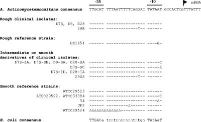
Figure 2 is a summary of the sequencing results of the −35 site, the spacer, and the −10 site. More detailed information on the entire sequenced regions may be accessed through GenBank. All four rough clinical isolates (D7S, D9, D28, and 29R) had a promoter essentially identical to that identified by Haase et al. (5). Two published flp sequences from strain CU1000 (GenBank AY157714) (12) and strain 310a (GenBank D83053) (10) also contain the same promoter sequence. Therefore, this sequence was designated as the consensus sequence. The rough-colony strain HK1651 exhibited a one-base variation in a semiconserved base at the −10 site (Fig. 2). In broth culture, strain HK1651 formed less compact aggregates than did other rough clinical strains. Presumably, this base variation of strain HK1651 may slightly reduce the flp promoter strength. The flanking regions of the promoter exhibited a greater sequence variation among strains, but the variations did not correlate with the rough-smooth conversion (data not shown).
FIG. 2.
The flp promoter sequences of the A. actinomycetemcomitans study strains. The entire sequenced regions can be accessed through GenBank: flp locus of strain D7S, AY262277; D9, AY460681; D28, AY460682; 29R, AY460683; JP2, AY460684; Y4, AY460685; ATCC 29522, AY460686; ATCC 29523, AY460687; ATCC 29524, AY460688. The consensus sequence of the E. coli promoter (7) is provided for comparison (uppercase, >60% conservation; lowercase, >43%). The sequence for ATCC 33384 was from GenBank (T. Tanimoto, unpublished data; accession number AB071167). Flag symbol, the transcription-initiation point; −, nucleotide identical to the parent; Δ, deletion.
Five of the eight smooth- or intermediate-colony derivative strains and three of the six smooth-colony reference strains exhibited sequence variations in the conserved −10 site. Six of these variations were due to a single-nucleotide substitution, and the remaining two variations were due to a single-nucleotide deletion. A frequent mutation was the transition of the most conserved T residue of the −10 site. This base was often called “invariable” T, as it was conserved in 97% of Escherichia coli promoters among 112 promoter sequences compiled, and mutations of this base were demonstrated to severely affect a number of promoters' activities in E. coli (7, 19). Strain JP2 exhibited a single-nucleotide deletion in the spacer region between the conserved −35 and −10 sites. Strain ATCC 29524 showed a deletion of the entire −35 and part of the spacer region. The promoter sequences of the remaining three derivative strains (strains D7S-ID, D28-IA, and 29-SS) and the smooth reference strain ATCC 29523 were identical to the consensus sequence or the sequence of the parental strain (i.e., strain 29R).
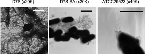
The bacterial morphology was examined by transmission electron microscopy (TEM) by a previously described protocol (17). As expected, the smooth-colony strain D7S-SA was nonfimbriated, in contrast to its parental rough-colony strain D7S (Fig. 3). We have examined 15 other smooth-derivative strains from clinical isolates and have not detected the presence of fimbriae (data not shown). However, TEM of strain ATCC 29523 showed a few thin fibrils that resembled fimbriae (Fig. 3). It is interesting that strain ATCC 29523 has a wild-type flp promoter. We also noted a tendency of strain 29523 to aggregate in broth cultures. The low expression of fimbria may explain the aggregation of ATCC 29523 in broth.
FIG. 3.
Transmission electron micrographs of the wild-type A. actinomycetemcomitans strain D7S, smooth-derivative strain D7S-SA, and reference strain ATCC 29523. Note that a few strands of fimbriae were present on the surface of strain ATCC 29523. Bars, 1 μm.
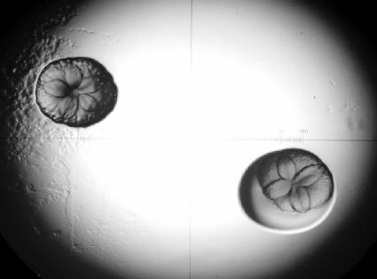
The mutated flp promoters of several smooth-colony variants were replaced with a wild-type promoter. A 1-kb PCR DNA fragment encompassing flp-1-flp-2 and the promoter was amplified from rough strain D7S using primers F-Ev24 and orfB-X (Fig. 1 shows primer locations). The DNA was then digested with EcoRV and XbaI, ligated to pTc-USS at the same sites, and transformed into smooth-colony strains D7S-SA and D7S-SB by a natural transformation protocol described previously (16). The plasmid pTc-USS is a pBluescript II KS (Stratagene) derivative that does not replicate in A. actinomycetemcomitans (17). Therefore, the Tcr transformants should have the plasmid containing the wild-type flp promoter integrated in the chromosome by a single crossover. If the point mutation in the flp promoter accounted for the rough-to-smooth conversion in strains D7S-SA and D7S-SB, insertion of a promoter (presumably any well-expressed promoter) upstream of the flp operon should restore the rough-colony phenotype. As expected, the transformants were predominantly of rough-colony type (an example is shown in Fig. 4). The integration of the plasmid in the chromosome was verified in selected rough colonies by PCR using two pairs of primers, F-Ev24/Umer and orfC-R/Rmer (data not shown). We also replaced the promoter region of the smooth variant D7S-SA with the corresponding promoter region from different A. actinomycetemcomitans strains. Again, the 1-kb promoter-flp-1-flp-2 region was amplified from the rough strains HK1651 and 29R and smooth strains ATCC 29523 and ATCC 33384. Each PCR amplicon DNA was ligated to pTc-USS and transformed into strain D7S-SA. The results showed that no rough transformants were obtained with the donor DNA from strain ATCC 33384, while most transformants with donor DNA from the other three strains were predominantly rough-colony type (e.g., 75% of the transformants were rough-colony type with donor DNA from strain ATCC 29523). The results indicated that random mutations in the promoter of the flp operon are a mechanism for the rough-to-smooth conversion of A. actinomycetemcomitans.
FIG. 4.
Colonies of A. actinomycetemcomitans strain D7S-SA transformed with pTc-USS carrying the wild-type flp promoter DNA. Colonies were 5 days old and ∼1 mm in size. The picture was taken under a light microscope with a 4× objective. Left, wild-type-like rough colony; right, chimeric colony, in which the smooth portion appeared after 3 days of culture.
Conversely, we replaced the flp promoter of the rough-colony strain D7S with the promoter obtained from smooth-colony variants. A 2.3-kb DNA fragment that included the flp promoter region was amplified by PCR from each of the smooth-colony strains JP2, Y4, and ATCC 29524 with primers orfC-R and flp-UE (5′-CTGAATTCTCGCTCAGATACGGA, which is located at 1.1 kb upstream of the promoter region) and directly used as the donor DNA to transform strain D7S. To enrich the nonfimbriated bacteria, the transformed bacteria were briefly incubated in broth. Fimbriated D7S bacteria formed aggregates that either settled to the bottom of the culture tube or adhered to the tube, while the nonfimbriated smooth-colony variants would grow as a single-cell suspension. The top portion of the undisturbed broth culture of the transformed D7S was collected and plated on agar. The results showed that the colonies were predominantly smooth. The flp promoters of four smooth-colony variants (two were transformed with the flp promoter from strain JP2 and one each was transformed with the flp promoter from strains Y4 and ATCC 29524) were amplified by PCR and sequenced. The results showed that each of the flp promoters of these smooth-colony variants carried the specific sequence variation of the flp promoter found in the corresponding donor strain JP2, Y4, or ATCC 29524. The smooth-colony transformant derived from transformation with donor DNA of strain ATCC 29524 displayed an additional insertion of two nucleotides downstream of the −10 site, which may have arisen from PCR amplification or transformation. However, the insertion occurred at the nonconserved area and was not expected to be the cause of the reduced expression of the flp operon. The results further substantiated the role of the flp promoter mutations in rough-to-smooth conversion.
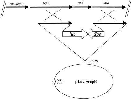
The expression levels of flp promoters in rough- and smooth-colony bacteria were measured by inserting a firefly luciferase gene, luc, as the reporter gene at the rcpB site of the flp operon (Fig. 5). Briefly, 3.2-kb DNA containing the Spe marker and the rcpB-flanking DNA was amplified from a previously constructed ΔrcpB::Spe mutant of strain D7S (unpublished data) with a primer at 1.1 kb upstream of rcpB and another primer at 1 kb downstream of rcpB. This 3.2-kb DNA was cloned in pBluescript II KS at the EcoRV site to produce pB-ΔrcpB. Separately, a luc-Spe cassette plasmid was constructed based on pBluescript II KS and pBRluc (4), and the plasmid was named pLuc-Spe2. A 3-kb luc-Spe reporter-marker was amplified from pLuc-Spe2 using the primer Luc-B1, 5′-AGGGATCCTAGGAAGCTTTCCATGGA (the luc start codon is underlined), and the primer Spe-USS, 5′-AAAGTGCGGTTTACACTTACTTTAGTTTT. This luc-Spe marker was then used to replace the 1.1-kb Spe marker in pB-ΔrcpB to create pLuc-ΔrcpB (Fig. 5), which was used to transform A. actinomycetemcomitans strains. Transformants were verified by PCR and tested for luciferase production. Briefly, bacteria were grown on serum trypticase soy broth agar overnight and resuspended in tryptic soy broth at an optical density (OD) at 600 nm of 0.5, and luciferase was assayed by mixing 20 μl of luciferin (1 mM in 0.1 M sodium citrate, pH 7.0) with 80 μl bacterial suspension (OD at 600 nm = 0.1 to 0.6) at room temperature for 5 min, and the light was counted twice with the BetaScout Liquid Scintillation Tester (Perkin-Elmer Life Sciences). Enzyme activity was defined as (photon counts in 10 s − background counts)/0.001 OD unit of bacteria. The results (Table 2) showed that the luc expression in strain D7S-SA, with a T-to-C transition in the flp promoter, was 80-fold lower than that in the wild type and that expression in strain D7S-SC, with a T deletion, was 130-fold lower than that in the wild type. Interestingly, the luc activity in strain ATCC 29523 was fourfold lower than that in strain D7S. Perhaps the low-level expression of the flp promoter in ATCC 29523 resulted in the scant expression of fimbria seen under TEM.
FIG. 5.
Genetic map of the plasmid pLuc-ΔrcpB (not in exact scale). The plasmid pLuc-ΔrcpB contains a 2.85-kb luc-Spe cassette and flanking DNA of rcpB. The plasmid was used to transform A. actinomycetemcomitans strains to generate transformants with luc-Spe replacing rcpB of the flp operon.
TABLE 2.
Luciferase activities of different A. actinomycetemcomitans strains
| Strain | Luciferase activitya |
|---|---|
| D7S (ΔrcpB::luc-Spe) | 14,421 ± 5,856 |
| D7S-SA (ΔrcpB::luc-Spe) | 181 ± 20 |
| D7S-SC (ΔrcpB::luc-Spe) | 108 ± 18 |
| ATCC 29523 (ΔrcpB::luc-Spe) | 3,524 ± 942 |
Mean ± standard deviation.
Our findings suggest that in vitro spontaneous rough-to-smooth conversion of A. actinomycetemcomitans commonly occurs due to mutations at the −35 site, the spacer region, or the −10 site of the flp promoter. Such mutations are not likely to be reversible at a significant rate and may explain the lack of smooth-to-rough conversion among A. actinomycetemcomitans strains. However, several smooth variants had an apparently wild-type flp promoter. Mutation of the flp promoter is not the only mechanism of the rough-to-smooth conversion of this bacterium.
Acknowledgments
This research was supported by NIDCR grant R01 DE12212.
We thank S. Goodman and O. Kay for their help in developing the luciferase reporter.
Editor: V. J. DiRita
REFERENCES
- 1.Asikainen, S., and C. Chen. 1999. Oral ecology and person-to-person transmission of Actinobacillus actinomycetemcomitans and Porphyromonas gingivalis. Periodontol. 2000 20:65-81. [DOI] [PubMed] [Google Scholar]
- 2.Fine, D. H., D. Furgang, J. Kaplan, J. Charlesworth, and D. H. Figurski. 1999. Tenacious adhesion of Actinobacillus actinomycetemcomitans strain CU1000 to salivary-coated hydroxyapatite. Arch. Oral Biol. 44:1063-1076. [DOI] [PubMed] [Google Scholar]
- 3.Gally, D. L., J. A. Bogan, B. I. Eisenstein, and I. C. Blomfield. 1993. Environmental regulation of the fim switch controlling type 1 fimbrial phase variation in Escherichia coli K-12: effects of temperature and media. J. Bacteriol. 175:6186-6193. [DOI] [PMC free article] [PubMed] [Google Scholar]
- 4.Goodman, S. D., and Q. Gao. 1999. Firefly luciferase as a reporter to study gene expression in Streptococcus mutans. Plasmid 42:154-157. [DOI] [PubMed] [Google Scholar]
- 5.Haase, E. M., J. O. Stream, and F. A. Scannapieco. 2003. Transcriptional analysis of the 5′ terminus of the flp fimbrial gene cluster from Actinobacillus actinomycetemcomitans. Microbiology 149:205-215. [DOI] [PubMed] [Google Scholar]
- 6.Haase, E. M., J. L. Zmuda, and F. A. Scannapieco. 1999. Identification and molecular analysis of rough-colony-specific outer membrane proteins of Actinobacillus actinomycetemcomitans. Infect. Immun. 67:2901-2908. [DOI] [PMC free article] [PubMed] [Google Scholar]
- 7.Hawley, D. K., and W. R. McClure. 1983. Compilation and analysis of Escherichia coli promoter DNA sequences. Nucleic Acids Res. 11:2237-2255. [DOI] [PMC free article] [PubMed] [Google Scholar]
- 8.Inoue, T., I. Tanimoto, H. Ohta, K. Kato, Y. Murayama, and K. Fukui. 1998. Molecular characterization of low-molecular-weight component protein, Flp, in Actinobacillus actinomycetemcomitans fimbriae. Microbiol. Immunol. 42:253-258. [DOI] [PubMed] [Google Scholar]
- 9.Inouye, T., H. Ohta, S. Kokeguchi, K. Fukui, and K. Kato. 1990. Colonial variation and fimbriation of Actinobacillus actinomycetemcomitans. FEMS Microbiol. Lett. 57:13-17. [DOI] [PubMed] [Google Scholar]
- 10.Ishihara, K., K. Honma, T. Miura, T. Kato, and K. Okuda. 1997. Cloning and sequence analysis of the fimbriae associated protein (fap) gene from Actinobacillus actinomycetemcomitans. Microb. Pathog. 23:63-69. [DOI] [PubMed] [Google Scholar]
- 11.Kachlany, S. C., P. J. Planet, M. K. Bhattacharjee, E. Kollia, R. DeSalle, D. H. Fine, and D. H. Figurski. 2000. Nonspecific adherence by Actinobacillus actinomycetemcomitans requires genes widespread in bacteria and archaea. J. Bacteriol. 182:6169-6176. [DOI] [PMC free article] [PubMed] [Google Scholar]
- 12.Kachlany, S. C., P. J. Planet, R. Desalle, D. H. Fine, D. H. Figurski, and J. B. Kaplan. 2001. flp-1, the first representative of a new pilin gene subfamily, is required for non-specific adherence of Actinobacillus actinomycetemcomitans. Mol. Microbiol. 40:542-554. [DOI] [PubMed] [Google Scholar]
- 13.Neidhardt, F. C., R. Curtiss III, J. L. Ingraham, E. C. C. Lin, K. B. Low, B. Magasanik, W. S. Reznikoff, M. Riley, M. Schaechter, and H. E. Umbarger (ed.). 1996. Escherichia coli and Salmonella: cellular and molecular biology, 2nd ed. ASM Press, Washington, D.C.
- 14.Planet, P. J., S. C. Kachlany, R. DeSalle, and D. H. Figurski. 2001. Phylogeny of genes for secretion NTPases: identification of the widespread tadA subfamily and development of a diagnostic key for gene classification. Proc. Natl. Acad. Sci. USA 98:2503-2508. [DOI] [PMC free article] [PubMed] [Google Scholar]
- 15.Rosan, B., J. Slots, R. J. Lamont, M. A. Listgarten, and G. M. Nelson. 1988. Actinobacillus actinomycetemcomitans fimbriae. Oral Microbiol. Immunol. 3:58-63. [DOI] [PubMed] [Google Scholar]
- 16.Wang, Y., S. D. Goodman, R. J. Redfield, and C. Chen. 2002. Natural transformation and DNA uptake signal sequences in Actinobacillus actinomycetemcomitans. J. Bacteriol. 184:3442-3449. [DOI] [PMC free article] [PubMed] [Google Scholar]
- 17.Wang, Y., W. Shi, W. Chen, and C. Chen. 2003. Type IV pilus gene homologs pilABCD are required for natural transformation in Actinobacillus actinomycetemcomitans. Gene 312:249-255. [DOI] [PubMed] [Google Scholar]
- 18.Weiser, J. N., S. T. Chong, D. Greenberg, and W. Fong. 1995. Identification and characterization of a cell envelope protein of Haemophilus influenzae contributing to phase variation in colony opacity and nasopharyngeal colonization. Mol. Microbiol. 17:555-564. [DOI] [PubMed] [Google Scholar]
- 19.Youderian, P., S. Bouvier, and M. M. Susskind. 1982. Sequence determinants of promoter activity. Cell 30:843-853. [DOI] [PubMed] [Google Scholar]
- 20.Zambon, J. J. 1985. Actinobacillus actinomycetemcomitans in human periodontal disease. J. Clin. Periodontol. 12:1-20. [DOI] [PubMed] [Google Scholar]


