Abstract
Epigenetics defines changes in cell function without involving alterations in DNA sequence. Neuroepigenetics bridges neuroscience and epigenetics by regulating gene expression in the nervous system and its impact on brain function. With the increase in research in recent years, it was observed that alterations in the gene expression did not always originate from changes in the genetic sequence, which has led to understanding the role of epigenetics in neurodegenerative diseases (NDDs) including Alzheimer’s disease (AD) and Parkinson’s disease (PD). Epigenetic alterations contribute to the aberrant expression of genes involved in neuroinflammation, protein aggregation, and neuronal death. Natural phytochemicals have shown promise as potential therapeutic agents against NDDs because of their antioxidant, anti-inflammatory, and neuroprotective effects in cellular and animal models. For instance, resveratrol (grapes), curcumin (turmeric), and epigallocatechin gallate (EGCG; green tea) exhibit neuroprotective effects through their influence on DNA methylation patterns, histone acetylation, and non-coding RNA expression profiles. Phytochemicals also aid in slowing disease progression, preserving neuronal function, and enhancing cognitive and motor abilities. The present review focuses on various epigenetic modifications involved in the pathology of NDDs, including AD and PD, gene expression regulation related to epigenetic alterations, and the role of specific polyphenols in influencing epigenetic modifications in AD and PD.
Keywords: epigenetic modulation, Alzheimer’s disease, Parkinson’s disease, phytochemicals, polyphenols, nutriepigenomics
1. Introduction
Epigenetics, an intricate field at the crossroads of genetics and environmental influences, is a pivotal avenue for unraveling the complexity inherent in gene regulation and cellular function. Going beyond the traditional focus on the linear sequence of DNA, epigenetics delves into reversible modifications on a cell’s DNA bases or histones, affecting gene expression that transpires without modifying the fundamental genetic code [1,2]. Understanding epigenetic mechanisms is crucial for unraveling the molecular intricacies governing cell differentiation, developmental and biological processes, response to environmental cues, and the manifestation of diverse phenotypes in human health and disease.
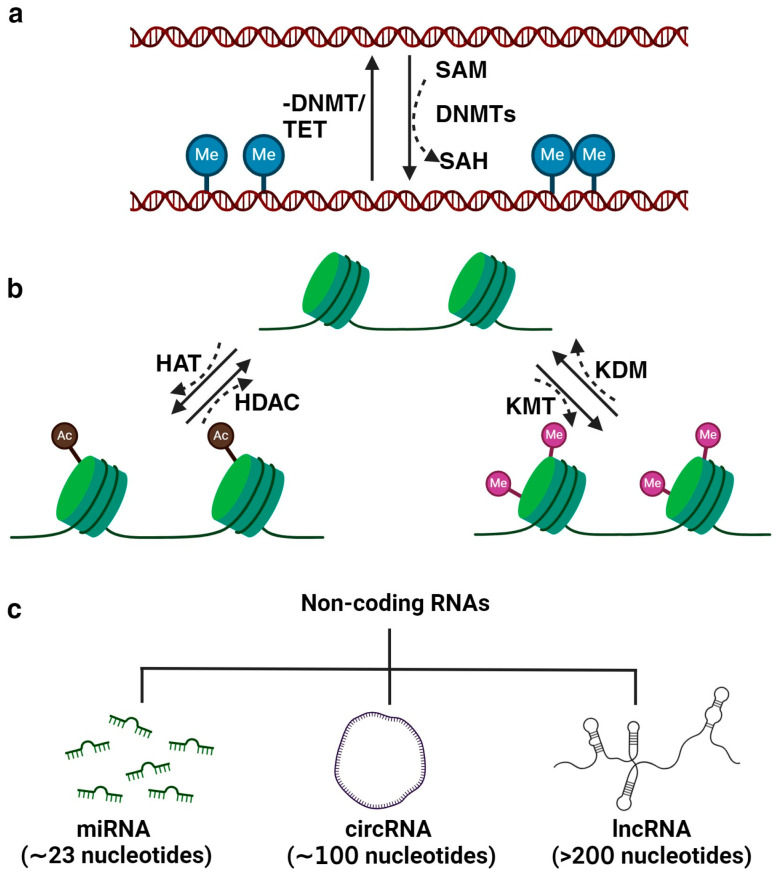
Three primary mechanisms of epigenetic regulation are illustrated and detailed in Figure 1. DNA methylation is the process of adding a methyl group to a cytosine base in DNA. This modification typically represses gene expression. In mammals, DNA methylation predominantly occurs on cytosines at CpG sites and is facilitated by DNA methyltransferases (DNMTs; DNMT1, DNMT3a, and DNMT3B), which catalyzes the transfer of methyl group from the critical methyl donor S-adenosylmethionine (SAM) [3]. While DNMTs primarily catalyze DNA methylation, the metabolic enzyme nicotinamide N-methyltransferase (NNMT) indirectly influences this process through its regulation of SAM levels [4,5]. NNMT regulates methylation potential within cells by catalyzing the methylation of nicotinamide (NAM) using SAM as a methyl donor. By controlling the availability of SAM, NNMT influences the cellular balance between methyl donor availability and utilization [6]. The interplay between NNMT, NAM, SAM, and cellular metabolism extends to epigenetic regulation. SAM is not only essential for DNA methylation, which is catalyzed by DNMTs, but also for histone methylation, mediated by HMTs [7]. NNMT-mediated depletion of SAM can disrupt these epigenetic modifications, leading to alterations in chromatin structure and gene expression profiles. Through its modulation of intracellular SAM levels, NNMT intricately governs the expression of genes, which involves a reduction in DNA CpG island methylation, leading to a pronounced upregulation of genes [8]. In essence, NNMT’s influence on SAM concentrations serves as a molecular switch, tipping the balance toward heightened gene expression. NNMT has been emerging as a promising therapeutic target in cancer treatment, and several NNMT inhibitors are under study for the therapeutic management of the disease [9,10,11]. DNA demethylation occurs through active processes involving a group of proteins known as ten-eleven translocation methylcytosine dioxygenases (TET). Alternatively, demethylation can happen passively, where 5-methylcytosine (5 mC) is lost during successive rounds of replication without functional DNMT1 [12]. Histone modification is another mechanism of epigenetic regulation. Histones are proteins around which DNA is packaged, forming a chromatin structure. Various chemical modifications can occur on histone proteins, including acetylation, methylation, and phosphorylation. Depending on the specific modification and location, these modifications can promote or inhibit gene expression [13]. The dynamic process of histone acetylation is intricately controlled by various enzymes, namely histone acetyltransferases (HATs) and histone deacetylases (HDACs). HATs (categorized under p300/CBP, GNATs, MYST, and the transcription factor-related family) attach acetyl groups to histones, resulting in a more relaxed chromatin structure that facilitates transcriptional activation [14]. Conversely, HDACs (Class I, II, III, and IV) remove acetyl groups from histones, leading to a more compact chromatin conformation and restricting gene transcription [15]. In addition to HATs and HDACs, there are two other types of enzymes that regulate histone modifications: histone methyltransferases (HMTs), which add methyl groups to lysine or arginine residues, and histone demethylases (HDMs), which act to eliminate them [16]. Non-coding RNA (ncRNA) is an RNA molecule that does not undergo translation into a protein. However, recent evidence has illuminated the significant role of ncRNAs, including microRNAs (miRNAs; ~23 nucleotides), long ncRNAs (lncRNAs; >200 nucleotides), and circular RNAs (circRNAs; ~100 nucleotides) in various biological as well as pathological processes [17].
Figure 1.
Illustration of essential epigenetic mechanisms governing gene expression by regulating transcriptional activation and repression. (a) DNA methylation/demethylation involves adding or removing methyl groups to DNA, respectively, particularly at CpG dinucleotides, influencing gene silencing or activation. (b) Histone modifications, including acetylation/deacetylation and methylation/demethylation, modulate chromatin structure, promoting either transcriptional activation or repression. (c) Additionally, non-coding RNAs (ncRNAs) such as miRNA, circRNA, and lncRNA participate in fine-tuning gene expression by targeting mRNA stability or acting as molecular decoys.
Epigenetic regulation is crucial for normal development and cellular differentiation. It plays a significant role in diverse biological processes, such as embryonic development, tissue-specific gene expression, and response to environmental stimuli. Epigenetic mechanisms can modify gene expression outcomes, thereby influencing the phenotypic characteristics of the cell [18]. It also has implications for health and disease, as alterations in epigenetic marks can contribute to various conditions, such as cancer, metabolic syndromes, and neurological disorders.
2. Neuroepigenetics
The nervous system orchestrates complex functions with unique epigenetic profiles meticulously tailored to specific roles. Due to this intricacy, the transcription machinery faces an arduous challenge in the nervous system, making it highly responsive to epigenetic disturbances [19]. Consequently, the significance of epigenetics in the nervous system is underscored by the occurrence of severe neurodegenerative diseases (NDDs) resulting from mutations in epigenetic genes [20]. The genetic and epigenetic code interplay has been extensively examined in learning and memory. Pioneering studies have confirmed the critical involvement of epigenetic changes in processes related to learning, memory, and synaptic plasticity [21]. It is also evident that disruptions in epigenetic mechanisms not only hinder normal brain functioning but are also linked to various neurological diseases, particularly Alzheimer’s disease (AD) and Parkinson’s disease (PD).
3. Epigenetic Changes in the Pathology of Alzheimer’s Disease
Alzheimer’s disease (AD) stands as the predominant type of dementia globally, representing an age-related neurodegenerative condition that significantly impacts memory and cognitive functions. While clinical symptoms typically surface in individuals aged 65 and above, the development of AD spans several decades, with initial pathogenic processes believed to commence several decades before noticeable symptoms emerge. AD is considered a heterogenous disorder with both familial and sporadic forms. Familial AD (FAD) or early onset AD (EOAD) occurs mainly due to mutations in the genes amyloid precursor proteins (APPs), Presenilin-1 and 2 (PSEN-1, PSEN-2) [22]. Sporadic AD (SAD) or late-onset AD (LOAD) is multifactorial, with heritability as high as 80% [23]. The APOE ε4 allele was supposed to be the only gene involved in LOAD for several years. However, many other risk genes involved in cellular pathways have also been identified, such as in the maintenance of synaptic plasticity (phosphatidylinositol binding clathrin assembly protein (PICALM), bridging integrator 1 (BIN1), CD2-associated protein (CD2AP)), in immune function (clusterin (CLU), complement C3b/C4b receptor 1 (CR1), CD33 molecule (CD33), ephrin receptor A1 (EPHA1)), in lipid metabolism (ATP binding cassette subfamily A member 7 (ABCA7)), and cell signaling (a membrane spanning four domains, A4A and A6E (MS4A4A, MS4A6E)) [24,25,26].
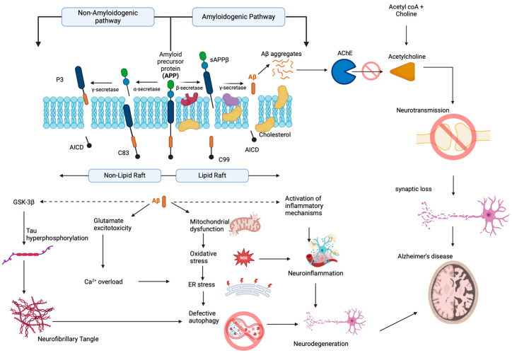
During AD progression, there occurs a profound loss of forebrain cholinergic neurons coupled with a reduction in acetylcholine in the synaptic cleft due to the aberrant action of cholinesterase enzymes (AChE) leading to cognitive deficits [27,28]. Moreover, there also exists a cross-talk between the cholinergic pathway and amyloid beta pathway (Aβ), and the aberrant disruption of either pathway influences the other [29]. The Aβ peptide is derived from APP, when sequential processing occurs by the enzymes β,γ-secretases instead of non-amyloidogenic processing by α,γ-secretases (Figure 2). The monomeric Aβ undergoes nucleation-dependent polymerization to form oligomer and fibrillar structures and accumulates as senile plaques [30,31,32]. Although under physiological conditions, there exists a balance between Aβ production and elimination through the low-density lipoprotein receptor-related protein (LRP) and enzymatic degradation by neprilysin, the mechanisms are severely impaired in AD patients, contributing to the severity of the disease [33]. Aβ can interact and bind to the receptor for advanced glycation end products (RAGE), which are overexpressed on the membrane of neurons and microglial cells during AD and invoke oxidative stress-mediated degeneration of neurons [34,35,36].
Figure 2.
Pathophysiological changes occurring during Alzheimer’s disease (created with BioRender.com).
Mounting evidence also suggests that Aβ deposits can invoke neuroinflammation, one of the underlying causes of AD, by activating astrocytes and microglia [37]. Astrocyte and microglial cells activate the nuclear factor kappa B (NF-κB) pathway that subsequently leads to the secretion of several proinflammatory factors, including inducible nitric oxide synthase (iNOS), interleukin-1β (IL-1β), and cyclooxygenase-2 (COX-2), which eventually contributes to neuronal damage and death [38,39,40,41]. In addition to the direct neurotoxic effect, reports show that activated inflammatory cells can help in Aβ accumulation [42]. The secreted cytokines also induce β-secretase expression and enzymatic activity, resulting in increased generation of Aβ [43].
Aβ aggregates show cross-talk between several signaling pathways to aggravate the disease condition. For instance, Aβ oligomers have high affinity toward the receptors including N-methyl-D-aspartate receptors (NMDAR), α-amino-3-hydroxy-5-methyl-4-isoxazole propionic acid receptor (AMPAR), α7 nicotinic acetylcholine receptor (nAChR) and mediate excitotoxicity causing long-term depression (LTD) [44,45,46,47,48]. Aβ oligomers can possibly influence the release of glutamate and inhibit the ability of cells to reuptake the neurotransmitter by the downregulation of glutamate transporters (GLAST-1, GLT-1). These alterations buildup the level of glutamate in the perisynaptic space, resulting in excitotoxicity associated with disruption of Ca2+ homeostasis and subsequent degeneration of neurons mediated by ER stress and unfolded protein response (UPR) mechanisms [49,50,51,52]. In addition, as the deposition of Aβ aggregates continues, it leads to the phosphorylation of tau, a microtubule-associated protein located on the axonal compartment that helps in the microtubule stabilization by promoting the assembly of tubulin to microtubules [53]. The aberrant hyperphosphorylation makes the tau protein resist proteolysis by proteases, which in turn impairs the capacity of tau to bind to tubulin, causing deterioration of the microtubule structure, destruction of axonal transport and synaptic metabolism [54]. These changes ultimately result in the disintegration of the cytoskeleton and cause neuronal death. In recent years, researchers have increasingly recognized the pivotal role of epigenetic mechanisms in the progression and course of AD.
3.1. DNA Methylation in AD
Mounting evidence indicates that neuroepigenetic modifications that occur naturally during aging due to the involvement of environmental-associated factors act as one of the etiologies for NDDs. Significant correlations were identified linking reduced DNA methylation to the advancement of AD. Elevated levels of NNMT are notably observed in the brains of individuals affected by AD, particularly within the neurons undergoing degeneration, indicating that the upsurge in NNMT expression could be a prevalent characteristic shared among individuals with AD and could lead to hypomethylation, suggesting NNMT-targeted therapy for the treatment [55]. Postmortem brain samples of AD patients have shown a reduction in global DNA methylation in the hippocampal region [56]. In several studies, APP and PSEN1 promoter hypomethylation have been found in AD patients, which is associated with increased amyloid beta peptide (Aβ) [57,58]. Hypomethylation at intron 1 of triggering receptor expressed on myeloid cells 2 (TREM2) is observed in AD patients and is associated with increased TREM2 expression [59]. CREB-regulated transcription coactivator 1 (CRTC1) is hypomethylated in the hippocampal region of AD individuals and is correlated with the deposition of p-tau [60]. In late-onset AD (LOAD) individuals, CpGs in BIN1 (which regulates presynaptic neurotransmitter release) are hypomethylated, leading to increased expression in peripheral blood [61]. A study by Chapuis et al. [62] demonstrated that increased BIN1 expression induces AD pathogenesis by modulating the tau pathway. Analysis of blood samples from AD patients showed decreased methylation in the mtDNA D-loop region as well as CpG island shores of AD genes CR1, CLU, and PICALM [63,64]. Hypomethylation has also been observed in apolipoprotein E (APOE) in the AD brain, which contributes to neural cell dysfunction [65].
The DNA methylation patterns in the human hippocampus revealed hypermethylation in the promoter of the dual-specificity phosphatase 22 (DUSP22), which plays a key role in mediating PKA-dependent tau phosphorylation and CREB activation [66]. Hypermethylation of tripartite motif-containing 59 (TRIM59) and Kruppel-like factor 14 (KLF14) were observed in the peripheral blood obtained from familial AD patients, and bioinformatics analysis revealed that the changes in hypermethylation pattern could induce proapoptotic signaling during AD [67]. Studies indicate that Aβ induced hypermethylation of neprilysin (NEP) in the promoter region, thereby suppressing the expression of the enzyme involved in the toxic peptide degradation [68]. Analysis of peripheral blood from AD individuals showed a significantly higher methylation ratio in the brain-derived neurotrophic factor (BDNF) promoter compared to the control, with a negative correlation with neuropsychological test subscores indicating the influence in AD manifestation [69,70]. Various studies have reported DNA hypermethylation of ankyrin 1 (ANK1) in the postmortem brain of AD individuals, followed by a reduction in expression during the early onset of the disease [71,72,73]. At the internal promoter of protein kinase C zeta (PRKCZ; involved in long-term potentiation), DNA hypermethylation was observed in AD individuals and is associated with reduced levels of PKMζ [74].
3.2. Histone Modification in AD
Recent epigenome-wide association studies and proteome studies have indicated an increase in histone H3 acetylation (H3K9ac, H3K27ac) in the AD brain, which in turn results in the transcriptional activation of several AD-associated genes to promote neurodegeneration [75,76,77]. A study by Takasu et al. [78] showed that the administration of a HDAC inhibitor in PSAPP mice restored gamma oscillation deficits, which is required for regulating synaptic plasticity and cognitive function with concomitant revitalization in the functionality of fast-spiking interneurons. A comparative study in postmortem brain tissue by Nativio et al. [79] showed H4K16ac enrichment associated with neuroplasticity in typically aged individuals and a reduction in AD individuals. Overexpression of HDACs, including HDAC2, 3, and 6, has been related to the repression of genes involved in mitochondrial biogenesis [80], vesicle trafficking [81], and the antioxidant mechanism [82,83,84,85]. Inhibition of class I HDACs in vitro restored Ca2+ homeostasis by regulating proteins that control the communication between endoplasmic reticulum and mitochondria [86]. Guan et al. [87] have shown that overexpression of HDAC2 in mice could cause impaired memory and synaptic plasticity, while knockout reversed the effect. Similarly, HDAC2 overexpression is also seen in mice models and AD individuals, which could block the expression of neuroplasticity-associated genes [84]. Further, the knockdown of HDAC2 in APP/PS1 mice increased BDNF levels and improved memory [88]. Narayan et al. [89] demonstrated an increase in the level of acetyl H3 and H4 in AD individuals, indicating histone acetylation is not always neuroprotective and aberrant histone modification is involved in neurodegeneration. Further, in support of this notion, RNA-seq analysis of AD postmortem brain revealed an increase in the HATs, which acetylates H3K27 and H3K9 compared to the age-matched control that could worsen Aβ42 toxicity besides changes in the methylation pattern of H3K36me1, H3K23me1, and H3K27me3 [77].
Changes in methylation of H2BK108 and H4R55 have been reported in the frontal cortex of the postmortem brain of AD individuals [90]. An increase in histone methylation of H3K9me2 in the NEP promoter-1, promoter-2, and HDAC1 was found to be correlated with the downregulation of neprilysin that is required for the degradation of Aβ under hypoxia [91]. Likewise, elevated H3K9me3 is associated with cognitive impairment and reduction of spine plasticity in aged mice [92]. Various studies have shown an aberrant cytoplasmic localization [93] of H3K4me3 as well as an elevated level of H3K4me3 and its catalyzing methyltransferases [94], accompanied by tau hyperphosphorylation. Further, inhibiting specified methyltransferases using inhibitors in AD mice restored synaptic functions and improved memory [94]. Brain tissues from AD mice models and individuals showed an increase of H3K9me2 enrichment at glutamate receptors, impairing synaptic and cognitive functions [95].
3.3. Non-Coding RNAs in AD
ncRNAs are involved in biological and pathological processes by interacting with DNA, RNA, and protein [96]. Pioneering studies have confirmed the aberrant expression of ncRNA during NDDs, which alters the transcriptional regulation of genes and modulates cell signaling pathways, further aggravating the disease [97]. For instance, upregulation of miR-206 and miR-613 in transgenic AD mice has been associated with reduced levels of BDNF contributing to AD pathology [98,99]. An increase in the miR-455-3p level in postmortem brains of AD individuals could be positively corroborated with Aβ1–42 levels [100,101]. In AD transgenic mice, expression of miR-137 is downregulated and coupled with an increase in calcium voltage-gated channel subunit alpha1 C (CACNA1C), and transfection with miR-137 mimics abrogated Aβ1–42-mediated tau phosphorylation [102]. Overexpression of miR-137 is also reported to inhibit the neurotoxic behavior of Aβ in neuronal cells by targeting the extracellular signal-regulated kinase 1/2 (ERK1/2) pathway [103]. Overexpression of miR-326 in AD mice improves cognitive function by reducing the level of Aβ and targeting vav guanine nucleotide exchange factor 1 (VAV1) to downregulate jun N-terminal kinase (JNK) signaling pathway causing neuroprotection [104]. Upregulation of miR-124 upon transfection with miR-124 mimics decreased the expression of beta-site amyloid precursor protein cleaving enzyme 1 (BACE-1) and prevented the degeneration of neurons against Aβ toxicity [105]. Delivery of miR-16 mimics to the brain of AD mice could effectively reduce BACE-1, tau, and ERK1/2, along with inflammatory and oxidative stress markers [106]. A significant reduction of miR-195 AD individuals carrying a single ApoE ε4 allele, while the overexpression rescued ApoE4-induced cognitive impairment and lysosomal deficits in iPSCs-derived brain cells of ApoE4+/+ AD individuals [107]. Pioneering studies have revealed the role of miRNAs in tau protein regulation. Deficiency of miR-132/212 and miR-219 increased tau phosphorylation and aggregation in AD triple transgenic mice and tau Drosophila model, respectively [108,109].
Transcriptomic analysis of brain tissues obtained from AD individuals and animal models showed a disrupted pattern in the lncRNA expression compared to control [110,111]. X inactive specific transcript (XIST), one of the most extensively studied lncRNAs, was reported to be upregulated in AD animal models. In the AD mice model, lncRNA XIST downregulated NEP expression and induced Aβ-mediated neuroinflammation, while the knockdown of XIST alleviated the effect [112]. A positive and negative correlation was observed between XIST-miR-124 and XIST-BACE1, respectively, and silencing of XIST attenuated BACE1, implicating the pathological influence of XIST in AD [113]. Apart from XIST, several lncRNAs, including lncRNA brain cytoplasmic 200 (BC200), lncRNA sortilin-related receptor 1 antisense RNA1 (SORL1-AS1), lncRNA SOX21 antisense divergent transcript 1 (SOX21-AS1), and lncRNA nuclear paraspeckle assembly transcript 1 (NEAT1) are also implicated in the pathological process of AD including synaptic impairment, APP processing, and Aβ production and tau phosphorylation [114,115,116,117].
4. Epigenetic Changes in the Pathology of Parkinson’s Disease
Parkinson’s disease (PD) is a prominent neurodegenerative condition that affects the motor system, leading to tremors, stiffness, and difficulties in coordination. Unlike AD, PD is not solely age-related, as it can manifest in individuals across a broad age spectrum, although it is more commonly diagnosed in older adults. The hallmark of PD is the progressive loss of dopaminergic neurons in the brain, contributing to the characteristic motor symptoms [118]. While the clinical signs become evident when a significant number of neurons are already compromised, the underlying pathological processes are believed to initiate long before symptoms become apparent.
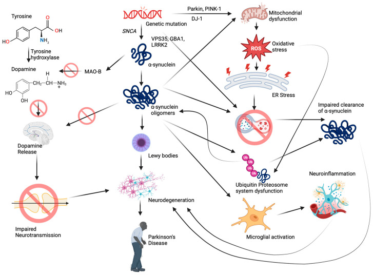
Similar to AD, mutations occurring in some of the important genes play a contributing role in the onset of PD, including synuclein alpha (SNCA/PARK1), leucine-rich repeat kinase 2 (LRRK2), PTEN-induced kinase 1 antisense RNA (PINK1), parkin RBR E3 ubiquitin protein ligase (PARK2), Parkinsonism associated deglycase (PARK7), vacuolar protein sorting 35 retromer complex component (VPS35), glucosylceramidase beta 1 (GBA1) that are involved in the regulation of autophagy, unfolded protein response, mitochondrial biogenesis, ubiquitin-proteasome degradation, and survival of dopaminergic neurons [119]. The pathological changes (Figure 3) occur with the accumulation of the oligomeric α-synuclein protein, forming Lewy bodies and resulting in the loss of dopaminergic neurons in the substantia nigra pars compacta [120]. Neuronal loss is further associated with the depletion of the neurotransmitter dopamine, which leads to impaired movement and motor coordination. In addition, there also exists a reduction in the level of serotonin (5-HT) and 5-HT transporter (SERT), affecting serotonergic neurotransmission and further contributing to PD [121]. The mitochondrial enzyme monoamine oxidase B (MAO-B) degrades excess dopamine under physiological conditions [122]. During PD, apart from age-related increase, α-synuclein also induces the expression of MAO-B and stimulates the enzymatic activity by directly binding to them, which in turn makes dopamine unavailable for neurotransmission [123,124].
Figure 3.
Neurobiological mechanisms involved in the pathogenesis of Parkinson’s disease (created with BioRender.com).
Genome-wide association studies in PD have highlighted the alterations of several genes and their proteins, which could compromise mitochondrial function, vesicular trafficking, lysosomal functions, and proteostasis [125,126,127]. Studies indicate that there is a deficiency in mitochondrial respiratory chain enzymes in PD individuals [128,129], and accumulation of α-synuclein impairs mitochondrial respiratory chain complexes and promotes oxidative stress [130,131]. Moreover, oligomeric α-synuclein causes oxidation of mitochondrial ATP synthase, resulting in mitochondrial permeability transition pore (mPTP) opening, membrane depolarization, and cell death [132,133]. Under physiological conditions, upon membrane depolarization, translocation of Pink-1 and Parkin to the mitochondria promotes the removal of damaged mitochondria through mitophagy [134,135], whereas pathogenic mutations in PINK1/PARKIN cause impaired mitophagy, resulting in the accumulation of defective mitochondria [136,137]. Moreover, extracellular α-synuclein causes Parkin S-nitrosylation, causing auto-ubiquitination and impaired mitophagy [138,139].
Accumulation of misfolded α-synuclein causes impairment of the ubiquitin-proteasome system (UPS) that maintains cellular protein homeostasis. Studies indicate that α-synuclein inhibits the chymotrypsin-like activity of the 20S complex and impairs the degradation system [140,141,142]. In addition, oligomeric α-synuclein interacts with 26S proteasome, inhibiting the proteosomal activity [141,143]. Proteome studies in yeast also show that there is a downregulation of proteins functioning as proteasome subunits upon α-synuclein expression [144]. Mounting evidence also suggests a defect in lysosomal functioning during PD. As the autophagy–lysosome pathway contributes to the clearance of defective/misfolded proteins, lysosomal dysfunction causes the aggregation and accumulation of α-synuclein [145]. Mutations in GBA, the gene coding for lysosomal hydrolase enzyme glucocerebrosidase (GCase), have been identified to be related to PD [146]. Knockout of GBA in SH-SY5Y cells shows aberrant buildup of enlarged autophagic vesicles and damaged cellular organelles, characteristics of lysosomal dysfunction coupled with deposition of oligomeric α-synuclein [147]. Moreover, reductions of lysosome-associated membrane protein 1 (LAMP-1) and cathepsin D (CatD) have also been reported in the postmortem brain of PD individuals, which acts as markers for lysosomal dysfunction [148]. Mutation or knockout of the ATP13A2 gene induces CatD deficiency, causing abnormal lysosomal function and preventing the degradation of α-synuclein [149,150]. Moreover, studies indicate that overexpression of α-synuclein downregulates CatD, which could be due to a reduction in mannose 6-phosphate receptor (MPR300) expression that is needed for the transport of cathepsin D from trans-Golgi network (TGN) to endosomes and thereby to lysosomes emphasizing impaired trafficking [151,152]. A30P mutation (familial PD mutation) in α-synuclein inhibits the binding of the protein to the vesicle, abrogating the clearance mechanisms [153]. Overexpression of α-synuclein in yeast was reported to cause vesicular trafficking (endocytosis, exocytosis) and endosomal anomalies [154]. Further, α-synuclein blocks endoplasmic reticulum-Golgi traffic, causing neuronal loss [155].
Chronic neuroinflammation is regarded as one of the pathological mechanisms behind PD. The postmortem brain samples of PD individuals show reactive microglia and elevated levels of TNF-α, IL-1β, and IFN-γ [156,157]. Reports show that α-synuclein can instigate inflammatory response, causing further neuronal loss [158,159]. In addition, PD-associated genes including LRRK2, Parkin, PINK1, and DJ-1 also contribute to neuroinflammation [160,161,162,163]. Recently, researchers have been delving into the role of epigenetic mechanisms in the onset and progression of PD, shedding light on potential avenues for therapeutic interventions.
4.1. DNA Methylation in PD
Differential patterns of DNA methylation have also been reported by various studies in the blood and brain samples of PD individuals linked to aberrant gene silencing or reactivation [164,165,166]. For instance, in the postmortem brain of PD individuals, hypomethylation of SNCA intron 1 was observed, resulting in increased α-synuclein expression [167]. Hypermethylation was observed in the promoter of PGC-1α and reduced mitochondrial biogenesis in the substantia nigra (SN) of PD mice model [168]. Longitudinal genome-wide methylation analysis in blood of PD individuals showed altered methylome patterns on the CpG sites of lamin tail domain-containing 1 (IFLTD1/LMNTD1), an intermediate filament protein, and delta-like non-canonical notch ligand 1 (DLK1), a transmembrane protein involved in differentiation of multiple cell types [169]. Cai et al. [170] have identified altered expression of clock genes in the leukocytes of PD individuals. Subsequently, methylation in the promoter of clock genes, including cryptochrome circadian regulator 1 (CRY1) and neuronal PAS domain protein 2 (NPAS2), with significantly decreased methylation frequency of the latter in PD individuals was identified, explaining the disturbed sleeping pattern [171]. Similar to AD, reports suggest an increase in NNMT expression in the degenerating neurons of PD individuals, correlating with hypomethylation [172,173]. Hypomethylation and overexpression of cytochrome P450 family 2 subfamily E member 1 (CYP2E1) gene has been reported in the postmortem brain samples of PD individuals, which could be correlated with the increased ROS generation during disease progression [174]. Additionally, SNCA protein expression is downregulated in the brain of CYP2E1 knockout mice [175]. In PD-like cellular and mice models, hypermethylation of autophagy and beclin 1 regulator 1 (AMBRA1) induced dopaminergic neuronal loss via mitophagy [176]. Methylome and transcriptome analysis of the blood of the PD individuals revealed differentially methylated regions, which were mapped near the genes, including nuclear transcription factor Y subunit alpha (NFYA), vault RNA 2-1 (VTRNA2-1), cytochrome P450 family 1 subfamily A member 1 (CYP1A1) and discoidin domain receptor tyrosine kinase 1 (DDR1) [166]. The study conducted by Wu et al. [177] demonstrated an upregulation in the level of cyclin-dependent kinase inhibitor 2A (CDKN2) following the overexpression of tet methylcytosine dioxygenase 2 (TET2). This increase in CDKN2 expression led to cell cycle arrest in dopaminergic neurons. Marshall et al. [178] identified 15 differentially methylated cytosine sites within the TET2 region in PD-connected neurons. Specifically, they observed hypermethylation within an enhancer region of the Tet2 gene body, while the promoter region of TET2 displayed hypomethylation. Additionally, further investigation revealed that TET2 inactivation conferred protection against dopaminergic neuronal loss induced by inflammation, suggesting a neuroprotective role for TET2 loss in PD pathogenesis.
4.2. Histone Modification in PD
Genome-wide analysis of histone acetylation in the brain samples of idiopathic PD individuals showed an increase in acetylation of H3K27 similar to that of AD individuals, and prediction analysis indicates a strong link between the altered acetylation and expression of PD-associated genes (synuclein alpha (SNCA), microtubule-associated protein tau (MAPT), and parkin RBR E3 ubiquitin protein ligase (PRKN)) [179]. In Drosophila melanogaster, overexpression of α-synuclein induced histone H3 methylation (H3K9me1, H3K9me2) and α-synuclein exposure in differentiated SH-SY5Y cells enhanced H3K9 methylation via EHMT2 resulting in the upregulation of H3K9me2 [180]. In the postmortem brain of PD patients, enrichment of H3K4me3 was observed in the regulatory region of SNCA and was positively correlated with α-synuclein level individuals, and the targeted attenuation reduced α-synuclein levels in the dopaminergic neurons [181]. Midbrain samples from PD individuals showed upregulation of histone acetylation (H2Ak5, H2Bk15, H3k9, and H4k5) compared to control individuals. Additionally, a significant reduction in HDAC1, HDAC2, HDAC6, and SIRT1 was observed in PD mice, indicating increased acetylation attributable to diminished HDACs [182].
4.3. Non-Coding RNAs in PD
In the case of PD, overexpression of miR-153 and miR-7 post-transcriptionally controls the SNCA expression and regulates the endogenous protein level [183]. Kabaria et al. [184] have reported a reduction in α-synuclein level upon overexpression of miR-34b and miR-34c in neuronal cells, and single nucleotide polymorphism (SNP) in the 3′-UTR of α-synuclein impedes the binding of miRNA’s and makes them resistant resulting in increased expression and aggregation of α-synuclein. Inhibition of miR-96 ameliorated behavioral and motor deficits in PD mice by inhibiting the p38 mitogen-activated protein kinases (MAPK) pathway while increasing calcium voltage-gated channel auxiliary subunit gamma 5 (CACNG5) expression [185]. Likewise, inhibition of miR-494-3p attenuated 1-methyl-4-phenylpyridinium (MPP+)-induced neurotoxicity by inducing the expression of SIRT-3 [186]. Overexpression of miR-181a, downregulated during PD, significantly reduced autophagy through the downregulation of p38, JNK pathway upon MPP+-induced toxicity in SK-N-SH cells [187]. miR-218-5p overexpression alleviated MPTP+-induced PD in vivo and suppressed inflammatory response by targeting DEAD-box helicase 41 (Ddx41) [188]. The miRNA-sequencing of peripheral blood mononuclear cells (PBMC) from PD individuals showed a reduction in miR-30e-5p, which is positively and negatively correlated with nuclear receptor subfamily 4 group A member 2 (NURR1) and NLR family pyrin domain-containing 3 (NLRP3) expressions respectively, indicating the role in inflammatory pathology during the disease progression [189]. Likewise, low miR-15b-5p expression induced inflammatory response, apoptosis, and CREB1-induced expression of the same targeted AXIN2 through the Wnt/β-catenin pathway [190]. MiR-218, a dopaminergic-specific miRNA, promoted dopaminergic differentiation, while the deletion altered dopamine release in mice brain [191].
While evaluating the lncRNA expression in the PBMC of PD individuals, Sarıekiz et al. [192] have observed 13 upregulated (including BTB domain and CNC homolog 1 intronic transcript 2 (BACH1-IT2), cAMP-dependent protein kinase inhibitor alpha antisense RNA 1 (PKIA-AS1)) and 31 downregulated (including par-3 family cell polarity regulator antisense RNA 1 (PARD3-AS1), neighbor of BRCA1 lncRNA 2 (NBR2) and MORC family CW-type zinc finger 2 antisense RNA 1 (MORC2-AS1)) lncRNAs, which are involved in playing roles in neuroinflammation, intracellular signal transduction and ATPase activity. Microarray analysis in the SN region of the PD individuals showed 87 differentially regulated lncRNAs, among which lncRNA AL049437 (upregulated) and lncRNA AK021630 (downregulated) altered significantly. Additionally, in vitro studies showed that AL049437 and AK021630 were involved in cell apoptosis and survival, respectively [193]. A significant downregulation in the expression of lncRNA AK127687 and lncRNA PINK1-AS1, along with their mRNAs LRRK2 and PINK1 were identified in the SN and cerebellum of PD individuals [194]. The expression of highly regulated lncRNA, NEAT1 is increased in MPTP-induced PD mice, and knockdown of the same in SH-SY5Y cells exposed to MPP+ resulted in a significant decrease in cell apoptosis and increase in dopamine content through the modulation of miR-124/KLF4 axis [195]. In addition, the downregulation of NEAT1 also repressed NLRP3 inflammasome activation through miR-1301-3p/gap junction protein beta 1 (GJB1) signaling pathway regulation, making it a possible target for PD treatment [196]. The lncRNA distal-less homeobox 6 antisense 1 (DLX6-AS1) induced microglial inflammatory response in PD mice models, and silencing alleviated the response through the modulation of miR-223–3p/neuropilin 1 (NRP1) axis [197]. LncRNA HOXA cluster antisense RNA 2 (HOXA-AS2) was considerably upregulated in peripheral blood mononuclear cells of PD individuals, which promoted neuroinflammation and is inversely correlated with peroxisome proliferator-activated receptor-gamma coactivator (PGC-1α) expression [198]. A decline in the level of lncRNA-T199678 upon α-synuclein toxicity, while overexpression of the same mitigated the effect and protected the dopaminergic neurons by targeting miR-101-3p indicating its essential role in PD [199,200]. A decline in the expression of lncRNA maternally expressed 3 (MEG3) and maternally expressed 8 (MEG8) has been reported in PD individuals, which is strongly correlated with non-motor symptoms, cognitive deficit, and inflammation, respectively [201,202].
5. Nutriepigenomics
Nutrigenomics involves studying how specific nutrients or dietary patterns like vitamins, minerals, and bioactive compounds interact with the epigenome and influence the activity of genes without altering the underlying DNA sequence [203]. Recent evidence underscores the influence of diet on key epigenetic mechanisms, including DNA methylation, histone modifications, and non-coding RNA activity. Adopting a healthy diet not only represents a critical opportunity for reducing disease risk but is especially significant in addressing neurological conditions such as AD and PD. Metabolic and dietary therapies, exemplified by the Mediterranean diet, rich in vegetables, fruits, fish, and oils, have shown potential in decreasing the risk of AD and mild cognitive impairment, emphasizing the significant impact of nutrition on the pathogenesis and progression of neurodegenerative disorders [204].
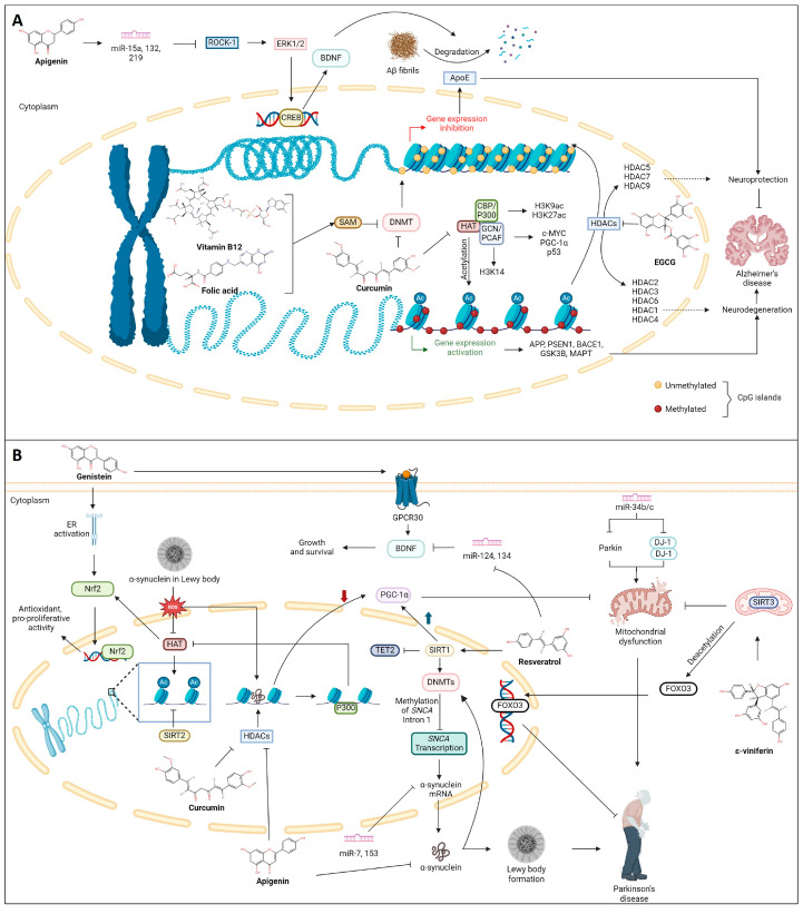
The sensitivity of the epigenome to diet stems from nutrients and metabolites acting as substrates and cofactors for enzymes involved in DNA, RNA, and histone modifications. This intricate interplay allows diet composition to alter the availability of cofactors, thereby influencing the activity of metabolic enzymes and the binding of gene-regulatory complexes to their substrates [205]. This discovery has transformed our comprehension of the connection between nutrition, health, and disease, opening fresh avenues for exploration and treatment, particularly in NDDs. For instance, the diverse array of components such as vitamins B12, choline, and folate, along with bioactive compounds from tea, garlic, soy, and various natural products, contribute to the modulation of epigenetic mechanisms [206]. As nutriepigenomics sheds light on how dietary factors influence the epigenome, it becomes evident that certain bioactive compounds, such as polyphenols, possess the potential to harness these epigenetic mechanisms for therapeutic benefits, allowing the introduction of the term “epigenetic diet” [207]. Various polyphenols like curcumin, resveratrol, and catechins are noted for their ability to influence NF-κB expression and chromatin remodeling by regulating the activity of HDACs and DNMTs [208]. In this context, the following section delves into the specific polyphenols that have demonstrated promise in influencing epigenetic modifications in AD and PD, primarily during in vitro and in vivo studies, paving the way for a comprehensive exploration of their potential in neuroprotection and disease management. Furthermore, a schematic representation of the influence of phytochemicals in various epigenetic modulations is shown in Figure 4 and listed in Table 1.
Figure 4.
Mechanism of phytochemicals in the regulation of epigenetic modifications in (A) Alzheimer’s Disease and (B) Parkinson’s Disease (created with BioRender.com).
Table 1.
Summary of the phytochemicals’ role in the epigenetic modulation of Alzheimer’s and Parkinson’s diseases.
| Polyphenol(s) | Structure(s) | Neurodegenerative Diseases | Epigenetic Target Mechanism(s) | Refs. |
|---|---|---|---|---|
| Tea polyphenols |


|
Alzheimer’s disease |
|
[209,210,211,212] |
| Resveratrol |

|
Parkinson’s disease |
|
[213] |
| ε-Viniferin |
|
Parkinson’s disease |
|
[214,215] |
| Curcumin |

|
Alzheimer’s disease |
|
[216,217,218,219,220,221,222] |
| Parkinson’s disease |
|
|||
| Folic acid and vitamin B12 |

|
Alzheimer’s disease |
|
[223,224,225] |
| Apigenin |

|
Alzheimer’s disease |
|
[226,227,228,229,230,231,232,233,234] |
| Parkinson’s disease |
|
|||
| Genistein |

|
Alzheimer’s disease |
|
[235,236,237,238] |
| Parkinson’s disease |
|
6. Role of Phytochemicals on the Epigenetic Modification in Combating AD and PD
6.1. Tea Polyphenols
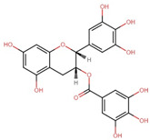
The primary tea polyphenols, including catechin, epicatechin, and (−)-epigallocatechin-3-gallate (EGCG), have been identified to inhibit DNMT-mediated DNA methylation in a dose-dependent manner leading to demethylation and gene reactivation [209]. Among these, EGCG, the most potent polyphenol found in tea, exerts its effect on DNA methylation directly or indirectly. It forms hydrogen bonds within the active site of DNMT, directly inhibiting its activity. Additionally, EGCG contributes to histone post-translational modifications by inhibiting HMTs [210]. In the context of tumor-related gene expression regulation, EGCG acts as an inhibitor of HDAC, as demonstrated in neuronal cells, and downregulates the expression of APP [211]. Further, EGCG in AD mice models promoted NEP expression in M146L cells while reducing Aβ level, which correlated with the compound’s inhibitory effect against HDAC1 [212]. It has also been speculated that EGCG, in addition to acting as an inhibitor, also attenuates the competition of HDAC1 with the APP intracellular domain (AICD) for binding to the NEP promoter, thereby enhancing neprilysin enzyme induction while decreasing APP and sAPPα levels [239,240].
6.2. Resveratrol
The epigenetic mechanisms of resveratrol, which is primarily found in grapes, mulberries, cranberries, and peanuts, occur through histone modifications. Resveratrol exhibits its neuroprotective effect and improves cognitive functions by activating the deacetylase enzyme SIRT1 [241,242]. It has also been reported to inhibit DNMT activity [208]. A study conducted in retinal pigment epithelial cells revealed that resveratrol prevented the decline in expression of DNMT1, DNMT3A, DNMT3B, and SIRT1 triggered by oxidative or inflammatory stimuli [243,244]. Activating endogenous SIRT1 by resveratrol triggers CDKN2A DNA hypermethylation by reducing TET2 protein levels. This reduction alleviates the inhibitory influence on cyclin-dependent kinase 4 (CDK4) and promotes the upregulation of pRb, fostering cell proliferation and growth. Correspondingly, analogous outcomes are noted upon inhibiting endogenous TET2 enzyme activity using a TET2 inhibitor, thus presenting a promising target for PD therapy [213].
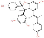
Additionally, resveratrol may play a pivotal role in regulating the expression of miRNAs, which have implications in neurodegenerative diseases [245]. Interestingly, in neurodegenerative diseases, there is an observed upregulation of miR-21, miR-125, miR-146, and miR-155. Tili et al. [246] demonstrated that resveratrol mitigates the heightened levels of pro-inflammatory miR-155 induced by LPS by enhancing the expression of miR-663. Furthermore, resveratrol was found to decrease the levels of miR-124 and miR-134, thereby promoting the synthesis of BDNF [247,248]. However, conflicting findings exist, as another study suggested that resveratrol treatment leads to an upregulation of miR-124 expression in T cells within the brain [249]. Epigenetic inheritance has been observed in the F1 and F2 generations of mice upon maternal resveratrol supplementation, which also improved cognitive functions with a significant increase in global DNA methylation as well as modulation of Dnmt3a/b and Tet2 expressions [250]. In a separate investigation, rat mothers receiving resveratrol supplementation during perinatal asphyxia showed promising neuroprotective outcomes in their offspring. This effect was characterized by a decrease in inflammatory markers, including interleukin-1β (IL-1β), tumor necrosis factor-α (TNF-α), and S-100 calcium-binding protein B (S-100B), which were regulated by specific miRNAs such as miR-124, miR-132, miR-134, miR-146, and miR-15a [243,251]. A compound structurally akin to resveratrol, ε-viniferin, has been shown to enhance SIRT3 expression and forkhead box O3 (FOXO3) deacetylation. This resulted in reduced mitochondrial depolarization induced by rotenone, decreased neuronal apoptosis, and restoration of the expression of proteins associated with mitochondrial homeostasis in a PD model using SH-SY5Y cells [214,215]. ε-Viniferin, a compound structurally akin to resveratrol, has enhanced SIRT3 expression and FOXO3 deacetylation, protecting against PD in SH-SY5Y cells [214,215].
6.3. Curcumin
The main ingredient in turmeric, curcumin, is used by traditional medicinal practitioners in India and China for its antioxidant, anti-inflammatory, antiangiogenic, and anticancer properties. Epidemiological studies performed in India reported that consumption of curcumin leads to a lower incidence of AD in the population [252]. Curcumin can alleviate neurodegeneration and neuroinflammation through multiple mechanisms, including epigenetic changes [253], proven in different experimental models [254]. Curcumin aids in decreasing the production of Aβ by inhibiting the activation of presenilin 1 [255] apart from inhibiting HAT, thereby decreasing histone H3 and H4 acetylation in brain cancer cells [216] and inhibiting DNMT1 activity by covalently binding to the active site of the enzyme [208,256]. In addition, curcumin has been reported to inhibit NNMT in colorectal cancer cells [257].
Curcumin was reported as a natural selective inhibitor of p300 in HAT. Additionally, it suppressed the expression of PS1 and BACE1 by inhibiting histone H3 acetylation in their promoter regions in N2a/APPswe cells, suggesting its role in epigenetic regulation [217]. Curcumin and related curcuminoid compounds could cross the blood-brain barrier and influence the epigenetic regulation in brain cells [220]. Curcumin was reported to reduce the level of methylation of histone H3 at lysine 27 (H3K27me3) jumonji domain-containing protein 3 (JMJD3) within the promoter region of the BDNF, which led to the decrease in the formation of amyloid aggregates thereby boosting mitochondrial function and limiting the Aβ aggregate accumulation [258]. Histone acetylation of the genes associated with AD plays a crucial role in regulating learning and memory and eventually plays a role in AD pathology. Curcumin was reported to alter gene expression by regulating HATs and HDACs activity [259]. It can selectively inhibit the intrinsic E1A-associated 300-kDa protein p300 (p300) in HATs, thereby suppressing the expression of AD-related genes PS1 and BACE1 by inhibiting H3 acetylation in their promoter regions [217].
Curcumin and its metabolites have been observed to inhibit the catalytic thiol group in DNA methyltransferase DNMT1, thereby reducing global DNA methylation levels [260]. Curcumin analogs have upregulated the activity of the Aβ-degrading enzyme neprilysin with DNA methylation modulation through DNMT1 inhibition [218,219]. Curcumin could also induce neuroprotective properties through Wnt pathway modulation [261] as demethoxycurcumin and bisdemethoxycurcumin, the curcuminoid compounds, induce demethylation of Wnt inhibitory factor-1 (WIF-1) promoter region via suppressing the activity of DNMT1, and thereby inhibiting the canonical Wnt pathway [220].
Additionally, the epigenetic mechanisms of curcumin have also been regulated through inflammatory pathways, including sirtuins and nuclear factor erythroid 2–related factor 2 (Nrf2) pathways [254]. Curcumin modulates several miRNAs, including the AD-associated apolipoprotein E gene. It regulates the elevation of miR-128 and miR-9, thereby diminishing levels of phosphorylated tau protein and clearing tau tangles from rat cortical neurons [221]. Altogether, curcumin can influence histone modifications, DNA methylation, and levels of miRNAs to modulate Wnt signaling, amyloid processing, and inflammatory pathways, thereby aiding a novel strategy in AD therapeutics [254]. Liposomal-formulated curcumin was reported to target HDAC, prevent apoptosis, and improve motor deficits in Park 7 (DJ-1)-knockout rat model of PD. The curcumin treatment improved motor behavior and motor impairment, blocked neuronal apoptosis, and stimulated DA neurons in the SN, indicating that the nanotechnology-based epigenetics-driven drug discovery platform toward efficacious therapeutics in PD [222].
Even though curcumin is one of the widely used phytochemicals for its health benefits, its safety level is still debatable. The lesser bioavailability of curcumin makes it difficult for it to extrapolate the effects observed in in vitro studies in clinical trials. It was also reported that higher or irregular doses of curcumin could induce DNA damage and ROS production, inactivate the tumor suppressor protein, affect systemic iron metabolism, and inhibit drug-metabolizing enzymes cytochrome P450, glutathione-S-transferase, and UDP-glucuronosyltransferase which, in turn, can hamper the survival of the host [262].
6.4. Folic Acid and Vitamin B12
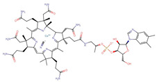
Impaired homocysteine metabolism and deregulation of critical methylation reactions can lead to phosphorylated tau and APP accumulation in the brain [263]. A lower bioavailability of SAM leads to changes in the expression of APP metabolism-regulating genes, which results in increased production and/or accumulation of Aβ peptide. SAM maintains the appropriate methylation of genes involved in APP processing. In that way, silencing these genes prevents the Aβ formation and accumulation, and altered SAM metabolism has been linked to the onset of AD [225]. Reducing folate and vitamin B12 in neuroblastoma cell lines may also lead to a decrease in SAM levels, thereby increasing PSEN1 and BACE levels and Aβ production [223], which the administration of SAM could restore to medium. The SAM-based inhibition of the progression of Alzheimer-like features has also been observed in vitamin-B-deficient animal models [224].
6.5. Apigenin
Apigenin (4′,5,7-trihydroxyflavone) is an abundantly found flavonoid in various herbal plants, fruits (oranges), and vegetables, including thyme, chamomile, basil, oranges, onions, and parsley, to name a few [264]. With a demonstrated ability to modify the epigenetics of various diseases, including neurodegenerative disorders, apigenin attenuates the activity of histone deacetylases (HDACs) and restores microRNA (miRNA) expression. It mitigates neuroinflammation [229,265], thereby improving overall cognitive functions. In a previous in vitro study on a preneoplastic JB6 P+ skin cancer cell line, apigenin was shown to reduce DNA methylation in the Nrf promoter via CpG demethylation while halting the expressions of DNMTs (DNMT1, DNMT3a, and DNMT3b) and HDACs (1–8) epigenetic proteins, which reestablished the expression of Nrf2 and elevated the levels of the NAD(P)H:quinone-oxidoreductase-1 (NQO1) gene [266]. The administration of apigenin can alleviate neuroinflammation and oxidative stress-induced neuronal apoptosis in PD and postoperative cognitive dysfunction (POCD) [229,267]. This flavonoid has proven to inhibit both the upregulation of NF-kB gene expression and neuroinflammation in substantia nigra pars compacta (SNpc). It also prevents the release of pro-inflammatory cytokines such as TNF-α, IL-β, IL-6, caspase-1, and the pro-inflammatory inducible nitric oxide synthase (iNOS-1) enzyme in PD, and downregulates the levels of IL-2, IL-4, and IL-10 in POCD [229,267,268]. Furthermore, apigenin treatment reduces the level of α-synuclein while positively regulating the protein expression of tyrosine hydrolase (TH) and dopamine D2 receptor, indicating that apigenin confers neuroprotectant properties that may serve as an alternative therapeutic for neurodegeneration disorders like PD [267,268].
Neurodegenerative disorders often lead to histone acetylation and the disruption of gene expression associated with normal cognitive functions [181,269]. One of the target genes of histone acetylation is BNDF, which plays a role in restoring normal cognitive functions. Recombinant BNDF can improve cognitive activity and enhance the synaptic density of the hippocampus in AD [226]. Alterations in BNDF expression levels will affect histone acetylation [227,228]. Apigenin is reported to be able to activate the acetylation of H3 lysine 9 (H3K9) and H4 lysine 12 (H4K12) at the P4 promoter of BNDF, followed by the upregulation of downstream calcium/calmodulin-stimulated protein kinase II (p-CAMK II), cAMP response element-binding protein (p-CREB), and extracellular signal-regulated kinase (p-ERK) signaling [229].
Other than cognitive function, histone acetylation is vital for normal memory function [270]. Changes in the plasticity of chromatin and histone acetylation are commonly correlated with memory impairment in aging. In a previous prostate cancer study, apigenin promotes H3K9 and H4K12 acetylation by attenuating the activity of HDAC and decreasing the protein expressions of HDAC1 and HDAC3 [265]. In parallel with cancer study, apigenin can reinstate acetylation levels of both H3 and H4 through the upregulation of CREB binding protein (CBP) and downregulation of HDAC2, proposing apigenin has a vital role in the regulation of histone acetylation in the hippocampus [270].
Moreover, miRNAs have been correlated with the regulation of memory and neurogenesis. Overexpression of miR-15a, miR-132, and miR-219 can enhance spatial memory and overall cognitive ability in AD mice, as AD brains were shown to have downregulation of miR-15a [230,231]. Rho-associated protein kinase-1 (ROCK-1) is essential in the synthesis of inflammatory cytokines in microglial and release by regulating the microglial activation as well as downregulatory effects on ERK 1/2, CREB, and BNDF [232,233]. One AD-related study has shown that miR-15a suppresses the ROCK-1 gene in hippocampus neurons, reducing abnormal tau hyperphosphorylation and neuronal cell morbidity [234]. The neuroprotective effect of apigenin was identified because it significantly increased the miR-15a gene expression and decreased ROCK-1 expression while reversing the activation of microglial and reducing the ionized calcium-binding adapter protein-1 (Iba-1) in methotrexate-treated rat hippocampus [230]. Notably, apigenin could be a potential treatment candidate for epigenetic therapy of neurodegenerative diseases in the future, such as AD and PD.
6.6. Genistein
Genistein (4′,5′,7-trihydroxyisoflavone) belongs to the isoflavone class of flavonoids commonly found in soybean-related products. Extensive research has demonstrated its potent anticancer properties, functioning as an inhibitor of epidermal growth factor receptor tyrosine kinase [271]. Beyond its anticancer effects, genistein has been described as having the ability to alleviate symptoms associated with central nervous system (CNS) disorders, including depression, AD, PD, epilepsy, and dementia [272,273,274]. Genistein exhibits promising blood-brain barrier (BBB) permeability, mimicking neuroprotective effects both in vitro and in vivo through its anti-inflammatory activity [275,276]. The neuroprotective effects of estrogens have been evident in various neuronal cell studies [277]. However, estrogen therapy poses potential risks, such as an increased likelihood of various cancers and harm to patients susceptible to blood clotting [271]. Consequently, ongoing research aims to identify estrogen-like phytoestrogens devoid of toxicities. Genistein, with its structural similarity to estrogen, is recognized as a potent phytoestrogen, acting as an agonist binding to estrogen receptors α and β (ERα and Erβ) [278].
Oxidative stress and neuroinflammation contribute to the demise of nigrostriatal dopaminergic neurons [279,280]. Genistein has been identified as a modifier of inflammatory molecules, improving conditions associated with these processes. Recent findings indicate that genistein exerts a neuroprotective effect in a human neuronal SH-SY5Y cell Parkinson’s disease model through ERs and the Nrf2 channels [235]. Activation of Nrf2 by genistein upregulates heme oxygenase 1 (HMOX1), conferring neuronal protection along with glutathione peroxidase. Moreover, genistein is observed to reduce or reverse mitochondrial oxidative stress-induced damage and neuronal cell death [235].
Furthermore, genistein mitigates elevated malondialdehyde (MDA) content without affecting nitrite content or superoxide dismutase (SOD) activity, indicating its ability to reverse Aβ-induced memory deficits by ameliorating oxidative stress [281]. Research by Li et al. [282] similarly describes genistein’s capacity to reduce cerebral infarction and mitigate neuronal damage and apoptosis in neonatal hypoxic-ischemic brain damage (HIBD) mice. This phytoestrogen achieves these effects by mitigating oxidative stress and neuroinflammation via the activation of the Nrf2/HMOX-1 pathway and attenuation of the NF-κB inflammatory pathway, resulting in a total reduction of pro-inflammatory cytokines such as IL-6, IL-1β, and TNF-α [282,283].
To date, limited reports have explored the influence of genistein on gene expression and memory in the brain via epigenetic regulation. Genistein shares similar effects with 17β-estradiol [278], binding to G protein-coupled receptor 30 (GPR30) and enhancing spatial memory function. It also improves the gene expressions of BDNF, insulin-like growth factor 1 (IGF-1), and miR-132 in the hippocampus tissue of ovariectomized rats [284]. Studies suggest that miR-132 expression is associated with neuronal cell growth, synaptic formation, and angiogenesis [238,285]. miR-132 plays a role in memory and learning cognitive function via downregulation of p250GAP, which controls Rac1-PAK-mediated dendrospinogenesis and restoration of the actin polymerization in the dendritic spine of the perirhinal cortex miR-132 plays a crucial role in memory and learning cognitive function by downregulating p250GAP, controlling Rac1-PAK-mediated dendrospinogenesis, and restoring actin polymerization in the dendritic spine of the perirhinal cortex [236,237,238]. These findings suggest that genistein could influence miRNA expression, potentially halting the progression of neurodegenerative diseases.
7. Conclusions and Future Perspectives
The exploration of nutriepigenomics within the context of AD and PD offers a compelling avenue for understanding and potentially mitigating the epigenetic dysregulation that underlies these complex neurological conditions. As we deepen our understanding of the intricate interplay between dietary factors and epigenetic mechanisms, we uncover novel opportunities for therapeutic intervention. Nutriepigenomics highlights the significant impact of diet on the epigenome and emphasizes the potential for personalized dietary strategies to modulate disease progression and improve outcomes for individuals with AD and PD. By identifying specific bioactive compounds found in natural sources, we can target key epigenetic pathways implicated in neurodegeneration, offering the possibility of tailored interventions that address the unique molecular profiles of each patient. In addition, the complementary use of phytochemicals alongside available commercial drugs presents a promising avenue for combinatorial treatment approaches. Moving forward, continued research efforts should focus on elucidating the precise mechanisms through which dietary factors exert their epigenetic effects and exploring the feasibility and efficacy of implementing personalized “epigenetic diets” in clinical practice. Additionally, collaborative endeavors between researchers, clinicians, and nutritionists will be essential for translating scientific insights into tangible therapeutic strategies that benefit patients.
In summary, the convergence of neuroepigenetics and nutriepigenomics provides a compelling framework for advancing therapeutic approaches in NDDs. By harnessing the power of dietary interventions to modulate epigenetic mechanisms, we can envision a future where personalized nutritional strategies play a pivotal role in slowing disease progression, preserving cognitive and motor function, and ultimately improving the quality of life for individuals affected by neurological disorders. The intricate interplay between neuroepigenetics and the therapeutic potential of phytochemicals offers a promising avenue for understanding and managing neurodegenerative diseases. However, most of the studies have been done predominantly in in vitro and in vivo models, which, when studied in clinical trials, showed lower bioavailability of certain phytochemicals, limiting their efficacy in reaching therapeutic concentrations in the brain. Additionally, the variability in phytochemical content among natural sources and batch-to-batch variations in supplements can pose challenges in standardizing dosages for consistent therapeutic outcomes. Moreover, some phytochemicals may exhibit side effects or interactions with other medications, necessitating cautious evaluation and monitoring in clinical settings. These factors highlight the need for further research and refinement in harnessing the full therapeutic potential of phytochemical interventions for neurodegenerative disorders. Therefore, future research endeavors should continue to unravel the intricate mechanisms underlying these interactions and their further effects, ultimately translating scientific insights into tangible clinical benefits for individuals grappling with the burdens of AD, PD, and related neurodegenerative disorders.
Acknowledgments
M.I.P. and C.S.Y.C. wish to thank the Ratchadaphiseksomphot Endowment Fund for their Postdoctoral Fellowship and Chulalongkorn University, Thailand, for their support. K.V. wishes to thank the Second Century Fund (C2F), Chulalongkorn University, Thailand, for their support. B.S.S. also wishes to thank Chiang Mai University, Thailand, for their support.
Author Contributions
Conceptualization, M.I.P., and A.P.; investigation and writing—original draft preparation, M.I.P., B.S.S., C.S.Y.C., K.V., J.M.B., and A.P.; writing—review and editing, M.I.P., B.S.S., C.S.Y.C., K.V., T.T., J.M.B., and A.P.; visualization, M.I.P.; project administration, A.P.; funding acquisition, T.T., J.M.B., and A.P. All authors have read and agreed to the published version of the manuscript.
Institutional Review Board Statement
Not applicable.
Informed Consent Statement
Not applicable.
Data Availability Statement
Not applicable.
Conflicts of Interest
The authors declare no conflicts of interest.
Funding Statement
This research was supported by the Ratchadapisek Somphot Endowment Fund (2023), Chulalongkorn University (Review_66_006_5300_001).
Footnotes
Disclaimer/Publisher’s Note: The statements, opinions and data contained in all publications are solely those of the individual author(s) and contributor(s) and not of MDPI and/or the editor(s). MDPI and/or the editor(s) disclaim responsibility for any injury to people or property resulting from any ideas, methods, instructions or products referred to in the content.
References
- 1.Ghosh P., Saadat A. Neurodegeneration and epigenetics: A review. Neurologia. 2021;38:e62–e68. doi: 10.1016/j.nrl.2021.01.016. [DOI] [PubMed] [Google Scholar]
- 2.Cheng Y., He C., Wang M., Ma X., Mo F., Yang S., Han J., Wei X. Targeting epigenetic regulators for cancer therapy: Mechanisms and advances in clinical trials. Signal Transduct. Target. Ther. 2019;4:62. doi: 10.1038/s41392-019-0095-0. [DOI] [PMC free article] [PubMed] [Google Scholar]
- 3.Zhang W., Xu J. DNA methyltransferases and their roles in tumorigenesis. Biomark. Res. 2017;5:1. doi: 10.1186/s40364-017-0081-z. [DOI] [PMC free article] [PubMed] [Google Scholar]
- 4.Campagna R., Vignini A. NAD+ Homeostasis and NAD+-Consuming Enzymes: Implications for Vascular Health. Antioxidants. 2023;12:376. doi: 10.3390/antiox12020376. [DOI] [PMC free article] [PubMed] [Google Scholar]
- 5.Patnaik E., Madu C., Lu Y. Epigenetic Modulators as Therapeutic Agents in Cancer. Int. J. Mol. Sci. 2023;24:14964. doi: 10.3390/ijms241914964. [DOI] [PMC free article] [PubMed] [Google Scholar]
- 6.Wang W., Yang C., Wang T., Deng H. Complex roles of nicotinamide N-methyltransferase in cancer progression. Cell Death Dis. 2022;13:267. doi: 10.1038/s41419-022-04713-z. [DOI] [PMC free article] [PubMed] [Google Scholar]
- 7.Roberti A., Fernández A.F., Fraga M.F. Nicotinamide N-methyltransferase: At the crossroads between cellular metabolism and epigenetic regulation. Mol. Metab. 2021;45:101165. doi: 10.1016/j.molmet.2021.101165. [DOI] [PMC free article] [PubMed] [Google Scholar]
- 8.Ulanovskaya O.A., Zuhl A.M., Cravatt B.F. NNMT promotes epigenetic remodeling in cancer by creating a metabolic methylation sink. Nat. Chem. Biol. 2013;9:300–306. doi: 10.1038/nchembio.1204. [DOI] [PMC free article] [PubMed] [Google Scholar]
- 9.van Haren M.J., Gao Y., Buijs N., Campagna R., Sartini D., Emanuelli M., Mateuszuk L., Kij A., Chlopicki S., Escudé Martinez de Castilla P., et al. Esterase-Sensitive Prodrugs of a Potent Bisubstrate Inhibitor of Nicotinamide N-Methyltransferase (NNMT) Display Cellular Activity. Biomolecules. 2021;11:1357. doi: 10.3390/biom11091357. [DOI] [PMC free article] [PubMed] [Google Scholar]
- 10.van Haren M.J., Zhang Y., Thijssen V., Buijs N., Gao Y., Mateuszuk L., Fedak F.A., Kij A., Campagna R., Sartini D., et al. Macrocyclic peptides as allosteric inhibitors of nicotinamide N-methyltransferase (NNMT) RSC Chem. Biol. 2021;2:1546–1555. doi: 10.1039/d1cb00134e. [DOI] [PMC free article] [PubMed] [Google Scholar]
- 11.Gao Y., van Haren M.J., Buijs N., Innocenti P., Zhang Y., Sartini D., Campagna R., Emanuelli M., Parsons R.B., Jespers W., et al. Potent Inhibition of Nicotinamide N-Methyltransferase by Alkene-Linked Bisubstrate Mimics Bearing Electron Deficient Aromatics. J. Med. Chem. 2021;64:12938–12963. doi: 10.1021/acs.jmedchem.1c01094. [DOI] [PMC free article] [PubMed] [Google Scholar]
- 12.Kumar S., Chinnusamy V., Mohapatra T. Epigenetics of modified DNA bases: 5-methylcytosine and beyond. Front. Genet. 2018;9:429871. doi: 10.3389/fgene.2018.00640. [DOI] [PMC free article] [PubMed] [Google Scholar]
- 13.Neganova M.E., Klochkov S.G., Aleksandrova Y.R., Aliev G. Seminars in Cancer Biology. Academic Press; Cambridge, MA, USA: 2022. Histone modifications in epigenetic regulation of cancer: Perspectives and achieved progress; pp. 452–471. [DOI] [PubMed] [Google Scholar]
- 14.Gujral P., Mahajan V., Lissaman A.C., Ponnampalam A.P. Histone acetylation and the role of histone deacetylases in normal cyclic endometrium. Reprod. Biol. Endocrinol. 2020;18:84. doi: 10.1186/s12958-020-00637-5. [DOI] [PMC free article] [PubMed] [Google Scholar]
- 15.Milazzo G., Mercatelli D., Di Muzio G., Triboli L., De Rosa P., Perini G., Giorgi F.M. Histone deacetylases (HDACs): Evolution, specificity, role in transcriptional complexes, and pharmacological actionability. Genes. 2020;11:556. doi: 10.3390/genes11050556. [DOI] [PMC free article] [PubMed] [Google Scholar]
- 16.Alaskhar Alhamwe B., Khalaila R., Wolf J., von Bülow V., Harb H., Alhamdan F., Hii C.S., Prescott S.L., Ferrante A., Renz H. Histone modifications and their role in epigenetics of atopy and allergic diseases. Allergy Asthma Clin. Immunol. 2018;14:39. doi: 10.1186/s13223-018-0259-4. [DOI] [PMC free article] [PubMed] [Google Scholar]
- 17.Burgos M., Hurtado A., Jimenez R., Barrionuevo F.J. Non-Coding RNAs: lncRNAs, miRNAs, and piRNAs in Sexual Development. Sex. Dev. 2021;15:335–350. doi: 10.1159/000519237. [DOI] [PubMed] [Google Scholar]
- 18.Paul S. Impact of epigenetics on human health and possible tool for remediation. Nucleus. 2021;64:255–258. doi: 10.1007/s13237-021-00379-8. [DOI] [Google Scholar]
- 19.Sanchez-Mut J.V., Gräff J. Epigenetic alterations in Alzheimer’s disease. Front. Behav. Neurosci. 2015;9:347. doi: 10.3389/fnbeh.2015.00347. [DOI] [PMC free article] [PubMed] [Google Scholar]
- 20.Qureshi I.A., Mehler M.F. Epigenetic mechanisms underlying nervous system diseases. Handb. Clin. Neurol. 2018;147:43–58. doi: 10.1016/B978-0-444-63233-3.00005-1. [DOI] [PMC free article] [PubMed] [Google Scholar]
- 21.Coda D.M., Graff J. Neurogenetic and Neuroepigenetic Mechanisms in Cognitive Health and Disease. Front. Mol. Neurosci. 2020;13:205. doi: 10.3389/fnmol.2020.589109. [DOI] [PMC free article] [PubMed] [Google Scholar]
- 22.Lanoiselée H.M., Nicolas G., Wallon D., Rovelet-Lecrux A., Lacour M., Rousseau S., Richard A.C., Pasquier F., Rollin-Sillaire A., Martinaud O., et al. APP, PSEN1, and PSEN2 mutations in early-onset Alzheimer disease: A genetic screening study of familial and sporadic cases. PLoS Med. 2017;14:e1002270. doi: 10.1371/journal.pmed.1002270. [DOI] [PMC free article] [PubMed] [Google Scholar]
- 23.Gatz M., Reynolds C.A., Fratiglioni L., Johansson B., Mortimer J.A., Berg S., Fiske A., Pedersen N.L. Role of genes and environments for explaining Alzheimer disease. Arch. Gen. Psychiatry. 2006;63:168–174. doi: 10.1001/archpsyc.63.2.168. [DOI] [PubMed] [Google Scholar]
- 24.Shen L., Jia J. An Overview of Genome-Wide Association Studies in Alzheimer’s Disease. Neurosci. Bull. 2016;32:183–190. doi: 10.1007/s12264-016-0011-3. [DOI] [PMC free article] [PubMed] [Google Scholar]
- 25.Nazarian A., Yashin A.I., Kulminski A.M. Genome-wide analysis of genetic predisposition to Alzheimer’s disease and related sex disparities. Alzheimer’s Res. Ther. 2019;11:5. doi: 10.1186/s13195-018-0458-8. [DOI] [PMC free article] [PubMed] [Google Scholar]
- 26.Shigemizu D., Mitsumori R., Akiyama S., Miyashita A., Morizono T., Higaki S., Asanomi Y., Hara N., Tamiya G., Kinoshita K., et al. Ethnic and trans-ethnic genome-wide association studies identify new loci influencing Japanese Alzheimer’s disease risk. Transl. Psychiatry. 2021;11:151. doi: 10.1038/s41398-021-01272-3. [DOI] [PMC free article] [PubMed] [Google Scholar]
- 27.Malar D.S., Devi K.P. Dietary polyphenols for treatment of Alzheimer’s disease--future research and development. Curr. Pharm. Biotechnol. 2014;15:330–342. doi: 10.2174/1389201015666140813122703. [DOI] [PubMed] [Google Scholar]
- 28.Nyakas C., Granic I., Halmy L.G., Banerjee P., Luiten P.G. The basal forebrain cholinergic system in aging and dementia. Rescuing cholinergic neurons from neurotoxic amyloid-β42 with memantine. Behav. Brain Res. 2011;221:594–603. doi: 10.1016/j.bbr.2010.05.033. [DOI] [PubMed] [Google Scholar]
- 29.Kar S., Quirion R. Amyloid beta peptides and central cholinergic neurons: Functional interrelationship and relevance to Alzheimer’s disease pathology. Prog. Brain Res. 2004;145:261–274. doi: 10.1016/s0079-6123(03)45018-8. [DOI] [PubMed] [Google Scholar]
- 30.Chen G.F., Xu T.H., Yan Y., Zhou Y.R., Jiang Y., Melcher K., Xu H.E. Amyloid beta: Structure, biology and structure-based therapeutic development. Acta Pharmacol. Sin. 2017;38:1205–1235. doi: 10.1038/aps.2017.28. [DOI] [PMC free article] [PubMed] [Google Scholar]
- 31.Wogulis M., Wright S., Cunningham D., Chilcote T., Powell K., Rydel R.E. Nucleation-dependent polymerization is an essential component of amyloid-mediated neuronal cell death. J. Neurosci. Off. J. Soc. Neurosci. 2005;25:1071–1080. doi: 10.1523/jneurosci.2381-04.2005. [DOI] [PMC free article] [PubMed] [Google Scholar]
- 32.Xue W.F., Homans S.W., Radford S.E. Systematic analysis of nucleation-dependent polymerization reveals new insights into the mechanism of amyloid self-assembly. Proc. Natl. Acad. Sci. USA. 2008;105:8926–8931. doi: 10.1073/pnas.0711664105. [DOI] [PMC free article] [PubMed] [Google Scholar]
- 33.Deane R., Bell R.D., Sagare A., Zlokovic B.V. Clearance of amyloid-beta peptide across the blood-brain barrier: Implication for therapies in Alzheimer’s disease. CNS Neurol. Disord. Drug Targets. 2009;8:16–30. doi: 10.2174/187152709787601867. [DOI] [PMC free article] [PubMed] [Google Scholar]
- 34.Sturchler E., Galichet A., Weibel M., Leclerc E., Heizmann C.W. Site-specific blockade of RAGE-Vd prevents amyloid-beta oligomer neurotoxicity. J. Neurosci. Off. J. Soc. Neurosci. 2008;28:5149–5158. doi: 10.1523/jneurosci.4878-07.2008. [DOI] [PMC free article] [PubMed] [Google Scholar]
- 35.Yan S.D., Chen X., Fu J., Chen M., Zhu H., Roher A., Slattery T., Zhao L., Nagashima M., Morser J., et al. RAGE and amyloid-beta peptide neurotoxicity in Alzheimer’s disease. Nature. 1996;382:685–691. doi: 10.1038/382685a0. [DOI] [PubMed] [Google Scholar]
- 36.Deane R., Singh I., Sagare A.P., Bell R.D., Ross N.T., LaRue B., Love R., Perry S., Paquette N., Deane R.J., et al. A multimodal RAGE-specific inhibitor reduces amyloid β-mediated brain disorder in a mouse model of Alzheimer disease. J. Clin. Investig. 2012;122:1377–1392. doi: 10.1172/jci58642. [DOI] [PMC free article] [PubMed] [Google Scholar]
- 37.Leng F., Edison P. Neuroinflammation and microglial activation in Alzheimer disease: Where do we go from here? Nat. Rev. Neurol. 2021;17:157–172. doi: 10.1038/s41582-020-00435-y. [DOI] [PubMed] [Google Scholar]
- 38.Montoliu-Gaya L., Mulder S.D., Herrebout M.A.C., Baayen J.C., Villegas S., Veerhuis R. Aβ-oligomer uptake and the resulting inflammatory response in adult human astrocytes are precluded by an anti-Aβ single chain variable fragment in combination with an apoE mimetic peptide. Mol. Cell. Neurosci. 2018;89:49–59. doi: 10.1016/j.mcn.2018.03.015. [DOI] [PubMed] [Google Scholar]
- 39.Garwood C.J., Pooler A.M., Atherton J., Hanger D.P., Noble W. Astrocytes are important mediators of Aβ-induced neurotoxicity and tau phosphorylation in primary culture. Cell Death Dis. 2011;2:e167. doi: 10.1038/cddis.2011.50. [DOI] [PMC free article] [PubMed] [Google Scholar]
- 40.Kaur D., Sharma V., Deshmukh R. Activation of microglia and astrocytes: A roadway to neuroinflammation and Alzheimer’s disease. Inflammopharmacology. 2019;27:663–677. doi: 10.1007/s10787-019-00580-x. [DOI] [PubMed] [Google Scholar]
- 41.Choi Y., Lee M.K., Lim S.Y., Sung S.H., Kim Y.C. Inhibition of inducible NO synthase, cyclooxygenase-2 and interleukin-1beta by torilin is mediated by mitogen-activated protein kinases in microglial BV2 cells. Br. J. Pharmacol. 2009;156:933–940. doi: 10.1111/j.1476-5381.2009.00022.x. [DOI] [PMC free article] [PubMed] [Google Scholar]
- 42.Guo J.T., Yu J., Grass D., de Beer F.C., Kindy M.S. Inflammation-dependent cerebral deposition of serum amyloid a protein in a mouse model of amyloidosis. J. Neurosci. Off. J. Soc. Neurosci. 2002;22:5900–5909. doi: 10.1523/jneurosci.22-14-05900.2002. [DOI] [PMC free article] [PubMed] [Google Scholar]
- 43.Sastre M., Dewachter I., Landreth G.E., Willson T.M., Klockgether T., van Leuven F., Heneka M.T. Nonsteroidal anti-inflammatory drugs and peroxisome proliferator-activated receptor-gamma agonists modulate immunostimulated processing of amyloid precursor protein through regulation of beta-secretase. J. Neurosci. Off. J. Soc. Neurosci. 2003;23:9796–9804. doi: 10.1523/jneurosci.23-30-09796.2003. [DOI] [PMC free article] [PubMed] [Google Scholar]
- 44.Müller M.K., Jacobi E., Sakimura K., Malinow R., von Engelhardt J. NMDA receptors mediate synaptic depression, but not spine loss in the dentate gyrus of adult amyloid Beta (Aβ) overexpressing mice. Acta Neuropathol. Commun. 2018;6:110. doi: 10.1186/s40478-018-0611-4. [DOI] [PMC free article] [PubMed] [Google Scholar]
- 45.Parameshwaran K., Sims C., Kanju P., Vaithianathan T., Shonesy B.C., Dhanasekaran M., Bahr B.A., Suppiramaniam V. Amyloid beta-peptide Abeta(1-42) but not Abeta(1-40) attenuates synaptic AMPA receptor function. Synapse. 2007;61:367–374. doi: 10.1002/syn.20386. [DOI] [PubMed] [Google Scholar]
- 46.Sadigh-Eteghad S., Talebi M., Farhoudi M., Golzari S.E.J., Sabermarouf B., Mahmoudi J. Beta-amyloid exhibits antagonistic effects on alpha 7 nicotinic acetylcholine receptors in orchestrated manner. J. Med. Hypotheses Ideas. 2014;8:49–52. doi: 10.1016/j.jmhi.2014.01.001. [DOI] [Google Scholar]
- 47.Fani G., Mannini B., Vecchi G., Cascella R., Cecchi C., Dobson C.M., Vendruscolo M., Chiti F. Aβ Oligomers Dysregulate Calcium Homeostasis by Mechanosensitive Activation of AMPA and NMDA Receptors. ACS Chem. Neurosci. 2021;12:766–781. doi: 10.1021/acschemneuro.0c00811. [DOI] [PMC free article] [PubMed] [Google Scholar]
- 48.Ni R., Marutle A., Nordberg A. Modulation of α7 nicotinic acetylcholine receptor and fibrillar amyloid-β interactions in Alzheimer’s disease brain. J. Alzheimer’s Dis. JAD. 2013;33:841–851. doi: 10.3233/jad-2012-121447. [DOI] [PubMed] [Google Scholar]
- 49.Wang R., Reddy P.H. Role of Glutamate and NMDA Receptors in Alzheimer’s Disease. J. Alzheimer’s Dis. JAD. 2017;57:1041–1048. doi: 10.3233/jad-160763. [DOI] [PMC free article] [PubMed] [Google Scholar]
- 50.Acosta C., Anderson H.D., Anderson C.M. Astrocyte dysfunction in Alzheimer disease. J. Neurosci. Res. 2017;95:2430–2447. doi: 10.1002/jnr.24075. [DOI] [PubMed] [Google Scholar]
- 51.Uddin M.S., Tewari D., Sharma G., Kabir M.T., Barreto G.E., Bin-Jumah M.N., Perveen A., Abdel-Daim M.M., Ashraf G.M. Molecular Mechanisms of ER Stress and UPR in the Pathogenesis of Alzheimer’s Disease. Mol. Neurobiol. 2020;57:2902–2919. doi: 10.1007/s12035-020-01929-y. [DOI] [PubMed] [Google Scholar]
- 52.Malar D.S., Prasanth M.I., Brimson J.M., Verma K., Prasansuklab A., Tencomnao T. Hibiscus sabdariffa extract protects HT-22 cells from glutamate-induced neurodegeneration by upregulating glutamate transporters and exerts lifespan extension in C. elegans via DAF-16 mediated pathway. Nutr. Healthy Aging. 2021;6:229–247. doi: 10.3233/NHA-210131. [DOI] [Google Scholar]
- 53.Mandelkow E.M., Mandelkow E. Biochemistry and cell biology of tau protein in neurofibrillary degeneration. Cold Spring Harb. Perspect. Med. 2012;2:a006247. doi: 10.1101/cshperspect.a006247. [DOI] [PMC free article] [PubMed] [Google Scholar]
- 54.Mietelska-Porowska A., Wasik U., Goras M., Filipek A., Niewiadomska G. Tau protein modifications and interactions: Their role in function and dysfunction. Int. J. Mol. Sci. 2014;15:4671–4713. doi: 10.3390/ijms15034671. [DOI] [PMC free article] [PubMed] [Google Scholar]
- 55.Kocinaj A., Chaudhury T., Uddin M.S., Junaid R.R., Ramsden D.B., Hondhamuni G., Klamt F., Parsons L., Parsons R.B. High Expression of Nicotinamide N-Methyltransferase in Patients with Sporadic Alzheimer’s Disease. Mol. Neurobiol. 2021;58:1769–1781. doi: 10.1007/s12035-020-02259-9. [DOI] [PMC free article] [PubMed] [Google Scholar]
- 56.Chouliaras L., Mastroeni D., Delvaux E., Grover A., Kenis G., Hof P.R., Steinbusch H.W., Coleman P.D., Rutten B.P., van den Hove D.L. Consistent decrease in global DNA methylation and hydroxymethylation in the hippocampus of Alzheimer’s disease patients. Neurobiol. Aging. 2013;34:2091–2099. doi: 10.1016/j.neurobiolaging.2013.02.021. [DOI] [PMC free article] [PubMed] [Google Scholar]
- 57.West R.L., Lee J.M., Maroun L.E. Hypomethylation of the amyloid precursor protein gene in the brain of an Alzheimer’s disease patient. J. Mol. Neurosci. 1995;6:141–146. doi: 10.1007/BF02736773. [DOI] [PubMed] [Google Scholar]
- 58.Monti N., Cavallaro R.A., Stoccoro A., Nicolia V., Scarpa S., Kovacs G.G., Fiorenza M.T., Lucarelli M., Aronica E., Ferrer I. CpG and non-CpG Presenilin1 methylation pattern in course of neurodevelopment and neurodegeneration is associated with gene expression in human and murine brain. Epigenetics. 2020;15:781–799. doi: 10.1080/15592294.2020.1722917. [DOI] [PMC free article] [PubMed] [Google Scholar]
- 59.Ozaki Y., Yoshino Y., Yamazaki K., Sao T., Mori Y., Ochi S., Yoshida T., Mori T., Iga J.-i., Ueno S.-i. DNA methylation changes at TREM2 intron 1 and TREM2 mRNA expression in patients with Alzheimer’s disease. J. Psychiatr. Res. 2017;92:74–80. doi: 10.1016/j.jpsychires.2017.04.003. [DOI] [PubMed] [Google Scholar]
- 60.Mendioroz M., Celarain N., Altuna M., Sánchez-Ruiz de Gordoa J., Zelaya M.V., Roldán M., Rubio I., Larumbe R., Erro M.E., Méndez I. CRTC1 gene is differentially methylated in the human hippocampus in Alzheimer’s disease. Alzheimer’s Res. Ther. 2016;8:15. doi: 10.1186/s13195-016-0183-0. [DOI] [PMC free article] [PubMed] [Google Scholar]
- 61.Salcedo-Tacuma D., Melgarejo J.D., Mahecha M.F., Ortega-Rojas J., Arboleda-Bustos C.E., Pardo-Turriago R., Arboleda H. Differential methylation levels in CpGs of the BIN1 gene in individuals with Alzheimer disease. Alzheimer Dis. Assoc. Disord. 2019;33:321–326. doi: 10.1097/WAD.0000000000000329. [DOI] [PubMed] [Google Scholar]
- 62.Chapuis J., Hansmannel F., Gistelinck M., Mounier A., Van Cauwenberghe C., Kolen K.V., Geller F., Sottejeau Y., Harold D., Dourlen P., et al. Increased expression of BIN1 mediates Alzheimer genetic risk by modulating tau pathology. Mol. Psychiatry. 2013;18:1225–1234. doi: 10.1038/mp.2013.1. [DOI] [PMC free article] [PubMed] [Google Scholar]
- 63.Stoccoro A., Siciliano G., Migliore L., Coppedè F. Decreased methylation of the mitochondrial D-loop region in late-onset Alzheimer’s disease. J. Alzheimer’s Dis. 2017;59:559–564. doi: 10.3233/JAD-170139. [DOI] [PubMed] [Google Scholar]
- 64.Mitsumori R., Sakaguchi K., Shigemizu D., Mori T., Akiyama S., Ozaki K., Niida S., Shimoda N. Lower DNA methylation levels in CpG island shores of CR1, CLU, and PICALM in the blood of Japanese Alzheimer’s disease patients. PLoS ONE. 2020;15:e0239196. doi: 10.1371/journal.pone.0239196. [DOI] [PMC free article] [PubMed] [Google Scholar]
- 65.Foraker J., Millard S.P., Leong L., Thomson Z., Chen S., Keene C.D., Bekris L.M., Yu C.-E. The APOE gene is differentially methylated in Alzheimer’s disease. J. Alzheimer’s Dis. 2015;48:745–755. doi: 10.3233/JAD-143060. [DOI] [PMC free article] [PubMed] [Google Scholar]
- 66.Sanchez-Mut J.V., Aso E., Heyn H., Matsuda T., Bock C., Ferrer I., Esteller M. Promoter hypermethylation of the phosphatase DUSP22 mediates PKA-dependent TAU phosphorylation and CREB activation in Alzheimer’s disease. Hippocampus. 2014;24:363–368. doi: 10.1002/hipo.22245. [DOI] [PMC free article] [PubMed] [Google Scholar]
- 67.Wezyk M., Spólnicka M., Pośpiech E., Pepłońska B., Zbieć-Piekarska R., Ilkowski J., Styczyńska M., Barczak A., Zboch M., Filipek-Gliszczynska A. Hypermethylation of TRIM59 and KLF14 influences cell death signaling in familial Alzheimer’s disease. Oxidative Med. Cell. Longev. 2018;2018:6918797. doi: 10.1155/2018/6918797. [DOI] [PMC free article] [PubMed] [Google Scholar]
- 68.Chen K.-L., Wang S.S.-S., Yang Y.-Y., Yuan R.-Y., Chen R.-M., Hu C.-J. The epigenetic effects of amyloid-β1–40 on global DNA and neprilysin genes in murine cerebral endothelial cells. Biochem. Biophys. Res. Commun. 2009;378:57–61. doi: 10.1016/j.bbrc.2008.10.173. [DOI] [PubMed] [Google Scholar]
- 69.Chang L., Wang Y., Ji H., Dai D., Xu X., Jiang D., Hong Q., Ye H., Zhang X., Zhou X., et al. Elevation of peripheral BDNF promoter methylation links to the risk of Alzheimer’s disease. PLoS ONE. 2014;9:e110773. doi: 10.1371/journal.pone.0110773. [DOI] [PMC free article] [PubMed] [Google Scholar]
- 70.Nagata T., Kobayashi N., Ishii J., Shinagawa S., Nakayama R., Shibata N., Kuerban B., Ohnuma T., Kondo K., Arai H. Association between DNA methylation of the BDNF promoter region and clinical presentation in Alzheimer’s disease. Dement. Geriatr. Cogn. Disord. Extra. 2015;5:64–73. doi: 10.1159/000375367. [DOI] [PMC free article] [PubMed] [Google Scholar]
- 71.Lunnon K., Smith R., Hannon E., De Jager P.L., Srivastava G., Volta M., Troakes C., Al-Sarraj S., Burrage J., Macdonald R. Methylomic profiling implicates cortical deregulation of ANK1 in Alzheimer’s disease. Nat. Neurosci. 2014;17:1164–1170. doi: 10.1038/nn.3782. [DOI] [PMC free article] [PubMed] [Google Scholar]
- 72.De Jager P.L., Srivastava G., Lunnon K., Burgess J., Schalkwyk L.C., Yu L., Eaton M.L., Keenan B.T., Ernst J., McCabe C. Alzheimer’s disease: Early alterations in brain DNA methylation at ANK1, BIN1, RHBDF2 and other loci. Nat. Neurosci. 2014;17:1156–1163. doi: 10.1038/nn.3786. [DOI] [PMC free article] [PubMed] [Google Scholar]
- 73.Smith A.R., Smith R.G., Burrage J., Troakes C., Al-Sarraj S., Kalaria R.N., Sloan C., Robinson A.C., Mill J., Lunnon K. A cross-brain regions study of ANK1 DNA methylation in different neurodegenerative diseases. Neurobiol. Aging. 2019;74:70–76. doi: 10.1016/j.neurobiolaging.2018.09.024. [DOI] [PubMed] [Google Scholar]
- 74.Pramio D.T., Vieceli F.M., Varella-Branco E., Goes C.P., Kobayashi G.S., da Silva Pelegrina D.V., de Moraes B.C., El Allam A., De Kumar B., Jara G. DNA methylation of the promoter region at the CREB1 binding site is a mechanism for the epigenetic regulation of brain-specific PKMζ. Biochim. Biophys. Acta (BBA)-Gene Regul. Mech. 2023;1866:194909. doi: 10.1016/j.bbagrm.2023.194909. [DOI] [PMC free article] [PubMed] [Google Scholar]
- 75.Marzi S.J., Leung S.K., Ribarska T., Hannon E., Smith A.R., Pishva E., Poschmann J., Moore K., Troakes C., Al-Sarraj S. A histone acetylome-wide association study of Alzheimer’s disease identifies disease-associated H3K27ac differences in the entorhinal cortex. Nat. Neurosci. 2018;21:1618–1627. doi: 10.1038/s41593-018-0253-7. [DOI] [PubMed] [Google Scholar]
- 76.Klein H.-U., McCabe C., Gjoneska E., Sullivan S.E., Kaskow B.J., Tang A., Smith R.V., Xu J., Pfenning A.R., Bernstein B.E. Epigenome-wide study uncovers large-scale changes in histone acetylation driven by tau pathology in aging and Alzheimer’s human brains. Nat. Neurosci. 2019;22:37–46. doi: 10.1038/s41593-018-0291-1. [DOI] [PMC free article] [PubMed] [Google Scholar]
- 77.Nativio R., Lan Y., Donahue G., Sidoli S., Berson A., Srinivasan A.R., Shcherbakova O., Amlie-Wolf A., Nie J., Cui X. An integrated multi-omics approach identifies epigenetic alterations associated with Alzheimer’s disease. Nat. Genet. 2020;52:1024–1035. doi: 10.1038/s41588-020-0696-0. [DOI] [PMC free article] [PubMed] [Google Scholar]
- 78.Takasu K., Niidome K., Hasegawa M., Ogawa K. Histone deacetylase inhibitor improves the dysfunction of hippocampal gamma oscillations and fast spiking interneurons in Alzheimer’s disease model mice. Front. Mol. Neurosci. 2021;14:782206. doi: 10.3389/fnmol.2021.782206. [DOI] [PMC free article] [PubMed] [Google Scholar]
- 79.Nativio R., Donahue G., Berson A., Lan Y., Amlie-Wolf A., Tuzer F., Toledo J.B., Gosai S.J., Gregory B.D., Torres C. Dysregulation of the epigenetic landscape of normal aging in Alzheimer’s disease. Nat. Neurosci. 2018;21:497–505. doi: 10.1038/s41593-018-0101-9. [DOI] [PMC free article] [PubMed] [Google Scholar]
- 80.Sheng B., Wang X., Su B., Lee H.g., Casadesus G., Perry G., Zhu X. Impaired mitochondrial biogenesis contributes to mitochondrial dysfunction in Alzheimer’s disease. J. Neurochem. 2012;120:419–429. doi: 10.1111/j.1471-4159.2011.07581.x. [DOI] [PMC free article] [PubMed] [Google Scholar]
- 81.Sebollela A., Freitas-Correa L., Oliveira F.F., Paula-Lima A.C., Saraiva L.M., Martins S.M., Mota L.D., Torres C., Alves-Leon S., De Souza J.M. Amyloid-β oligomers induce differential gene expression in adult human brain slices. J. Biol. Chem. 2012;287:7436–7445. doi: 10.1074/jbc.M111.298471. [DOI] [PMC free article] [PubMed] [Google Scholar]
- 82.Caldeira G.L., Ferreira I.L., Rego A.C. Impaired transcription in Alzheimer’s disease: Key role in mitochondrial dysfunction and oxidative stress. J. Alzheimer’s Dis. 2013;34:115–131. doi: 10.3233/JAD-121444. [DOI] [PubMed] [Google Scholar]
- 83.Ding H., Dolan P.J., Johnson G.V. Histone deacetylase 6 interacts with the microtubule-associated protein tau. J. Neurochem. 2008;106:2119–2130. doi: 10.1111/j.1471-4159.2008.05564.x. [DOI] [PMC free article] [PubMed] [Google Scholar]
- 84.Gräff J., Rei D., Guan J.-S., Wang W.-Y., Seo J., Hennig K.M., Nieland T.J., Fass D.M., Kao P.F., Kahn M. An epigenetic blockade of cognitive functions in the neurodegenerating brain. Nature. 2012;483:222–226. doi: 10.1038/nature10849. [DOI] [PMC free article] [PubMed] [Google Scholar]
- 85.Zhu X., Wang S., Yu L., Jin J., Ye X., Liu Y., Xu Y. HDAC 3 negatively regulates spatial memory in a mouse model of Alzheimer’s disease. Aging Cell. 2017;16:1073–1082. doi: 10.1111/acel.12642. [DOI] [PMC free article] [PubMed] [Google Scholar]
- 86.Marinho D., Ferreira I.L., Lorenzoni R., Cardoso S.M., Santana I., Rego A.C. Reduction of class I histone deacetylases ameliorates ER-mitochondria cross-talk in Alzheimer’s disease. Aging Cell. 2023;22:e13895. doi: 10.1111/acel.13895. [DOI] [PMC free article] [PubMed] [Google Scholar]
- 87.Guan J.-S., Haggarty S.J., Giacometti E., Dannenberg J.-H., Joseph N., Gao J., Nieland T.J., Zhou Y., Wang X., Mazitschek R. HDAC2 negatively regulates memory formation and synaptic plasticity. Nature. 2009;459:55–60. doi: 10.1038/nature07925. [DOI] [PMC free article] [PubMed] [Google Scholar]
- 88.Hsiao Y.-H., Hung H.-C., Yu Y.-J., Su C.-L., Chen S.-H., Gean P.-W. Co-housing reverses memory decline by epigenetic regulation of brain-derived neurotrophic factor expression in an animal model of Alzheimer’s disease. Neurobiol. Learn. Mem. 2017;141:1–8. doi: 10.1016/j.nlm.2017.02.020. [DOI] [PubMed] [Google Scholar]
- 89.Narayan P.J., Lill C., Faull R., Curtis M.A., Dragunow M. Increased acetyl and total histone levels in post-mortem Alzheimer’s disease brain. Neurobiol. Dis. 2015;74:281–294. doi: 10.1016/j.nbd.2014.11.023. [DOI] [PubMed] [Google Scholar]
- 90.Anderson K.W., Turko I.V. Histone post-translational modifications in frontal cortex from human donors with Alzheimer’s disease. Clin. Proteom. 2015;12:26. doi: 10.1186/s12014-015-9098-1. [DOI] [PMC free article] [PubMed] [Google Scholar]
- 91.Wang Z., Yang D., Zhang X., Li T., Li J., Tang Y., Le W. Hypoxia-induced down-regulation of neprilysin by histone modification in mouse primary cortical and hippocampal neurons. PLoS ONE. 2011;6:e19229. doi: 10.1371/journal.pone.0019229. [DOI] [PMC free article] [PubMed] [Google Scholar]
- 92.Snigdha S., Prieto G.A., Petrosyan A., Loertscher B.M., Dieskau A.P., Overman L.E., Cotman C.W. H3K9me3 inhibition improves memory, promotes spine formation, and increases BDNF levels in the aged hippocampus. J. Neurosci. 2016;36:3611–3622. doi: 10.1523/JNEUROSCI.2693-15.2016. [DOI] [PMC free article] [PubMed] [Google Scholar]
- 93.Mastroeni D., Delvaux E., Nolz J., Tan Y., Grover A., Oddo S., Coleman P.D. Aberrant intracellular localization of H3k4me3 demonstrates an early epigenetic phenomenon in Alzheimer’s disease. Neurobiol. Aging. 2015;36:3121–3129. doi: 10.1016/j.neurobiolaging.2015.08.017. [DOI] [PMC free article] [PubMed] [Google Scholar]
- 94.Cao Q., Wang W., Williams J.B., Yang F., Wang Z.-J., Yan Z. Targeting histone K4 trimethylation for treatment of cognitive and synaptic deficits in mouse models of Alzheimer’s disease. Sci. Adv. 2020;6:eabc8096. doi: 10.1126/sciadv.abc8096. [DOI] [PMC free article] [PubMed] [Google Scholar]
- 95.Zheng Y., Liu A., Wang Z.-J., Cao Q., Wang W., Lin L., Ma K., Zhang F., Wei J., Matas E. Inhibition of EHMT1/2 rescues synaptic and cognitive functions for Alzheimer’s disease. Brain. 2019;142:787–807. doi: 10.1093/brain/awy354. [DOI] [PMC free article] [PubMed] [Google Scholar]
- 96.Idda M.L., Munk R., Abdelmohsen K., Gorospe M. Noncoding RNAs in Alzheimer’s disease. Wiley Interdiscip. Rev. RNA. 2018;9:e1463. doi: 10.1002/wrna.1463. [DOI] [PMC free article] [PubMed] [Google Scholar]
- 97.Zhang Y., Zhao Y., Ao X., Yu W., Zhang L., Wang Y., Chang W. The role of non-coding RNAs in Alzheimer’s disease: From regulated mechanism to therapeutic targets and diagnostic biomarkers. Front. Aging Neurosci. 2021;13:654978. doi: 10.3389/fnagi.2021.654978. [DOI] [PMC free article] [PubMed] [Google Scholar]
- 98.Tian N., Cao Z., Zhang Y. MiR-206 decreases brain-derived neurotrophic factor levels in a transgenic mouse model of Alzheimer’s disease. Neurosci. Bull. 2014;30:191–197. doi: 10.1007/s12264-013-1419-7. [DOI] [PMC free article] [PubMed] [Google Scholar]
- 99.Li W., Li X., Xin X., Kan P.-C., Yan Y. MicroRNA-613 regulates the expression of brain-derived neurotrophic factor in Alzheimer’s disease. Biosci. Trends. 2016;10:372–377. doi: 10.5582/bst.2016.01127. [DOI] [PubMed] [Google Scholar]
- 100.Kumar S., Reddy P.H. MicroRNA-455-3p as a potential biomarker for Alzheimer’s disease: An update. Front. Aging Neurosci. 2018;10:41. doi: 10.3389/fnagi.2018.00041. [DOI] [PMC free article] [PubMed] [Google Scholar]
- 101.Cosín-Tomás M., Antonell A., Lladó A., Alcolea D., Fortea J., Ezquerra M., Lleó A., Martí M.J., Pallàs M., Sanchez-Valle R. Plasma miR-34a-5p and miR-545-3p as early biomarkers of Alzheimer’s disease: Potential and limitations. Mol. Neurobiol. 2017;54:5550–5562. doi: 10.1007/s12035-016-0088-8. [DOI] [PubMed] [Google Scholar]
- 102.Jiang Y., Xu B., Chen J., Sui Y., Ren L., Li J., Zhang H., Guo L., Sun X. Micro-RNA-137 inhibits tau hyperphosphorylation in Alzheimer’s disease and targets the CACNA1C gene in transgenic mice and human neuroblastoma SH-SY5Y cells. Med. Sci. Monit. Int. Med. J. Exp. Clin. Res. 2018;24:5635. doi: 10.12659/MSM.908765. [DOI] [PMC free article] [PubMed] [Google Scholar]
- 103.Nong W., Wei Z.-q., Mo X.-N., Wu L., Tang N. miR-137 overexpression protects neurons from Aβ-induced neurotoxicity via ERK1/2. All Life. 2021;14:522–529. doi: 10.1080/26895293.2021.1932612. [DOI] [Google Scholar]
- 104.He B., Chen W., Zeng J., Tong W., Zheng P. MicroRNA-326 decreases tau phosphorylation and neuron apoptosis through inhibition of the JNK signaling pathway by targeting VAV1 in Alzheimer’s disease. J. Cell. Physiol. 2020;235:480–493. doi: 10.1002/jcp.28988. [DOI] [PubMed] [Google Scholar]
- 105.Fang M., Wang J., Zhang X., Geng Y., Hu Z., Rudd J.A., Ling S., Chen W., Han S. The miR-124 regulates the expression of BACE1/β-secretase correlated with cell death in Alzheimer’s disease. Toxicol. Lett. 2012;209:94–105. doi: 10.1016/j.toxlet.2011.11.032. [DOI] [PubMed] [Google Scholar]
- 106.Parsi S., Smith P.Y., Goupil C., Dorval V., Hébert S.S. Preclinical evaluation of miR-15/107 family members as multifactorial drug targets for Alzheimer’s disease. Molecular Therapy-Nucleic Acids. 2015;4:E256. doi: 10.1038/mtna.2015.33. [DOI] [PMC free article] [PubMed] [Google Scholar]
- 107.Cao J., Huang M., Guo L., Zhu L., Hou J., Zhang L., Pero A., Ng S., El Gaamouch F., Elder G. MicroRNA-195 rescues ApoE4-induced cognitive deficits and lysosomal defects in Alzheimer’s disease pathogenesis. Mol. Psychiatry. 2021;26:4687–4701. doi: 10.1038/s41380-020-0824-3. [DOI] [PMC free article] [PubMed] [Google Scholar]
- 108.Hernandez-Rapp J., Rainone S., Goupil C., Dorval V., Smith P.Y., Saint-Pierre M., Vallée M., Planel E., Droit A., Calon F. microRNA-132/212 deficiency enhances Aβ production and senile plaque deposition in Alzheimer’s disease triple transgenic mice. Sci. Rep. 2016;6:30953. doi: 10.1038/srep30953. [DOI] [PMC free article] [PubMed] [Google Scholar]
- 109.Santa-Maria I., Alaniz M.E., Renwick N., Cela C., Fulga T.A., Van Vactor D., Tuschl T., Clark L.N., Shelanski M.L., McCabe B.D. Dysregulation of microRNA-219 promotes neurodegeneration through post-transcriptional regulation of tau. J. Clin. Investig. 2015;125:681–686. doi: 10.1172/JCI78421. [DOI] [PMC free article] [PubMed] [Google Scholar]
- 110.Crist A.M., Hinkle K.M., Wang X., Moloney C.M., Matchett B.J., Labuzan S.A., Frankenhauser I., Azu N.O., Liesinger A.M., Lesser E.R. Transcriptomic analysis to identify genes associated with selective hippocampal vulnerability in Alzheimer’s disease. Nat. Commun. 2021;12:2311. doi: 10.1038/s41467-021-22399-3. [DOI] [PMC free article] [PubMed] [Google Scholar]
- 111.Tang L., Liu L., Li G., Jiang P., Wang Y., Li J. Expression profiles of long noncoding RNAs in intranasal LPS-mediated Alzheimer’s disease model in mice. BioMed Res. Int. 2019;2019:9642589. doi: 10.1155/2019/9642589. [DOI] [PMC free article] [PubMed] [Google Scholar]
- 112.Yan X.-W., Liu H.-J., Hong Y.-X., Meng T., Du J., Chang C. lncRNA XIST induces Aβ accumulation and neuroinflammation by the epigenetic repression of NEP in Alzheimer’s disease. J. Neurogenet. 2022;36:11–20. doi: 10.1080/01677063.2022.2028784. [DOI] [PubMed] [Google Scholar]
- 113.Yue D., Guanqun G., Jingxin L., Sen S., Shuang L., Yan S., Minxue Z., Ping Y., Chong L., Zhuobo Z. Silencing of long noncoding RNA XIST attenuated Alzheimer’s disease-related BACE1 alteration through miR-124. Cell Biol. Int. 2020;44:630–636. doi: 10.1002/cbin.11263. [DOI] [PubMed] [Google Scholar]
- 114.Li H., Zheng L., Jiang A., Mo Y., Gong Q. Identification of the biological affection of long noncoding RNA BC200 in Alzheimer’s disease. Neuroreport. 2018;29:1061–1067. doi: 10.1097/WNR.0000000000001057. [DOI] [PubMed] [Google Scholar]
- 115.Ciarlo E., Massone S., Penna I., Nizzari M., Gigoni A., Dieci G., Russo C., Florio T., Cancedda R., Pagano A. An intronic ncRNA-dependent regulation of SORL1 expression affecting Aβ formation is upregulated in post-mortem Alzheimer’s disease brain samples. Dis. Models Mech. 2013;6:424–433. doi: 10.1242/dmm.009761. [DOI] [PMC free article] [PubMed] [Google Scholar]
- 116.Xu W., Li K., Fan Q., Zong B., Han L. Knockdown of long non-coding RNA SOX21-AS1 attenuates amyloid-β-induced neuronal damage by sponging miR-107. Biosci. Rep. 2020;40:BSR20194295. doi: 10.1042/BSR20194295. [DOI] [PMC free article] [PubMed] [Google Scholar]
- 117.Ke S., Yang Z., Yang F., Wang X., Tan J., Liao B. Long noncoding RNA NEAT1 aggravates Aβ-induced neuronal damage by targeting miR-107 in Alzheimer’s disease. Yonsei Med. J. 2019;60:640. doi: 10.3349/ymj.2019.60.7.640. [DOI] [PMC free article] [PubMed] [Google Scholar]
- 118.Ramesh S., Arachchige A.S.P.M. Depletion of dopamine in Parkinson’s disease and relevant therapeutic options: A review of the literature. AIMS Neurosci. 2023;10:200. doi: 10.3934/Neuroscience.2023017. [DOI] [PMC free article] [PubMed] [Google Scholar]
- 119.Zeng X.S., Geng W.S., Jia J.J., Chen L., Zhang P.P. Cellular and Molecular Basis of Neurodegeneration in Parkinson Disease. Front. Aging Neurosci. 2018;10:109. doi: 10.3389/fnagi.2018.00109. [DOI] [PMC free article] [PubMed] [Google Scholar]
- 120.Malar D.S., Prasanth M.I., Brimson J.M., Sharika R., Sivamaruthi B.S., Chaiyasut C., Tencomnao T. Neuroprotective Properties of Green Tea (Camellia sinensis) in Parkinson’s Disease: A Review. Molecules. 2020;25:3926. doi: 10.3390/molecules25173926. [DOI] [PMC free article] [PubMed] [Google Scholar]
- 121.Caligiore D., Montedori F., Buscaglione S., Capirchio A. Increasing Serotonin to Reduce Parkinsonian Tremor. Front. Syst. Neurosci. 2021;15:682990. doi: 10.3389/fnsys.2021.682990. [DOI] [PMC free article] [PubMed] [Google Scholar]
- 122.Finberg J.P., Rabey J.M. Inhibitors of MAO-A and MAO-B in Psychiatry and Neurology. Front. Pharmacol. 2016;7:340. doi: 10.3389/fphar.2016.00340. [DOI] [PMC free article] [PubMed] [Google Scholar]
- 123.Jia C., Cheng C., Li T., Chen X., Yang Y., Liu X., Li S., Le W. α-Synuclein Up-regulates Monoamine Oxidase A Expression and Activity via Trans-Acting Transcription Factor 1. Front. Aging Neurosci. 2021;13:653379. doi: 10.3389/fnagi.2021.653379. [DOI] [PMC free article] [PubMed] [Google Scholar]
- 124.Kang S.S., Ahn E.H., Zhang Z., Liu X., Manfredsson F.P., Sandoval I.M., Dhakal S., Iuvone P.M., Cao X., Ye K. α-Synuclein stimulation of monoamine oxidase-B and legumain protease mediates the pathology of Parkinson’s disease. EMBO J. 2018;37:e98878. doi: 10.15252/embj.201798878. [DOI] [PMC free article] [PubMed] [Google Scholar]
- 125.Vázquez-Vélez G.E., Zoghbi H.Y. Parkinson’s Disease Genetics and Pathophysiology. Annu. Rev. Neurosci. 2021;44:87–108. doi: 10.1146/annurev-neuro-100720-034518. [DOI] [PubMed] [Google Scholar]
- 126.Kia D.A., Zhang D., Guelfi S., Manzoni C., Hubbard L., Reynolds R.H., Botía J., Ryten M., Ferrari R., Lewis P.A., et al. Identification of Candidate Parkinson Disease Genes by Integrating Genome-Wide Association Study, Expression, and Epigenetic Data Sets. JAMA Neurol. 2021;78:464–472. doi: 10.1001/jamaneurol.2020.5257. [DOI] [PMC free article] [PubMed] [Google Scholar]
- 127.Nalls M.A., Blauwendraat C., Vallerga C.L., Heilbron K., Bandres-Ciga S., Chang D., Tan M., Kia D.A., Noyce A.J., Xue A., et al. Identification of novel risk loci, causal insights, and heritable risk for Parkinson’s disease: A meta-analysis of genome-wide association studies. Lancet Neurol. 2019;18:1091–1102. doi: 10.1016/s1474-4422(19)30320-5. [DOI] [PMC free article] [PubMed] [Google Scholar]
- 128.Mizuno Y., Ohta S., Tanaka M., Takamiya S., Suzuki K., Sato T., Oya H., Ozawa T., Kagawa Y. Deficiencies in complex I subunits of the respiratory chain in Parkinson’s disease. Biochem. Biophys. Res. Commun. 1989;163:1450–1455. doi: 10.1016/0006-291x(89)91141-8. [DOI] [PubMed] [Google Scholar]
- 129.Shinde S., Pasupathy K. Respiratory-chain enzyme activities in isolated mitochondria of lymphocytes from patients with Parkinson’s disease: Preliminary study. Neurol. India. 2006;54:390–393. doi: 10.4103/0028-3886.28112. [DOI] [PubMed] [Google Scholar]
- 130.Devi L., Raghavendran V., Prabhu B.M., Avadhani N.G., Anandatheerthavarada H.K. Mitochondrial import and accumulation of alpha-synuclein impair complex I in human dopaminergic neuronal cultures and Parkinson disease brain. J. Biol. Chem. 2008;283:9089–9100. doi: 10.1074/jbc.M710012200. [DOI] [PMC free article] [PubMed] [Google Scholar]
- 131.Hsu L.J., Sagara Y., Arroyo A., Rockenstein E., Sisk A., Mallory M., Wong J., Takenouchi T., Hashimoto M., Masliah E. alpha-synuclein promotes mitochondrial deficit and oxidative stress. Am. J. Pathol. 2000;157:401–410. doi: 10.1016/s0002-9440(10)64553-1. [DOI] [PMC free article] [PubMed] [Google Scholar]
- 132.Ludtmann M.H.R., Angelova P.R., Horrocks M.H., Choi M.L., Rodrigues M., Baev A.Y., Berezhnov A.V., Yao Z., Little D., Banushi B., et al. α-synuclein oligomers interact with ATP synthase and open the permeability transition pore in Parkinson’s disease. Nat. Commun. 2018;9:2293. doi: 10.1038/s41467-018-04422-2. [DOI] [PMC free article] [PubMed] [Google Scholar]
- 133.Park J.H., Burgess J.D., Faroqi A.H., DeMeo N.N., Fiesel F.C., Springer W., Delenclos M., McLean P.J. Alpha-synuclein-induced mitochondrial dysfunction is mediated via a sirtuin 3-dependent pathway. Mol. Neurodegener. 2020;15:5. doi: 10.1186/s13024-019-0349-x. [DOI] [PMC free article] [PubMed] [Google Scholar]
- 134.Narendra D., Tanaka A., Suen D.F., Youle R.J. Parkin is recruited selectively to impaired mitochondria and promotes their autophagy. J. Cell Biol. 2008;183:795–803. doi: 10.1083/jcb.200809125. [DOI] [PMC free article] [PubMed] [Google Scholar]
- 135.Hammerling B.C., Najor R.H., Cortez M.Q., Shires S.E., Leon L.J., Gonzalez E.R., Boassa D., Phan S., Thor A., Jimenez R.E., et al. A Rab5 endosomal pathway mediates Parkin-dependent mitochondrial clearance. Nat. Commun. 2017;8:14050. doi: 10.1038/ncomms14050. [DOI] [PMC free article] [PubMed] [Google Scholar]
- 136.Geisler S., Holmström K.M., Treis A., Skujat D., Weber S.S., Fiesel F.C., Kahle P.J., Springer W. The PINK1/Parkin-mediated mitophagy is compromised by PD-associated mutations. Autophagy. 2010;6:871–878. doi: 10.4161/auto.6.7.13286. [DOI] [PubMed] [Google Scholar]
- 137.López-Doménech G., Howden J.H., Covill-Cooke C., Morfill C., Patel J.V., Bürli R., Crowther D., Birsa N., Brandon N.J., Kittler J.T. Loss of neuronal Miro1 disrupts mitophagy and induces hyperactivation of the integrated stress response. EMBO J. 2021;40:e100715. doi: 10.15252/embj.2018100715. [DOI] [PMC free article] [PubMed] [Google Scholar]
- 138.Wilkaniec A., Lenkiewicz A.M., Czapski G.A., Jęśko H.M., Hilgier W., Brodzik R., Gąssowska-Dobrowolska M., Culmsee C., Adamczyk A. Extracellular Alpha-Synuclein Oligomers Induce Parkin S-Nitrosylation: Relevance to Sporadic Parkinson’s Disease Etiopathology. Mol. Neurobiol. 2019;56:125–140. doi: 10.1007/s12035-018-1082-0. [DOI] [PMC free article] [PubMed] [Google Scholar]
- 139.Shaltouki A., Hsieh C.H., Kim M.J., Wang X. Alpha-synuclein delays mitophagy and targeting Miro rescues neuron loss in Parkinson’s models. Acta Neuropathol. 2018;136:607–620. doi: 10.1007/s00401-018-1873-4. [DOI] [PMC free article] [PubMed] [Google Scholar]
- 140.Stefanis L., Larsen K.E., Rideout H.J., Sulzer D., Greene L.A. Expression of A53T mutant but not wild-type alpha-synuclein in PC12 cells induces alterations of the ubiquitin-dependent degradation system, loss of dopamine release, and autophagic cell death. J. Neurosci. Off. J. Soc. Neurosci. 2001;21:9549–9560. doi: 10.1523/jneurosci.21-24-09549.2001. [DOI] [PMC free article] [PubMed] [Google Scholar]
- 141.Snyder H., Mensah K., Theisler C., Lee J., Matouschek A., Wolozin B. Aggregated and monomeric alpha-synuclein bind to the S6’ proteasomal protein and inhibit proteasomal function. J. Biol. Chem. 2003;278:11753–11759. doi: 10.1074/jbc.M208641200. [DOI] [PubMed] [Google Scholar]
- 142.Zondler L., Kostka M., Garidel P., Heinzelmann U., Hengerer B., Mayer B., Weishaupt J.H., Gillardon F., Danzer K.M. Proteasome impairment by α-synuclein. PLoS ONE. 2017;12:e0184040. doi: 10.1371/journal.pone.0184040. [DOI] [PMC free article] [PubMed] [Google Scholar]
- 143.Emmanouilidou E., Stefanis L., Vekrellis K. Cell-produced alpha-synuclein oligomers are targeted to, and impair, the 26S proteasome. Neurobiol. Aging. 2010;31:953–968. doi: 10.1016/j.neurobiolaging.2008.07.008. [DOI] [PubMed] [Google Scholar]
- 144.Popova B., Galka D., Häffner N., Wang D., Schmitt K., Valerius O., Knop M., Braus G.H. α-Synuclein Decreases the Abundance of Proteasome Subunits and Alters Ubiquitin Conjugates in Yeast. Cells. 2021;10:2229. doi: 10.3390/cells10092229. [DOI] [PMC free article] [PubMed] [Google Scholar]
- 145.Webb J.L., Ravikumar B., Atkins J., Skepper J.N., Rubinsztein D.C. Alpha-Synuclein is degraded by both autophagy and the proteasome. J. Biol. Chem. 2003;278:25009–25013. doi: 10.1074/jbc.M300227200. [DOI] [PubMed] [Google Scholar]
- 146.Sidransky E., Nalls M.A., Aasly J.O., Aharon-Peretz J., Annesi G., Barbosa E.R., Bar-Shira A., Berg D., Bras J., Brice A., et al. Multicenter analysis of glucocerebrosidase mutations in Parkinson’s disease. N. Engl. J. Med. 2009;361:1651–1661. doi: 10.1056/NEJMoa0901281. [DOI] [PMC free article] [PubMed] [Google Scholar]
- 147.Bae E.J., Yang N.Y., Lee C., Lee H.J., Kim S., Sardi S.P., Lee S.J. Loss of glucocerebrosidase 1 activity causes lysosomal dysfunction and α-synuclein aggregation. Exp. Mol. Med. 2015;47:e153. doi: 10.1038/emm.2014.128. [DOI] [PMC free article] [PubMed] [Google Scholar]
- 148.Chu Y., Dodiya H., Aebischer P., Olanow C.W., Kordower J.H. Alterations in lysosomal and proteasomal markers in Parkinson’s disease: Relationship to alpha-synuclein inclusions. Neurobiol. Dis. 2009;35:385–398. doi: 10.1016/j.nbd.2009.05.023. [DOI] [PubMed] [Google Scholar]
- 149.Matsui H., Sato F., Sato S., Koike M., Taruno Y., Saiki S., Funayama M., Ito H., Taniguchi Y., Uemura N., et al. ATP13A2 deficiency induces a decrease in cathepsin D activity, fingerprint-like inclusion body formation, and selective degeneration of dopaminergic neurons. FEBS Lett. 2013;587:1316–1325. doi: 10.1016/j.febslet.2013.02.046. [DOI] [PubMed] [Google Scholar]
- 150.Nyuzuki H., Ito S., Nagasaki K., Nitta Y., Matsui N., Saitoh A., Matsui H. Degeneration of dopaminergic neurons and impaired intracellular trafficking in Atp13a2 deficient zebrafish. IBRO Rep. 2020;9:1–8. doi: 10.1016/j.ibror.2020.05.002. [DOI] [PMC free article] [PubMed] [Google Scholar]
- 151.Pohlmann R., Boeker M.W., von Figura K. The two mannose 6-phosphate receptors transport distinct complements of lysosomal proteins. J. Biol. Chem. 1995;270:27311–27318. doi: 10.1074/jbc.270.45.27311. [DOI] [PubMed] [Google Scholar]
- 152.Matrone C., Dzamko N., Madsen P., Nyegaard M., Pohlmann R., Søndergaard R.V., Lassen L.B., Andresen T.L., Halliday G.M., Jensen P.H., et al. Mannose 6-Phosphate Receptor Is Reduced in -Synuclein Overexpressing Models of Parkinsons Disease. PLoS ONE. 2016;11:e0160501. doi: 10.1371/journal.pone.0160501. [DOI] [PMC free article] [PubMed] [Google Scholar]
- 153.Jensen P.H., Nielsen M.S., Jakes R., Dotti C.G., Goedert M. Binding of alpha-synuclein to brain vesicles is abolished by familial Parkinson’s disease mutation. J. Biol. Chem. 1998;273:26292–26294. doi: 10.1074/jbc.273.41.26292. [DOI] [PubMed] [Google Scholar]
- 154.Sancenon V., Lee S.A., Patrick C., Griffith J., Paulino A., Outeiro T.F., Reggiori F., Masliah E., Muchowski P.J. Suppression of α-synuclein toxicity and vesicle trafficking defects by phosphorylation at S129 in yeast depends on genetic context. Hum. Mol. Genet. 2012;21:2432–2449. doi: 10.1093/hmg/dds058. [DOI] [PMC free article] [PubMed] [Google Scholar]
- 155.Cooper A.A., Gitler A.D., Cashikar A., Haynes C.M., Hill K.J., Bhullar B., Liu K., Xu K., Strathearn K.E., Liu F., et al. Alpha-synuclein blocks ER-Golgi traffic and Rab1 rescues neuron loss in Parkinson’s models. Science. 2006;313:324–328. doi: 10.1126/science.1129462. [DOI] [PMC free article] [PubMed] [Google Scholar]
- 156.Kouli A., Camacho M., Allinson K., Williams-Gray C.H. Neuroinflammation and protein pathology in Parkinson’s disease dementia. Acta Neuropathol. Commun. 2020;8:211. doi: 10.1186/s40478-020-01083-5. [DOI] [PMC free article] [PubMed] [Google Scholar]
- 157.Amin J., Holmes C., Dorey R.B., Tommasino E., Casal Y.R., Williams D.M., Dupuy C., Nicoll J.A.R., Boche D. Neuroinflammation in dementia with Lewy bodies: A human post-mortem study. Transl. Psychiatry. 2020;10:267. doi: 10.1038/s41398-020-00954-8. [DOI] [PMC free article] [PubMed] [Google Scholar]
- 158.Su X., Maguire-Zeiss K.A., Giuliano R., Prifti L., Venkatesh K., Federoff H.J. Synuclein activates microglia in a model of Parkinson’s disease. Neurobiol. Aging. 2008;29:1690–1701. doi: 10.1016/j.neurobiolaging.2007.04.006. [DOI] [PMC free article] [PubMed] [Google Scholar]
- 159.Watson M.B., Richter F., Lee S.K., Gabby L., Wu J., Masliah E., Effros R.B., Chesselet M.F. Regionally-specific microglial activation in young mice over-expressing human wildtype alpha-synuclein. Exp. Neurol. 2012;237:318–334. doi: 10.1016/j.expneurol.2012.06.025. [DOI] [PMC free article] [PubMed] [Google Scholar]
- 160.Gillardon F., Schmid R., Draheim H. Parkinson’s disease-linked leucine-rich repeat kinase 2(R1441G) mutation increases proinflammatory cytokine release from activated primary microglial cells and resultant neurotoxicity. Neuroscience. 2012;208:41–48. doi: 10.1016/j.neuroscience.2012.02.001. [DOI] [PubMed] [Google Scholar]
- 161.Frank-Cannon T.C., Tran T., Ruhn K.A., Martinez T.N., Hong J., Marvin M., Hartley M., Treviño I., O’Brien D.E., Casey B., et al. Parkin deficiency increases vulnerability to inflammation-related nigral degeneration. J. Neurosci. Off. J. Soc. Neurosci. 2008;28:10825–10834. doi: 10.1523/jneurosci.3001-08.2008. [DOI] [PMC free article] [PubMed] [Google Scholar]
- 162.Akundi R.S., Huang Z., Eason J., Pandya J.D., Zhi L., Cass W.A., Sullivan P.G., Büeler H. Increased mitochondrial calcium sensitivity and abnormal expression of innate immunity genes precede dopaminergic defects in Pink1-deficient mice. PLoS ONE. 2011;6:e16038. doi: 10.1371/journal.pone.0016038. [DOI] [PMC free article] [PubMed] [Google Scholar]
- 163.Waak J., Weber S.S., Waldenmaier A., Görner K., Alunni-Fabbroni M., Schell H., Vogt-Weisenhorn D., Pham T.T., Reumers V., Baekelandt V., et al. Regulation of astrocyte inflammatory responses by the Parkinson’s disease-associated gene DJ-1. FASEB J. Off. Publ. Fed. Am. Soc. Exp. Biol. 2009;23:2478–2489. doi: 10.1096/fj.08-125153. [DOI] [PubMed] [Google Scholar]
- 164.Masliah E., Dumaop W., Galasko D., Desplats P. Distinctive patterns of DNA methylation associated with Parkinson disease: Identification of concordant epigenetic changes in brain and peripheral blood leukocytes. Epigenetics. 2013;8:1030–1038. doi: 10.4161/epi.25865. [DOI] [PMC free article] [PubMed] [Google Scholar]
- 165.Shu L., Qin L., Min S., Pan H., Zhong J., Guo J., Sun Q., Yan X., Chen C., Tang B. Genetic analysis of DNA methylation and hydroxymethylation genes in Parkinson’s disease. Neurobiol. Aging. 2019;84:242.e13–242.e16. doi: 10.1016/j.neurobiolaging.2019.02.025. [DOI] [PubMed] [Google Scholar]
- 166.Henderson A.R., Wang Q., De Both M., Huentelman M.J. DNA methylation and expression profiles of whole blood in Parkinson’s disease. Front. Genet. 2021;12:640266. doi: 10.3389/fgene.2021.640266. [DOI] [PMC free article] [PubMed] [Google Scholar]
- 167.Jowaed A., Schmitt I., Kaut O., Wüllner U. Methylation regulates alpha-synuclein expression and is decreased in Parkinson’s disease patients’ brains. J. Neurosci. 2010;30:6355–6359. doi: 10.1523/JNEUROSCI.6119-09.2010. [DOI] [PMC free article] [PubMed] [Google Scholar]
- 168.Su X., Chu Y., Kordower J.H., Li B., Cao H., Huang L., Nishida M., Song L., Wang D., Federoff H.J. PGC−1α promoter methylation in Parkinson’s disease. PLoS ONE. 2015;10:e0134087. doi: 10.1371/journal.pone.0134087. [DOI] [PMC free article] [PubMed] [Google Scholar]
- 169.Henderson-Smith A., Fisch K.M., Hua J., Liu G., Ricciardelli E., Jepsen K., Huentelman M., Stalberg G., Edland S.D., Scherzer C.R. DNA methylation changes associated with Parkinson’s disease progression: Outcomes from the first longitudinal genome-wide methylation analysis in blood. Epigenetics. 2019;14:365–382. doi: 10.1080/15592294.2019.1588682. [DOI] [PMC free article] [PubMed] [Google Scholar]
- 170.Cai Y., Liu S., Sothern R., Xu S., Chan P. Expression of clock genes Per1 and Bmal1 in total leukocytes in health and Parkinson’s disease. Eur. J. Neurol. 2010;17:550–554. doi: 10.1111/j.1468-1331.2009.02848.x. [DOI] [PubMed] [Google Scholar]
- 171.Lin Q., Ding H., Zheng Z., Gu Z., Ma J., Chen L., Chan P., Cai Y. Promoter methylation analysis of seven clock genes in Parkinson’s disease. Neurosci. Lett. 2012;507:147–150. doi: 10.1016/j.neulet.2011.12.007. [DOI] [PubMed] [Google Scholar]
- 172.Parsons R.B., Smith M.L., Williams A.C., Waring R.H., Ramsden D.B. Expression of nicotinamide N-methyltransferase (E.C. 2.1.1.1) in the Parkinsonian brain. J. Neuropathol. Exp. Neurol. 2002;61:111–124. doi: 10.1093/jnen/61.2.111. [DOI] [PubMed] [Google Scholar]
- 173.Parsons R.B., Smith S.W., Waring R.H., Williams A.C., Ramsden D.B. High expression of nicotinamide N-methyltransferase in patients with idiopathic Parkinson’s disease. Neurosci. Lett. 2003;342:13–16. doi: 10.1016/s0304-3940(03)00218-0. [DOI] [PubMed] [Google Scholar]
- 174.Kaut O., Schmitt I., Wüllner U. Genome-scale methylation analysis of Parkinson’s disease patients’ brains reveals DNA hypomethylation and increased mRNA expression of cytochrome P450 2E1. Neurogenetics. 2012;13:87–91. doi: 10.1007/s10048-011-0308-3. [DOI] [PubMed] [Google Scholar]
- 175.Kaut O., Schmitt I., Stahl F., Fröhlich H., Hoffmann P., Gonzalez F.J., Wüllner U. Epigenome-wide analysis of DNA methylation in Parkinson’s disease cortex. Life. 2022;12:502. doi: 10.3390/life12040502. [DOI] [PMC free article] [PubMed] [Google Scholar]
- 176.He S., Qu Q., Chen X., Zhao L., Jiao Z., Wan Z., Kwok H.F., Qu S. Downregulation of Ambra1 by altered DNA methylation exacerbates dopaminergic neuron damage in a fenpropathrin-induced Parkinson-like mouse model. Ecotoxicol. Environ. Saf. 2024;271:115995. doi: 10.1016/j.ecoenv.2024.115995. [DOI] [PubMed] [Google Scholar]
- 177.Wu T.-T., Liu T., Li X., Chen Y.-J., Chen T.-J., Zhu X.-Y., Chen J.-L., Li Q., Liu Y., Feng Y. TET2-mediated Cdkn2A DNA hydroxymethylation in midbrain dopaminergic neuron injury of Parkinson’s disease. Hum. Mol. Genet. 2020;29:1239–1252. doi: 10.1093/hmg/ddaa022. [DOI] [PubMed] [Google Scholar]
- 178.Marshall L.L., Killinger B.A., Ensink E., Li P., Li K.X., Cui W., Lubben N., Weiland M., Wang X., Gordevicius J. Epigenomic analysis of Parkinson’s disease neurons identifies Tet2 loss as neuroprotective. Nat. Neurosci. 2020;23:1203–1214. doi: 10.1038/s41593-020-0690-y. [DOI] [PubMed] [Google Scholar]
- 179.Toker L., Tran G.T., Sundaresan J., Tysnes O.-B., Alves G., Haugarvoll K., Nido G.S., Dölle C., Tzoulis C. Genome-wide histone acetylation analysis reveals altered transcriptional regulation in the Parkinson’s disease brain. Mol. Neurodegener. 2021;16:31. doi: 10.1186/s13024-021-00450-7. [DOI] [PMC free article] [PubMed] [Google Scholar]
- 180.Sugeno N., Jäckel S., Voigt A., Wassouf Z., Schulze-Hentrich J., Kahle P.J. α-Synuclein enhances histone H3 lysine-9 dimethylation and H3K9me2-dependent transcriptional responses. Sci. Rep. 2016;6:36328. doi: 10.1038/srep36328. [DOI] [PMC free article] [PubMed] [Google Scholar]
- 181.Guhathakurta S., Kim J., Adams L., Basu S., Song M.K., Adler E., Je G., Fiadeiro M.B., Kim Y.S. Targeted attenuation of elevated histone marks at SNCA alleviates α-synuclein in Parkinson’s disease. EMBO Mol. Med. 2021;13:e12188. doi: 10.15252/emmm.202012188. [DOI] [PMC free article] [PubMed] [Google Scholar]
- 182.Park G., Tan J., Garcia G., Kang Y., Salvesen G., Zhang Z. Regulation of histone acetylation by autophagy in Parkinson disease. J. Biol. Chem. 2016;291:3531–3540. doi: 10.1074/jbc.M115.675488. [DOI] [PMC free article] [PubMed] [Google Scholar]
- 183.Doxakis E. Post-transcriptional regulation of α-synuclein expression by mir-7 and mir-153. J. Biol. Chem. 2010;285:12726–12734. doi: 10.1074/jbc.M109.086827. [DOI] [PMC free article] [PubMed] [Google Scholar]
- 184.Kabaria S., Choi D.C., Chaudhuri A.D., Mouradian M.M., Junn E. Inhibition of miR-34b and miR-34c enhances α-synuclein expression in Parkinson’s disease. FEBS Lett. 2015;589:319–325. doi: 10.1016/j.febslet.2014.12.014. [DOI] [PMC free article] [PubMed] [Google Scholar]
- 185.Dong Y., Han L.-L., Xu Z.-X. Suppressed microRNA-96 inhibits iNOS expression and dopaminergic neuron apoptosis through inactivating the MAPK signaling pathway by targeting CACNG5 in mice with Parkinson’s disease. Mol. Med. 2018;24:61. doi: 10.1186/s10020-018-0059-9. [DOI] [PMC free article] [PubMed] [Google Scholar]
- 186.Geng L., Zhang T., Liu W., Chen Y. miR-494-3p modulates the progression of in vitro and in vivo Parkinson’s disease models by targeting SIRT3. Neurosci. Lett. 2018;675:23–30. doi: 10.1016/j.neulet.2018.03.037. [DOI] [PubMed] [Google Scholar]
- 187.Liu Y., Song Y., Zhu X. MicroRNA-181a regulates apoptosis and autophagy process in Parkinson’s disease by inhibiting p38 mitogen-activated protein kinase (MAPK)/c-Jun N-terminal kinases (JNK) signaling pathways. Med. Sci. Monit. Int. Med. J. Exp. Clin. Res. 2017;23:1597. doi: 10.12659/MSM.900218. [DOI] [PMC free article] [PubMed] [Google Scholar]
- 188.Wang D., Gao H., Qin Q., Li J., Zhao J., Qu Y., Li J., Xiong Y., Min Z., Mao Z. MicroRNA-218-5p-Ddx41 axis restrains microglia-mediated neuroinflammation through downregulating type I interferon response in a mouse model of Parkinson’s disease. J. Transl. Med. 2024;22:63. doi: 10.1186/s12967-024-04881-w. [DOI] [PMC free article] [PubMed] [Google Scholar]
- 189.Li T., Tan X., Tian L., Jia C., Cheng C., Chen X., Wei M., Wang Y., Hu Y., Jia Q. The role of Nurr1-miR-30e-5p-NLRP3 axis in inflammation-mediated neurodegeneration: Insights from mouse models and patients’ studies in Parkinson’s disease. J. Neuroinflamm. 2023;20:274. doi: 10.1186/s12974-023-02956-x. [DOI] [PMC free article] [PubMed] [Google Scholar]
- 190.Liu T., Li G. miR-15b-5p transcription mediated by CREB1 protects against inflammation and apoptosis in Parkinson disease models by inhibiting AXIN2 and activating Wnt/β-catenin. J. Neuropathol. Exp. Neurol. 2023;82:995–1009. doi: 10.1093/jnen/nlad084. [DOI] [PubMed] [Google Scholar]
- 191.Pulcrano S., De Gregorio R., De Sanctis C., Volpicelli F., Piscitelli R.M., Speranza L., Perrone-Capano C., Di Porzio U., Caiazzo M., Martini A. miR-218 promotes dopaminergic differentiation and controls neuron excitability and neurotransmitter release through the regulation of a synaptic-related genes network. J. Neurosci. 2023;43:8104–8125. doi: 10.1523/JNEUROSCI.0431-23.2023. [DOI] [PMC free article] [PubMed] [Google Scholar]
- 192.Sarıekiz F.G., Tomatır A.G., Tokgün P.E., Bir L.S. Evaluation of Long Non-Coding RNA Expression Profiles in Peripheral Blood Mononuclear Cells of Patients with Parkinson’s Disease. Mol. Neurobiol. 2023;60:6201–6211. doi: 10.1007/s12035-023-03470-0. [DOI] [PubMed] [Google Scholar]
- 193.Ni Y., Huang H., Chen Y., Cao M., Zhou H., Zhang Y. Investigation of long non-coding RNA expression profiles in the substantia nigra of Parkinson’s disease. Cell. Mol. Neurobiol. 2017;37:329–338. doi: 10.1007/s10571-016-0373-0. [DOI] [PMC free article] [PubMed] [Google Scholar]
- 194.Elkouris M., Kouroupi G., Vourvoukelis A., Papagiannakis N., Kaltezioti V., Matsas R., Stefanis L., Xilouri M., Politis P.K. Long non-coding RNAs associated with neurodegeneration-linked genes are reduced in Parkinson’s disease patients. Front. Cell. Neurosci. 2019;13:58. doi: 10.3389/fncel.2019.00058. [DOI] [PMC free article] [PubMed] [Google Scholar]
- 195.Liu Y., Lu Z. Long non-coding RNA NEAT 1 mediates the toxic of Parkinson’s disease induced by MPTP/MPP+ via regulation of gene expression. Clin. Exp. Pharmacol. Physiol. 2018;45:841–848. doi: 10.1111/1440-1681.12932. [DOI] [PubMed] [Google Scholar]
- 196.Sun Q., Zhang Y., Wang S., Yang F., Cai H., Xing Y., Chen Z., Chen J. NEAT1 decreasing suppresses Parkinson’s disease progression via acting as miR-1301-3p sponge. J. Mol. Neurosci. 2021;71:369–378. doi: 10.1007/s12031-020-01660-2. [DOI] [PubMed] [Google Scholar]
- 197.Liu L., Zhou T., Li T., Liang Z., Luo X. LncRNA DLX6-AS1 promotes microglial inflammatory response in Parkinson’s disease by regulating the miR-223–3p/NRP1 axis. Behav. Brain Res. 2022;431:113923. doi: 10.1016/j.bbr.2022.113923. [DOI] [PubMed] [Google Scholar]
- 198.Yang X., Zhang Y., Chen Y., He X., Qian Y., Xu S., Gao C., Mo C., Chen S., Xiao Q. LncRNA HOXA-AS2 regulates microglial polarization via recruitment of PRC2 and epigenetic modification of PGC-1α expression. J. Neuroinflamm. 2021;18:197. doi: 10.1186/s12974-021-02267-z. [DOI] [PMC free article] [PubMed] [Google Scholar]
- 199.Lin D., Liang Y., Jing X., Chen Y., Lei M., Zeng Z., Zhou T., Wu X., Peng S., Zheng D. Microarray analysis of an synthetic α-synuclein induced cellular model reveals the expression profile of long non-coding RNA in Parkinson’s disease. Brain Res. 2018;1678:384–396. doi: 10.1016/j.brainres.2017.11.007. [DOI] [PubMed] [Google Scholar]
- 200.Bu L.-L., Xie Y.-Y., Lin D.-Y., Chen Y., Jing X.-N., Liang Y.-R., Peng S.-D., Huang K.-X., Tao E.-X. LncRNA-T199678 mitigates α-synuclein-induced dopaminergic neuron injury via miR-101-3p. Front. Aging Neurosci. 2020;12:599246. doi: 10.3389/fnagi.2020.599246. [DOI] [PMC free article] [PubMed] [Google Scholar]
- 201.Quan Y., Wang J., Zhao J. Association of the plasma long non-coding RNA MEG3 with Parkinson’s disease. Front. Neurol. 2020;11:532891. doi: 10.3389/fneur.2020.532891. [DOI] [PMC free article] [PubMed] [Google Scholar]
- 202.Lin X., Tao T., He X., Mao L., Pan L., Chen L. LncRNA MEG8 ameliorates Parkinson’s disease neuro-inflammation through miR-485-3p/FBXO45 axis. Acta Neurol. Belg. 2023;124:549–557. doi: 10.1007/s13760-023-02388-7. [DOI] [PubMed] [Google Scholar]
- 203.Ahluwalia M.K. Nutrigenetics and nutrigenomics—A personalized approach to nutrition. Adv. Genet. 2021;108:277–340. doi: 10.1016/bs.adgen.2021.08.005. [DOI] [PubMed] [Google Scholar]
- 204.Arora S., Santiago J.A., Bernstein M., Potashkin J.A. Diet and lifestyle impact the development and progression of Alzheimer’s dementia. Front. Nutr. 2023;10:1213223. doi: 10.3389/fnut.2023.1213223. [DOI] [PMC free article] [PubMed] [Google Scholar]
- 205.Huo M., Zhang J., Huang W., Wang Y. Interplay among metabolism, epigenetic modifications, and gene expression in cancer. Front. Cell Dev. Biol. 2021;9:793428. doi: 10.3389/fcell.2021.793428. [DOI] [PMC free article] [PubMed] [Google Scholar]
- 206.Choi S.-W., Friso S. Epigenetics: A new bridge between nutrition and health. Adv. Nutr. 2010;1:8–16. doi: 10.3945/an.110.1004. [DOI] [PMC free article] [PubMed] [Google Scholar]
- 207.Leri M., Scuto M., Ontario M.L., Calabrese V., Calabrese E.J., Bucciantini M., Stefani M. Healthy effects of plant polyphenols: Molecular mechanisms. Int. J. Mol. Sci. 2020;21:1250. doi: 10.3390/ijms21041250. [DOI] [PMC free article] [PubMed] [Google Scholar]
- 208.Hardy T.M., Tollefsbol T.O. Epigenetic diet: Impact on the epigenome and cancer. Epigenomics. 2011;3:503–518. doi: 10.2217/epi.11.71. [DOI] [PMC free article] [PubMed] [Google Scholar]
- 209.Fang M., Chen D., Yang C.S. Dietary polyphenols may affect DNA methylation. J. Nutr. 2007;137:223S–228S. doi: 10.1093/jn/137.1.223S. [DOI] [PubMed] [Google Scholar]
- 210.Link A., Balaguer F., Goel A. Cancer chemoprevention by dietary polyphenols: Promising role for epigenetics. Biochem. Pharmacol. 2010;80:1771–1792. doi: 10.1016/j.bcp.2010.06.036. [DOI] [PMC free article] [PubMed] [Google Scholar]
- 211.Hu Q., Chang X., Yan R., Rong C., Yang C., Cheng S., Gu X., Yao H., Hou X., Mo Y. (−)-Epigallocatechin-3-gallate induces cancer cell apoptosis via acetylation of amyloid precursor protein. Med. Oncol. 2015;32:390. doi: 10.1007/s12032-014-0390-0. [DOI] [PubMed] [Google Scholar]
- 212.Chang X., Rong C., Chen Y., Yang C., Hu Q., Mo Y., Zhang C., Gu X., Zhang L., He W., et al. (−)-Epigallocatechin-3-gallate attenuates cognitive deterioration in Alzheimer’s disease model mice by upregulating neprilysin expression. Exp. Cell Res. 2015;334:136–145. doi: 10.1016/j.yexcr.2015.04.004. [DOI] [PubMed] [Google Scholar]
- 213.Li X., Liu T., Wu T.-T., Feng Y., Peng S.-J., Yin H., Wu Y.-C. SIRT1 deacetylates TET2 and promotes its ubiquitination degradation to achieve neuroprotection against Parkinson’s disease. Front. Neurol. 2021;12:652882. doi: 10.3389/fneur.2021.652882. [DOI] [PMC free article] [PubMed] [Google Scholar]
- 214.Tian J., Jin L., Liu H., Hua Z. Stilbenes: A promising small molecule modulator for epigenetic regulation in human diseases. Front. Pharmacol. 2023;14:1326682. doi: 10.3389/fphar.2023.1326682. [DOI] [PMC free article] [PubMed] [Google Scholar]
- 215.Zhang S., Ma Y., Feng J. Neuroprotective mechanisms of ε-viniferin in a rotenone-induced cell model of Parkinson’s disease: Significance of SIRT3-mediated FOXO3 deacetylation. Neural Regen. Res. 2020;15:2143–2153. doi: 10.4103/1673-5374.282264. [DOI] [PMC free article] [PubMed] [Google Scholar]
- 216.Kang S.-K., Cha S.-H., Jeon H.-G. Curcumin-induced histone hypoacetylation enhances caspase-3-dependent glioma cell death and neurogenesis of neural progenitor cells. Stem Cells Dev. 2006;15:165–174. doi: 10.1089/scd.2006.15.165. [DOI] [PubMed] [Google Scholar]
- 217.Lu X., Deng Y., Yu D., Cao H., Wang L., Liu L., Yu C., Zhang Y., Guo X., Yu G. Histone acetyltransferase p300 mediates histone acetylation of PS1 and BACE1 in a cellular model of Alzheimer’s disease. PLoS ONE. 2014;9:e103067. doi: 10.1371/journal.pone.0103067. [DOI] [PMC free article] [PubMed] [Google Scholar]
- 218.Deng Y., Lu X., Wang L., Li T., Ding Y., Cao H., Zhang Y., Guo X., Yu G. Curcumin inhibits the AKT/NF-κB signaling via CpG demethylation of the promoter and restoration of NEP in the N2a cell line. AAPS J. 2014;16:649–657. doi: 10.1208/s12248-014-9605-8. [DOI] [PMC free article] [PubMed] [Google Scholar]
- 219.Chen P.-T., Chen Z.-t., Hou W.-C., Yu L.-C., Chen R.P.-Y. Polyhydroxycurcuminoids but not curcumin upregulate neprilysin and can be applied to the prevention of Alzheimer’s disease. Sci. Rep. 2016;6:29760. doi: 10.1038/srep29760. [DOI] [PMC free article] [PubMed] [Google Scholar]
- 220.Liu Y.-L., Yang H.-P., Zhou X.-D., Gong L., Tang C.-L., Wang H.-J. The hypomethylation agent bisdemethoxycurcumin acts on the WIF-1 promoter, inhibits the canonical Wnt pathway and induces apoptosis in human non-small-cell lung cancer. Curr. Cancer Drug Targets. 2011;11:1098–1110. doi: 10.2174/156800911798073041. [DOI] [PubMed] [Google Scholar]
- 221.Patil S.P., Tran N., Geekiyanage H., Liu L., Chan C. Curcumin-induced upregulation of the anti-tau cochaperone BAG2 in primary rat cortical neurons. Neurosci. Lett. 2013;554:121–125. doi: 10.1016/j.neulet.2013.09.008. [DOI] [PMC free article] [PubMed] [Google Scholar]
- 222.Chiu S., Terpstra K.J., Bureau Y., Hou J., Raheb H., Cernvosky Z., Badmeav V., Copen J., Husni M., Woodbury-Farina M. Liposomal-formulated curcumin [Lipocurc™] targeting HDAC (histone deacetylase) prevents apoptosis and improves motor deficits in park 7 (DJ-1)-knockout rat model of Parkinson’s disease: Implications for epigenetics-based nanotechnology-driven drug platform. J. Complement. Integr. Med. 2013;10:75–88. doi: 10.1515/jcim-2013-0020. [DOI] [PubMed] [Google Scholar]
- 223.Fuso A., Cavallaro R.A., Zampelli A., D’Anselmi F., Piscopo P., Confaloni A., Scarpa S. γ-Secretase is differentially modulated by alterations of homocysteine cycle in neuroblastoma and glioblastoma cells. J. Alzheimer’s Dis. 2007;11:275–290. doi: 10.3233/JAD-2007-11303. [DOI] [PubMed] [Google Scholar]
- 224.Fuso A., Nicolia V., Ricceri L., Cavallaro R.A., Isopi E., Mangia F., Fiorenza M.T., Scarpa S. S-adenosylmethionine reduces the progress of the Alzheimer-like features induced by B-vitamin deficiency in mice. Neurobiol. Aging. 2012;33:1482.e1–1482.e16. doi: 10.1016/j.neurobiolaging.2011.12.013. [DOI] [PubMed] [Google Scholar]
- 225.Scarpa S., Fuso A., D’Anselmi F., Cavallaro R.A. Presenilin 1 gene silencing by S-adenosylmethionine: A treatment for Alzheimer disease? FEBS Lett. 2003;541:145–148. doi: 10.1016/S0014-5793(03)00277-1. [DOI] [PubMed] [Google Scholar]
- 226.Blurton-Jones M., Kitazawa M., Martinez-Coria H., Castello N.A., Müller F.-J., Loring J.F., Yamasaki T.R., Poon W.W., Green K.N., LaFerla F.M. Neural stem cells improve cognition via BDNF in a transgenic model of Alzheimer disease. Proc. Natl. Acad. Sci. USA. 2009;106:13594–13599. doi: 10.1073/pnas.0901402106. [DOI] [PMC free article] [PubMed] [Google Scholar]
- 227.Ji M., Dong L., Jia M., Liu W., Zhang M., Ju L., Yang J., Xie Z., Yang J. Epigenetic enhancement of brain-derived neurotrophic factor signaling pathway improves cognitive impairments induced by isoflurane exposure in aged rats. Mol. Neurobiol. 2014;50:937–944. doi: 10.1007/s12035-014-8659-z. [DOI] [PMC free article] [PubMed] [Google Scholar]
- 228.Jalali Mashayekhi F., Rasti M., Rahvar M., Mokarram P., Namavar M.R., Owji A.A. Expression levels of the BDNF gene and histone modifications around its promoters in the ventral tegmental area and locus ceruleus of rats during forced abstinence from morphine. Neurochem. Res. 2012;37:1517–1523. doi: 10.1007/s11064-012-0746-9. [DOI] [PubMed] [Google Scholar]
- 229.Chen L., Xie W., Xie W., Zhuang W., Jiang C., Liu N. Apigenin attenuates isoflurane-induced cognitive dysfunction via epigenetic regulation and neuroinflammation in aged rats. Arch. Gerontol. Geriatr. 2017;73:29–36. doi: 10.1016/j.archger.2017.07.004. [DOI] [PubMed] [Google Scholar]
- 230.Taha M., Eldemerdash O.M., Elshaffei I.M., Yousef E.M., Soliman A.S., Senousy M.A. Apigenin attenuates hippocampal microglial activation and restores cognitive function in methotrexate-treated rats: Targeting the miR-15a/ROCK-1/ERK1/2 pathway. Mol. Neurobiol. 2023;60:3770–3787. doi: 10.1007/s12035-023-03299-7. [DOI] [PMC free article] [PubMed] [Google Scholar]
- 231.Yang S., Wang G., Ma Z.F., Qin L.q., Zhai Y.j., Yu Z.L., Xue M., Zhang Y.h., Wan Z. DietaryAdvancedGlycationEnd Products–InducedCognitive impairment in aged ICR mice: Protective role of quercetin. Mol. Nutr. Food Res. 2020;64:1901019. doi: 10.1002/mnfr.201901019. [DOI] [PubMed] [Google Scholar]
- 232.Hensel N., Rademacher S., Claus P. Chatting with the neighbors: Crosstalk between Rho-kinase (ROCK) and other signaling pathways for treatment of neurological disorders. Front. Neurosci. 2015;9:145678. doi: 10.3389/fnins.2015.00198. [DOI] [PMC free article] [PubMed] [Google Scholar]
- 233.Koch J.C., Tatenhorst L., Roser A.-E., Saal K.-A., Tönges L., Lingor P. ROCK inhibition in models of neurodegeneration and its potential for clinical translation. Pharmacol. Ther. 2018;189:1–21. doi: 10.1016/j.pharmthera.2018.03.008. [DOI] [PubMed] [Google Scholar]
- 234.Hébert S.S., Papadopoulou A.S., Smith P., Galas M.-C., Planel E., Silahtaroglu A.N., Sergeant N., Buée L., De Strooper B. Genetic ablation of Dicer in adult forebrain neurons results in abnormal tau hyperphosphorylation and neurodegeneration. Hum. Mol. Genet. 2010;19:3959–3969. doi: 10.1093/hmg/ddq311. [DOI] [PubMed] [Google Scholar]
- 235.Wu H.-C., Hu Q.-L., Zhang S.-J., Wang Y.-M., Jin Z.-K., Lv L.-F., Zhang S., Liu Z.-L., Wu H.-L., Cheng O.-M. Neuroprotective effects of genistein on SH-SY5Y cells overexpressing A53T mutant α-synuclein. Neural Regen. Res. 2018;13:1375–1383. doi: 10.4103/1673-5374.235250. [DOI] [PMC free article] [PubMed] [Google Scholar]
- 236.Impey S., Davare M., Lasiek A., Fortin D., Ando H., Varlamova O., Obrietan K., Soderling T.R., Goodman R.H., Wayman G.A. An activity-induced microRNA controls dendritic spine formation by regulating Rac1-PAK signaling. Mol. Cell. Neurosci. 2010;43:146–156. doi: 10.1016/j.mcn.2009.10.005. [DOI] [PMC free article] [PubMed] [Google Scholar]
- 237.Lambert T.J., Storm D.R., Sullivan J.M. MicroRNA132 modulates short-term synaptic plasticity but not basal release probability in hippocampal neurons. PLoS ONE. 2010;5:e15182. doi: 10.1371/journal.pone.0015182. [DOI] [PMC free article] [PubMed] [Google Scholar]
- 238.Scott H.L., Tamagnini F., Narduzzo K.E., Howarth J.L., Lee Y.B., Wong L.F., Brown M.W., Warburton E.C., Bashir Z.I., Uney J.B. MicroRNA-132 regulates recognition memory and synaptic plasticity in the perirhinal cortex. Eur. J. Neurosci. 2012;36:2941–2948. doi: 10.1111/j.1460-9568.2012.08220.x. [DOI] [PMC free article] [PubMed] [Google Scholar]
- 239.Grimm M.O., Mett J., Stahlmann C.P., Haupenthal V.J., Zimmer V.C., Hartmann T. Neprilysin and Aβ Clearance: Impact of the APP Intracellular Domain in NEP Regulation and Implications in Alzheimer’s Disease. Front. Aging Neurosci. 2013;5:98. doi: 10.3389/fnagi.2013.00098. [DOI] [PMC free article] [PubMed] [Google Scholar]
- 240.Nalivaeva N.N., Turner A.J. Targeting amyloid clearance in Alzheimer’s disease as a therapeutic strategy. Br. J. Pharmacol. 2019;176:3447–3463. doi: 10.1111/bph.14593. [DOI] [PMC free article] [PubMed] [Google Scholar]
- 241.Chen J., Zhou Y., Mueller-Steiner S., Chen L.-F., Kwon H., Yi S., Mucke L., Gan L. SIRT1 protects against microglia-dependent amyloid-β toxicity through inhibiting NF-κB signaling. J. Biol. Chem. 2005;280:40364–40374. doi: 10.1074/jbc.M509329200. [DOI] [PubMed] [Google Scholar]
- 242.Huber K., Superti-Furga G. After the grape rush: Sirtuins as epigenetic drug targets in neurodegenerative disorders. Bioorganic Med. Chem. 2011;19:3616–3624. doi: 10.1016/j.bmc.2011.01.018. [DOI] [PubMed] [Google Scholar]
- 243.Grinan-Ferre C., Bellver-Sanchis A., Izquierdo V., Corpas R., Roig-Soriano J., Chillón M., Andres-Lacueva C., Somogyvári M., Sőti C., Sanfeliu C. The pleiotropic neuroprotective effects of resveratrol in cognitive decline and Alzheimer’s disease pathology: From antioxidant to epigenetic therapy. Ageing Res. Rev. 2021;67:101271. doi: 10.1016/j.arr.2021.101271. [DOI] [PubMed] [Google Scholar]
- 244.Maugeri A., Barchitta M., Mazzone M.G., Giuliano F., Basile G., Agodi A. Resveratrol modulates SIRT1 and DNMT functions and restores LINE-1 methylation levels in ARPE-19 cells under oxidative stress and inflammation. Int. J. Mol. Sci. 2018;19:2118. doi: 10.3390/ijms19072118. [DOI] [PMC free article] [PubMed] [Google Scholar]
- 245.McCubrey J.A., Lertpiriyapong K., Steelman L.S., Abrams S.L., Yang L.V., Murata R.M., Rosalen P.L., Scalisi A., Neri L.M., Cocco L. Effects of resveratrol, curcumin, berberine and other nutraceuticals on aging, cancer development, cancer stem cells and microRNAs. Aging. 2017;9:1477. doi: 10.18632/aging.101250. [DOI] [PMC free article] [PubMed] [Google Scholar]
- 246.Tili E., Michaille J.-J., Adair B., Alder H., Limagne E., Taccioli C., Ferracin M., Delmas D., Latruffe N., Croce C.M. Resveratrol decreases the levels of miR-155 by upregulating miR-663, a microRNA targeting JunB and JunD. Carcinogenesis. 2010;31:1561–1566. doi: 10.1093/carcin/bgq143. [DOI] [PMC free article] [PubMed] [Google Scholar]
- 247.Zhao Y.-N., Li W.-F., Li F., Zhang Z., Dai Y.-D., Xu A.-L., Qi C., Gao J.-M., Gao J. Resveratrol improves learning and memory in normally aged mice through microRNA-CREB pathway. Biochem. Biophys. Res. Commun. 2013;435:597–602. doi: 10.1016/j.bbrc.2013.05.025. [DOI] [PubMed] [Google Scholar]
- 248.Shen J., Xu L., Qu C., Sun H., Zhang J. Resveratrol prevents cognitive deficits induced by chronic unpredictable mild stress: Sirt1/miR-134 signalling pathway regulates CREB/BDNF expression in hippocampus in vivo and in vitro. Behav. Brain Res. 2018;349:1–7. doi: 10.1016/j.bbr.2018.04.050. [DOI] [PubMed] [Google Scholar]
- 249.Gandy K.A.O., Zhang J., Nagarkatti P., Nagarkatti M. Resveratrol (3, 5, 4′-trihydroxy-trans-stilbene) attenuates a mouse model of multiple sclerosis by altering the miR-124/sphingosine kinase 1 axis in encephalitogenic T cells in the brain. J. Neuroimmune Pharmacol. 2019;14:462–477. doi: 10.1007/s11481-019-09842-5. [DOI] [PMC free article] [PubMed] [Google Scholar]
- 250.Izquierdo V., Palomera-Ávalos V., López-Ruiz S., Canudas A.-M., Pallàs M., Griñán-Ferré C. Maternal resveratrol supplementation prevents cognitive decline in senescent mice offspring. Int. J. Mol. Sci. 2019;20:1134. doi: 10.3390/ijms20051134. [DOI] [PMC free article] [PubMed] [Google Scholar]
- 251.Isac S., Panaitescu A.M., Spataru A., Iesanu M., Totan A., Udriste A., Cucu N., Peltecu G., Zagrean L., Zagrean A.-M. Trans-resveratrol enriched maternal diet protects the immature hippocampus from perinatal asphyxia in rats. Neurosci. Lett. 2017;653:308–313. doi: 10.1016/j.neulet.2017.06.003. [DOI] [PubMed] [Google Scholar]
- 252.Chandra V., Pandav R., Dodge H., Johnston J., Belle S., DeKosky S., Ganguli M. Incidence of Alzheimer’s disease in a rural community in India: The Indo–US study. Neurology. 2001;57:985–989. doi: 10.1212/WNL.57.6.985. [DOI] [PubMed] [Google Scholar]
- 253.Hatami M., Abdolahi M., Soveyd N., Djalali M., Togha M., Honarvar N.M. Molecular mechanisms of curcumin in neuroinflammatory disorders: A mini review of current evidences. Endocr. Metab. Immune Disord.-Drug Targets (Former. Curr. Drug Targets-Immune Endocr. Metab. Disord.) 2019;19:247–258. doi: 10.2174/1871530319666181129103056. [DOI] [PubMed] [Google Scholar]
- 254.Abdul-Rahman T., Awuah W.A., Mikhailova T., Kalmanovich J., Mehta A., Ng J.C., Coghlan M.A., Zivcevska M., Tedeschi A.J., de Oliveira E.C. Antioxidant, anti-inflammatory and epigenetic potential of curcumin in Alzheimer’s disease. BioFactors. 2024 doi: 10.1002/biof.2039. [DOI] [PubMed] [Google Scholar]
- 255.Zhang X., Zhang H., Si L., Li Y. Curcumin mediates presenilin-1 activity to reduce β-amyloid production in a model of Alzheimer’s disease. Pharmacol. Rep. 2011;63:1101–1108. doi: 10.1016/s1734-1140(11)70629-6. [DOI] [PubMed] [Google Scholar]
- 256.Sezgin Z., Dincer Y. Alzheimer’s disease and epigenetic diet. Neurochem. Int. 2014;78:105–116. doi: 10.1016/j.neuint.2014.09.012. [DOI] [PubMed] [Google Scholar]
- 257.Li G., Fang S., Shao X., Li Y., Tong Q., Kong B., Chen L., Wang Y., Yang J., Yu H., et al. Curcumin Reverses NNMT-Induced 5-Fluorouracil Resistance via Increasing ROS and Cell Cycle Arrest in Colorectal Cancer Cells. Biomolecules. 2021;11:1295. doi: 10.3390/biom11091295. [DOI] [PMC free article] [PubMed] [Google Scholar]
- 258.Li J., Wang S., Zhang S., Cheng D., Yang X., Wang Y., Yin H., Liu Y., Liu Y., Bai H. Curcumin slows the progression of Alzheimer’s disease by modulating mitochondrial stress responses via JMJD3-H3K27me3-BDNF axis. Am. J. Transl. Res. 2021;13:13380. [PMC free article] [PubMed] [Google Scholar]
- 259.Bouyahya A., El Hachlafi N., Aanniz T., Bourais I., Mechchate H., Benali T., Shariati M.A., Burkov P., Lorenzo J.M., Wilairatana P. Natural bioactive compounds targeting histone deacetylases in human cancers: Recent updates. Molecules. 2022;27:2568. doi: 10.3390/molecules27082568. [DOI] [PMC free article] [PubMed] [Google Scholar]
- 260.Liu Z., Xie Z., Jones W., Pavlovicz R.E., Liu S., Yu J., Li P.-k., Lin J., Fuchs J.R., Marcucci G. Curcumin is a potent DNA hypomethylation agent. Bioorganic Med. Chem. Lett. 2009;19:706–709. doi: 10.1016/j.bmcl.2008.12.041. [DOI] [PubMed] [Google Scholar]
- 261.Ashrafizadeh M., Ahmadi Z., Mohamamdinejad R., Yaribeygi H., Serban M.-C., Orafai H.M., Sahebkar A. Curcumin therapeutic modulation of the wnt signaling pathway. Curr. Pharm. Biotechnol. 2020;21:1006–1015. doi: 10.2174/1389201021666200305115101. [DOI] [PubMed] [Google Scholar]
- 262.Burgos-Morón E., Calderón-Montaño J.M., Salvador J., Robles A., López-Lázaro M. The dark side of curcumin. Int. J. Cancer. 2010;126:1771–1775. doi: 10.1002/ijc.24967. [DOI] [PubMed] [Google Scholar]
- 263.Sontag E., Nunbhakdi-Craig V., Sontag J.-M., Diaz-Arrastia R., Ogris E., Dayal S., Lentz S.R., Arning E., Bottiglieri T. Protein phosphatase 2A methyltransferase links homocysteine metabolism with tau and amyloid precursor protein regulation. J. Neurosci. 2007;27:2751–2759. doi: 10.1523/JNEUROSCI.3316-06.2007. [DOI] [PMC free article] [PubMed] [Google Scholar]
- 264.Salehi B., Venditti A., Sharifi-Rad M., Kręgiel D., Sharifi-Rad J., Durazzo A., Lucarini M., Santini A., Souto E.B., Novellino E. The therapeutic potential of apigenin. Int. J. Mol. Sci. 2019;20:1305. doi: 10.3390/ijms20061305. [DOI] [PMC free article] [PubMed] [Google Scholar]
- 265.Pandey M., Kaur P., Shukla S., Abbas A., Fu P., Gupta S. Plant flavone apigenin inhibits HDAC and remodels chromatin to induce growth arrest and apoptosis in human prostate cancer cells: In vitro and in vivo study. Mol. Carcinog. 2012;51:952–962. doi: 10.1002/mc.20866. [DOI] [PMC free article] [PubMed] [Google Scholar]
- 266.Paredes-Gonzalez X., Fuentes F., Su Z.-Y., Kong A.-N.T. Apigenin reactivates Nrf2 anti-oxidative stress signaling in mouse skin epidermal JB6 P+ cells through epigenetics modifications. AAPS J. 2014;16:727–735. doi: 10.1208/s12248-014-9613-8. [DOI] [PMC free article] [PubMed] [Google Scholar]
- 267.Anusha C., Sumathi T., Joseph L.D. Protective role of apigenin on rotenone induced rat model of Parkinson’s disease: Suppression of neuroinflammation and oxidative stress mediated apoptosis. Chem.-Biol. Interact. 2017;269:67–79. doi: 10.1016/j.cbi.2017.03.016. [DOI] [PubMed] [Google Scholar]
- 268.Patel M., Singh S. Apigenin attenuates functional and structural alterations via targeting NF-kB/Nrf2 signaling pathway in LPS-induced parkinsonism in experimental rats: Apigenin attenuates LPS-induced Parkinsonism in experimental rats. Neurotox. Res. 2022;40:941–960. doi: 10.1007/s12640-022-00521-7. [DOI] [PubMed] [Google Scholar]
- 269.Wan G., Zhou W., Hu Y., Ma R., Jin S., Liu G., Jiang Q. Transcriptional regulation of lncRNA genes by histone modification in Alzheimer’s disease. BioMed Res. Int. 2016;2016:3164238. doi: 10.1155/2016/3164238. [DOI] [PMC free article] [PubMed] [Google Scholar]
- 270.Peleg S., Sananbenesi F., Zovoilis A., Burkhardt S., Bahari-Javan S., Agis-Balboa R.C., Cota P., Wittnam J.L., Gogol-Doering A., Opitz L. Altered histone acetylation is associated with age-dependent memory impairment in mice. Science. 2010;328:753–756. doi: 10.1126/science.1186088. [DOI] [PubMed] [Google Scholar]
- 271.Subedi L., Ji E., Shin D., Jin J., Yeo J.H., Kim S.Y. Equol, a dietary daidzein gut metabolite attenuates microglial activation and potentiates neuroprotection in vitro. Nutrients. 2017;9:207. doi: 10.3390/nu9030207. [DOI] [PMC free article] [PubMed] [Google Scholar]
- 272.Arbabi E., Hamidi G., Talaei S.A., Salami M. Estrogen agonist genistein differentially influences the cognitive and motor disorders in an ovariectomized animal model of Parkinsonism. Iran. J. Basic Med. Sci. 2016;19:1285. doi: 10.22038/ijbms.2016.7911. [DOI] [PMC free article] [PubMed] [Google Scholar]
- 273.Chang M., Zhang L., Dai H., Sun L. Genistein acts as antidepressant agent against chronic mild stress-induced depression model of rats through augmentation of brain-derived neurotrophic factor. Brain Behav. 2021;11:e2300. doi: 10.1002/brb3.2300. [DOI] [PMC free article] [PubMed] [Google Scholar]
- 274.Viña J., Escudero J., Baquero M., Cebrián M., Carbonell-Asíns J.A., Muñoz J.E., Satorres E., Meléndez J.C., Ferrer-Rebolleda J., Cózar-Santiago M.d.P. Genistein effect on cognition in prodromal Alzheimer’s disease patients. The GENIAL clinical trial. Alzheimer’s Res. Ther. 2022;14:164. doi: 10.1186/s13195-022-01097-2. [DOI] [PMC free article] [PubMed] [Google Scholar]
- 275.Johnson S.L., Kirk R.D., DaSilva N.A., Ma H., Seeram N.P., Bertin M.J. Polyphenol microbial metabolites exhibit gut and blood–brain barrier permeability and protect murine microglia against LPS-induced inflammation. Metabolites. 2019;9:78. doi: 10.3390/metabo9040078. [DOI] [PMC free article] [PubMed] [Google Scholar]
- 276.Schreihofer D.A., Redmond L. Soy phytoestrogens are neuroprotective against stroke-like injury in vitro. Neuroscience. 2009;158:602–609. doi: 10.1016/j.neuroscience.2008.10.003. [DOI] [PMC free article] [PubMed] [Google Scholar]
- 277.Morale M.C., Serra P.A., L’Episcopo F., Tirolo C., Caniglia S., Testa N., Gennuso F., Giaquinta G., Rocchitta G., Desole M.S. Estrogen, neuroinflammation and neuroprotection in Parkinson’s disease: Glia dictates resistance versus vulnerability to neurodegeneration. Neuroscience. 2006;138:869–878. doi: 10.1016/j.neuroscience.2005.07.060. [DOI] [PubMed] [Google Scholar]
- 278.Hammond R., Mauk R., Ninaci D., Nelson D., Gibbs R. Chronic treatment with estrogen receptor agonists restores acquisition of a spatial learning task in young ovariectomized rats. Horm. Behav. 2009;56:309–314. doi: 10.1016/j.yhbeh.2009.06.008. [DOI] [PMC free article] [PubMed] [Google Scholar]
- 279.Du Z.-R., Gu Y., Xie X.-M., Zhang M., Jiang G.-Y., Chen W.-F. GPER and IGF-1R mediate the anti-inflammatory effect of genistein against lipopolysaccharide (LPS)-induced nigrostriatal injury in rats. J. Steroid Biochem. Mol. Biol. 2021;214:105989. doi: 10.1016/j.jsbmb.2021.105989. [DOI] [PubMed] [Google Scholar]
- 280.Zhou C., Huang Y., Przedborski S. Oxidative stress in Parkinson’s disease: A mechanism of pathogenic and therapeutic significance. Ann. N. Y. Acad. Sci. 2008;1147:93–104. doi: 10.1196/annals.1427.023. [DOI] [PMC free article] [PubMed] [Google Scholar]
- 281.Bagheri M., Joghataei M.-T., Mohseni S., Roghani M. Genistein ameliorates learning and memory deficits in amyloid β (1–40) rat model of Alzheimer’s disease. Neurobiol. Learn. Mem. 2011;95:270–276. doi: 10.1016/j.nlm.2010.12.001. [DOI] [PubMed] [Google Scholar]
- 282.Li Y., Zhang J.-J., Chen R.-J., Chen L., Chen S., Yang X.-F., Min J.-W. Genistein mitigates oxidative stress and inflammation by regulating Nrf2/HO-1 and NF-κB signaling pathways in hypoxic-ischemic brain damage in neonatal mice. Ann. Transl. Med. 2022;10:32. doi: 10.21037/atm-21-4958. [DOI] [PMC free article] [PubMed] [Google Scholar]
- 283.Qian Y., Cao L., Guan T., Chen L., Xin H., Li Y., Zheng R., Yu D. Protection by genistein on cortical neurons against oxidative stress injury via inhibition of NF-kappaB, JNK and ERK signaling pathway. Pharm. Biol. 2015;53:1124–1132. doi: 10.3109/13880209.2014.962057. [DOI] [PubMed] [Google Scholar]
- 284.Habibi P., Babri S., Ahmadiasl N., Yousefi H. Effects of genistein and swimming exercise on spatial memory and expression of microRNA 132, BDNF, and IGF-1 genes in the hippocampus of ovariectomized rats. Iran. J. Basic Med. Sci. 2017;20:856. doi: 10.22038/IJBMS.2017.9106. [DOI] [PMC free article] [PubMed] [Google Scholar]
- 285.Matsuda S., Minami A., Ono Y., Kitagishi Y. Diet and Nutrition in Dementia and Cognitive Decline. Elsevier; Amsterdam, The Netherlands: 2015. Neuroprotection of genistein in Alzheimer’s disease; pp. 1003–1010. [Google Scholar]
Associated Data
This section collects any data citations, data availability statements, or supplementary materials included in this article.
Data Availability Statement
Not applicable.


