Abstract
Purpose
Post-induction hypotension (PIH) is an independent risk factor for prolonged postoperative stay and hospital death. Patients undergoing transcatheter aortic valve implantation (TAVI) are prone to develop PIH. This study aimed to develop a predictive model for PIH in patients undergoing TAVI.
Methods
This single-center retrospective observational study included 163 patients who underwent TAVI. PIH was defined as at least one measurement of systolic arterial pressure <90 mmHg or at least one incident of norepinephrine infusion at a rate >6 µg/min from anesthetic induction until 20 min post-induction. Multivariate logistic regression analysis was performed to develop a predictive model for PIH in patients undergoing TAVI.
Results
In total, 161 patients were analyzed. The prevalence of PIH was 57.8%. Multivariable logistic regression analysis showed that baseline mean arterial pressure ≥90 mmHg [adjusted odds ratio (aOR): 0.413, 95% confidence interval (95% CI): 0.193–0.887; p=0.023] and higher doses of fentanyl (per 1-µg/kg increase, aOR: 0.619, 95% CI: 0.418–0.915; p=0.016) and ketamine (per 1-mg/kg increase, aOR: 0.163, 95% CI: 0.062–0.430; p=0.002) for induction were significantly associated with lower risk of PIH. A higher dose of propofol (per 1-mg/kg increase, aOR: 3.240, 95% CI: 1.320–7.920; p=0.010) for induction was significantly associated with higher risk of PIH. The area under the curve (AUC) for this model was 0.802.
Conclusion
The present study developed predictive models for PIH in patients who underwent TAVI. This model may be helpful for anesthesiologists in preventing PIH in patients undergoing TAVI.
Keywords: Predict model, Post-induction hypotension, Transcatheter aortic valve implantation
Introduction
Aortic stenosis (AS), which can cause not only heart frailty but also sudden death, is the most common heart valve disease in developed countries with an aging population [1]. Although surgical aortic valve replacement (SAVR) has been the standard treatment for decades, transcatheter aortic valve implantation (TAVI) has become an alternative treatment for elderly patients with severe AS at moderate and high operative risk over the last 10 years [2, 3]. Thus, elderly patients with severe aortic stenosis with many comorbidities who are not candidates for SAVR can now be treated with TAVI. However, as it is difficult to maintain the hemodynamics of these patients after anesthetic induction and during the operation, anesthetic management of these patients is challenging for anesthesiologists.
The prevalence of post-induction hypotension (PIH) is reported to be 9.0–36.5% even though the definition of PIH and the patients included in each study differed [4–6]. As PIH is an independent risk factor for prolonged postoperative stay and hospital death [4, 7], it is important for anesthesiologists to predict and prevent the development of PIH. Previous studies have shown that lower baseline arterial pressure, older age, American Society of Anesthesiologists physical status (ASA-PS) III or IV, and the presence of diabetes mellitus type II were associated with a higher risk of development of PIH according to patients’ background [4–6]. Additionally, the use of propofol for anesthetic induction and increasing the induction dosage of fentanyl were also associated with a higher risk of developing PIH with regard to the anesthetic method [4]. However, as these studies were mostly patients with ASA-PS I or II and were younger than patients undergoing TAVI, these results may not be applicable to patients undergoing TAVI. Thus, developing predictive models for PIH in patients undergoing TAVI is needed, which would help anesthesiologists prevent the development of PIH and improve postoperative outcomes in patients undergoing TAVI.
This study aimed to develop a predictive model for PIH in patients undergoing TAVI. In addition, we evaluated the association between PIH and postoperative outcomes in patients undergoing TAVI.
Methods
Study procedures and patients
This single-center, retrospective observational study was approved by the Ethics Committee of the Hirosaki University Graduate School of Medicine, Hirosaki, Japan and was published on our department and hospital homepage (2022–063). The requirement for written informed consent from each patient was waived because of the retrospective nature of the study, and the Ethics Committee approved the waiver.
We included 163 patients who underwent TAVI at Hirosaki University Hospital between November 5, 2019 and August 9, 2022. Patients who underwent TAVI with monitored anesthesia care (MAC) were excluded.
PIH was defined as at least one measurement of systolic arterial pressure (SAP) <90 mmHg or at least one incident of norepinephrine infusion at a rate >6 µg/min from anesthetic induction until 20 min post-induction, which is same as a previous study [5].
Patients with PIH were assigned to the PIH group and those without PIH were assigned to the non-PIH group.
Data collection
The following data were obtained from the medical and anesthesia records: age, sex, body mass index, ASA-PS, New York Heart Association (NYHA) functional class, Society of Thoracic Surgeons (STS) risk score, past medical history, type of antihypertensive agents, cardiac echocardiogram data, amount of anesthetics for induction, amount of vasopressor use, heart rate (HR) (pre-induction, minimum from induction of general anesthesia until 20 min post-induction), duration of anesthesia and surgery, amount of intraoperative blood loss, amount of intraoperative urine output, amount of intraoperative fluid infusion, length of intensive care unit (ICU) stay, and length of hospital stay.
Anesthetic induction procedure
All patients were premedicated with 75 mg roxatidine acetate hydrochloride. All antihypertensive agents were continued on the day of the surgery. A radial arterial line was placed prior to anesthetic induction in all cases. The choice of anesthetic was not standardized and was performed by an anesthesiologist. For anesthetic induction, we used a combination of propofol or remimazolam, remifentanil, and/or fentanyl, with or without ketamine and rocuronium. For maintenance anesthesia, we used a combination of propofol or remimazolam, remifentanil, and/or fentanyl, with or without ketamine and rocuronium, or a combination of desflurane and remifentanil with/without fentanyl and rocuronium.
Statistical analyses
The patients’ characteristic data, intraoperative data, and postoperative outcomes were presented as the median (25th to 75th percentile) and the number (percentage of each group). Statistical differences between the two groups were assessed using Fisher’s exact test for categorical variables and the Mann–Whitney U test for continuous variables.
We performed a multivariable logistic regression analysis to develop a predictive model for PIH in patients undergoing TAVI. To estimate the optimal cutoff value of continuous variables for predicting the development of PIH in multivariate logistic regression analyses, a receiver operating characteristic (ROC) curve analysis was conducted for each continuous variable. The STS risk score was adjusted for age, sex, comorbidities, and cardiac function. Baseline blood pressure, age, ASA-PS, and the presence of type II diabetes mellitus have been reported to be associated with a higher risk of developing PIH [4–6]. However, as age and the presence of type II diabetes mellitus were used to calculate STS risk scores, these variables were not included. Additionally, ASA-PS was also not included because 96.3% of patients in the study were ASA-PS 3. Therefore, only the baseline blood pressure was included. A mean pressure gradient of ≥60 mmHg is reported to be associated with an increased risk of severe AS [8] and was included in the definition of very severe AS [9]. Thus, as a mean pressure gradient of ≥60 mmHg may be associated with PIH, it was also included. The type and amount of anesthesia used for induction were also included. The variance inflation factor (VIF) was used to check for multicollinearity among the variables. Discrimination was measured using the area under the curve (AUC). The results are expressed as adjusted odds ratios (aORs) with corresponding 95% confidence intervals (CIs).
Additionally, we performed Kaplan–Meier curve analysis with the log-rank test to investigate the effect of PIH on the length of hospital stay, and we compared the probability of hospital stay between the PIH and non-PIH groups.
All data analyses were performed using EZR software ver. 1.61 (Saitama Medical Center, Jichi Medical University, Saitama, Japan). Statistical significance was set at p < 0.05.
Results
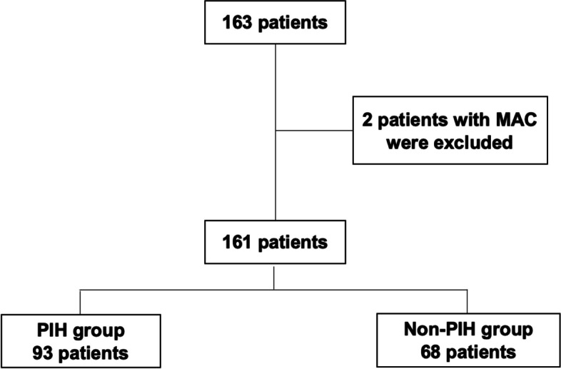
Of 163 patients, 161 were finally analyzed after exclusion (Fig. 1), with 93 and 68 patients assigned to the PIH and non-PIH groups, respectively. The prevalence of PIH in patients who underwent TAVI was 57.8% in the present study. There are not any patients who needed norepinephrine infusion at a rate >6 µg/min from anesthetic induction until 20 min post-induction.
Fig 1.

Flow chart of this study cohort. MAC: monitored anesthesia care, PIH: post-induction hypotension
Patient characteristics are shown in Table 1. There were no significant differences between the two groups. Perioperative data of the patients are presented in Table 2. The doses of propofol [PIH group vs. non-PIH group, median (25th to 75th percentile); 0.44 (0.00, 0.88) mg/kg vs. 0.00 (0.00, 0.73) mg/kg, p=0.016] and remifentanil [0.20 (0.10, 0.20) µg/kg/min vs. 0.10 (0.00, 0.20) µg/kg/min, p=0.002] were higher in the PIH group than in the non-PIH group. The doses of ketamine [0.45 (0.00, 0.74) mg/kg vs. 0.79 (0.37, 1.00) mg/kg, p<0.001] and fentanyl [0.00 (0.00, 1.02) µg/kg vs. 1.07 (0.00, 1.74) µg/kg, p<0.001] were lower in the PIH group than in the non-PIH group. In the present study, the maximum doses of fentanyl, ketamine, and propofol were 4.27 µg/kg, 2.10 mg/kg, and 1.71 mg/mg, respectively. No significant differences were observed between the type of valve, duration of surgery and anesthesia, blood loss, urine output, fluid infusion, length of ICU and hospital stay, hospital death, and perioperative stroke. Hemodynamic parameters from induction of general anesthesia until 20 min post-induction are shown in Table 3. The baseline SAP, mean arterial pressure (MAP), and diastolic arterial pressure (DAP) were significantly lower in the PIH group than in the non-PIH group. The minimum SAP, MAP, and DAP before and after tracheal intubation were significantly lower in the PIH group than in the non-PIH group. The minimum HR after tracheal intubation was significantly lower in the PIH group than that in the non-PIH group. The amounts of ephedrine and phenylephrine used during this period were significantly higher in the PIH group than in the non-PIH group.
Table 1.
Patients’ characteristics
| PIH group | Non-PIH group | P value | |
|---|---|---|---|
| N | 93 | 68 | N.A. |
| Male, n | 35 (37.6%) | 20 (29.4%) | 0.315 |
| Age, years | 84.0 (80.0, 86.0) | 84.0 (81.0, 86.0) | 0.611 |
| BMI, kg/m2 | 23.7 (21.1, 25.9) | 23.7 (20.4, 26.0) | 0.242 |
| ASA-PS | |||
| 3, n | 91 (97.8%) | 64 (94.1%) | |
| 4, n | 2 (2.2%) | 4 (5.9%) | |
| NYHA ≥3, n | 22 (23.7%) | 11 (16.2%) | 0.323 |
| STS risk score, % | 12.8 (9.6, 21.3) | 13.2 (9.9, 21.8) | 0.770 |
| Medical history | |||
| Hypertension, n | 78 (83.9%) | 54 (79.4%) | 0.535 |
| Diabetes mellitus, n | 17 (18.3%) | 18 (26.5%) | 0.248 |
| Af, n | 28 (30.1%) | 19 (27.9%) | 0.861 |
| Stroke, n | 11 (11.8%) | 15 (22.1%) | 0.088 |
| CAD, n | 31 (33.3%) | 20 (29.4%) | 0.612 |
| Antihypertensive agents | |||
| ACE inhibitor, n | 13 (14.0%) | 11 (16.2%) | 0.823 |
| AT1 blocker, n | 38 (40.9%) | 26 (38.2%) | 0.748 |
| Alpha blocker, n | 2 (2.2 %) | 2 (2.9%) | 1.000 |
| Alpha-beta blocker, n | 10 (10.8%) | 8 (11.8%) | 1.000 |
| Beta blocker, n | 14 (15.1%) | 13 (19.1%) | 0.527 |
| Calcium antagonist, n | 39 (41.9%) | 34 (50.0%) | 0.339 |
| ARNI, n | 4 (4.3%) | 2 (2.9%) | 1.000 |
| Cardiac echocardiogram | |||
| LVEF, % | 65.0 (56.0, 69.0) | 63.0 (55.5, 69.3) | 0.792 |
| mPG, mmHg | 53.0 (39.0, 65.0) | 51.0 (42.8, 63.5) | 0.828 |
| AVAi, cm2/m2 | 0.43 (0.36, 0.54) | 0.46 (0.36, 0.51) | 0.524 |
| Vmax, m/s | 4.8 (4.1, 5.2) | 4.6 (4.2, 5.1) | 0.616 |
| Moderate or severe AR, n | 63 (67.7%) | 38 (55.9%) | 0.140 |
| Moderate or severe MR, n | 52 (55.9%) | 43 (63.2%) | 0.418 |
Differences between the PIH and non-PIH groups were estimated using Fisher’s exact test for categorical variables and the Mann–Whitney test for continuous variables. Data are shown as number (percentage of each group) or median (25th–75th percentile)
BMI body mass index, ASA-PS American Society of Anesthesiologists Physical Status, NYHA, New York Heart Association functional class, STS Society of Thoracic Surgeons, Af atrial fibrillation, CAD coronary artery disease, ACE angiotensin-converting enzyme, AT1 angiotensin II type 1 receptor, ARNI angiotensin receptor neprilysin inhibitor, LVEF left ventricle ejection fraction, mPG mean pressure gradient, AVAi aortic valve area index, Vmax maximal velocity, AR aortic valve regurgitation, MR mitral valve regurgitation
Table 2.
Perioperative data of the patients
| PIH group | Non-PIH group | P value | |
|---|---|---|---|
| Dose of anesthetics for induction | |||
| Rm, mg/kg | 0.00 (0.00, 0.08) | 0.03 (0.0, 0.08) | 0.070 |
| Prop, mg/kg | 0.44 (0.00, 0.88) | 0.00 (0.00, 0.73) | 0.016* |
| Keta, mg/kg | 0.45 (0.00, 0.74) | 0.79 (0.37, 1.00) | <0.001* |
| Remi, µg/kg/min | 0.20 (0.10, 0.20) | 0.10 (0.00, 0.20) | 0.002* |
| Fent, µg/kg | 0.00 (0.00, 1.02) | 1.07 (0.00, 1.74) | <0.001* |
| Type of valve | 0.228 | ||
| SAPIEN 3, n | 73 (78.5%) | 58 (85.3%) | |
| Evolut, n | 20 (21.5%) | 10 (14.7%) | |
| Duration of surgery, h | 1.6 (1.4, 1.8) | 1.6 (1.3, 1.8) | 0.763 |
| Duration of anesthesia, h | 2.9 (2.5, 3.2) | 2.8 (2.5, 3.1) | 0.436 |
| Blood loss, g/kg/h | 0.2 (0.0, 0.3) | 0.1 (0.0, 0.3) | 0.754 |
| Urine output, mL/kg/h | 3.1 (1.8, 4.8) | 3.9 (1.7, 4.8) | 0.348 |
| Fluid infusion, mL/kg/h | 9.9 (7.9, 13.4) | 11.0 (9.2, 13.5) | 0.197 |
| Length of ICU stay, day | 2.0 (2.0, 2.0) | 2.0 (2.0, 2.0) | 0.632 |
| hospital day, day | 7.0 (6.0, 8.0) | 6.0 (6.0, 7.0) | 0.754 |
| Hospital death, n | 0 (0.0%) | 1 (1.5%) | 1.000 |
| Perioperative stroke, n | 1 (1.1%) | 1 (1.5%) | 1.000 |
Differences between the PIH and non-PIH groups were estimated using Fisher's exact test for categorical variables and the Mann–Whitney U-test for continuous variables. Data are presented as number (percentage of each group) or median (25th–75th percentile)
Rm remimazolam, Prop propofol, Keta ketamine, Remi remifentanil, Fent fentanyl
* significant difference
Table 3.
Hemodynamic parameters from induction of general anesthesia until 20 min post-induction
| PIH group | Non-PIH group | P value | |
|---|---|---|---|
| Baseline | |||
| SAP, mmHg | 143.0 (127.0, 159.0) | 158.5 (138.0, 173.3) | 0.011* |
| MAP, mmHg | 87.0 (78.3, 99.0) | 95.0 (87.0, 102.8) | 0.006* |
| DAP, mmHg | 60.0 (52.0, 67.0) | 63.5 (55.0,70.0) | 0.042* |
| HR, bpm | 71.0 (63.0, 78.0) | 70.0 (64.8, 80.0) | 0.587 |
| Pre-tracheal intubation | |||
| min SAP, mmHg | 92.0 (72.0, 106.0) | 115.0 (101.0, 128.8) | <0.001* |
| min MAP, mmHg | 61.0 (500, 67.0) | 71.0 (64.3, 79.3) | <0.001* |
| min DAP, mmHg | 43.0 (37.0, 49.0) | 51.0 (45.0, 55.0) | <0.001* |
| min HR, bpm | 61.0 (53.0, 69.0) | 63.0 (56.0, 70.5) | 0.221 |
| Post-tracheal intubation | |||
| min SAP, mmHg | 75.0 (66.0, 82.0) | 104.0 (95.0, 116.3) | <0.001* |
| min MAP, mmHg | 50.0 (43.3, 52.0) | 67.0 (59.5, 75.0) | <0.001* |
| min DAP, mmHg | 36(32.0, 40.0) | 48.0 (41.0, 55.0) | 0.001* |
| min HR, bpm | 56.0 (49.0, 64.0) | 61.0 (50.0, 70.0) | 0.039* |
| Vasopressors use | |||
| Ephedrine, mg | 0.0 (0.0, 4.0) | 0.0 (0.0, 0.0) | 0.001* |
| Phenylephrine, mg | 0.1 (0.0, 0.2) | 0.0 (0.0, 0.05) | <0.001* |
| Noradrenaline, µg | 0.0 (0.0, 0.0) | 0.0 (0.0, 0.0) | 0.194 |
| Atropin, mg | 0.0 (0.0, 0.0) | 0.0 (0.0, 0.0) | 0.293 |
Differences between the PIH and non-PIH groups were estimated using Fisher's exact test for categorical variables and the Mann–Whitney U-test for continuous variables. Data are presented as number (percentage of each group) or median (25th–75th percentile)
SAP systolic arterial pressure, MAP mean arterial pressure, DAP diastolic arterial pressure, HR heart rate, min minimal
* Sgnificant difference
The ROC curves revealed that the cut-off values for STS risk score and baseline MAP to predict the development of PIH were and 6.5% and 90 mmHg, respectively. The AUC values of the STS risk score and MAP were 0.515 and 0.626, respectively, indicating low accuracies.
The result of the multivariable logistic regression analysis to develop a predictive model for PIH in patients undergoing TAVI is shown in Table 4. Baseline MAP ≥90 mmHg (aOR: 0.413; 95% CI: 0.193–0.887; p=0.023) and higher doses of fentanyl (per 1-µg/kg increase, aOR: 0.619, 95% CI: 0.418–0.915; p=0.016) and ketamine (per 1-mg/kg increase, aOR: 0.163, 95% CI: 0.062–0.430; p=0.002) for anesthetic induction were significantly associated with a lower risk of the development of PIH. A higher dose of propofol (per 1-mg/kg increase, aOR: 3.240, 95% CI: 1.320–7.920; p=0.010) for anesthetic induction was significantly associated with a higher risk of the development of PIH. The area under the curve (AUC) for this model was 0.802.
Table 4.
Predictive model for post-induction hypotension in patients undergoing transcatheter aortic valve implantation
| aOR | 95%CI | P value | |
|---|---|---|---|
| (Intercept) | 4.960 | 1.750, 14.00 | 0.003* |
| STS risk score ≥6.5% | 3.030 | 0.908, 10.10 | 0.071 |
| mPG ≥60 mmHg | 1.610 | 0.724, 3.950 | 0.242 |
| Baseline MAP ≥90 mmHg | 0.413 | 0.193, 0.887 | 0.023* |
| Dose of rm for induction (per 1 mg/kg increase) | 1.070 | 0.009, 126.0 | 0.977 |
| Dose of prop for induction (per 1 mg/kg increase) | 3.240 | 1.320, 7.920 | 0.010* |
| Dose of fent for induction (per 1 µg/kg increase) | 0.619 | 0.418, 0.915 | 0.016* |
| Dose of remi for induction (per 1 µg/kg/min increase) | 0.485 | 0.137, 1.710 | 0.261 |
| Dose of keta for induction (per 1 mg/kg increase) | 0.163 | 0.062, 0.430 | 0.002* |
Multivariate logistic regression analyses were performed to develop predictive models for post-induction hypotension in patients who underwent transcatheter aortic valve implantation. No variance inflation factor value was up to 10, indicating no collinearity in the model. The area under the curve was 0.802 (95% CI: 0.732, 0.873)
aOR adjusted odds ratio, CI confidence interval, STS Society of Thoracic Surgeons, mPG mean pressure gradient, MAP mean arterial pressure, Rm remimazolam, Prop propofol, Keta ketamine, Remi remifentanil, Fent fentanyl
* significant difference
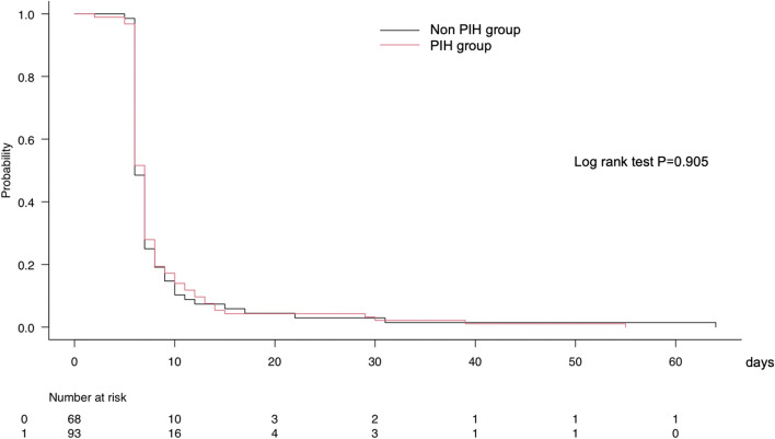
Figure 2 shows the results of the Kaplan–Meier curve analysis with log-rank test to compare the length of hospital stay between the PIH and non-PIH groups. There was no significant difference in the length of hospital stay between the two groups.
Fig 2.
Kaplan–Meier curve analysis with log-rank test to compare the probability of hospital stay among the patients with and without PIH. PIH: post-induction hypotension; *, statistical significance
Discussion
The present study developed a predictive model for PIH in TAVI patients. A baseline MAP ≥90 mmHg and higher doses of fentanyl and ketamine for anesthetic induction were significantly associated with a lower risk of developing PIH. A higher dose of propofol for anesthetic induction is significantly associated with a higher risk of developing PIH. The AUC value of this model was 0.802, indicating that its discrimination ability was good. There were no significant differences in the length of ICU and hospital stays between the PIH and non-PIH groups.
To the best of our knowledge, this is the first study to develop a predictive model for PIH in TAVI patients. In the present model, STS risk score was used as a variable for evaluating the preoperative condition. STS risk score was calculated using the patients' background such as age, sex, height, body weight, laboratory data, medical history, chronic medication, and cardiac function, and has been used to predict the mortality risk and complications after adult cardiac surgery [10]. Thus, we were able to adjust for many patient characteristics in the model, despite the small sample size. Additionally, the STS risk score is reported to be associated with mortality after TAVI [11]. However, the STS risk score was not a significant predictor of PIH in patients undergoing TAVI. Indeed, the short-term outcomes after TAVI were not significantly different between the PIH and non-PIH groups in the present study. However, as the 95% CI and p-value of STS risk score ≥6.5% were 0.908–10.10 and 0.071, respectively, increasing the sample size may change the results.
Consistent with the results of previous studies [4–6], baseline arterial pressure was also associated with PIH in this study. Although these studies included SAP as the baseline arterial pressure in the model, the present study included MAP as the baseline arterial pressure in the model. Generally, as patients who undergo TAVI are prone to have aortic regurgitation, the baseline arterial pressure should be assessed not only by SAP but also by DAP. Thus, MAP was included in the model and was an independent predictor of PIH in patients undergoing TAVI. In our institution, all antihypertensive agents were continued on the day of surgery because some of them are also used to treat heart failure. Therefore, caution is needed when interpreting this result.
In the present study, a higher dose of propofol for anesthetic induction was significantly associated with a higher risk of PIH. This result was consistent with that of previous studies [4, 12]. Propofol anesthesia is more likely to cause hypotension than remimazolam in ASA I or II patients [13]. Additionally, in the present study, higher doses of fentanyl and ketamine for anesthetic induction were significantly associated with a lower risk of PIH. A previous study showed that fentanyl with propofol induction was less likely to cause PIH than remifentanil with propofol induction [14]. However, an excessive amount of fentanyl for anesthetic induction can cause PIH. Indeed, a previous study showed that a higher dose of fentanyl can cause PIH [4]. The previous study showed that 1.51–5.0 µg/kg and >5.0 µg/kg of fentanyl for induction were more likely to cause PIH than 0–1.5 µg/kg of fentanyl for induction. In this study, the maximum dose of fentanyl was 4.27 µg/kg and the patient who was infused this dose of fentanyl was in the non-PIH group. The median doses (25th to 75th percentile) of fentanyl for induction were 0.00 (0.00, 1.02) µg/kg in the PIH group and 1.07 (0.00, 1.74) µg/kg in the non-PIH group, which means the patients in this study was treated with relatively low dose of fentanyl. Thus, clinicians need caution when interpreting this result about fentanyl. Ketamine is effective in maintaining hemodynamic stability during anesthetic induction [15]. However, ketamine can cause tachycardia and increase oxygen consumption owing to increased sympathetic stimulation [16, 17]. As these effects of ketamine are not preferable for patients with severe AS, it should be used carefully and with other anesthetics. Additionally, tracheal intubation under insufficient depth of anesthesia also causes sympathetic stimulation and adversely affects hemodynamics in patients with AS. Therefore, we do not recommend anesthetic induction with only ketamine and fentanyl in patients who undergo TAVI, because it tends to be insufficient in terms of the depth of anesthesia. Thus, it is important to use ketamine and fentanyl in addition to propofol or remimazolam for a well-balanced anesthetic induction.
This study has several limitations. First, as this was a single-center, retrospective observational study with a small sample size, selection bias and undetected confounding factors may have affected the results. Indeed, as the patients’ background was adjusted for only the STS risk score, we were not able to find any other variables that were associated with PIH except for baseline MAP. Second, as the choice of anesthetics was not standardized and was up to the anesthesiologist’s discretion, the development of PIH may have depended on the competence of the anesthesiologist. However, all cases were managed or supervised by anesthesia specialists. Third, as we did not follow the patients’ courses after hospital discharge, postoperative complications after hospital discharge and long-term outcomes could not be assessed. Although the prevalence of postoperative complications was not significantly different between the PIH and non-PIH groups, PIH might affect the long-term outcomes.
In conclusion, this study developed a predictive model for PIH in patients undergoing TAVI. A baseline MAP ≥90 mmHg and higher doses of fentanyl and ketamine for anesthetic induction were significantly associated with a lower risk of PIH. Of course, excessive amount of these two drugs should be avoided. A higher dose of propofol for anesthetic induction is significantly associated with a higher risk of PIH. This model may be helpful for anesthesiologists in preventing PIH in patients undergoing TAVI. However, as this was a single-center, retrospective observational study with a small sample size, these results should be treated with caution and further studies are required to strengthen this evidence.
Acknowledgements
We would like to thank Editage (www.editage.com) for English language editing.
Abbreviations
- AS
Aortic stenosis
- SAVR
Surgical aortic valve replacement
- TAVI
Transcatheter aortic valve implantation
- PIH
Post-induction hypotension
- ASA-PS
American Society of Anesthesiologists physical status
- MAC
Monitored anesthesia care
- SAP
Systolic arterial pressure
- NYHA
New York Heart Association
- STS
Society of Thoracic Surgeons
- HR
Heart rate
- ICU
Intensive care unit
- ROC
Receiver operating characteristic
- VIF
Variance inflation factor
- AUC
Area under the curve
- OR
Odds ratios
- CI
Confidence interval
- MAP
Mean arterial pressure
- DAP
Diastolic arterial pressure
Authors’ contributions
KN, SU, HK, and DT collected data. DT analyzed the data and drafted the manuscript. TK and KH made critical revisions. All authors have read and approved the final manuscript for submission.
Funding
None.
Availability of data and material
Data will be made available on reasonable request.
Declarations
Ethics approval and consent to participate
This single-center, retrospective observational study was approved by the Ethics Committee of the Hirosaki University Graduate School of Medicine, Hirosaki, Japan and publicized on our department homepage using an opt out approach that participants are included in the research unless they give their express decision to be excluded. The requirement for written informed consent from each patient was waived because of the retrospective nature of the study, and the Ethics Committee approved the waiver.
Consent for publication
We used an opt out approach. The requirement for written informed consent from each patient was waived because of the retrospective nature of the study, and the Ethics Committee approved the waiver.
Conmpeting interests
None.
Footnotes
Publisher's Note
Springer Nature remains neutral with regard to jurisdictional claims in published maps and institutional affiliations.
References
- 1.Baumgartner H, Falk V, Bax JJ, et al. ESC/EACTS Guidelines for the management of valvular heart disease. Eur Heart J. 2017;38(36):2739–91. doi: 10.1093/eurheartj/ehx391. [DOI] [PubMed] [Google Scholar]
- 2.Smith CR, Leon MB, Mack MJ, Miller DC, Moses JW, Svensson LG, Tuzcu EM, Webb JG, Fontana GP, Makkar RR, Williams M, Dewey T, Kapadia S, Babaliaros V, Thourani VH, Corso P, Pichard AD, Bavaria JE, Herrmann HC, Akin JJ, Anderson WN, Wang D, Pocock SJ. PARTNER Trial Investigators. Transcatheter versus surgical aortic-valve replacement in high-risk patients. N Engl J Med. 2011;364(23):2187–98. doi: 10.1056/NEJMoa1103510. [DOI] [PubMed] [Google Scholar]
- 3.The UK TAVI Trial Investigators Effect of transcatheter aortic valve implantation vs surgical aortic valve replacement on all-cause mortality in patients with aortic stenosis: a randomized clinical trial. JAMA. 2022;327(19):1875–87. doi: 10.1001/jama.2022.5776. [DOI] [PMC free article] [PubMed] [Google Scholar]
- 4.Reich DL, Hossain S, Krol M, Baez B, Patel P, Bernstein A, Bodian CA. Predictors of hypotension after induction of general anesthesia. Anesth Analg. 2005;101(3):622–8. doi: 10.1213/01.ANE.0000175214.38450.91. [DOI] [PubMed] [Google Scholar]
- 5.Südfeld S, Brechnitz S, Wagner JY, Reese PC, Pinnschmidt HO, Reuter DA, Saugel B. Post-induction hypotension and early intraoperative hypotension associated with general anaesthesia. Br J Anaesth. 2017;119(1):57–64. doi: 10.1093/bja/aex127. [DOI] [PubMed] [Google Scholar]
- 6.Jor O, Maca J, Koutna J, Gemrotova M, Vymazal T, Litschmannova M, Sevcik P, Reimer P, Mikulova V, Trlicova M, Cerny V. Hypotension after induction of general anesthesia: occurrence, risk factors, and therapy. A prospective multicentre observational study. J Anesth. 2018;32(5):673–80. doi: 10.1007/s00540-018-2532-6. [DOI] [PubMed] [Google Scholar]
- 7.Green RS, Butler MB. Postintubation hypotension in general anesthesia: a retrospective analysis. J Intensive Care Med. 2016;31(10):667–75. doi: 10.1177/0885066615597198. [DOI] [PubMed] [Google Scholar]
- 8.Bohbot Y, Kowalski C, Rusinaru D, Ringle A, Marechaux S, Tribouilloy C. Impact of mean transaortic pressure gradient on long-term outcome in patients with severe aortic stenosis and preserved left ventricular ejection fraction. J Am Heart Assoc. 2017;6(6):e005850. doi: 10.1161/JAHA.117.005850. [DOI] [PMC free article] [PubMed] [Google Scholar]
- 9.Tribouilloy C, Rusinaru D, Bohbot Y, Maréchaux S, Vanoverschelde JL, Enriquez-Sarano M. How should very severe aortic stenosis be defined in asymptomatic individuals? J Am Heart Assoc. 2019;8(3):e011724. doi: 10.1161/JAHA.118.011724. [DOI] [PMC free article] [PubMed] [Google Scholar]
- 10.Ad N, Holmes SD, Patel J, Pritchard G, Shuman DJ, Halpin L. Comparison of EuroSCORE II, original EuroSCORE, and The Society of Thoracic Surgeons risk score in cardiac surgery patients. Ann Thorac Surg. 2016;102(2):573–9. doi: 10.1016/j.athoracsur.2016.01.105. [DOI] [PubMed] [Google Scholar]
- 11.Ishizu K, Shirai S, Isotani A, Hayashi M, Kawaguchi T, Taniguchi T, Ando K, Yashima F, Tada N, Yamawaki M, Naganuma T, Yamanaka F, Ueno H, Tabata M, Mizutani K, Takagi K, Watanabe Y, Yamamoto M, Hayashida K. OCEAN-TAVI Investigators. Long-term prognostic value of the Society of Thoracic Surgery risk score in patients undergoing transcatheter aortic valve implantation (from the OCEAN-TAVI Registry) Am J Cardiol. 2021;149:86–94. doi: 10.1016/j.amjcard.2021.03.027. [DOI] [PubMed] [Google Scholar]
- 12.Schonberger RB, Dai F, Michel G, Vaughn MT, Burg MM, Mathis M, Kheterpal S, Akhtar S, Shah N, Bardia A. Association of propofol induction dose and severe pre-incision hypotension among surgical patients over age 65. J Clin Anesth. 2022;80:110846. doi: 10.1016/j.jclinane.2022.110846. [DOI] [PMC free article] [PubMed] [Google Scholar]
- 13.Doi M, Morita K, Takeda J, Sakamoto A, Yamakage M, Suzuki T. Efficacy and safety of remimazolam versus propofol for general anesthesia: a multicenter, single-blind, randomized, parallel-group, phase IIb/III trial. J Anesth. 2020;34(4): 543–53. doi: 10.1007/s00540-020-02788-6. [DOI] [PubMed] [Google Scholar]
- 14.Wilhelm W, Biedler A, Huppert A, Kreuer S, Bücheler O, Ziegenfuss T, Larsen R. Comparison of the effects of remifentanil or fentanyl on anaesthetic induction characteristics of propofol, thiopental or etomidate. Eur J Anaesthesiol. 2002;19(5):350–6. doi: 10.1017/s026502150200056x. [DOI] [PubMed] [Google Scholar]
- 15.Hosseinzadeh H, Eidy M, Golzari SE, Vasebi M. Hemodynamic stability during induction of anesthesia in elderly patients: propofol + ketamine versus propofol + etomidate. J Cardiovasc Thorac Res. 2013;5(2):51–4. doi: 10.5681/jcvtr.2013.011. [DOI] [PMC free article] [PubMed] [Google Scholar]
- 16.Green SM, Roback MG, Kennedy RM, Krauss B. Clinical practice guideline for emergency department ketamine dissociative sedation: 2011 update. Ann Emerg Med. 2011;57(5):449–61. doi: 10.1016/j.annemergmed.2010.11.030. [DOI] [PubMed] [Google Scholar]
- 17.Stefánsson T, Wickström I, Haljamäe H. Hemodynamic and metabolic effects of ketamine anesthesia in the geriatric patient. Acta Anaesthesiol Scand. 1982;26(4):371–7. doi: 10.1111/j.1399-6576.1982.tb01785.x. [DOI] [PubMed] [Google Scholar]
Associated Data
This section collects any data citations, data availability statements, or supplementary materials included in this article.
Data Availability Statement
Data will be made available on reasonable request.


