Abstract
The haloalkane-degrading bacteria Rhodococcus rhodochrous NCIMB13064, Pseudomonas pavonaceae 170, and Mycobacterium sp. strain GP1 share a highly conserved haloalkane dehalogenase gene (dhaA). Here, we describe the extent of the conserved dhaA segments in these three phylogenetically distinct bacteria and an analysis of their flanking sequences. The dhaA gene of the 1-chlorobutane-degrading strain NCIMB13064 was found to reside within a 1-chlorobutane catabolic gene cluster, which also encodes a putative invertase (invA), a regulatory protein (dhaR), an alcohol dehydrogenase (adhA), and an aldehyde dehydrogenase (aldA). The latter two enzymes may catalyze the oxidative conversion of n-butanol, the hydrolytic product of 1-chlorobutane, to n-butyric acid, a growth substrate for many bacteria. The activity of the dhaR gene product was analyzed in Pseudomonas sp. strain GJ1, in which it appeared to function as a repressor of dhaA expression. The 1,2-dibromoethane-degrading strain GP1 contained a conserved DNA segment of 2.7 kb, which included dhaR, dhaA, and part of invA. A 12-nucleotide deletion in dhaR led to constitutive expression of dhaA in strain GP1, in contrast to the inducible expression of dhaA in strain NCIMB13064. The 1,3-dichloropropene-degrading strain 170 possessed a conserved DNA segment of 1.3 kb harboring little more than the coding region of the dhaA gene. In strains 170 and GP1, a putative integrase gene was found next to the conserved dhaA segment, which suggests that integration events were responsible for the acquisition of these DNA segments. The data indicate that horizontal gene transfer and integrase-dependent gene acquisition were the key mechanisms for the evolution of catabolic pathways for the man-made chemicals 1,3-dichloropropene and 1,2-dibromoethane.
Synthetic haloalkanes form an important class of environmental pollutants because of their widespread use in industry and agriculture, persistence in the environment, and potential carcinogenicity. The poor biodegradability of these chemicals is mainly due to the inability of microorganisms to effectively metabolize these unnatural compounds. Nevertheless, microbial communities exposed to synthetic haloalkanes often respond by expressing specific pathways that degrade these molecules in order to exploit them as growth substrates. Since synthetic haloalkanes are xenobiotic compounds of recent origin, the way in which genes have been assembled to form functional catabolic pathways is an interesting subject for studying microbial evolution and gene transfer.
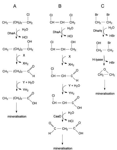
Rhodococcus rhodochrous NCIMB13064, isolated in the United Kingdom from a soil sample obtained from an industrial site which had previously been exposed to chlorinated alkanes, is capable of utilizing 1-chlorobutane and several other haloalkanes as the sole carbon and energy source (9). The cleavage of the carbon-halogen bond in 1-chlorobutane, which is the key step in its catabolism, is catalyzed by an inducible hydrolytic haloalkane dehalogenase (DhaA) and results in the formation of n-butanol. This intermediate is subsequently oxidized in two steps to n-butyric acid (Fig. 1), which can serve as a growth substrate for many bacteria. The haloalkane dehalogenase gene (dhaA) was shown to be located on the autotransmissible plasmid pRTL1 (22) and was cloned and sequenced (23).
FIG. 1.
Catabolic pathways for haloaliphatics. (A) 1-Chlorobutane in R. rhodochrous NCIMB13064. (B) 1,3-Dichloropropene in P. pavonaceae 170. (C) 1,2-Dibromoethane in Mycobacterium sp. strain GP1. Abbreviations: DhaA and DhaAf, haloalkane dehalogenases; H-lyase, halohydrin halogen-halide lyase; CaaD, 3-chloroacrylic acid dehalogenase; X, alcohol dehydrogenase cofactor; Y, aldehyde dehydrogenase cofactor.
The gram-negative 1,3-dichloropropene-utilizing bacterium Pseudomonas pavonaceae 170, isolated in The Netherlands from soil that was repeatedly treated with the nematocidic soil fumigant 1,3-dichloropropene, was shown by PCR amplification to possess a haloalkane dehalogenase gene identical to the dhaA gene of the gram-positive strain NCIMB13064 (35). In contrast to the inducible production of DhaA in strain NCIMB13064, DhaA is constitutively produced in strain 170 and catalyzes the first step in the degradation of 1,3-dichloropropene (Fig. 1).
Recently, we demonstrated that the 1,2-dibromoethane-degrading organism Mycobacterium sp. strain GP1, which was isolated by prolonged batch enrichment from a mixed bacterial culture, also contains a haloalkane dehalogenase gene (dhaAf) that is very similar to the dhaA gene found in strain NCIMB13064 (36). The haloalkane dehalogenase encoded by dhaAf is identical to DhaA, except for three amino acid substitutions and a 14-amino-acid extension at the C terminus. Nucleotide sequence analysis indicated that the dhaAf gene was formed by a fusion of a dhaA gene with the last 42 nucleotides of a hheB gene, which encodes a haloalcohol dehalogenase (50). The haloalkane dehalogenase (DhaAf) is constitutively produced in strain GP1 and catalyzes the conversion of 1,2-dibromoethane to 2-bromoethanol, which is further metabolized via ethylene oxide (Fig. 1).
The presence of a highly conserved dhaA gene in the three phylogenetically different organisms R. rhodochrous NCIMB13064, P. pavonaceae 170, and Mycobacterium sp. strain GP1 suggests that dhaA has been distributed among these organisms by horizontal transfer. To determine the size of the transferred DNA fragments and to identify the mechanisms that were involved in the distribution process, we have analyzed the DNA regions flanking the dhaA gene in these three haloalkane-utilizing strains. The results suggest that the haloalkane dehalogenase gene regions of strains 170 and GP1 originate from a 1-chlorobutane catabolic gene cluster similar to the one that is present on plasmid pRTL1 in strain NCIMB13064. Horizontal gene transfer and integrase-dependent acquisition of existing DNA fragments harboring the dhaA gene were probably the key steps during the evolution of 1,3-dichloropropene- and 1,2-dibromoethane-degradative pathways. Furthermore, the constitutive expression of dhaA in strains 170 and GP1, in contrast to the inducible expression of dhaA in strain NCIMB13064, is explained by the absence or inactivation of the regulatory gene dhaR.
MATERIALS AND METHODS
Materials.
Restriction enzymes, Taq DNA polymerase, T4 DNA ligase, the DNA-packaging kit, and materials used for Southern blot hybridization were purchased from Boehringer Mannheim (Mannheim, Germany). 1,2-Dibromoethane was supplied by Acros Organics (Geel, Belgium). The oligonucleotides used as primers were supplied by Eurosequence BV (Groningen, The Netherlands).
Bacterial strains, plasmids, and growth conditions.
The characteristics of the 1,3-dichloropropene-degrading bacterium P. pavonaceae 170, formerly known as Pseudomonas cichorii 170 (44), and of the 1,2-dibromoethane-degrading organism Mycobacterium sp. strain GP1 are given elsewhere (35, 36). R. rhodochrous NCIMB13064 contains the large catabolic plasmid pRTL1 (22) and is able to use 1-chlorobutane as the sole carbon and energy source (9). Escherichia coli strains HB101 (7) and JM101 (49) and plasmid pBluescript SK(−) (Stratagene, Leusden, The Netherlands) were used for routine cloning experiments. E. coli HB101(pRK600) (13) was the helper strain used for mobilizing pLAFR3/5- and pDSK519-derived plasmids in triparental matings with the recipient Pseudomonas sp. strain GJ1 (17). Plasmid pDSK519 and cosmids pLAFR3 and pLAFR5 are mobilizable broad-host-range vectors (19, 39). Recombinant cosmid pLTL1k contains the 1-chlorobutane catabolic gene cluster of strain NCIMB13064 (23) and was used as template for DNA sequencing and as the source for some cloning and expression experiments described here. Recombinant cosmids pGP1-4B5, which contains the dhaAf gene region of strain GP1 (36), and pPC33, which contains the dhaA gene region of strain 170 (this study), were used as templates for DNA sequencing.
R. rhodochrous NCIMB13064 and Mycobacterium sp. strain GP1 were grown at 30°C on nutrient broth or on mineral medium (17) supplemented with 5 mM n-propanol, respectively. Pseudomonas and E. coli strains were grown at 30°C on Luria-Bertani medium (38). When required, Difco agar (15 g/liter) was added to the medium. LBZ medium, used for qualitative dehalogenase activity determination, was solid Luria-Bertani medium without NaCl. Antibiotics were added in the following amounts: ampicillin, 100 μg/ml; kanamycin, 50 μg/ml; chloramphenicol, 50 μg/ml; and tetracycline, 12.5 μg/ml. When necessary, media were supplemented with 5-bromo-4-chloro-3-indolyl-β-d-galactopyranoside (X-Gal; 40 μg/ml) and isopropyl-β-d-thiogalactopyranoside (IPTG; 0.4 mM).
DNA techniques.
General procedures for cloning, transformation, and DNA manipulation were performed essentially as described by Sambrook et al. (38). Triparental matings were carried out as described by Janssen et al. (18). Isolation of total genomic DNA from strains NCIMB13064, 170, and GP1 was performed according to the phenol extraction procedure described previously (35). The IS2112- and IS1071-specific probes used in hybridization experiments were obtained by PCR amplification by using primers and conditions as previously described (21, 48). DNA fragments were purified by using the Qiaquick PCR purification kit (Qiagen). Southern blot hybridization experiments were performed as described previously (21).
Crude extracts and dehalogenase assays.
Pseudomonas and E. coli cells were harvested in the late-exponential growth phase by centrifugation (10 min at 10,000 × g), were washed with 1 volume of 50 mM Tris-sulfate buffer (pH 8.2), and were disrupted at 4°C in an appropriate amount of this buffer by sonication (10 s per ml of suspension at a 70 W output in a Vibra cell sonicator). A crude extract was obtained by centrifugation (45 min at 16,000 × g).
Haloalkane dehalogenase activities were measured by incubating an appropriate amount of cell extract with 3 ml of 5 mM 1,2-dibromoethane in 50 mM Tris-sulfate buffer (pH 8.2) at 30°C. Halide liberation was monitored colorimetrically as described previously (20). All dehalogenase activities are expressed as units per miligram; 1 U was defined as the amount of enzyme that catalyzes the production of 1 μmol of halide per min. Protein concentrations were estimated with Coomassie brillant blue by using bovine serum albumin as the standard. Enzyme assays were carried out twice, and the differences in specific activities were less than 10%.
Cloning of the dhaA gene region from P. pavonaceae 170.
A genomic library of P. pavonaceae 170 was constructed by using cosmid vector pLAFR3 according to a previously described procedure (39). Individual cosmid clones were screened for dehalogenase activity by monitoring halide production upon incubation with 1,2-dibromoethane. Out of 5,000 E. coli HB101 clones tested, two dehalogenase-positive clones were found. Recombinant cosmids pPC8 and pPC33 encoding haloalkane dehalogenase were isolated from these two HB101 clones and were digested with BamHI. Both cosmids had a 3-kb BamHI fragment in common. The 3-kb BamHI fragment of pPC33 was ligated into the BamHI site of pBluescript SK(−). The ligation mixture was used to transform cells of E. coli JM101, and transformants were plated on LBZ plates containing ampicillin, X-Gal, and IPTG. Ampicillin-resistant white colonies displaying haloalkane dehalogenase activity with 1,2-dibromoethane were selected. Plasmid DNA (pSK45) of one of these colonies was isolated and used as template for DNA sequencing. The nucleotide sequences of DNA regions flanking the 3-kb BamHI fragment were determined by using cosmid pPC33 directly as a template for DNA sequencing.
Nucleotide sequencing and analysis.
DNA sequencing was performed as previously described (21, 36). The nucleotide sequence data were analyzed by using the programs supplied in the DNASTAR software package (DNASTAR Inc., Madison, Wis.) or those supplied in the PC/GENE software package (Genofit, Geneva, Switzerland). Searches for nucleotide and amino acid sequence similarities were carried out by using the BLAST program (3) and the DDBJ, EMBL, and GenBank databases. Protein sequences were aligned by using CLUSTAL W (41), and alignments of nucleotide sequences were made by using LALIGN (Institut de Génétique Humaine, Montpellier, France).
Nucleotide sequence accession numbers.
The nucleotide sequence data of the haloalkane dehalogenase gene regions from R. rhodochrous NCIMB13064, P. pavonaceae 170, and Mycobacterium sp. strain GP1 have been submitted to the DDBJ, EMBL, and GenBank databases under accession no. L49435, AJ250371, and AJ250372, respectively.
RESULTS AND DISCUSSION
Sequence analysis of the dhaA gene region from R. rhodochrous NCIMB13064.
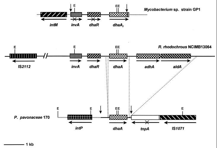
The dhaA gene of the 1-chlorobutane-degrading bacterium R. rhodochrous NCIMB13064 is located on the 100-kb plasmid pRTL1 (23). A 13.1-kb DNA fragment of pRTL1, including a 8.2-kb region upstream and a 4-kb region downstream of dhaA, was sequenced (Fig. 2). The dhaA gene sequence and the analysis of insertion element IS2112, which is located approximately 6 kb upstream of dhaA (Fig. 2), were described previously (21, 23).
FIG. 2.
Organization of the haloalkane dehalogenase gene regions of R. rhodochrous NCIMB13064, P. pavonaceae 170, and Mycobacterium sp. strain GP1. Genes are shown as hatched boxes, and arrows indicate the direction of transcription. Identical hatching indicates identical genes. Incomplete ORFs are indicated by a cross through the arrow. The borders of the conserved DNA segments in strains 170 and GP1, which are highly similar to segments of the 1-chlorobutane-catabolic gene cluster of strain NCIMB13064, are indicated by vertical arrows. The two deletions within the conserved region of strain 170 are indicated by vertical dotted lines. The 42-nucleotide extension of the dhaA ORF in strain GP1, which is the result of a fusion between the dhaA segment and haloalcohol dehalogenase HheB encoding DNA (36), is indicated by a shaded box. EcoRI restriction sites (E) are shown.
Two complete open reading frames (ORFs), designated invA and dhaR, were found upstream of dhaA (Fig. 2 and 3). InvA shares extensive similarity with proteins belonging to the invertase family of site-specific recombinases. The inversion reaction is a site-specific recombination between inverted repeat sequences which flank the invertable DNA fragment and is carried out by invertases. Several examples of invertable DNA that can serve as a genetic switch between the expression of alternative sets of genes have been described (14). InvA is most similar to the invertases Pin of E. coli (34) and Hin of Salmonella enterica serovar Typhimurium (51) (Table 1). The high amino acid sequence identity with these DNA invertases implies a common phylogenetic origin, although invertase action has yet to be demonstrated for InvA.
FIG. 3.
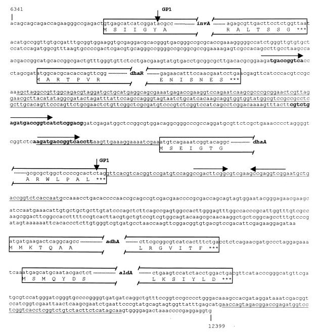
Partial nucleotide sequence of the 1-chlorobutane-degradative gene cluster found in pRTL1 of R. rhodochrous NCIMB13064. The borders of the conserved DNA segment in Mycobacterium sp. strain GP1 are indicated by vertical arrows. DNA stretches identical to the nucleotide sequences flanking the dhaA gene in P. pavonaceae 170 are underlined. Inverted and directed repeats are indicated by horizontal arrows above the sequence. Palindromic sequences are shown in boldface.
TABLE 1.
Localization of genes, the corresponding gene products, and identities with other proteins
| Strain | Gene | Region of nucleotide sequencea | Gene product | Deduced mol wt (kDa) | % Identity with other proteins (references) |
|---|---|---|---|---|---|
| NCIMB13064 | invA | 6369–6929 | Putative invertase | 20.6 | Pin, 50 (34); Hin, 48 (51) |
| dhaR | 7209–7815 | Repressor-type regulator | 22.7 | ArpA, 20 (29); BarA, 20 (28); FarA, 20 (45) | |
| dhaA | 8244–9125 | Haloalkane dehalogenase | 33.2 | See reference 23 | |
| adhA | 9543–10655 | Putative alcohol dehydrogenase | 38.8 | AdhD, 50 (8); AdhB, 48 (8); AdhI, 34 (33); AdhS, 33 (32) | |
| aldA | 10687–12135 | Putative aldehyde dehydrogenase | 50.7 | Rv0223C, 47 (8); CymC, 38 (11); DhaL, 38 (46); AldA, 35 (8) | |
| 170 | intP | 587–1726b | Putative integrase | 40.4 | See Fig. 6A |
| dhaA | 2641–3522 | Haloalkane dehalogenase | 33.2 | See reference 23 | |
| tnpAΔ | 3879–5024b | Disrupted transposase | TnpA, 51c | ||
| tnpA of IS1071 | 5355–>6900b | Transposase | 108.4 | See reference 26 | |
| GP1 | intM | 82–1437b | Putative integrase | 49.1 | See Fig. 6B |
| dhaAf | 3482–4405 | Haloalkane dehalogenase | 34.7 | See reference 36 |
The nucleotide sequence numbers refer to the sequences of the haloalkane dehalogenase gene regions of strains NCIMB13064, 170, and GP1 that have been deposited at the DDBJ, EMBL, and GenBank databases under accession no. L49435, AJ250371, and AJ250372, respectively.
Located on the complementary DNA strand when compared to the haloalkane dehalogenase gene.
Accession no. AF028594.
Database searches with the deduced amino acid sequence of the dhaR gene revealed no proteins with significant similarity to the entire sequence of the dhaR product. However, DhaR contains a region near the N terminus which resembles the helix-turn-helix (HTH) DNA binding motifs of a number of transcriptional regulators (10, 30). Considerable similarity was found with the HTH motifs of the Streptomyces autoregulator receptors ArpA, BarA, and FarA (Fig. 4), which were proposed to act as repressor-type regulators for secondary metabolism and morphogenesis (28, 29, 45). The overall sequence similarity to these three Streptomyces regulators is low (Table 1).
FIG. 4.
Alignment of the N-terminal amino acid sequences of FarA, BarA, ArpA, and DhaR. Amino acids conserved in all four proteins are indicated by asterisks, whereas amino acids conserved in three out of four proteins are indicated by dots. The sequences corresponding to helix 2 and helix 3 in the HTH DNA binding motif are boxed.
Cells of strain NCIMB13064 grown in n-butanol do not possess DhaA activity, but growth on 1-chlorobutane induces the expression of the dehalogenase DhaA (9). The presence of a region resembling the HTH DNA binding motifs in DhaR and the localization of its encoding ORF directly upstream of dhaA suggest that DhaR modulates transcription of dhaA by binding directly to its promoter region in response to 1-chlorobutane. Sequence analysis of the dhaR-dhaA intergenic region revealed the presence of two identical directly repeated sequences of 13 bp (Fig. 3). The repeated sequences contain a 10-bp palindromic sequence (TGACCGGTCA) and are both part of a larger (imperfect) palindrome. The same 13-bp repeated sequence was also found upstream of dhaR (Fig. 3). The presence of these putative binding sites for DhaR upstream of both the dhaR and dhaA genes suggests that this protein regulates its own expression and that of DhaA. However, promoter sequences for Rhodococcus genes are not well characterized (24), and the promoter responsible for dhaA expression has not been identified.
Two ORFs, designated adhA and aldA, were found downstream of dhaA (Fig. 2 and 3). AdhA shares considerable similarity with several proteins that belong to the alcohol dehydrogenase subgroup which contains the NAD(P)- and zinc-dependent long chain alcohol dehydrogenases (Table 1). The highest similarity was found with putative alcohol dehydrogenases from Mycobacterium tuberculosis H37Rv (Table 1). Structural and catalytic residues that are conserved among almost all microbial alcohol dehydrogenases (37) are also present in AdhA. AldA shares considerable similarity with putative aldehyde dehydrogenases from M. tuberculosis H37Rv and with several known NAD(P)-dependent aldehyde dehydrogenases (Table 1). The cysteine residue (C302, family position) that is strictly conserved among all available sequences of the aldehyde dehydrogenase superfamily, and is proposed to be the active site nucleophile (6, 12), is also conserved in AldA.
Curragh and coworkers (9) showed that 1-chlorobutane was metabolized by strain NCIMB13064 via n-butanol and n-butyric acid (Fig. 1). 1-Chlorobutane is converted to n-butanol by DhaA (23). It therefore appears very likely that adhA and aldA, which are located downstream of dhaA (Fig. 2), encode the alcohol and aldehyde dehydrogenases that are involved in the oxidative conversion of n-butanol to n-butyric acid. The genes for the initial steps in the degradation of 1-chlorobutane thus appear to be located in a cluster on plasmid pRTL1.
The extent of the conserved dhaA gene fragments in P. pavonaceae 170 and Mycobacterium sp. strain GP1.
When the haloalkane dehalogenase gene regions of P. pavonaceae 170 and R. rhodochrous NCIMB13064 are compared, the conserved DNA fragment in strain 170 can be seen to include a region of about 1.3 kb that harbors little more than the coding region of the dhaA gene (Fig. 2). The dhaA genes of strains 170 and NCIMB13064 are completely identical. The first 37 nucleotides upstream of the start codon of dhaA are also identical in both strains, after which there is a deletion of 98 nucleotides in the Pseudomonas sequence when compared to the Rhodococcus sequence (Fig. 2 and 3). Upstream of this deletion, the sequences continue to be identical for 268 nucleotides and then abruptly become completely unrelated. The 98-nucleotide deletion in the Pseudomonas sequence includes exactly the DNA sequence between the 13-bp directed repeat as well as one of the repeated sequences itself (Fig. 3). The formation of this deletion may be explained by DNA strand slippage, which allows one repeated sequence to mispair with the complement of the other (2).
The two sequences are identical for only 77 nucleotides downstream of the dhaA gene. The following 60 nucleotides in the Pseudomonas sequence (nucleotides 3600 to 3659) are identical to a fragment downstream of aldA (nucleotides 12313 to 12372) in the Rhodococcus sequence (Fig. 3). This suggests that a large deletion of about 3.2 kb, including the alcohol and aldehyde dehydrogenase genes, has occured in the Pseudomonas sequence when compared to the Rhodococcus sequence (Fig. 2). No similarity between the two sequences was found further downstream.
When the haloalkane dehalogenase gene regions of Mycobacterium sp. strain GP1 and R. rhodochrous NCIMB13064 are compared, the conserved DNA fragment in strain GP1 appears to include a region of about 2.7 kb which harbors dhaA, dhaR, and part of invA (Fig. 2). The similarity of the dehalogenase gene region of strain GP1 to that of strain NCIMB13064 starts at nucleotide 17 of the invA ORF and ends exactly before the stop codon of the dhaA ORF (Fig. 3). This DNA segment in strain GP1 is identical to the corresponding segment in strain NCIMB13064, except for three nucleotide substitutions in dhaA and a 12-nucleotide deletion in the Rhodococcus regulatory gene dhaR. No further similarity was found between the two sequences.
The haloalkane dehalogenase gene regions of the three phylogenetically different bacteria R. rhodochrous NCIMB13064, P. pavonaceae 170, and Mycobacterium sp. strain GP1 thus appear to have a common genetic origin. The globally distributed 1-chlorobutane catabolic gene cluster on plasmid pRTL1 (G. J. Poelarends, M. Zandstra, T. Bosma, L. A. Kulakov, M. J. Larkin, J. R. Marchesi, A. J. Weightman, and D. B. Janssen, submitted for publication) seems to be ancestral to the DNA fragments harboring the dhaA gene in strains 170 and GP1. These dhaA gene fragments probably originated from a catabolic gene cluster similar to the one present on pRTL1 in strain NCIMB13064 and were horizontally transferred to the present hosts. Since the structural genes are identical, we conclude that horizontal transfer of the dhaA gene fragment to strain 170 has occurred naturally and recently, resulting in the formation of a newly evolved pathway for 1,3-dichloropropene degradation (Fig. 1). The hydrolytic product of 1,3-dichloropropene, 3-chloroallyl alcohol, can be used as a carbon source by several gram-negative bacteria (5, 43). We also propose that the 1,2-dibromoethane-degrading pathway of strain GP1 arose by horizontal transfer of a DNA fragment harboring dhaA from a donor strain present in the original mixed enrichment culture to a 2-bromoethanol-degrading host. The host that obtained a complete degradation pathway for 1,2-dibromoethane in this way gained a selective advantage and became the predominant organism in the mixed culture.
The role of the dhaR gene in the regulation of dhaA expression.
The sequence analysis indicated that expression of the dhaA gene in R. rhodochrous NCIMB13064 may be regulated by the dhaR gene product. If dhaR encodes a regulatory protein, its inactivation should lead to either constitutive expression or noninducibility of the dhaA gene, depending on the negative or positive nature of the regulation, respectively. E. coli HB101 harboring recombinant cosmid pLTL1k, which carried intact dhaR and dhaA genes (Fig. 5), constitutively expressed dhaA (Table 2), suggesting that expression of dhaA is not regulated in E. coli. Introduction of pLTL1k into Pseudomonas sp. strain GJ1 did not lead to constitutive expression of dhaA. We therefore used strain GJ1 to analyze the role of the dhaR gene in the regulation of dhaA expression.
FIG. 5.
Schematic overview of the 1-chlorobutane-degradative gene cluster in recombinant cosmid pLTL1k and of the two subclones used for dhaA expression studies in Pseudomonas sp. strain GJ1. Genes are indicated by hatched boxes, and arrows indicate the direction of transcription. Cosmid pLTL1k was cut at the indicated restriction sites, and the corresponding dhaA gene fragments were isolated and inserted into the BamHI or SalI site of pDSK519, yielding plasmids pDSKB1 and pDSKS4, respectively. The direction of the pDSK519-localized lac promoter is indicated. BamHI and SalI restriction sites are indicated by B and S, respectively.
TABLE 2.
Dehalogenase activities in crude extracts of E. coli HB101 and Pseudomonas sp. strain GJ1 harboring different constructs
| Strain | Dehalogenase sp act (mU/mg of protein)a |
|---|---|
| HB101 (pLTL1k) | 120 |
| GJ1 (pLTL1k) | <20 |
| GJ1 (pDSKB1) | <20 |
| GJ1 (pDSKS4) | 1,400 |
| GJ1 (pDSKS5) | 1,380 |
| GJ1 (pGP1-4B5) | 1,330 |
Specific activities with 1,2-dibromoethane (5 mM) were determined with extracts prepared from cells grown on Luria broth.
Plasmids that carried either dhaA and an intact dhaR gene (pDSKB1) or dhaA and the dhaR gene with a deletion in its proximal part (pDSKS4) were constructed (Fig. 5). In contrast to cell extracts prepared from strains GJ1(pLTL1k) and GJ1(pDSKB1), cell extract from strain GJ1(pDSKS4) displayed haloalkane dehalogenase activity (Table 2). Cell extract from strain GJ1 carrying pDSKS5, in which the SalI insert that is present in pDSKS4 was placed in the direction opposite of that of the lac promoter of pDSK519, also displayed dehalogenase activity (Table 2), indicating that the constitutive expression of dhaA was controlled by its own promoter and not by the lac promoter of pDSK519. These results show that inactivation of dhaR leads to constitutive expression of the dhaA gene, indicating that the dhaR gene product putatively acts as a repressor of dhaA expression.
In contrast to the negatively regulated expression of dhaA in R. rhodochrous NCIMB13064, the haloalkane dehalogenase genes in P. pavonaceae 170 and Mycobacterium sp. strain GP1 are constitutively expressed (35, 36). The constitutive expression of dhaA in strain 170 may be caused by the absence of a dhaR gene. Although strain GP1 possesses the dhaR gene in front of dhaAf, the 12-nucleotide deletion present in dhaR may inactivate the regulatory protein (DhaR), leading to constitutive expression. To confirm that this deletion in dhaR inactivated its gene product, recombinant cosmid pGP1-4B5, which carried the dhaAf gene region of strain GP1 (Fig. 2), was introduced into Pseudomonas sp. strain GJ1. In contrast to cell extract prepared from strain GJ1 harboring cosmid pLTL1k, which carried the dhaA gene region of strain NCIMB13064 (Fig. 5), cell extract of strain GJ1(pGP1-4B5) displayed haloalkane dehalogenase activity (Table 2), indicating that the deletion in dhaR had inactivated its gene product. This further emphasizes the negative regulatory role of DhaR.
In strains 170 and GP1, a putative DNA integrase gene is present next to the conserved dhaA gene fragment.
The sequence comparisons indicated that novel catabolic pathways for 1,3-dichloropropene and 1,2-dibromoethane were built by adding existing DNA fragments harboring dhaA to the genomes of P. pavonaceae 170 and Mycobacterium sp. strain GP1, respectively. This suggests the involvement of a mechanism for the acquisition of distinct DNA fragments. Gene acquisition by bacterial genomes could occur either by excisive-integrative recombination events mediated by insertion elements or by site-specific integration events mediated by DNA integrases (40, 42, 47).
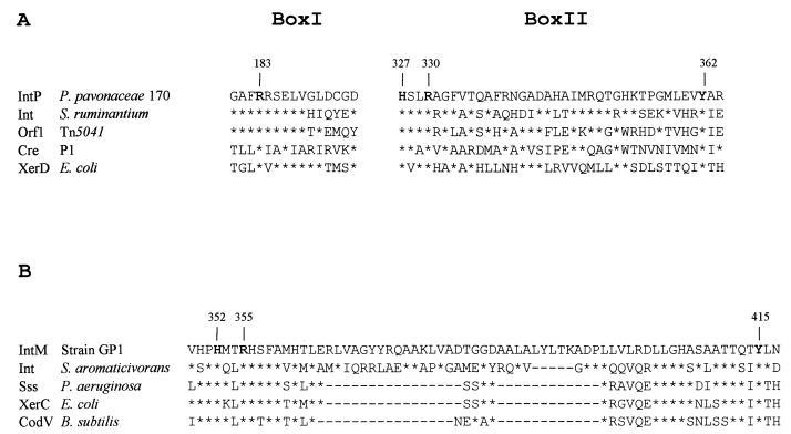
Interestingly, an ORF encoding a putative DNA integrase (intP) was found upstream of the conserved dhaA gene fragment in strain 170 (Fig. 2 and Table 1). IntP shares significant similarity with proteins belonging to the integrase (Int) family of site-specific recombinases (Fig. 6A). Although the members of the Int family exhibit a large diversity in their sequences, they all harbor two regions of marked sequence similarity, called box I and box II (27). IntP shows 55% identity to its nearest neighbor, the integrase-like protein of Selenomonas ruminantium, in these two conserved domains (Fig. 6A). The tetrad R-H-R-Y, which includes the active-site tyrosine (31), is conserved in almost all members of the Int family (1, 4, 27) and is present as Arg-183, His-327, Arg-330, and Tyr-362 in IntP.
FIG. 6.
(A) Amino acid sequence alignment of IntP with the conserved segments of the recombinases Cre of bacteriophage P1 (accession no. P06956) and XerD of E. coli (M54884) and the integrase-like proteins of S. ruminantium (AB011029) and Tn5041 (X98999). Asterisks represent bases identical to those in the upper sequence. Invariant amino acid residues which are believed to be involved in catalysis are numbered (IntP numbering) and shown in boldface. (B) Amino acid sequence alignment of IntM with the C termini of the recombinases Sss of P. aeruginosa (S61402), XerC of E. coli (P22885), and CodV of Bacillus subtilis (P39776) and the putative phage type integrase of Sphingomonas aromaticivorans (AF079317). Asterisks represent bases identical to those in the upper sequence. Dashes represent bases absent in other sequences. The conserved residues His-352, Arg-355, and Tyr-415 are numbered (IntM numbering) and shown in boldface.
An ORF encoding a putative DNA integrase (intM) was also found upstream of the conserved dhaA gene fragment in strain GP1 (Fig. 2 and Table 1). The C-terminal region of IntM shares considerable similarity with proteins of the Int family (Fig. 6B). The conserved tetrad R-H-R-Y of the Int family (1, 4, 27) is present as Arg-260, His-352, Arg-355, and Tyr-415 in IntM (Fig. 6B).
The presence of an integrase gene in the vicinity of the conserved dhaA gene fragment both in strain 170 and in strain GP1 suggests acquisition of these DNA fragments by site-specific integration. Assuming that no additional recombinations have taken place after the primary insertion event, comparisons of the boundaries between the conserved dhaA gene fragments and the sequences unique for strain 170 or GP1 define the insertion site. No short duplication of bases on either side of the presumed point of insertion or regions of dyad symmetry that could serve as integrase binding sites were found when the flanking sequences were compared. The absence of these features could be due to an adjacent deletion removing the border segment on one side. Such a deletion could also explain the fusion of the dhaA gene to the 3′ end of the haloalcohol dehalogenase gene hheB (Fig. 2).
The presence of insertion elements in the vicinity of the conserved dhaA gene fragments.
Many catabolic genes are associated with transposons (42, 47), and transposition is clearly a major mechanism for the acquisition of catabolic genes by bacterial genomes. Sequence analysis of the regions (>4 kb) adjacent to the conserved dhaA gene fragment in Mycobacterium sp. strain GP1 showed that these regions do not encode any proteins related to known transposases. Distal to the dhaA gene fragment in P. pavonaceae 170, however, a large ORF (tnpA) was found (Fig. 2) of which the deduced amino acid sequence showed extensive similarity with the putative transposase of Pseudomonas pseudoalcaligenes JS45 (Table 1). The tnpA ORF, however, appeared to be interrupted by an insertion element, identified as IS1071 (Fig. 2), causing a truncation of its product. Nucleotide sequencing of a 1.7-kb fragment of IS1071 revealed no differences with the known sequence of IS1071, which is involved in transposition of the chlorobenzoate genes (26). PCR amplification, using a primer specific for both ends of IS1071 (due to the inverted repeats at its termini) and recombinant cosmid pPC33 as template DNA, showed the formation of a 3.2-kb PCR product corresponding to the size of IS1071 (26). Thus, a complete copy of IS1071 was present as an insertion in the tnpA gene approximately 1.6 kb downstream of dhaA.
Southern hybridization analysis revealed five copies of IS1071 in strain 170 but only two copies in strain 170M4, a spontaneous mutant of strain 170 that has lost the dhaA gene (35) (results not shown). The 60-kb plasmid pPC170, that was previously identified in strain 170 (44), is still present in strain 170M4, showing that the dhaA gene is not plasmid localized. Assuming that the mutation in strain 170M4 was caused by a single deletion event, this could suggest that the dhaA gene region in strain 170 is flanked by several copies of IS1071, forming a composite class I element, and that a homologous recombination event between two insertion sequences was responsible for the loss of the dhaA gene and three copies of IS1071. Strain 170M4 also lacked the integrase gene intP, which is consistent with such a deletion event.
Many antibiotic resistance genes found on transposons in gram-negative bacteria are located within a conserved DNA sequence (15, 25, 40). These conserved elements, called integrons (16, 40), are formally distinct from other genetic elements in that they determine site-specific integration functions, a DNA integrase and a recombination site, and are thus able to acquire resistance genes at the specific site without the need for the presence of insertion sequences or integrase genes in the DNA segments that are acquired. If the integrase gene intP in P. pavonaceae 170 is flanked by IS1071 sequences and confers on this transposon a capability of taking up individual and unrelated catabolic genes by integrase-mediated recombination, strain 170 may possess a mobile DNA element similar to the previously identified integrons which are capable of taking up antibiotic resistance genes (16, 40).
No sequences similar to IS1071 were found in strains NCIMB13064 and GP1, indicating that this insertion element was not involved in distribution of the dhaA gene among these strains. Similarly, insertion sequence IS2112, which is located approximately 6 kb upstream of the dhaA gene in strain NCIMB13064 (Fig. 2) (21), is not present in strains 170 and GP1. These results are consistent with the hypothesis that site-specific integration events mediated by IntP and IntM were responsible for the acquisition of the dhaA gene fragments, rather than excisive-integrative recombination events mediated by insertion elements. However, the presence of multiple copies of an active mobile element (IS1071) around the dhaA gene in strain 170, combined with the fact that strain 170 was recently isolated from a 1,3-dichloropropene-contaminated environment in which natural genetic exchange is likely to be important (44), suggests that transposition has played a part in the mobilization of the dhaA gene among members of the microbial community present in this environment.
Concluding remarks.
Our data provide further support for previous studies suggesting that horizontal transfer of genes involved in pollutant biodegradation may play an important role in the evolution of catabolic pathways and the adaptation of microbial communities to different environmental contaminants. Up to now, catabolic genes were thought to be transferred mainly by means of conjugative plasmids and transposons (42, 47). The results presented here suggest that genes specifying adaptation to xenobiotics can also spread as integrons, as has been proposed in the case of genes specifying antibiotic resistance (16, 40). The widespread use of antibiotics and the introduction of xenobiotics into the environment seem to lead to adaptation by similar molecular mechanisms.
ACKNOWLEDGMENTS
This study was supported by the Life Sciences Foundation (SLW), which is subsidized by the Netherlands Organization for Scientific Research (NWO), and by the EC Environment and Climate Research Program contract ENV4-CT95-0086.
We thank P. Terpstra (BioMedical Technology Centre, University of Groningen, Groningen, The Netherlands) for his assistance in DNA sequencing and analysis.
REFERENCES
- 1.Abremski K E, Hoess R H. Evidence for a second conserved arginine residue in the integrase family of recombination proteins. Protein Eng. 1992;5:87–91. doi: 10.1093/protein/5.1.87. [DOI] [PubMed] [Google Scholar]
- 2.Albertini A M, Hofer M, Calos M P, Miller J H. On the formation of spontaneous deletions: the importance of short sequence homologies in the generation of large deletions. Cell. 1982;29:319–328. doi: 10.1016/0092-8674(82)90148-9. [DOI] [PubMed] [Google Scholar]
- 3.Altschul S F, Madden T L, Schäffer A A, Zhang J, Zhang Z, Miller W, Lipman D J. Gapped BLAST and PSI-BLAST: a new generation of protein database search programs. Nucleic Acids Res. 1997;25:3389–3402. doi: 10.1093/nar/25.17.3389. [DOI] [PMC free article] [PubMed] [Google Scholar]
- 4.Argos P, Landy A, Abremski K, Egan J B, Haggard-Ljungquist E, Hoess R H, et al. The integrase family of site-specific recombinases: regional similarities and global diversity. EMBO J. 1986;5:433–440. doi: 10.1002/j.1460-2075.1986.tb04229.x. [DOI] [PMC free article] [PubMed] [Google Scholar]
- 5.Belser N O, Castro C E. Biodehalogenation — the metabolism of the nematocides cis- and trans-3-chloroallyl alcohol by a bacterium isolated from soil. J Agric Food Chem. 1971;19:23–26. doi: 10.1021/jf60173a047. [DOI] [PubMed] [Google Scholar]
- 6.Blatter E E, Abriola D P, Pietruszko R. Aldehyde dehydrogenase. Covalent intermediate in aldehyde dehydrogenation and ester hydrolysis. Biochem J. 1992;282:353–360. doi: 10.1042/bj2820353. [DOI] [PMC free article] [PubMed] [Google Scholar]
- 7.Boyer H W, Roulland-Dussoix D. A complementation analysis of the restriction and modification of DNA in Escherichia coli. J Mol Biol. 1969;41:459–472. doi: 10.1016/0022-2836(69)90288-5. [DOI] [PubMed] [Google Scholar]
- 8.Cole S T, Brosch R, Parkhill J, Garnier T, Churcher C, Harris D, et al. Deciphering the biology of Mycobacterium tuberculosis from the complete genome sequence. Nature. 1998;393:537–544. doi: 10.1038/31159. [DOI] [PubMed] [Google Scholar]
- 9.Curragh H, Flynn O, Larkin M J, Stafford T M, Hamilton J T G, Harper D B. Haloalkane degradation and assimilation by Rhodococcus rhodochrous NCIMB13064. Microbiology. 1994;140:1433–1442. doi: 10.1099/00221287-140-6-1433. [DOI] [PubMed] [Google Scholar]
- 10.Dodd I B, Egan J B. Systematic method for the detection of potential 8 Cro-like DNA-binding regions in proteins. J Mol Biol. 1987;194:557–564. doi: 10.1016/0022-2836(87)90681-4. [DOI] [PubMed] [Google Scholar]
- 11.Eaton R W. p-Cumate catabolic pathway in Pseudomonas putida F1: cloning and characterization of DNA carrying the cmt operon. J Bacteriol. 1996;178:1351–1362. doi: 10.1128/jb.178.5.1351-1362.1996. [DOI] [PMC free article] [PubMed] [Google Scholar]
- 12.Farrés J, Wang T T Y, Cunningham S J, Weiner H. Investigation of the active site cysteine residue of rat liver mitochondrial aldehyde dehydrogenase by site-directed mutagenesis. Biochemistry. 1995;34:2592–2598. doi: 10.1021/bi00008a025. [DOI] [PubMed] [Google Scholar]
- 13.Finan T M, Kunkel B, De Vos G F, Signer E R. Second symbiotic megaplasmid in Rhizobium meliloti carrying exopolysaccharide and thiamine synthesis genes. J Bacteriol. 1986;167:66–72. doi: 10.1128/jb.167.1.66-72.1986. [DOI] [PMC free article] [PubMed] [Google Scholar]
- 14.Glasgow A C, Hughes K T, Simon M I. Bacterial DNA inversion systems. In: Berg D E, Howe M M, editors. Mobile DNA. Washington, D.C.: American Society for Microbiology; 1989. pp. 637–659. [Google Scholar]
- 15.Hall R M, Brookes D E, Stokes H W. Site-specific insertion of genes into integrons: role of the 59-base element and determination of the recombination cross-over point. Mol Microbiol. 1991;5:1941–1959. doi: 10.1111/j.1365-2958.1991.tb00817.x. [DOI] [PubMed] [Google Scholar]
- 16.Hall R M, Stokes H W. Integrons: novel DNA elements which capture genes by site-specific recombination. Genetica (The Hague) 1993;90:115–132. doi: 10.1007/BF01435034. [DOI] [PubMed] [Google Scholar]
- 17.Janssen D B, Scheper A, Witholt B. Biodegradation of 2-chloroethanol and 1,2-dichloroethane by pure bacterial cultures. Prog Ind Microbiol. 1984;20:169–178. [Google Scholar]
- 18.Janssen D B, Pries F, Van der Ploeg J, Kazemier B, Terpstra P, Witholt B. Cloning of 1,2-dichloroethane degradation genes of Xanthobacter autotrophicus GJ10 and expression and sequencing of the dhlA gene. J Bacteriol. 1989;171:6791–6799. doi: 10.1128/jb.171.12.6791-6799.1989. [DOI] [PMC free article] [PubMed] [Google Scholar]
- 19.Keen N T, Tamaki S, Kobayashi D, Trollinger D. Improved broad-host-range plasmids for DNA cloning in Gram-negative bacteria. Gene. 1988;70:191–197. doi: 10.1016/0378-1119(88)90117-5. [DOI] [PubMed] [Google Scholar]
- 20.Keuning S, Janssen D B, Witholt B. Purification and characterization of hydrolytic haloalkane dehalogenase from Xanthobacter autotrophicus GJ10. J Bacteriol. 1985;163:635–639. doi: 10.1128/jb.163.2.635-639.1985. [DOI] [PMC free article] [PubMed] [Google Scholar]
- 21.Kulakov L A, Poelarends G J, Janssen D B, Larkin M J. Characterization of IS2112, a new insertion sequence from Rhodococcus, and its relationship with mobile elements belonging to the IS110 family. Microbiology. 1999;145:561–568. doi: 10.1099/13500872-145-3-561. [DOI] [PubMed] [Google Scholar]
- 22.Kulakova A N, Stafford T M, Larkin M J, Kulakov L A. Plasmid pRTL1 controlling 1-chloroalkane degradation by Rhodococcus rhodochrous NCIMB13064. Plasmid. 1995;33:208–217. doi: 10.1006/plas.1995.1022. [DOI] [PubMed] [Google Scholar]
- 23.Kulakova A N, Larkin M J, Kulakov L A. The plasmid-located haloalkane dehalogenase gene from Rhodococcus rhodochrous NCIMB13064. Microbiology. 1997;143:109–115. doi: 10.1099/00221287-143-1-109. [DOI] [PubMed] [Google Scholar]
- 24.Larkin M J, De Mot R, Kulakov L A, Nagy I. Applied aspects of Rhodococcus genetics. Antonie Leeuwenhoek. 1998;74:133–153. doi: 10.1023/a:1001776500413. [DOI] [PubMed] [Google Scholar]
- 25.Martinez E, De la Cruz F. Genetic elements involved in Tn21 site-specific integration, a novel mechanism for the dissemination of antibiotic resistance genes. EMBO J. 1990;9:1275–1281. doi: 10.1002/j.1460-2075.1990.tb08236.x. [DOI] [PMC free article] [PubMed] [Google Scholar]
- 26.Nakatsu C, Ng J, Singh R, Straus N, Wyndham C. Chlorobenzoate catabolic transposon Tn5271 is a composite class I element with flanking class II insertion sequences. Proc Natl Acad Sci USA. 1991;88:8312–8316. doi: 10.1073/pnas.88.19.8312. [DOI] [PMC free article] [PubMed] [Google Scholar]
- 27.Nunes-Düby S E, Kwon H J, Tirumalai R S, Ellenberger T, Landy A. Similarity and differences among 105 members of the Int family of site-specific recombinases. Nucleic Acids Res. 1998;26:391–406. doi: 10.1093/nar/26.2.391. [DOI] [PMC free article] [PubMed] [Google Scholar]
- 28.Okamoto S, Nakamura K, Nihira T, Yamada Y. Virginiae butanolide binding protein from Streptomyces virginiae: evidence that VbrA is not the virginiae butanolide binding protein and reidentification of the true binding protein. J Biol Chem. 1995;270:12319–12326. doi: 10.1074/jbc.270.20.12319. [DOI] [PubMed] [Google Scholar]
- 29.Onaka H, Ando N, Nihira T, Yamada Y, Beppu T, Horinouchi S. Cloning and characterization of the A-factor receptor gene from Streptomyces griseus. J Bacteriol. 1995;177:6083–6092. doi: 10.1128/jb.177.21.6083-6092.1995. [DOI] [PMC free article] [PubMed] [Google Scholar]
- 30.Pabo C O, Sauer R T. Protein-DNA recognition. Annu Rev Biochem. 1984;53:293–321. doi: 10.1146/annurev.bi.53.070184.001453. [DOI] [PubMed] [Google Scholar]
- 31.Pargellis C A, Nunes-Düby S E, De Vargas L M, Landy A. Suicide recombination substrates yield covalent lambda integrase-DNA complexes and lead to identification of the active site tyrosine. J Biol Chem. 1988;263:7678–7685. [PubMed] [Google Scholar]
- 32.Park D H, Plapp B V. Isoenzymes of horseliver alcohol dehydrogenase active on ethanol and steroids. cDNA cloning, expression, and comparison of active sites. J Biol Chem. 1991;266:13296–13302. [PubMed] [Google Scholar]
- 33.Persson B, Bergman T, Keung W M, Waldenstrom U, Holmquist B, Vallee B L, Jornvall H. Basic features of class-I alcohol dehydrogenase: variable and constant segments coordinated by inter-class and intra-class variability. Conclusions from characterization of the alligator enzyme. Eur J Biochem. 1993;216:49–56. doi: 10.1111/j.1432-1033.1993.tb18115.x. [DOI] [PubMed] [Google Scholar]
- 34.Plasterk R H, Brinkman A, Van de Putte P. DNA inversions in the chromosome of Escherichia coli and in bacteriophage Mu: relationship to other site-specific recombination systems. Proc Natl Acad Sci USA. 1983;80:5355–5358. doi: 10.1073/pnas.80.17.5355. [DOI] [PMC free article] [PubMed] [Google Scholar]
- 35.Poelarends G J, Wilkens M, Larkin M J, Van Elsas J D, Janssen D B. Degradation of 1,3-dichloropropene by Pseudomonas cichorii 170. Appl Environ Microbiol. 1998;64:2931–2936. doi: 10.1128/aem.64.8.2931-2936.1998. [DOI] [PMC free article] [PubMed] [Google Scholar]
- 36.Poelarends G J, van Hylckama Vlieg J E T, Marchesi J R, Freitas dos Santos L M, Janssen D B. Degradation of 1,2-dibromoethane by Mycobacterium sp. strain GP1. J Bacteriol. 1999;181:2050–2058. doi: 10.1128/jb.181.7.2050-2058.1999. [DOI] [PMC free article] [PubMed] [Google Scholar]
- 37.Reid M F, Fewson C A. Molecular characterization of microbial alcohol dehydrogenases. Crit Rev Microbiol. 1994;20:13–56. doi: 10.3109/10408419409113545. [DOI] [PubMed] [Google Scholar]
- 38.Sambrook J, Fritsch E F, Maniatis T. Molecular cloning: a laboratory manual. 2nd ed. Cold Spring Harbor, N.Y: Cold Spring Harbor Laboratory; 1989. [Google Scholar]
- 39.Staskawicz B, Dahlbeck D, Keen N, Napoli C. Molecular characterization of cloned avirulence genes from race 0 and race 1 of Pseudomonas syringae pv. glycinea. J Bacteriol. 1987;169:5789–5794. doi: 10.1128/jb.169.12.5789-5794.1987. [DOI] [PMC free article] [PubMed] [Google Scholar]
- 40.Stokes H W, Hall R M. A novel family of potentially mobile DNA elements encoding site-specific gene-integration functions: integrons. Mol Microbiol. 1989;3:1669–1683. doi: 10.1111/j.1365-2958.1989.tb00153.x. [DOI] [PubMed] [Google Scholar]
- 41.Thompson J D, Higgens D G, Gibson T J. CLUSTAL W: improving the sensitivity of progressive multiple sequence alignment through sequence weighting, position-specific gap penalties and weight matrix choice. Nucleic Acids Res. 1994;22:4673–4680. doi: 10.1093/nar/22.22.4673. [DOI] [PMC free article] [PubMed] [Google Scholar]
- 42.Tschäpe H. The spread of plasmids as a function of bacterial adaptability. FEMS Microbiol Ecol. 1994;15:23–32. [Google Scholar]
- 43.Van der Waarde J J, Kok R, Janssen D B. Degradation of 2-chloroallyl alcohol by a Pseudomonas sp. Appl Environ Microbiol. 1993;59:528–535. doi: 10.1128/aem.59.2.528-535.1993. [DOI] [PMC free article] [PubMed] [Google Scholar]
- 44.Verhagen C, Smit E, Janssen D B, Van Elsas J D. Bacterial dichloropropene degradation in soil; screening of soils and involvement of plasmids carrying the dhlA gene. Soil Biol Biochem. 1995;27:1547–1557. [Google Scholar]
- 45.Waki M, Nihira T, Yamada Y. Cloning and characterization of the gene (farA) encoding the receptor for an extracellular regulatory factor (IM-2) from Streptomyces sp. strain FRI-5. J Bacteriol. 1997;179:5131–5137. doi: 10.1128/jb.179.16.5131-5137.1997. [DOI] [PMC free article] [PubMed] [Google Scholar]
- 46.Willuhn J, Schmitt-Wrede H P, Otto A, Wunderlich F. Cadmium-detoxification in the earthworm Enchytraeus: specific expression of a putative aldehyde dehydrogenase. Biochem Biophys Res Commun. 1996;226:128–134. doi: 10.1006/bbrc.1996.1321. [DOI] [PubMed] [Google Scholar]
- 47.Wyndham R C, Cashore A E, Nakatsu C H, Peel M C. Catabolic transposons. Biodegradation. 1994;5:323–342. doi: 10.1007/BF00696468. [DOI] [PubMed] [Google Scholar]
- 48.Xia X, Smith A R W, Bruce I J. Identification and sequencing of a novel insertion sequence, IS1471, in Burkholderia cepacia strain 2a. FEMS Microbiol Lett. 1996;144:203–206. doi: 10.1111/j.1574-6968.1996.tb08531.x. [DOI] [PubMed] [Google Scholar]
- 49.Yanisch-Perron C, Vieira J, Messing J. Improved M13 phage cloning vectors and host strains: nucleotide sequences of the M13mp18 and pUC19 vectors. Gene. 1985;33:103–109. doi: 10.1016/0378-1119(85)90120-9. [DOI] [PubMed] [Google Scholar]
- 50.Yu F, Nakamura T, Mizunashi W, Watanabe I. Cloning of two halohydrin halogen-halide lyase genes from Corynebacterium sp. strain N-1074 and structural comparison of the genes and gene products. Biosci Biotechnol Biochem. 1994;58:1451–1457. doi: 10.1271/bbb.58.1451. [DOI] [PubMed] [Google Scholar]
- 51.Zieg J, Simon M. Analysis of the nucleotide sequence of an invertable controlling element. Proc Natl Acad Sci USA. 1980;77:4196–4200. doi: 10.1073/pnas.77.7.4196. [DOI] [PMC free article] [PubMed] [Google Scholar]


