From a medical point of view, lasers are a convenient but sophisticated source of light in the visible, ultraviolet, and infrared parts of the spectrum. They are easy to control, and the light beam (of a single colour) can be focused to a small spot and in many cases can be transmitted via thin flexible fibres, making internal delivery of light feasible. The range of clinical applications is enormous, from the simple carbon dioxide laser, used as a non-contact scalpel or for superficial tissue ablation, to the precision of the excimer laser, used for reshaping corneas, and the flash lamp pumped dye laser, used to close the small blood vessels of disfiguring port wine birthmarks. This review looks at how the precision of light delivery and the predictability of biological response possible with laser therapy is starting to be exploited for the in situ destruction of diseased tissue and how these techniques might be developed in the future.
The first requirement for successful clinical use of lasers is to understand how light at the wavelength used can interact with living tissue. Most of the simple applications are thermal, but the effect produced depends on how much heat is delivered, how fast it is delivered, and the volume of tissue in which it is absorbed. Increasingly, however, the new technique of photodynamic therapy (non-thermal effects from combining laser light and a photosensitising drug) is attracting interest. This review considers particularly the effects of low power thermal treatment and photodynamic therapy (see table).
Thermal laser therapy
The carbon dioxide laser (wavelength 10 600 nm in the far infrared) is well established as a non-contact scalpel in relatively inaccessible areas like the brain and upper airways and for ablating small lesions as on the skin. However, the beam cannot be transmitted via flexible fibres and can only produce haemostasis in vessels well below 1 mm in diameter.
Possible futures
Image guided laser treatments in minimally invasive destruction of a wide range of tumours
Greater use of interstitial laser photocoagulation for lesions in solid organs, with potential targets including benign prostatic hypertrophy and small localised cancers or benign lesions of the liver, breast, uterus, and other organs
Increased use of photodynamic therapy with potential targets including dysplasia and localised tumours of the skin, mouth, oesophagus, major bronchi, bladder, and vulva.
Other potential applications of photodynamic therapy include preventing restenosis after balloon angioplasty or stent, to ablate the uterine endometrium, as an adjunct to cancer surgery, and treating macular degeneration and localised infections with resistant organisms
The next generation of reliable and cheap lasers will make most of these techniques accessible to all large hospitals
Light in the near infrared part of the spectrum—as from a NdYAG laser at 1064 nm or a semiconductor diode laser at 805 nm—penetrates tissue much better, producing effects through up to 10 mm of tissue. Immediately under a high power NdYAG laser beam, tissue is vaporised. Below the surface, tissue is coagulated, with effective haemostasis, and may later slough or heal with fibrosis. This beam can be transmitted via thin fibres, so the technique is of particular value for endoscopic palliative debulking of advanced cancers of the upper and lower gastrointestinal tract and major airways. Used in conjunction with brachytherapy (single dose intraluminal radiotherapy), this can provide excellent palliation for extended periods and is complementary to insertion of a stent.1 This application is relatively crude, but well established and effective.
The same principle (although with much less immediate vaporisation) is applied to cystoscopic laser treatment of benign prostatic hypertrophy. Special side firing laser fibres are used to direct the beam at the urethral surface of the prostate under direct vision. This is fairly widely used as an alternative to conventional transurethral resection, particularly in North America. However, more sophisticated ways of using these lasers are now emerging.
Interstitial laser photocoagulation
In this technique laser light is delivered to lesions in the centre of solid organs via fibres positioned through needles inserted percutaneously under image guidance. At low power (typically about 3 W, so there is no tissue vaporisation, compared with the 60-80 W used endoscopically), the diseased tissue is gently coagulated over a few minutes in such a way that the dead tissue can be resorbed by normal healing mechanisms without the need for further intervention. There is no effect on the overlying normal tissue, no cumulative toxicity (so treatment can be repeated if necessary), and no surgical wound to heal so recovery is rapid. However, the keys to success are positioning the fibres in the right place, matching the extent of laser induced necrosis to the limits of the lesion being treated, and ensuring that all treated areas (normal or abnormal) will heal safely. The whole process is critically dependent on imaging.
Hepatic metastases
—The best established application is for treating small, isolated metastases in the liver (mainly from previously resected colorectal primary cancers) in patients who are unsuitable for surgery.2 Under local anaesthesia and sedation, the needles are inserted percutaneously under computed tomographic guidance, the result being assessed on contrast enhanced computed tomograms taken 24 hours later. The technique is more controllable than percutaneous alcohol injection and simpler than cryotherapy.
Breast cancer
—The potential application attracting most interest is using interstitial laser photocoagulation in the initial treatment of small breast cancers as an alternative to lumpectomy—this would leave no scar or cosmetic deformity and could be a simple outpatient procedure performed under local anaesthetic. Contrast enhanced magnetic resonance imaging is extremely good at defining the limits of breast cancers and the limits of laser induced necrosis in cancers treated with interstitial laser photocoagulation a few days before definitive surgery.3 Further, if the procedure is undertaken with the patient in a magnetic resonance imager, changes can be seen on the images as the laser is firing, so the positions of the fibres can be adjusted if treatment is incomplete or in the wrong place. However, there is a long way to go before it will be clear whether interstitial laser photocoagulation can have a role in the routine management of breast cancer, as it is so crucial to be sure that all the cancer has been destroyed before the laser necrosed tissue can be safely left in situ.
Benign disease
—The technique may have a role much sooner in managing benign fibroadenomas of the breast. Many of these need no treatment, but, for those that do, interstitial laser photocoagulation is a simple alternative to excision that should leave no scar, which is particularly attractive for patients inclined to formation of keloids. Early results from clinical trials are promising. Similarly, interstitial laser photocoagulation is being explored for treating small, symptomatic uterine fibroids and as an alternative laser technique for treating benign prostatic hypertrophy.4
In principle, interstitial laser photocoagulation is applicable to well defined lesions in any solid organ in which the effect can be well enough localised not to cause any unacceptable damage to the surrounding normal tissue.
Photodynamic therapy
The form of light activated treatment that probably has the greatest overall potential is photodynamic therapy, although no applications are yet firmly established. This technique involves treatment with low power red light (usually from a laser) after administration of a photosensitising drug. There is no increase in tissue temperature. The real attraction is the nature of the tissue damage. Unlike thermal damage, connective tissues like collagen and elastin are largely unaffected, so there is much less risk to the mechanical integrity of hollow organs and healing takes place with more regeneration and less scarring. However, photodynamic therapy is more complicated as it involves delivery of both drug and light, and, for best results, close collaboration between scientists and clinicians is essential.5,6
Photodynamic therapy first attracted widespread attention because many photosensitisers are taken up slightly more by cancers than by the adjacent normal tissue. Unfortunately, the dream that this might be exploited to give selective necrosis of cancers without damaging adjacent tissues has not been realised, and, in general, the area necrosed is the area exposed to the light. Nevertheless, damage by photodynamic therapy in many normal tissues heals so well that the final result may effectively be selective necrosis of small tumours.
Neoplasia of hollow organs
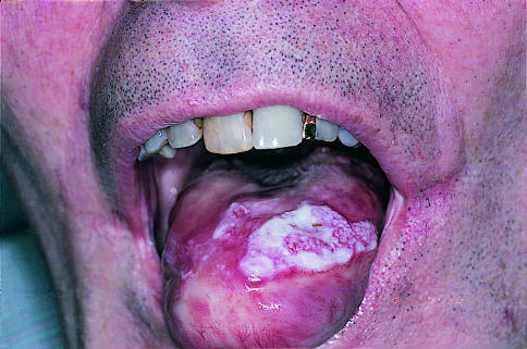
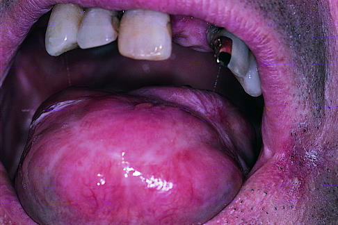
Photodynamic therapy is probably most useful for early invasive cancers in patients who are unsuitable for surgery. Most work has been done on localised cancers of the oral cavity with photosensitisers like porfimer sodium (Photofrin) and meso-tetra hydroxyphenyl chlorin (mTHPC, Foscan). These agents show no selectivity of necrotic effect between mucosa and underlying tissues, and the depth of necrosis can be 5 mm or more, but treated areas heal remarkably well (see fig 1).7 Good results have been reported for endoscopic photodynamic therapy for small cancers of the major airways, oesophagus, stomach, and colon, but it cannot treat tumour that has spread beyond the wall of the organ of origin.8 Experimental work suggests that normal bone is remarkably resistant to photodynamic therapy, so it may be a useful treatment for oral cancers that have invaded the mandible or maxilla, avoiding the need for mutilating surgery or radical radiotherapy.
Figure 1.
Early invasive squamous carcinoma of the tongue (top) and after photodynamic therapy (bottom). (Reproduced courtesy of C Hopper)
With the photosensitising agent 5-amino laevulinic acid (ALA, Levulan), the mucosa of the aerodigestive and urogenital tracts is sensitised more than the underlying layers, so that it is possible to achieve necrosis of the mucosa (normal or dysplastic) without damaging the underlying muscle. This is being studied for treating areas of dysplasia in the mouth, Barrett’s oesophagus, major bronchi, bladder, and vulva.9 Use of topical 5-amino laevulinic acid seems promising for a range of skin conditions from basal cell carcinomas to actinic keratoses and has even been tried for psoriasis.10 The effect is only superficial, however, and the depth of necrosis must be shown to be adequate, particularly for basal cell carcinomas. This is probably best done with high frequency ultrasound scanning, although few studies on this have yet been done.
Photodynamic therapy is not without problems. Treating sensitive areas like the mouth and skin can be painful, and healing may take several weeks. All photosensitisers given systemically cause some skin photosensitivity to sunlight. With porfimer sodium this can last 2-3 months, although with meso-tetra hydroxyphenyl chlorin and tin etiopurpurin (SnET2) it is more like 2-3 weeks, and with 5-amino laevulinic acid only 1-2 days. An advantage of meso-tetra hydroxyphenyl chlorin is that it requires smaller light doses, so treatment times are shorter (typically less than 10 minutes). Porfimer sodium is the only photosensitiser yet licensed for sale anywhere (in the United States, Canada, France, Holland, and Japan) and only for limited applications. No photosensitiser is yet licensed in Britain.
Neoplasia of solid organs
Recently, research has focused on the potential of interstitial photodynamic therapy with meso-tetra hydroxyphenyl chlorin for treating cancers in solid organs, particularly the prostate and pancreas. As more early prostate cancers are being found in younger asymptomatic men with an elevated blood level of prostate specific antigen, so the need increases for a potentially curative treatment that carries less risk of incontinence and impotence than radical prostatectomy or radical radiotherapy. Clinical trials of laser therapy have just started and are limited to patients with locally recurrent cancers after radiotherapy, but in some the levels of prostate specific antigen have been reduced to levels seen after radical prostatectomy and the incidence of complications has been low. If photodynamic therapy proves effective as primary treatment for localised disease with a low incidence of complications then this could be an important advance.
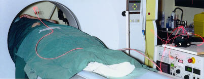
For inoperable pancreatic cancers, the therapeutic options are limited, even for lesions that are still localised to the pancreas. Experiments have shown that the normal pancreas and surrounding tissues can tolerate photodynamic therapy, and clinical trials have started, with the light being delivered by fibres inserted percutaneously under computed tomographic guidance (fig 2). Only a handful of patients have been treated, but none experienced serious problems and follow up scans showed large areas of tumour necrosis that resolved safely without further intervention. For selected patients this could prove a useful treatment.
Figure 2.
Interstitial photodynamic therapy for patient with pancreatic cancer. Three days after injection of photosensitising drug, needles have been positioned in the tumour under computed tomographic guidance and fibres from a diode laser inserted though the needles. (Reproduced courtesy of D E Whitelaw and W R Lees)
Interstitial photodynamic therapy could be applied to unresectable lesions in other solid organs such as the lungs. Another possibility is to use photodynamic therapy as adjuvant therapy after conventional surgery to destroy small deposits of tumour that may not be visible to the surgeon or that involve vital structures which cannot be removed. This has been described with resection of primary brain tumours.11
Vascular disease
One of the main problems after balloon angioplasty or insertion of a stent for obstructive vascular disease is restenosis, which is related at least in part to proliferation of smooth muscle cells from the media. Experiments have shown that photodynamic therapy with 5-amino laevulinic acid effectively suppresses this cell proliferation without increasing the risk of thrombosis or weakening the mechanical strength of the arterial wall.12 Clinical trials have just started. Conventional balloon angioplasties of stenosed superficial femoral arteries have been undertaken in photosensitised patients, and the guide wire then replaced by a thin laser fibre to deliver light to the treated area. The technique seems to be feasible and safe, but it is too early to judge efficacy. Interest among cardiologists currently centres on suppressing smooth muscle proliferation with brachytherapy (local radiotherapy), but if photodynamic therapy could produce the same effect it would be preferable as it would avoid the use of ionising radiation. The potential of this application is enormous as it could mean using photodynamic therapy as adjuvant treatment for most endoluminal procedures on coronary and peripheral arteries.
Another promising line of research on vascular disease uses photodynamic therapy with the photosensitiser benzoporphyrin derivative monoacid (BPD-MA, Verteporfin) for macular degeneration in the eye.13 Clinical trials are under way to try to slow down the visual deterioration associated with this condition.
Localised infections
Microorganisms are another potential target. Indeed, the first description of photodynamic therapy dates back to 1900 in a story reminiscent of the discovery of penicillin.14 Oscar Raab in Munich was assessing the toxicity of the dye acridine on Paramecium. On a sunny day it was toxic, but not during a great thunderstorm. He quickly realised the importance of light.
Many bacteria and larger organisms like Candida take up photosensitisers and can be killed with red light, but, for this to be clinically relevant, it must be possible to get drug and light to wherever the organisms are, which limits the technique to localised infections. This might be suitable for skin ulcers infected with resistant organisms like methicillin resistant Staphylococcus aureus (MRSA) or the mouth, where so much pathology is related to bacterial infections.
In theory any superficial localised infection might be amenable to photodynamic therapy. A possible example is infection of the upper gastrointestinal tract with Helicobacter pylori. H pylori can be photosensitised by a substance as simple as methylene blue, and everywhere the organism resides is readily accessible endoscopically. Thus, photodynamic therapy might be used to eradicate infection with H pylori, but considerable technical ingenuity would be needed to get adequate doses of drug and light to all relevant sites. Viruses present more of a challenge, but it may be possible to sensitise and destroy infected cells—such as in treating genital warts—as tissue necrosed by photodynamic therapy in these areas heals so well with little scarring and treatment can be repeated if necessary.
Menorrhagia
Photodynamic therapy could also be used to destroy essentially normal tissue, and the procedure attracting most interest at present is endometrial ablation to control menorrhagia. The idea is to inject the photosensitiser directly into the uterine cavity and then activate it by red light delivered by soft, flexible, optical devices that can be easily and safely inserted through the cervix, preferably without dilatation, to illuminate the entire endometrium.15 Many techniques have been tried to control menorrhagia without the need for hysterectomy, with varying degrees of success, and photodynamic therapy is only just starting clinical trials. However, it does have the advantages of not requiring advanced surgical skills and being unlikely to damage any adjacent organs, as the low power red light used would not penetrate through the myometrium. To take this procedure to its logical conclusion, if photodynamic therapy could produce complete endometrial ablation patients would probably become sterile. If this proved safe and reliable, it would be a simple and cheap method for female sterilisation that could rapidly be made available almost anywhere.
Conclusions
Clinical lasers used to be large, expensive, unreliable, and difficult to operate. Many are now the size of a briefcase, can be plugged into a standard power socket, and are rugged enough to be easily moved between locations for many uses. As demand increases, so the price is coming down.
For low power applications in which transmission of the light by fibre is not essential (as for photodynamic therapy of the skin and some internal sites where it may be possible to use a broader light delivery system such as in the mouth and the uterine endometrium), cheaper non-laser sources can be used such as light emitting diodes (LEDs) and suitably filtered beams from a xenon lamp. Thus, many of the future treatments suggested here could become accessible to most hospitals. The ones that would have to stay in specialist centres are those requiring sophisticated image guidance such as the treatment of breast, prostate, and pancreatic cancers.
Most of the new applications of lasers described here are already in early clinical trials. Others are more speculative, but for all, the basic validity of the required biological effects has been established. Work is still needed to identify which applications will find a place in routine medical practice and how these techniques will compare with other therapeutic options. Nevertheless, the evidence is mounting that laser treatments can offer considerable advantages over other options for a range of conditions.
Table.
Characteristics of interstitial laser photocoagulation and photodynamic therapy
| Interstitial laser photocoagulation | Photodynamic therapy | |
|---|---|---|
| Laser wavelength | Near infrared (805-1064 nm) | Red (630-730 nm) |
| Laser type | NdYAG | Diode |
| Diode | Dye | |
| Laser power (typical per fibre) | 3-5 W | 0.1-0.3 W (higher in hollow organs) |
| Nature of biological damage | Thermal | Photochemical |
| Effect on collagen and elastin | Destroyed | Largely unaffected |
| Inherent selectivity of necrosis between tumour and surrounding healthy tissue | None | Minimal (some selectivity between different layers of normal tissues) |
| Healing | Resorption and scarring | Regeneration, sometimes with scarring |
| Cumulative toxicity | None | None |
Acknowledgments
Conflict of interest: Research at the National Medical Laser Centre is supported by several commercial companies (including Scotia Quanta Nova, DUSA pharmaceuticals, Miravant/Pharmacia-Upjohn Diomed, Hamamatsu Photonics, and Siemens) and charities (including the Association for International Cancer Research, the Wolfson Foundation, the Stanley Thomas Johnson Foundation, and the Sir Jules Thorn Trust).
References
- 1.Spencer GM, Thorpe S, Sargeant IR, Blackman GM, Solano J, Tobias JS, et al. Laser and brachytherapy in the palliation of adenocarcinoma of the oesophagus and gastric cardia. Gut. 1996;39:726–731. doi: 10.1136/gut.39.5.726. [DOI] [PMC free article] [PubMed] [Google Scholar]
- 2.Amin Z, Donald JJ, Masters A, Kant R, Steger A, Bown SG, et al. Hepatic metastases: interstitial laser photocoagulation with real time ultrasound monitoring and dynamic CT evaluation of treatment. Radiology. 1993;187:339–347. doi: 10.1148/radiology.187.2.8475270. [DOI] [PubMed] [Google Scholar]
- 3.Mumtaz H, Hall-Craggs MA, Wotherspoon A, Paley M, Buonaccorsi G, Amin Z, et al. Laser therapy for breast cancer: MR imaging and histopathologic correlation. Radiology. 1996;200:651–658. doi: 10.1148/radiology.200.3.8756910. [DOI] [PubMed] [Google Scholar]
- 4.De la Rosette JJ, Muschter R, Lopez MA, Gillatt D. Interstitial laser coagulation in the treatment of benign prostatic hyperplasia using a diode laser system with temperature feedback. Br J Urol. 1997;80:433–438. doi: 10.1046/j.1464-410x.1997.00369.x. [DOI] [PubMed] [Google Scholar]
- 5.Wilson BC, Patterson MS, Lilge L. Implicit and explicit dosimetry in photodynamic therapy: a new paradigm. Lasers Med Sci. 1997;12:182–199. doi: 10.1007/BF02765099. [DOI] [PubMed] [Google Scholar]
- 6.Bown SG. Photodynamic therapy to scientists and clinicians—one world or two? J Photochem Photobiol. 1990;6:1–12. doi: 10.1016/1011-1344(90)85069-9. [DOI] [PubMed] [Google Scholar]
- 7.Fan KFM, Hopper C, Speight P, Buonaccorsi G, Bown SG. Photodynamic therapy using mTHPC for malignant disease in the oral cavity. Int J Cancer. 1997;72:1–7. doi: 10.1002/(sici)1097-0215(19970926)73:1<25::aid-ijc5>3.0.co;2-3. [DOI] [PubMed] [Google Scholar]
- 8.Sibille A, Lambert R, Souquet JC, Sabben G, Descos F. Long term survival after photodynamic therapy for oesophageal cancer. Gastroenterology. 1995;108:337–344. doi: 10.1016/0016-5085(95)90058-6. [DOI] [PubMed] [Google Scholar]
- 9.Barr H, Shepherd NA, Dix A, Roberts DJH, Tan WC, Krasner N. Eradication of high grade dysplasia in columnar lined (Barrett’s) oesophagus by photodynamic therapy with endogenously generated protoporphyrin IX. Lancet. 1996;348:584–585. doi: 10.1016/s0140-6736(96)03054-1. [DOI] [PubMed] [Google Scholar]
- 10.Svanberg K, Andersson T, Killander D, Wang I, Stenram U, Andersson-Engels S, et al. Photodynamic therapy of non-melanoma malignant tumours of the skin using topical delta-amino levulinic acid sensitisation and laser irradiation. Br J Dermatol. 1994;130:743–751. doi: 10.1111/j.1365-2133.1994.tb03412.x. [DOI] [PubMed] [Google Scholar]
- 11.Muller PJ, Wilson BC. Photodynamic therapy for recurrent supratentorial gliomas. Semin Surg Oncol. 1995;11:346–354. doi: 10.1002/ssu.2980110504. [DOI] [PubMed] [Google Scholar]
- 12.Nyamekye I, Buonaccorsi G, McEwan J, MacRobert A, Bown SG, Bishop C. Photodynamic therapy of normal and balloon injured rat carotid arteries using 5-amino levulinic acid. Circulation. 1995;91:417–425. doi: 10.1161/01.cir.91.2.417. [DOI] [PubMed] [Google Scholar]
- 13.Lin SC, Lin CP, Feld JR, Duker JS, Puliafito CA. The photodynamic occlusion of choroidal vessels using benzoporphyrin derivative. Curr Eye Res. 1994;13:513–522. doi: 10.3109/02713689408999883. [DOI] [PubMed] [Google Scholar]
- 14.Raab O. Ueber die wirkung fluorescierenden stoffe auf infusiorien. Z Biol. 1900;39:524–546. [Google Scholar]
- 15.Fehr MK, Wyss P, Tromberg BJ, Krasieva T, DiSaia PJ, Lin F, et al. Selective photosensitiser localisation in the human endometrium after intrauterine application of 5-amino levulinic acid. Am J Obstet Gynecol. 1996;175:1253–1259. doi: 10.1016/s0002-9378(96)70037-6. [DOI] [PubMed] [Google Scholar]


