Abstract
Objectives
The aim of the study was to utilise a large database available from a UK-based, commercial veterinary diagnostic laboratory to ascertain the prevalence of different forms of cutaneous neoplasia within the feline population, and to detect any breed, sex or age predilections for the more common tumours.
Methods
Records from the laboratory were searched for feline submissions received between 31 May 2006 and 31 October 2013. For masses arising within the skin for which histopathology had been performed, the diagnosis was recorded together with the breed, age, sex and neuter status of the cat. Odds ratios for breed predisposition to skin tumours overall, to histologically malignant tumours and to the more commonly occurring tumours were calculated, with the non-pedigree cat population as the control.
Results
Of the 219,083 feline samples submitted, masses arising within the skin comprised 4.4% and there were 89 different diagnoses recorded for these masses. Just 6.6% of these cases were non-neoplastic in nature, and, of neoplastic masses, 52.7% were considered histologically malignant. The 10 most common skin tumour types accounted for 80.7% of cases, with the four most common being basal cell tumours, fibrosarcomas, squamous cell carcinomas and mast cell tumours.
Conclusions and relevance
Despite the large number of different diagnoses in this study, a relatively small number of tumour types accounts for the majority of skin masses occurring in cats, most of which are neoplastic in nature. There are a number of breed predispositions for the more common tumour types, although no pedigree breed had increased odds of developing a malignant tumour compared with the non-pedigree cat population; several breeds had significantly decreased odds. Just over half of the neoplastic masses in this study were considered histologically malignant.
Introduction
The skin and subcutis are the most common anatomical locations for tumours to arise in the cat. 1 As both the largest and the most exposed organ of the body, the skin is particularly susceptible to external insults in a variety of forms, and it is also the most easily visualised and palpable. While there have been several major studies regarding the prevalence of feline tumours in other countries, including both the USA and Switzerland,2,3 there is little current information available as to the prevalence of cutaneous tumours specific to the UK cat population.
There is some variation between these studies, but the general consensus is that the four most common skin tumour types are fibrosarcoma, squamous cell carcinoma (SCC), mast cell tumour (MCT) and the tumours that fall under the umbrella term of ‘basal cell tumour’,2–5 with some differences in the order of prevalence depending on the particular study.
The purpose of this study was to utilise a large dataset from a commercial veterinary diagnostic laboratory to determine the prevalence of different forms of cutaneous tumours in the UK cat population, during the period from 31 May 2006 to 31 October 2013, and to detect any breed, sex or age predilections for the more common tumours.
Material and methods
Records from a large, UK-based commercial diagnostic laboratory (Finn Pathologists, Diss, UK) were searched for feline submissions received between 31 May 2006 and 31 October 2013, including samples submitted for various blood tests, cytology and histopathology. Histopathology samples taken from masses arising within the skin were then searched for according to the diagnosis made by the histopathologist originally reporting the case. Masses submitted from cats based outside the UK, or any tumour not located within the epidermis, dermis, subcutis or skin appendages, were excluded. Tumours arising from the mammary glands, oral cavity and third eyelid were also excluded, while tumours arising from the ears and anal gland region were included for the purposes of this study. Cats with multiple samples taken from the same tumour were recorded only once. For all cases included in this study the breed, age, and sex and neuter status of the cat were recorded, where this data was available from the original submission form, as well as the histopathological diagnosis of the mass.
A total of 35 different feline breeds were recorded. Domestic shorthair (DSH), domestic longhair (DLH), domestic cat and ‘crossbreed’ cats were amalgamated under the term ‘non-pedigree’, and cats of unspecified breed were recorded as ‘unknown’. Cats classified as non-pedigree were used as the standard for comparison both for determining whether pedigree breeds had a statistically significant different odds ratio with regard to developing skin tumours and the odds of having a malignant tumour. Gender was recorded as one of the following: male, male neutered, female, female neutered, unknown.
Cutaneous masses were classified into either neoplastic or non-neoplastic (including cysts, hamartomas, inflammatory, hyperplastic or pigmentary growths forming a mass-type lesion). Masses were then further categorised into one of four groups based upon their embryological origin; epithelial, mesenchymal, melanocytic or haematopoietic. Any remaining neoplasms that were either metastatic tumours or did not fit these categories, and any skin tumours of indeterminable origin, were classified as metastatic/other. The term ‘basal cell tumour’ was used to encompass all forms of benign basal cell tumour, including trichoblastomas and apocrine ductal adenomas.
Statistical analysis was performed using Prism 7 for Mac OS X (Version 7.0a). Data were systematically tested for normality and tested accordingly with D’Agostino and Pearson normality or Kruskall–Wallis tests as appropriate. Odds ratios (ORs) for breed predisposition to skin tumours, malignant tumours and certain types of tumours were calculated using an odds ratio calculator (MedCalc,Version 16.8.4) at a 95% confidence interval (CI) with non-pedigree (OR = 1) as the control against which all other breeds were assessed. A P value less than 0.05 was considered to be significant.
Results
The total number of feline submissions to the laboratory over the time period 31 May 2006 to 31 October 2013 was 219,083, including blood samples, cytology and histopathology submissions. Of these, masses arising within the skin comprised 4.4% (9683) affecting a total of 9,200 individual cats, and for which there were 89 different diagnoses recorded. Of these skin masses, 6.6% (636 of the 9683) were non-neoplastic in nature; the three most common diagnoses were follicular cysts (307 cases), apocrine gland cysts (178 cases) and dermoid cysts (44 cases), accounting for 83% of all the non-neoplastic masses in this study.
The remaining masses were deemed neoplastic in nature (9047 of the 9683; 93.4%), with 47.6% of these categorised as benign and 52.7% as malignant based on the original histopathology report (the remaining five masses could not be clearly categorised as either benign or malignant).
For the benign cutaneous neoplasms, those of epithelial origin were the most frequent subtype, accounting for 66% of all benign tumours in this study (2823 out of 4275 masses), followed by tumours of mesenchymal origin (17.5%; 748 out of 4275 masses), tumours of haemato-poietic origin (15.0%; 642 out of 4275) and tumours of melanocytic origin (1.5%; 62 out of 4275). For malignant neoplasms, tumours of mesenchymal origin were the most common subtype, comprising 49% of all malignant cutaneous masses (2342 out of 4767 masses), followed closely by those of epithelial origin (39.4%, 1877 out of 4767). Metastatic and other tumours accounted for 8% (381 of 4767) and the remainder were of melanocytic (2.2%, 107 from 4767) or haematopoietic origin (1.3%, 60 out of 4767; Table 1).
Table 1.
Cutaneous tumours according to embryonic origin and malignancy
| Origin | Benign (n = 4275) | Malignant (n = 4767) | Total (n = 9042) |
|---|---|---|---|
| Epithelial | 2823 | 1877 | 4700 |
| Mesenchymal | 748 | 2342 | 3090 |
| Haematopoietic | 642 | 60 | 702 |
| Melanocytic | 62 | 107 | 169 |
| Metastatic/other | n/a | 381 | 381 |
n/a = not applicable
Breed
Cats classified as non-pedigree (including DSH, DLH, ‘domestic cat’ and ‘crossbreed’) accounted for 88.2% of all feline sample submissions to the laboratory, while cats with no breed recorded (‘unclassified’) accounted for 3%, and the remainder of the cat population (9%) comprised various pedigree breeds.
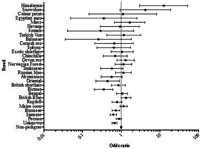
When considering all cutaneous neoplasms, both benign and malignant, certain breeds had statistically significant increased odds of developing a skin tumour when compared with the non-pedigree cat population (Figure 1). These included the British Blue (P = 0.05, OR = 1.33 [CI 1.00; 1.78]) and the Himalayan breeds (P = 0.0005, OR = 12.65 [CI 3.02; 52.92]). Several other breeds had statistically significant decreased odds of developing a skin tumour when compared with the non-pedigree cat population, including the Siamese (P <0.0001, OR = 0.63 [CI 0.52; 0.76]), Burmese (P = 0.0045, OR = 0.72 [CI 0.58; 0.90]), Birman (P = 0.0002, OR = 0.35 [CI 0.20; 0.61]) and Oriental breeds (P = 0.023, OR = 0.44 [CI 0.22; 0.89]).
Figure 1.
Odds ratios for all cutaneous masses in pedigree breeds compared with the non-pedigree population. OR = odds ratio; 95% confidence interval, non-pedigree population OR = 1; n = 9683
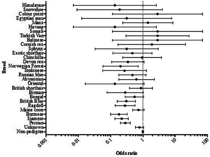
When only malignant neoplasms are considered, no pedigree breeds had statistically significant increased odds of developing a malignant tumour when compared with the non-pedigree cat population, but several breeds had statistically significant decreased odds (Figure 2); these included the Persian (P <0.0001, OR = 0.29 [CI 0.20; 0.41]), Siamese (P <0.0001, OR = 0.22 [CI 0.14; 0.36]), Burmese (P <0.0001, OR = 0.18 [CI 0.10; 0.33]), Ragdoll (P = 0.0004, OR = 0.28 [CI 0.14; 0.57]), British Blue (P = 0.0004, OR = 0.31 [CI 0.16; 0.59]), Birman (P = 0.056, OR = 0.28 [CI 0.078; 1.034]) and Norwegian Forest Cat breeds (P = 0.012, OR = 0.20 [CI 0.058; 0.71]).
Figure 2.
Odds ratios for malignant cutaneous neoplasms in pedigree breeds compared with the non-pedigree cat population. OR = odds ratio; 95% confidence interval, non-pedigree population OR = 1; n = 4767
Age and gender
Ages of affected cats ranged from under 1 year up to 25 years, with a median age of 11 years at the time of diagnosis. Seven cutaneous masses were from cats less than 1 year of age; two of these were MCTs and the remainder were non-neoplastic (two follicular hamartomas, two dermoid cysts and one cutaneous horn of feline paw-pad). There was no significant difference between the ages at which male and female animals were diagnosed with neoplastic skin tumours (P = 0.013). Malignant tumours were seen in an older population of animals (median age 12 years) compared with benign tumours (median age 11 years; P <0.0001), and this difference was more pronounced in male cats. Male neutered cats also tended to develop benign tumours at an earlier age than female neutered cats (P = 0.0128), but no significant difference in age was seen between the genders when developing malignant tumours, nor with benign tumours arising in male entire and female entire cats.
Diagnosis
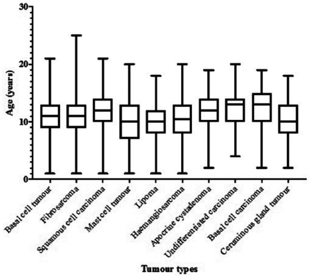
The 10 most common skin tumour types accounted for 80.7% (7300 out of 9047 masses) of all the neoplastic skin masses in this study, both benign and malignant. In order of descending prevalence these were: basal cell tumours (2189; 22.6%), fibrosarcomas (1766; 19.5%), SCCs (1031; 11.4%), MCTs (618; 6.8%), lipomas (516; 5.7%), haemangiosarcomas (404; 4.5%), apocrine cystadenomas (269; 3.0%), undifferentiated carcinomas (255; 2.8%), basal cell carcinomas (252; 2.8%) and ceruminous gland tumours (183; 2.0%) arising within the ear canals (Table 2). The gender and age distribution of affected cats with these 10 most commonly occurring neoplasms are summarised in Table 2 and Figure 3.
Table 2.
Ten most common types of skin tumours from a total of 9046 submissions, with the number of affected cats, male:female ratio, median and mean age
| Neoplasm | Number of cases (%) | Male:female ratio | Median age (range) | Mean age (SD) | |
|---|---|---|---|---|---|
| 1 | Basal cell tumour | 2189 (22.6) | 0.98 | 11 (1–21) | 11.0 (3.32) |
| 2 | Fibrosarcoma | 1766 (19.5) | 0.96 | 11 (1–25) | 11.0 (3.37) |
| 3 | Squamous cell carcinoma | 1031 (11.4) | 1.24 | 12 (1–21) | 12.2 (3.30) |
| 4 | Mast cell tumour | 618 (6.8) | 1.19 | 10 (<1–20) | 9.9 (4.00) |
| 5 | Lipoma | 516 (5.7) | 1.15 | 10 (1–18) | 9.6 (3.24) |
| 6 | Haemangiosarcoma | 404 (4.5) | 1.17 | 10.5 (1–20) | 10.5 (3.59) |
| 7 | Apocrine cystadenoma | 269 (3.0) | 0.88 | 12 (2–19) | 12.0 (2.93) |
| 8 | Carcinoma, undifferentiated | 255 (2.8) | 0.97 | 13 (4–20) | 12.1 (3.01) |
| 9 | Basal cell carcinoma | 252 (2.8) | 1.17 | 13 (2–19) | 12.5 (3.14) |
| 10 | Ceruminous gland tumour | 183 (2.0) | 1.02 | 10 (2–18) | 10.3 (3.57) |
Figure 3.

Age distribution (median, interquartile range, minimum and maximum) of cats presenting with the 10 most common types of neoplastic cutaneous tumour, starting with the most common neoplasm on the left side
Basal cell tumours
Basal cell tumours were the most common skin tumour in this study, accounting for 22.6% of all neoplastic skin masses (2189 cases out of 9047). Within this category, the majority were sub-classified as apocrine ductular adenomas (648 cases; 29.6% of all basal cell tumours) and the second most common sub-classification was trichoblastoma (610 cases; 27.9% of all basal cell tumours). If these two most common forms of basal cell tumour had been categorised as separate, distinct entities they would have ranked as the third most common skin tumour in the case of apocrine ductular adenomas, and the fifth most common in the case of trichoblastomas, just above and below MCTs, respectively. The remaining basal cell tumours in the database were either not further sub-classified (521 cases; 23.8%), or diagnosed as undifferentiated (315 cases; 14.4%), or as differentiating to apocrine glands (48 cases; 2.2%), squamous cells (19 cases; 0.9%), sebaceous glands (15 cases; 0.7%) or with multiple differentiation (11 cases; 0.5%).
The Persian (P = 0.0002, OR = 1.84 [CI 1.33; 2.54]), British Blue (P = 0.041, OR = 1.85 [CI 1.03; 3.34]) and Norwegian Forest Cat (P = 0.0011, OR = 4.98 [CI 1.89; 13.11]) breeds had statistically significant increased odds of having basal cell tumours compared with the non-pedigree population, while the Siamese breed (P = 0.030, OR = 0.55 [CI 0.32; 0.94]) had significantly decreased odds. The median age of cats developing basal cell tumours was 11 years (ranging from 1–21 years) at the time of diagnosis (Table 2), and no gender predisposition was detected.
Fibrosarcoma
Fibrosarcoma was the second most commonly diagnosed neoplastic skin tumour (19.5%; 1766 cases out of 9047). The Chinchilla breed (P = 0.040, OR = 4.27 [CI 1.07; 17.08]) had statistically significant increased odds of developing fibrosarcoma compared with the non-pedigree population, although the sample size for this breed was small (n = 8). The Persian (P <0.0001, OR = 0.24 [CI 0.12; 0.47]), Siamese (P = 0.0003, OR = 0.16 [CI 0.059; 0.43]), Burmese (P = 0.0021, OR = 0.16 [CI 0.052; 0.52]) and British Blue breeds (P = 0.017, OR = 0.09 [CI 0.012; 0.64]) had statistically significant decreased odds of developing fibrosarcoma compared with the non-pedigree population. The median age of cats diagnosed with fibrosarcoma was also 11 years (ranging from 1–25 years; Table 2), and no gender predisposition was found.
Squamous cell carcinoma (SCC)
SCC was the third most commonly diagnosed neoplastic skin tumour in this study (11.4%, 1031 cases out of 9047). Of these cases, 556 (53.9%) affected male cats, whether entire or neutered; male cats were at a 1.54 greater odds of developing SCC than females (P = 0.0012). Three breeds had statistically significant decreased odds of developing SCC compared with the non-pedigree cat population, including the Persian (P = 0.0055, OR = 0.34 [CI 0.16; 0.73]), the Siamese (P = 0.0093, OR = 0.22 [CI 0.069; 0.69]) and the Burmese (P = 0.042, OR = 0.30 [CI 0.095; 0.96]). The median age of cats diagnosed with SCC was 12 years (ranging from 1–21 years; Table 2).
Mast cell tumour (MCT)
MCT was the fourth most common neoplastic skin tumour in this study (6.8%, 618 cases out of 9047), and included both the mastocytic and histocytic forms. Several breeds had statistically significant increased odds of developing an MCT compared with the non-pedigree cat population, including the Siamese (P <0.0001, OR = 5.37 [CI 3.4695; 8.3223]), the Burmese (P = 0.0013, OR = 2.77 [CI 1.491; 5.1466]), the Maine Coon (P = 0.0495, OR = 1.94 [CI 1.0015; 3.7677]) and the Ragdoll (P <0.0001, OR = 7.43 [CI 3.9172; 14.1053]). The Oriental (P = 0.0412, OR = 5.31 [CI 1.069; 26.3717]), the Russian Blue (P = 0.0077, OR = 4.55 [CI 1.4947; 13.8752]) and the Havana breeds (P = 0.0047, OR = 31.86 [CI 2.8838; 351.9177]) also had statistically significant increased odds of developing an MCT compared with the non-pedigree population, although the sample sizes for these breed were very small (n = 2, n = 4 and n = 2, respectively). The median age of cats diagnosed with MCTs was 10 years (ranging from under 1 year to 20 years; Table 2).
Discussion
Neoplasia, arising at any anatomical location, is the fourth most common cause of death for cats presenting to primary care veterinary practices within England according to a recent study, 6 accounting for up to one quarter of deaths in the older cat population. Several studies have found that the skin and subcutis are the most common locations for tumours in cats,1,2 with the proportion varying from 29.6–41.5% of all tumours arising within these tissues. Furthermore, a significant number of these tumours are malignant; the current study found that 52.7% of skin tumours in cats were histologically malignant, and another recent Swiss study 3 found 76.1% were malignant. The difference in the proportion of malignant skin tumours between these two studies may in part be due to the differences in the two study populations; the current study utilises data from a commercial diagnostic laboratory covering the years 2006–2011 and for which primary care veterinary practices comprise the vast majority of clients, while the former study comprises data from two university-based diagnostic laboratories as well as a private laboratory and was gathered over a time span of over 40 years.
In the current study, the 10 most common skin tumours accounted for 80.7% of all cutaneous neoplasms, despite the large number of different diagnoses given for masses arising at this site (89 in total). Both in this study and in three other large studies2,3,4 looking at the prevalence of different skin tumours in cats, the four most commonly diagnosed tumours are very consistent, despite the differences in geographical location and time periods covered by the studies. These four skin tumours are basal cell tumours, MCTs, fibrosarcomas and SCCs, although the precise order of prevalence varies depending on the particular study. These four most common diagnoses account for 60.3% all cutaneous tumours in UK cats in the present study, while in an earlier UK-based study 4 these accounted for 65.3%, in an American-based study 2 they accounted for 77.1% and in the recent Swiss study 3 it was 71.5%.
When all tumours are considered (both benign and malignant), two breeds have statistically significant increased odds of developing cutaneous neoplasia when compared with the non-pedigree cat population, namely the British Blue and the Himalayan breeds, while four breeds have decreased odds, including the Persian, Burmese, Birman and Oriental breeds. When only malignant tumours are included, no breed has statistically significant increased odds of developing neoplasia compared with the non-pedigree cat population but several have decreased odds, including the Persian, Siamese, Burmese, Ragdoll, British Blue, Birman and Norwegian Forest Cat breeds. The recent Swiss study 3 found that the European Shorthair cat (the most common breed in that study) had the highest odds of developing a tumour in the skin or subcutis, while several other pedigree breeds had significantly lower odds ratios, overall in agreement with the present study. Studies looking for breed predispositions to various tumours in cats are always hampered to some degree by the predominance of non-pedigree cats, with far fewer representatives of some pedigree breeds being present within the feline population as a whole.
The most commonly diagnosed tumour arising with the skin of cats in this study was basal cell tumour, comprising 22.6% of the total. This was also the most commonly diagnosed tumour in the previous American-based study 2 (26.1%), while basal cell tumours ranked second most common in the Swiss study 3 (14.4%) and third in the earlier UK-based study 4 (14.8%). Another study 7 looking at 124 feline basal cell tumours found they comprised 10.9% of all feline skin neoplasms. However, basal cell tumours are a rather heterogonous group of neoplasms, encompassing several distinct types of tumour including apocrine ductular adenomas and trichoblastomas (both of which themselves have several different histological subtypes). Both apocrine ductular adenomas and trichoblastomas appear to be common feline skin tumours in their own right, but inconsistencies in the classification of basal cell tumours makes their true incidence in this and in other studies difficult to determine. Longhaired cats were previously reported to be predisposed to developing basal cell tumours, 7 while in this study the Persian, British Blue and Norwegian Forest Cat breeds were found to have significantly increased odds of developing these tumours and the Siamese breed had decreased odds. No gender predisposition was noted, similar to previous studies.2,7
Fibrosarcomas were found to be the second most common skin neoplasm in this study (19.5%), and were the most common skin tumour in both previous European studies (25.4%, 4 38.7% 3 ) but fourth in the American study. 2 In the present study, this category would undoubtedly include a proportion of feline injection-site sarcomas (FISS) with the histological phenotype of a fibrosarcoma; however, the incidence of FISS in the UK and the US is thought to be relatively low. 8 In this study only one breed – the Chinchilla – was found to have significantly increased odds of developing fibrosarcoma compared with the non-pedigree cat population, although the sample size for this breed is very small. Several breeds, including the Persian, Siamese, Burmese and British Blue breeds had significantly lower odds of developing fibrosarcoma compared with non-pedigree cats.
SCC was the third most common neoplastic skin tumour in this study (11.4%), and was also the third most common tumour in the American 2 (15.2%) and Swiss studies 3 (11.7%). SCC ranked second most common in the earlier UK-based study 4 (17.4%); the prevalence of SCC appears relatively consistent despite the different geographical locations of these studies. Both geography and skin pigmentation would be expected to influence the prevalence of SCC in different feline populations due to the relationship between SCC development and exposure to solar radiation. For example, breeds that typically have darker pigmentation of ear pinnae, eyelids and nasal planum (such as the Siamese) have previously been found to be at decreased risk of developing SCC,2,3 and the current study also supports this finding. The Burmese and Persian breeds also had a decreased risk of developing SCC compared with the non-pedigree cat population in this study; unfortunately coat colour is rarely recorded in these cases, making it impossible to determine if there is any correlation between the degree of pigmentation and the likelihood of tumour development. Male cats (neutered or entire) were found to have significantly higher odds of developing SCC than females in this study, a finding not previously reported. 2
MCTs were the fourth most common skin tumour in this study, accounting for 6.8% of all feline skin neoplasia, comparable with the two previous European studies; MCTs were also the fourth most common tumour in the earlier UK-based study 4 (7.7%), and were fifth most common in the Swiss study (6.7%). 3 In contrast, MCTs were the second most common skin tumour in the American study, 2 accounting for 21.1% of all skin neoplasia. Several breeds have previously been reported to be predisposed to developing MCTs, including the Siamese,2,9 Burmese, Russian Blue and Ragdoll breeds. 9 The present study confirms these particular breed predispositions, and in addition the Maine Coon, Oriental and Havana breeds also appear to have an increased risk of developing MCTs. The two neoplastic masses arising in cats less than 1 year of age were also both MCTs, similar to previous studies describing MCTs arising in very young cats. 10 The MCTs diagnosed in the cats in this study included both the mastocytic and histiocytic forms, as detailed in a previous study. 9
Conclusions
This large, UK-based retrospective study of feline cutaneous tumours supports the findings of previous studies, both American- and European-based, in that the four most common skin tumours in cats are basal cell tumours, fibrosarcomas, SCCs and MCTs, although there are interesting differences in the prevalence of these different tumours between the studies. Despite the large number of different diagnoses in this study, a relatively small number of tumour types accounts for the majority of skin masses occurring in cats. The study also confirms a number of apparent breed predispositions; for example, the Siamese, Burmese, Russian Blue, Ragdoll, Maine Coon, Oriental and Havana breeds appear to be predisposed to developing MCTs. When considering all forms of skin neoplasia, just over half of the masses in this study were considered histologically malignant, highlighting the importance of prompt and thorough diagnostic investigation of cutaneous masses in cats.
Acknowledgments
The authors would like to thank all of the staff at Finn Pathologists for their assistance with accessing the database. This research was performed as part of a final year research project (NH) supported by the Royal Veterinary College.
Footnotes
Accepted: 21 February 2017
The authors declared no potential conflicts of interest with respect to the research, authorship, and/or publication of this article.
Funding: The authors received no financial support for the research, authorship, and/or publication of this article.
References
- 1. Graf R, Grüntzig K, Hässig M, et al. Swiss Feline Cancer Registry: a retrospective study of the occurrence of tumours in cats in Switzerland from 1965 to 2008. J Comp Pathol 2015; 153: 266–277. [DOI] [PubMed] [Google Scholar]
- 2. Miller MA, Nelson SL, Turk JR, et al. Cutaneous neoplasia in 340 cats. Vet Pathol 1991; 28: 389–395. [DOI] [PubMed] [Google Scholar]
- 3. Graf R, Grüntzig K, Boo G, et al. Swiss Feline Cancer Registry 1965–2008: the influence of sex, breed and age on tumour types and tumour locations. J Comp Pathol 2016; 154: 195–210. [DOI] [PubMed] [Google Scholar]
- 4. Bostock DE. Neoplasms of the skin and subcutaneous tissues in dogs and cats. Br Vet J 1986; 142: 1–19. [DOI] [PubMed] [Google Scholar]
- 5. Jörger K. Skin tumors in cats. Occurrence and frequency in the research material (biopsies from 1984–1987) of the Institute for Veterinary Pathology, Zurich. Schweiz Arch Tierheilkd 1988; 130: 559–569. [PubMed] [Google Scholar]
- 6. O’Neill DG, Church DB, McGreevy PD, et al. Longevity and mortality of cats attending primary care veterinary practices in England. J Feline Med Surg 2015; 17: 125–133. [DOI] [PMC free article] [PubMed] [Google Scholar]
- 7. Diters RW, Walsh KM. Feline basal cell tumors: a review of 124 cases. Vet Pathol 1984; 21: 51–56. [DOI] [PubMed] [Google Scholar]
- 8. Dean RS, Pfeiffer DU, Adams VJ. The incidence of feline injection site sarcomas in the United Kingdom. BMC Vet Res 2013; 9: 17. [DOI] [PMC free article] [PubMed] [Google Scholar]
- 9. Melville K, Smith KC, Dobromylskyj MJ. Feline cutaneous mast cell tumours: a UK-based study comparing signalment and histological features with long-term outcomes. J Feline Med Surg 2015; 17: 486–493. [DOI] [PMC free article] [PubMed] [Google Scholar]
- 10. Chastain CB, Turk MAM, O’Brien D. Benign cutaneous mastocytomas in two litters of Siamese kittens. J Am Vet Med Assoc 1988; 193: 959–960. [PubMed] [Google Scholar]


